Brief Report

Extraction of the Anticancer and Antimicrobial Agent, Prodigiosin, from Vibrio gazogenes PB1 and Its Identification by 1D and 2D NMR

Department of Chemistry, College of Science, United Arab Emirates University, Al Ain P.O. Box 15551, United Arab Emirates
*
Authors to whom correspondence should be addressed.
Submission received: 17 August 2022 / Revised: 4 September 2022 / Accepted: 6 September 2022 / Published: 16 September 2022
(This article belongs to the Special Issue Spectroscopic and Spectrometric Techniques for Structural Analysis)

Abstract

:
Prodigiosin is a secondary metabolite produced in several species of bacteria. It exhibits antimicrobial and anticancer properties. Methods for the extraction and identification of prodigiosin and their related derivatives from bacterial cultures typically depend on solvent-based extractions followed by NMR spectroscopy. The estuarine bacterium, V. gazogenes PB1, was previously shown to produce prodigiosin. This conclusion, however, was based on analytical data obtained from ultraviolet-visible absorption spectrophotometry and infrared spectroscopy. Complete dependence on these techniques would be considered inadequate for the accurate identification of the various members of the prodiginine family of compounds, which possess very similar chemical structures and near-identical optical properties. In this study, we extracted prodigiosin from a culture of Vibrio gazogenes PB1 cultivated in minimal media, and for the first time, confirmed the synthesis of prodigiosin Vibrio gazogenes PB1 using NMR techniques. The chemical structure was validated by 1H and 13C NMR spectroscopy, and further corroborated by 2D NMR, which included 1H-1H-gDQFCOSY, 1H-13C-gHSQC, and 1H-13C-gHMBC, as well as 1H-1H-homonuclear decoupling experiments. Based on this data, previous NMR spectral assignments of prodigiosin are reaffirmed and in some cases, corrected. The findings will be particularly relevant for experimental work relating to the use of V. gazogenes PB1 as a host for the synthesis of prodigiosin.

1. Introduction

Prodigiosin is a secondary metabolite that belongs to the prodiginine family and has received renewed attention due to its diverse biotechnological applications [1]. It has potential uses as an anticancer agent [2], antibiotic [3], and fungicide [4]. The structure of prodigiosin was first ascertained from total synthesis and later from product isolation [5]. The distinctive red colour of prodigiosin is attributed to the highly conjugated system of its three pyrrole rings (A–C). Ring B bears a methoxy substituent at the C3′ position, while ring C is attached to an exocyclic olefin at C5’’ and is substituted at the C2’’ and C3’’ by a methyl group and a hydrophobic pentyl chain, respectively (Figure 1A).
Prodigiosin is produced in several species of bacteria, including Serratia marcescens and Hahella chejuensis, and Pseudoalteromonas rubra [6,7,8]. By far, the most commonly employed bacterium for the production of prodigiosin is S. marcescens [1,3,5,9,10,11,12,13]. We recently demonstrated that Vibrio gazogenes PB1, previously known as Beneckea gazogenes [14], possesses excellent physiological traits as a bacterial host for the production of prodigiosin in minimal media [15]. As a host for prodigiosin production, V. gazogenes PB1 is appealing for a number of reasons. Firstly, this particular bacterial species is widely available from several culture collections around the world, including the Korean Collection for Type Cultures (KCTC: 12695). Secondly, V. gazogenes can tolerate and grow in saline conditions. This is a useful trait from an industrial perspective, as it would hinder the growth of microbial contaminants during a scale-up [16]. Thirdly, V. gazogenes is a biosafety level 1 organism, and would therefore pose a low risk for large-scale use and for research purposes. In contrast, the commonly used S. marcescens is an opportunistic pathogen known to causes infections in humans [17].
At the time of its discovery, only ultraviolet-visible absorption spectrophotometry and infrared spectroscopy were used to confirm the synthesis of prodigiosin in V. gazogenes PB1 [14]. Though such techniques are relatively quick, simple, non-destructive, and require only a small amount of sample, complete dependence on these techniques would be insufficient for the accurate identification of prodiginine compounds, which possess very similar chemical structures and near-identical optical properties. For example, the maximum absorption wavelength of prodigiosin and undecylprodigiosin compounds are 535 nm and 533 nm, respectively [18,19]. Furthermore, both of these prodiginine analogs possess exactly the same number and type of functional groups and would be difficult to distinguish using basic analytical techniques.
For the structural validation of secondary metabolites such as prodigiosin, one-dimensional nuclear magnetic resonance (1D NMR) has been a long-favored method [18]. The overlap of signals encountered in 1D NMR, however, can sometimes confound the validation of structures for even small complex molecules; this can be overcome with the use of two-dimensional NMR (2D NMR) techniques such as double-quantum filtered correlation spectroscopy (gDQFCOSY), heteronuclear single-quantum correlation spectroscopy (gHSQC) and heteronuclear multiple-bond correlation (HMBCAD), which offer a superior level of structural details [20]. Herein, we confirm for the first time the synthesis of prodigiosin isolated from a culture of V. gazogenes PB1 using both 1D and 2D NMR techniques. Additionally, we provide evidence based on 1D and 2D NMR analyses to correct current chemical assignments for prodigiosin, as well as reassign inaccurate ones.

2. Results

2.1. Extraction of Prodigiosin from Bacterial Cultures

For the extraction of prodigiosin, the following commonly used laboratory organic solvents were employed: acetone, methanol, ethanol, chloroform, and dichloromethane. In agreement with an earlier study [21], acetone was indeed found to be the most superior solvent for the extraction of prodigiosin, with both ethanol and methanol offering very similar extraction performances (Figure S1A, Supplementary Materials). With respect to drying times, acetone offered the quickest drying time of around 2 h, while ethanol and methanol required 4 to 5 h of drying time (Figure S1B, Supplementary Materials). The amount of prodigiosin extracted was determined by recording the absorbance at 535 nm [18] and comparing this to the amount of prodigiosin extracted using acetone, which had previously been shown to result in the best extraction (Figure S2, Supplementary Materials) [21]. Having ascertained that acetone was the most superior extraction solvent, this solvent was therefore used to prepare the prodigiosin extract for NMR analysis.

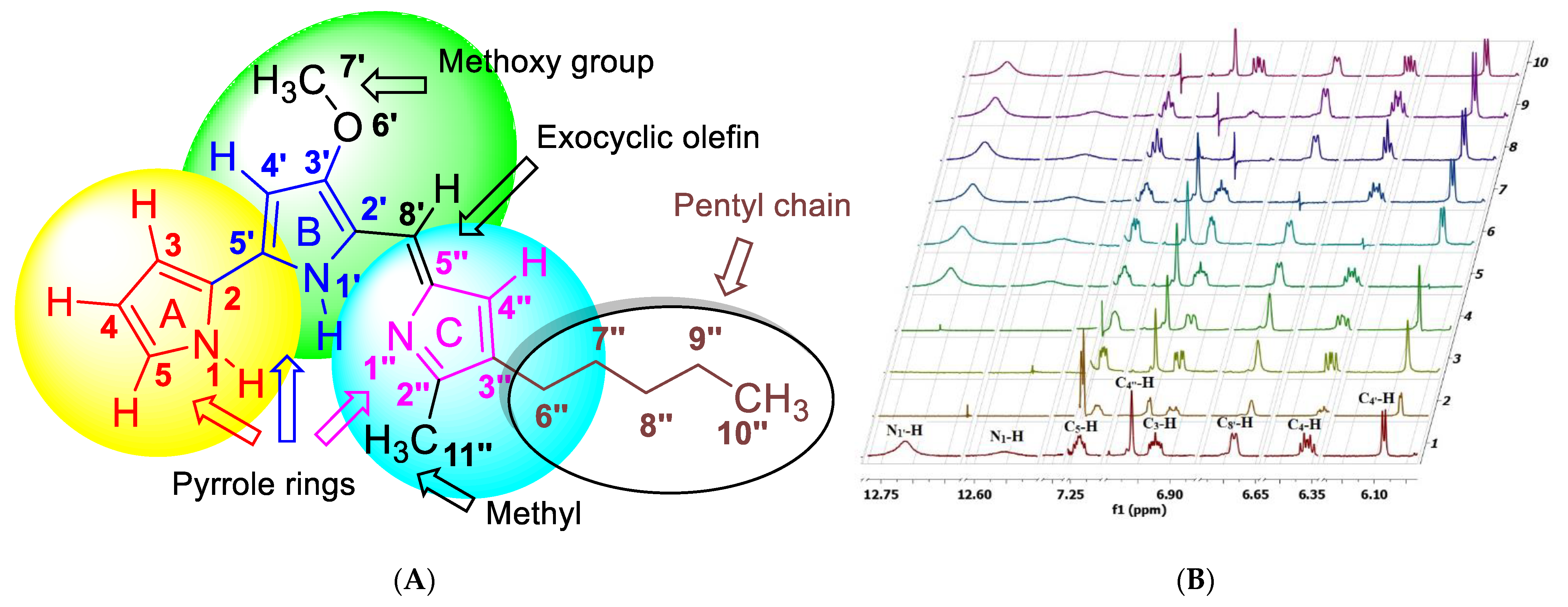
2.2. 1D NMR Analysis of Prodigiosin

An examination of the 13C-CRisis-Attached Proton Test (13C-CRAPT) NMR confirmed the presence of the expected 20 carbon signals: five aromatic CHs, one olefinic CH, seven aromatic sp2 hybridized quaternary carbons, one methoxy, two methyls, and four methylene carbons, which is consistent with all carbon atoms being magnetically nonequivalent (Table 1, Figures S3 and S4). The 13C-CRAPT spectrum showed seven signals in the aliphatic region, thus, confirming the presence of the four pentyl chain methylenes as indicated by the positive phases of the corresponding signals for C6’’-C9’’ (δ 31.4, 29.8, 25.3, 22.5 ppm). In the same region, three signals with negative phases supported the presence of a methoxy group (C7’: δ 58.7 ppm) and two methyl carbons C10’’ and C11’’ (δ 14.3 & 12.5 ppm). The methoxy group was readily assigned given its distinctive downfield chemical shift at δ 58.7 ppm. The integration of the 1HNMR spectrum confirmed the presence of 25 protons: six aromatic and olefinic Hs, two NHs, and 17 aliphatic protons (Table 1, Figure S5, Supplementary Materials). The peaks obtained for the 13C NMR analyses (101 MHz, deuterated chloroform (CDCl3) and 1H NMR (400 MHz, CDCl3) are summarized in Table 1.

2.3. 2D NMR Analysis of Prodigiosin

To fully assign the 1H NMR resonances of the two methyl groups on the aliphatic pentyl chain (C10’’) and pyrrole C ring (C11’’) and correlate their chemical shifts to the respective attached carbons, we resorted to 2D NMR by employing several techniques. Based on the 1H-13C-gHSQC 2D spectrum, the C10’’H3 methyl (δ 0.89), which appears as a triplet in the 1H-NMR, was correlated to the carbon at δ 14.3 ppm (off-diagonal cross peak at δ 0.89/14.3 ppm), while the C11’’H3 methyl singlet in the 1H-NMR matched the carbon at δ 12.5 ppm (off-diagonal cross peak at δ 2.54/12.5 ppm) (Figure S6, Supplementary Materials). The pentyl methylenes were identified in the 1H-NMR based on their multiplicity pattern and were correlated using 1H-1H-gDQFCOSY. They were matched with the corresponding carbon signals using 1H-13C-gHSQC. The distinctive and only expected methylene triplet of C6’’H2 at δ 2.39 ppm (t, 2H, J = 7.6 Hz) triggered the assignment of the remaining CH2′s (C7’’H2- C9’’H2) using 1H-1H-gDQFCOSY (Figure S7A–D, Supplementary Materials). Thus, a strong contour between C6’’H2 and the adjacent C7’’H2 (δ 2.39/1.53 ppm) confirmed their vicinal relationship and is further supported by the anticipated quintet appearance of C7’’H2 (quint, 2H, J = 7.6 Hz) and matching scalar couplings. The latter is further correlated to C8’’H2 and C9’’H2, which appear as a broad complex multiplet with a chemical shift range between δ 1.37–1.21 ppm. This multiplet clearly correlates to the terminal pentyl methyl C10’’H3 with a strong cross peak (δ 1.37–1.21/0.89 ppm). Further, 1H-1H-gDQFCOSY correlated the right-hand side of the complex signal (δ 1.53/1.29–1.21 ppm) with C7’’H2, confirming the chemical shift of C8’’H2 as the latter, which was also correlated through 1H-13C-gHSQC to C8’’ (δ 1.29–1.21/31.4 ppm). On the other hand, the left-hand side portion of the broad signal at δ 1.37–1.21 ppm (δ 1.37–1.29 ppm) was assigned to C9’’H2 and correlated, via 1H-13C-gHSQC, to C9’’ (δ 1.37–1.29/22.5 ppm). With these assignments in hand, matching the methylene protons C6’’H2-C9’’H2 to the corresponding carbon atoms using 1H-13C-gHSQC and 1H-1H-gDQFCOSY was straightforward (C6’’H2: δ 25.3; C7’’H2: δ 29.8; C8’’H2: δ 31.4; C9’’H2: δ 22.5) (Figures S6 and S7A–D).
The aromatic CHs of ring A (C3H-C5H) were clearly distinguished as one spin system in the 1H-1H-gDQFCOSY spectrum, which shows two contours correlating C4H with both, C3H (δ 6.92/6.36 ppm) and C5H (δ 7.23/6.36 ppm) (Figure S7A–D). Although 1H-1H-gDQFCOSY correlations confirmed the chemical shift of C4H, distinguishing between C3H and C5H was more challenging. In principle, 1H-1H-ROESY correlation between N1-H and C5H could identify the chemical shift of the latter. Unfortunately, 1H-1H-ROESY (Figure S8, Supplementary Materials) displayed very weak signals due to low sample concentration and could not be used for data analysis. The suggested assignment of C3H and C5H is supported by considering the negative inductive effect (-I) of N1 as well as its negative mesomeric effect. Based on the latter phenomenon, C5 (directly bonded to the more electronegative N atom compared to C3, which is connected to the C atom) is expected to resonate much further downfield in the 13C than C3. Therefore, the 13C signals at δ 117.0 and 127.0 ppm, which were correlated through the 1H-13C-HSQC to the unassigned proton chemical shifts at δ 6.92 and 7.23 ppm, respectively, suggest that the cross-peak at δ 6.92/117 ppm stems from C3H and the contour at (δ 7.23/127 ppm) belongs to the more shifted C5H. Further strong evidence supporting this assignment is based on the negative mesomeric effect of N1, which delocalizes electrons to C3, causing a significant 13C upfield chemical shift of C3 (δ 117.0 ppm). Notably, the C3H is also shifted upfield relative to C5H. Such high upfield shifts are well documented for carbons and protons in the ortho position of aromatic alcohols, alkoxy, and amine compounds where 13C chemical shifts resonate typically around δ 116 ppm.
The unexpected observation of two strong contours in the 1H-1H-gDQFCOSY between C3H (δ 6.92)/C5H (δ 7.23) and the chemical shift of a broad signal at δ 12.56 ppm confirmed the latter absorption as the N1-H of ring A. Most intriguing was the contribution of N1’-H (δ 12.71 ppm) of the pyrrole ring B, which led to the most challenging task of assigning the chemical shifts of C4’H, C8’H, and C4’’H.
The presence of two strong cross-peaks due to long-range coupling (4JN’H-CH) in the 1H-1H-gDQFCOSY between N1’-H and the two doublets at δ 6.68 (J = 2.4 Hz) and 6.08 (J = 2.0 Hz) ppm immediately established that these chemical shifts are related to C4’H and C8’H. Hence, the only remaining and unassigned signal at δ 6.96 must belong to the C4’’H singlet, which is clearly an isolated uncoupled spin system. Interestingly, the C4’’H showed 1H-13C heteronuclear multiple-bond correlation (3JH-C-13) in the 1H-13C-HMBCAD spectrum with C8’ (δ 128.4 ppm), which in turn was traced to δ 6.68 using gHSQC and assigned to C8’H, indicating that C4’H resonates at δ 6.08 (13C: δ 92.8 ppm) (Figure S9A–E, Supplementary Materials). The methoxy group is well known to cause an extreme upfield shift of the 13C and 1H chemical shifts of ortho carbons and protons, as observed with C4’H, thereby lending further support to the suggested assignment. The proton chemical shifts in the skeletal structure of prodigiosin were subsequently traced to the attached carbons using gHSQC, as summarized in Table 1.
The quaternary carbons of ring C, C2’’, C3’’, and C5’’ were assigned δ 128.5, 147.1, and 125.2 ppm, respectively, based on the strong 1H-13C-HMBC correlation of C11’’-H3 with C3’’ (3JCH: δ 12.5/147.1 ppm), C6’’-H2/ C4’’-H with C2’’ (3JCH: δ 6.96 & 2.39/128.5 ppm), and C8’-H with C5’’ (2JCH: δ 6.68/125.2 ppm). On the other hand, the quaternary carbons of ring B, C2’, C3’, and C5’ and were assigned δ 120.7, 165.7, and 147.7 ppm, respectively, based on strong 1H-13C-HMBC correlation of C4’-H with C2’ (3JCH: δ 6.08/120.7 ppm) and C5’ (2JCH: δ 6.08/147.7 ppm), and C7’-H3 with C3’ (3JCH: δ 4.01/165.7 ppm). Notably, the 3JCH correlation contour between C4’-H and C2’ (3JCH: δ 6.08/120.7 ppm) was much stronger compared to the 2JCH cross-peak observed between C4’-H and C5’ (2JCH: δ 6.08/147.7 ppm), which is typical in HMBC spectra. Further evidence to support such an assignment is based on the phenomenon of negative mesomeric effect of the methoxy substituent, which severely shifts carbon and proton signals upfield. Such high upfield shifts are well documented for carbons and protons in the ortho position of aromatic alkoxides where 13C chemical shifts typically appear around δ 116 ppm. In this case, C2′ resonates at 120.7 ppm. Furthermore, the same double mesomeric effect of the methoxy substituent and nitrogen atom on C4′ caused even a more severe upfield shift of C4′ to 92.8 ppm. Another convincing factor is that C5′ is directly connected to a more electronegative atom (N1′-C5′), causing extreme downfield shift of the C5′ to 147.7 ppm. Finally, the remaining unassigned chemical shift at δ 122.3 was attributed to C2 of Ring A.

2.4. 1H-1H-Homonuclear Decoupling Experiments

The 1H-1H-gDQFCOSY cross-peaks were insufficient to totally correlate the C3-H-C5-H spin system and unambiguously match the 1HNMR chemical shifts of ring A to the C3-H-C5-H protons because of the weak correlation of C5-H with C4-H in the gDQFCOSY, and the close overlap between the ddd signals for each of the ring A protons. As some of the key coupling constants required for unequivocal assignments were still unresolved, homonuclear decoupling experiments were carried out to match the chemical shifts of ring A protons with their respective positions on the aromatic ring, calculate the J-values, and provide further evidence for the remaining assignments (Figure 1B and Figure S10A–K).
The original, full spectrum is shown in Figure 1B as spectrum 1. Starting with ring A protons, irradiation of C5-H (Figure 1B spectrum 10), C4-H (Figure 1B, spectrum 6), C3-H (Figure 1B, spectrum 8), and N1-H (Figure 1B, spectrum 3) resulted in C4-H and C3-H collapsing into a dd (Figure 1B, spectrum 10), C3-H and C5-H collapsing into a dd (Figure 1B, spectrum 6), C4-H and C5-H collapsing into a dd (Figure 1B, spectrum 8), and C3-H, C4-H, and C5-H collapsing into a dd (Figure 1B, spectrum 3), respectively. Even more intriguing was the collapse of C4’-H and C8’-H into singlets (Figure 1B, spectrum 4) when N1’-H was decoupled, proving their proximity and they comprise an independent long-range coupled spin system.
As expected, decoupling of C4’-H (Figure 1B, spectrum 5) and C8’-H (Figure 1B, spectrum 7) had no impact. The preceding experiments led to the unambiguous assignment of the chemical shifts for ring A and B protons through their scalar coupling constants and splitting pattern. Based on spectrum 10, the long-range coupling constant 4J (N1-H-C3-H) and 4J (N1-H-C4-H) is 2.4 Hz and 3J (C3-H-C4-H) is 4.0 Hz. From spectrum 3, 3J (C3-H-C4-H) is 4.0 Hz, 3J (C4-H-C5-H) is 2.4 Hz, and 4J (C3-H-C5-H) is 1.6 Hz. According to spectrum 6, 3J (N1-H-C5-H) is 1.2 Hz. Thus, the total J-coupling values are δ 6.92 (ddd, J = 4.0, 2.4, 1.2 Hz), 6.36 (ddd, J = 4.0, 2.4, 2.4 Hz), and 7.23 (ddd, J = 2.4, 1.6, 1.2 Hz). The J values support the earlier assignment of ring A-C proton positions and confirm the presence of 3J and long-range couplings of N1-H and N1’-H, which were key to full and successful structural elucidation. As expected, the decoupling of the C4’’-H singlet (Figure 1B, spectrum 9) had no impact on the spectrum compared to the uncoupled one (Figure 1B, spectrum 1).

3. Discussion

In this study, we show that an acetone-based extraction method for prodigiosin from a culture of V. gazogenes PB1 was adequate for downstream NMR analysis, which is often a critical step in validating the structure of secondary metabolites [22]. NMR was employed for the following reasons: (i) it is well suited for target chemicals that are not commercially available or costly to source [23], (ii) it does not require derivatization, nor extensive sample separation, [23], and (iii) it does not depend on an elaborate instrument set-up [23].
The miscible solvents (acetone, ethanol and methanol) were found to be the most effective solvents for prodigiosin extraction in accordance with an earlier study [21]. Of the three, acetone is ideal given its faster drying time. Surprisingly, the sole use of the immiscible solvents, chloroform, and dichloromethane, resulted in the poorest extraction even though these solvents possess a hydrophobic nature like that of prodigiosin. The ‘like-for-like’ approach for extraction clearly does not hold well in this case. This seemingly peculiar result is most likely attributed to the physiology of bacterial membranes. Bacteria naturally secrete a hydrophilic layer of polysaccharides over their membrane surface, known as the capsule [24]. This can serve a number of functions, including cell-to-cell attachment, cell-to-cell communication and even evading the immune response of its host. Immiscible solvents such as chloroform will be unable to effectively strip away this hydrophilic layer due to the lack of hydrogen bonding. Much of the prodigiosin will most probably remain trapped within the hydrophobic portion of the bacterial membrane. Miscible solvents such as acetone will likely be more effective in dismantling the capsule due to their excellent capacity to form hydrogen bonds, whilst also solubilizing prodigiosin, via hydrophobic interaction [25].
Several publications have reported on the isolation of prodigiosin and have summarized the proton and carbon NMR data [26,27,28,29]. However, none have provided clear 2D NMR evidence to assign and match all chemical shifts to the respective H and C nuclei. While some have only reported 1H NMR data [27], others have provided fully assigned NMR data either without the use of 2D NMR techniques [29,30], or used insufficient 2D NMR data (COSY and HMQC only) [26]. In certain cases [26,30], a numbered structure was not provided in a figure to cross-reference the experimental 1H and 13C data provided by the authors with the respective H and C nuclei of prodigiosin. In all cases, 2D NMR data was not made available as supporting information. The lack of adequate evidence from the available NMR analyses performed in earlier studies led to the trivial assignment of certain 1H and 13C chemical shifts. For example, the methylenes of the pentyl chain, methines of ring A, or some quaternary carbons were misassigned in earlier studies. To remove any uncertainty, herein we provide the full 2D NMR data (see supporting information section) and make convincing and complete H and C assignments of prodigiosin.
Here, we provide for the first time NMR data, based on 1D (1H and 13C CRAPT NMR), 2D NMR (1H-1H-gDQFCOSY, 1H-13C-gHSQC, and 1H-13C-gHMBC, and 1H-1H-homonuclear decoupling experiments, for the structural validation of prodigiosin isolated from a culture of V. gazogenes PB1. Our data is in agreement with several publications that have reported on the NMR characterization of prodigiosin in different deuterated solvents; inaccurate and erroneous assignments for prodigiosin were corrected [18,26,27,28,29]. Moreover, in contrast to previous work [26,27,28], structural assignments in this study were corroborated by 2D NMR and homonuclear decoupling experiments.

4. Materials and Methods

4.1. Extraction of Prodigiosin from V. gazogenes PB1

V. gazogenes PB1, obtained from the Korean Collection for Type Cultures (KCTC: 12695), was used as the bacterial source for the prodigiosin metabolite. The bacterium can also be sourced from other culture collections, as described previously by Vijay et al. [15]. For prodigiosin production, the bacterium was cultured in 20 mL minimal media supplemented with 2% (v/v) glucose and 3% (w/v) NaCl over two days in 100 mL conical flasks.
To evaluate the drying times of different organic solvents or combinations thereof for prodigiosin extraction, a 1 mL volume of bacterial culture was microfuged at 5000 g for 1 min at room temperature. After the removal of the supernatant, a 0.5 mL volume of the organic solvent was added to the pelleted cell. Cells were resuspended in the solvent by repeated pipetting, and vortexed vigorously for 20 s. All samples were microfuged at 15,000g for 5 min to spin down the cell debris. The red-coloured prodigiosin pigment present within the supernatant was quantified. A 400 µL aliquot of the supernatant was transferred to a 1.5 mL microtube and the samples were air-dried at 40 °C to determine which solvents offered the fastest drying times.
For NMR analysis, the prodigiosin was extracted from V. gazogenes PB1 by resuspending V. gazogenes PB1 cells, pelleted from a 20 mL bacterial culture in 10 mL acetone and vortexing vigorously for 30 s. After centrifugation (15,000g for 5 min), the supernatant, containing the prodigiosin compound, was transferred to a 10 cm glass dish. Acetone was evaporated off overnight (~18 h) at room temperature (21 °C).

4.2. Quantification of Prodigiosin

The levels of extracted prodigiosin were determined by absorption spectrophotometry. A 20 µL sample containing the extracted prodigiosin was added to acidified ethanol (180 µL) (1 M HCl dissolved in absolute ethanol) in a microplate well, in accordance with a previous study [18]. Absorbance was recorded at 535 nm. The amount of prodigiosin extracted for each solvent was expressed as a percentage, relative to the amount of prodigiosin extracted using acetone. This was calculated as follows: (absorbance values obtained for each solvent/absorbance values obtained with acetone) × 100. The absolute amount of prodigiosin extracted from V. gazogenes PB1 cell cultures was determined using the extinction coefficient of 139,800 M−1cm−1, as established in an earlier study [18]. Based on this extinction coefficient, a 0.5 to 2 mg sample was typically used for NMR analysis.

4.3. 1H and 13C NMR Characterisation of Prodigiosin

The dried prodigiosin extract (0.5–2 mg) was dissolved in 0.6 mL of chloroform-d (CDCl3), transferred to a 5 mm (OD) NMR tube and analyzed by 1H and 13C NMR at 298 K for structure verification. 1H and 13C NMR spectra were recorded on a Varian 400 spectrometer (9.4 T), observing 1H and 13C at 400 and 101 MHz, respectively, using tetramethylsilane (TMS) as the internal standard. The NMR chemical shifts (δ) are reported in parts per million (ppm) relative to the residual solvent peak (1H-NMR δ 7.26 for CDCl3; 13C-NMR δ 77.0 for CDCl3). The following abbreviations were used to explain NMR peak multiplicities: br s = broad signal, s = singlet, d = doublet, t = triplet, q = quartet, p = pentet, sept = septet, app = apparent, and m = multiplet.

5. Conclusions

In summary, this study describes the extraction of prodigiosin from a bacterial culture of V. gazogenes PB1, followed by its structural validation using 1D and 2D NMR techniques. The findings will be particularly relevant for experimental work relating to the use of V. gazogenes PB1 as a host for the production of prodigiosin.

Supplementary Materials

The following are available online at https://0-www-mdpi-com.brum.beds.ac.uk/article/10.3390/molecules27186030/s1, Figure S1A–1B: Extraction of prodigiosin, Figure S2: Absorption spectrum of acetone-extracted prodigiosin, Figure S3: 13C CRAPT NMR (CDCl3) spectrum of prodigiosin, Figure S4: 13C CRAPT NMR (CDCl3) spectrum of prodigiosin (processed with resolution booster and auto-cut spectrum), Figure S5: 1H NMR (CDCl3) spectrum of prodigiosin, Figure S6: 1H-13C-gHSQC NMR (CDCl3) spectrum of prodigiosin, Figure S7A–7D: 1H-1H-gDQFCOSY NMR (CDCl3) spectrum of prodigiosin. Figure S8: 1H-1H-ROESYAD NMR (CDCl3) spectrum of prodigiosin, Figure S9A–9E: 1H-13C-gHMBC NMR (CDCl3) spectrum of prodigiosin, Figure S10A–10K: 1H-1H-Homonuclear decoupled NMR spectrum of (CDCl3) spectrum of prodigiosin.

Author Contributions

M.K.A. conceptualized the study. D.V. carried out the in vivo experiments. D.V. carried out the genomic extraction and purification. D.V. and N.S.A. carried out the extraction and quantification of prodigiosin. Z.M. carried out the NMR analyses. D.V., Z.M., and M.K.A. analyzed and presented the results. M.K.A. and Z.M. drafted the manuscript. M.K.A. and Z.M. edited the final manuscript. All authors have read and agreed to the published version of the manuscript.

Funding

This research was funded by the UAEU Program for Advanced Research Funds awarded to M.K.A. (grant code: G00003439; fund code:12S011), start-up fund awarded to Z.M. (grant code: 00003291; fund code: 31S401) and the ‘SDG research program’ awarded to M.K.A. The APC was funded by United Arab Emirates University.

Institutional Review Board Statement

Not applicable.

Informed Consent Statement

Not applicable.

Data Availability Statement

Data is contained within the article or supplementary material.

Conflicts of Interest

The authors declare no conflict of interest.

Sample Availability

Samples of the compounds are available from the authors.

References

  1. Darshan, N.; Manonmani, H.K. Prodigiosin and its potential applications. J. Food Sci. Technol. 2015, 52, 5393–5407. [Google Scholar] [CrossRef] [PubMed]
  2. Perez-Tomas, R.; Vinas, M. New insights on the antitumoral properties of prodiginines. Curr. Med. Chem. 2010, 17, 2222–2231. [Google Scholar] [CrossRef]
  3. Lapenda, J.C.; Silva, P.A.; Vicalvi, M.C.; Sena, K.X.F.R.; Nascimento, S.C. Antimicrobial activity of prodigiosin isolated from Serratia marcescens UFPEDA 398. World J. Microbiol. Biotechnol. 2015, 31, 399–406. [Google Scholar] [CrossRef] [PubMed]
  4. John Jimtha, C.; Jishma, P.; Sreelekha, S.; Chithra, S.; Radhakrishnan, E. Antifungal properties of prodigiosin producing rhizospheric Serratia sp. Rhizosphere 2017, 3, 105–108. [Google Scholar] [CrossRef]
  5. Rapoport, H.; Holden, K.G. The synthesis of prodigiosin. J. Am. Chem. Soc. 1960, 82, 5510–5511. [Google Scholar] [CrossRef]
  6. Wasserman, H.H.; McKeon, J.E.; Santer, U.V. Studies related to the biosynthesis of prodigiosin in Serratia marcescens. Biochem. Biophys. Res. Commun. 1960, 3, 146–149. [Google Scholar] [CrossRef]
  7. Kim, D.; Lee, J.S.; Park, Y.K.; Kim, J.F.; Jeong, H.; Oh, T.K.; Kim, B.S.; Lee, C.H. Biosynthesis of antibiotic prodiginines in the marine bacterium Hahella chejuensis KCTC 2396. J. Appl. Microbiol. 2007, 102, 937–944. [Google Scholar] [CrossRef]
  8. Setiyono, E.; Adhiwibawa, M.A.S.; Indrawati, R.; Prihastyanti, M.N.U.; Shioi, Y.; Brotosudarmo, T.H.P. An Indonesian marine macterium, Pseudoalteromonas rubra, produces antimicrobial prodiginine pigments. ACS Omega 2020, 5, 4626–4635. [Google Scholar] [CrossRef]
  9. Morrison, D.A. Prodigiosin synthesis in mutants of Serratia marcesens. J. Bacteriol. 1966, 91, 1599–1604. [Google Scholar] [CrossRef]
  10. Williams, R.P. Biosynthesis of prodigiosin, a secondary metabolite of Serratia marcescens. Appl. Microbiol. 1973, 25, 396–402. [Google Scholar] [CrossRef]
  11. Gerber, N.N. Prodigiosin-like pigments. Crit. Rev. Microbiol. 1975, 3, 469–485. [Google Scholar] [CrossRef] [PubMed]
  12. Woodhams, D.C.; LaBumbard, B.C.; Barnhart, K.L.; Becker, M.H.; Bletz, M.C.; Escobar, L.A.; Flechas, S.V.; Forman, M.E.; Iannetta, A.A.; Joyce, M.D.; et al. Prodigiosin, violacein, and volatile organic compounds produced by widespread cutaneous bacteria of amphibians can inhibit two batrachochytrium fungal pathogens. Microb. Ecol. 2018, 75, 1049–1062. [Google Scholar] [CrossRef] [PubMed]
  13. Wang, S.L.; Wang, S.L.; Nguyen, V.B.; Doan, C.T.; Doan, C.T.; Tran, T.N.; Nguyen, M.T.; Nguyen, A.D. Production and potential applications of bioconversion of chitin and protein-containing fishery byproducts into prodigiosin: A review. Molecules 2020, 25, 2744. [Google Scholar] [CrossRef]
  14. Harwood, C.S. Beneckea gazogenes sp. nov., a red, facultatively anaerobic, marine bacterium. Curr. Microbiol. 1978, 1, 233–238. [Google Scholar] [CrossRef]
  15. Vijay, D.; Baby, B.; Alhayer, M.S.; Vijayan, R.; Akhtar, M.K. native production of prodigiosin in the estuarine bacterium, Vibrio gazogenes PB1, and identification of the associated pig genes. Front. Mar. Sci. 2022, 9, 940888. [Google Scholar] [CrossRef]
  16. Skinner, K.A.; Leathers, T.D. Bacterial contaminants of fuel ethanol production. J. Ind. Microbiol. Biotechnol. 2004, 31, 401–408. [Google Scholar] [CrossRef]
  17. Hejazi, A.; Falkiner, F.R.  Serratia marcescens. J. Med. Microbiol. 1997, 46, 903–912. [Google Scholar] [CrossRef]
  18. Domröse, A.; Klein, A.S.; Hage-Hülsmann, J.; Thies, S.; Svensson, V.; Classen, T.; Pietruszka, J.; Jaeger, K.E.; Drepper, T.; Loeschcke, A. Efficient recombinant production of prodigiosin in Pseudomonas putida. Front. Microbiol. 2015, 6, 972. [Google Scholar] [CrossRef]
  19. Umeyama, T.; Tanabe, Y.; Aigle, B.D.; Horinouchi, S. Expression of the Streptomyces coelicolor A3(2) ptpA gene encoding a phosphotyrosine protein phosphatase leads to overproduction of secondary metabolites in S. Zividans. Microbiol. Lett. FEMS Microbiol. Lett. 1996, 144, 177–184. [Google Scholar] [CrossRef]
  20. Silverstein, R.M.; Webster, F.X.; Kiemle, D.J.; Bryce, D.L. Spectrometric Identification of Organic Compounds, 8th ed.; John Wiley & Sons, Inc.: Hoboken, NJ, USA, 2014; pp. 1–464. [Google Scholar]
  21. Park, H.-Y.; Kim, T.-K.; Han, S.-J.; Yim, J.-H. Enhancement of the stability and solubility of prodigiosin using β-cyclodextrin in seawater. KSBB J. 2012, 27, 109–113. [Google Scholar] [CrossRef] [Green Version]
  22. Thiele, H.; McLeod, G.; Niemitz, M.; Kühn, T. Structure verification of small molecules using mass spectrometry and NMR spectroscopy. Mon. Chem. 2011, 142, 717–730. [Google Scholar] [CrossRef]
  23. Emwas, A.H.M. The strengths and weaknesses of NMR spectroscopy and mass spectrometry with particular focus on metabolomics research. Methods Mol. Biol. 2015, 1277, 161–193. [Google Scholar] [CrossRef] [PubMed]
  24. Willis, L.M.; Whitfield, C. Structure, biosynthesis, and function of bacterial capsular polysaccharides synthesized by ABC transporter-dependent pathways. Carbohydr. Res. 2013, 378, 35–44. [Google Scholar] [CrossRef]
  25. Bayer, M.E.; Thurow, H. Polysaccharide capsule of Escherichia coli: Microscope study of its size, structure, and sites of synthesis. J. Bacteriol. 1977, 130, 911–936. [Google Scholar] [CrossRef] [PubMed]
  26. Kimyon, Ö.; Das, T.; Ibugo, A.I.; Kutty, S.K.; Ho, K.K.; Tebben, J.; Kumar, N.; Manefield, M. Serratia secondary metabolite prodigiosin inhibits Pseudomonas aeruginosa biofilm development by producing reactive oxygen species that damage biological molecules. Front. Microbiol. 2016, 7, 972. [Google Scholar] [CrossRef]
  27. Lin, P.B.; Shen, J.; Ou, P.Y.; Liu, L.Y.; Chen, Z.Y.; Chu, F.J.; Wang, J.; Jin, X.B. Prodigiosin isolated from Serratia marcescens in the Periplaneta americana gut and its apoptosis-inducing activity in HeLa cells. Oncol. Rep. 2019, 41, 3377–3385. [Google Scholar] [CrossRef]
  28. Song, M.J.; Bae, J.; Lee, D.S.; Kim, C.H.; Kim, J.S.; Kim, S.W.; Hong, S.I. Purification and characterization of prodigiosin produced by integrated bioreactor from Serratia sp. KH-95. J. Biosci. Bioeng. 2006, 101, 157–161. [Google Scholar] [CrossRef]
  29. Alihosseini, F.; Lango, J.; Ju, K.S.; Hammock, B.D.; Sun, G. Mutation of bacterium Vibrio gazogenes for selective preparation of colorants. Biotechnol. Prog. 2010, 26, 352–360. [Google Scholar] [CrossRef]
  30. Boger, D.L.; Patel, M. Total synthesis of prodigiosin, prodigiosene, and desmethoxyprodigiosin: Diels-Alder reactions of heterocyclic azadienes and development of an effective palladium(II)-promoted 2,2’-bipyrrole coupling procedure. J. Org. Chem. 1988, 53, 1405–1415. [Google Scholar] [CrossRef]
Figure 1. NMR analysis of prodigiosin. (A) Chemical structure of prodigiosin indicating the numbers and positions of the individual atoms. (B) 1H-1H-Homonuclear decoupling experiments of prodigiosin. (1) Non-decoupled spectrum; (2) multifrequency decoupling of N1-H and N1’-H; (3) irradiation of N1-H; (4) irradiation of N1’-H; (5) irradiation of C4’-H; (6) irradiation of C4-H; (7) irradiation of C8’-H; (8) irradiation of C3-H; (9) irradiation of C4’’-H; (10) irradiation of C5-H.
Molecules 27 06030 g001
Table 1. 1H- and 13C-NMR data for prodigiosin.
1H-NMRProton NumberProdigiosin Protons ppmProton NumberProdigiosin Protons ppm
112.56 (s, 1H, NH)8′6.68 (d, 1H, J = 2.4 Hz, CH)
36.92 (ddd, J = 4.0, 2.4, 1.2 Hz)4′′6.96 (s, 1H, CH)
46.36 (ddd, J = 4.0, 2.4, 2.4 Hz)6′′2.39 (t, 2H, J = 7.6 Hz)
57.23 (ddd, J = 2.4, 1.6, 1.2 Hz)7′′1.53 (quint, 2H, J= 7.6 Hz)
1′12.71 (s, 1H, NH)8′′ & 9′′1.37–1.21 (m, 4H)
4′6.08 (d, 1H, J = 2.0 Hz, CH)10′′0.89 (s, 3H, J = 7.6 Hz, Me)
7′4.01 (s, 3H, OMe)11′′2.54 (s, 3H, Me)
13C-NMRCarbon NumberProdigiosin Carbons ppmCarbon NumberProdigiosin Carbons ppm
2122.32′′128.5
3117.03′′147.1
4111.74′′116.0
5127.05′′125.2
2′120.76′′25.3
3′165.87′′29.8
4′92.88′′31.4
5′147.79′′22.5
7′58.710′′14.3
8′128.411′′12.5
Publisher’s Note: MDPI stays neutral with regard to jurisdictional claims in published maps and institutional affiliations.

