Article

Anti-Obesity Effects of a Mixture of Atractylodes macrocephala and Amomum villosum Extracts on 3T3-L1 Adipocytes and High-Fat Diet-Induced Obesity in Mice

Research Institute, NUON Co., Ltd., Jungwon-gu, Seongnam 13201, Korea
*
Author to whom correspondence should be addressed.
Submission received: 28 December 2021 / Revised: 26 January 2022 / Accepted: 26 January 2022 / Published: 28 January 2022
(This article belongs to the Special Issue Functional Food and Their Pharmaceutical Properties)

Abstract

:
Since the potential of (3:1) mixtures of Atractylodes macrocephala and Amomum villosum extracts has been proposed in the management of obesity, the purpose of present study was to investigate the effects of AME:AVE (3:1) mixture on weight loss, obesity-related biochemical parameters, adipogenesis and lipogenesis related proteins in 3T3-L1 cells and HFD-induced obesity in a mouse model. Treatment with AME:AVE (3:1) mixture inhibited lipid accumulation. Furthermore, the treatment with 75 and 150 mg/kg of AME:AVE (3:1) significantly decreased the body weight gain, white adipose tissue (WAT) weight, and plasma glucose level in HFD-induced obese mice. Moreover, treatment with 75 and 150 mg/kg AME:AVE (3:1) also significantly lowered the size of adipocytes in adipose tissue and reduced the lipid accumulation in liver. AME:AVE (3:1) treatment significantly decreased the expression of proteins related to adipogenesis and lipogenesis in 3T3-L1 adipocytes and WAT of HFD-induced obese mice. These results suggest that the AME:AVE herbal mixture (3:1) has anti-obesity effects, which may be elicited by regulating the expression of adipogenesis and lipogenesis-related proteins in adipocytes and WAT in HFD-induced obesity in mice.

1. Introduction

Obesity has become a significant clinical problem in recent decades due to its role in type 2 diabetes, hypertension, hyperlipidemia, and cardiovascular disease [1,2]. It is characterized by increased fat mass, which is associated with increased cell number (hyperplasia) and size (hypertrophy) [3,4]. For this reason, obesity has become a serious public health concern worldwide [5].
Adipocytes are important cells of fatty tissues. Excessive amounts of lipid (triglycerides) can accumulate in adipose tissues accompanied by increased levels of adipogenesis and lipogenesis, which leads to enhanced body weight gain [6]. Obesity and related diseases are strongly linked to adipocyte differentiation and fat accumulation [7]. 3T3-L1 cells are widely used in anti-obesity studies to investigate key molecular markers of adipocyte differentiation [8,9,10].
The different pharmacological approaches to prevent and treat obesity have several limitations such as undesirable and adverse effects [11,12]. Public attention is increasingly focused on using natural products to prevent and treat obesity with various plants such as fruits, vegetables, and herbs, with minimal side effects [13].
Atractylodes macrocephala is a medicinal herb that has been widely used in Chinese traditional medicine for approximately 2000 years [14,15]. It is rich in bioactive compounds such as atractylenolides I, II, and III [16,17] and exhibits various pharmacological properties, including antioxidant [18], anti-inflammatory [19], antimutation [20], and antitumor effects [21].
Amomum villosum Lour. (Zingiberaceae, a legal source of Amomi Fructus) is a classic traditional Chinese herb [22]. Pharmacological studies have shown that Amomi Fructus has strong anti-ulcer, anti-diarrhea, anti-inflammatory, and antimicrobial activities [23]. Recently, it has been reported that a mixture of Atractylodes macrocephala and Amomum villosum extracts significantly inhibited OP9 adipocyte differentiation [24] and weight gain induced by high-fat diet [25,26]. Especially, Kim et al. [26] reported that the anti-obesity effects mediated by decreased expression of adipogenesis- and lipogenesis-related genes expressions was confirmed in the diet-induced obesity 1 (DIO1) model induced with AME:AVE (3:1). Based on these studies, we hypothesized that AME:AVE (3:1) might also have anti-obesity effects in 3T3-L1 cells and obesity-induced DIO2 models.
Therefore, in this study, the anti-obesity effects and the underlying mechanisms of adipogenesis and lipogenesis in 3T3-L1 cells and HFD-induced obesity in mice model were investigated using a 3:1 mixture of Atractylodes macrocephala and Amomum villosum extracts reported by Kim et al. [24,25,26].

2. Results

2.1. Cytotoxic Effects of the Mixture of Atractylodes macrocephala and Amomum villosum Extracts (3:1) in 3T3-L1 Preadipocytes

The cytotoxic effect of AME:AVE (3:1) on the viability of 3T3-L1 preadipocytes was evaluated using the MTT assay. AME:AVE (3:1) did not exhibit cytotoxicity at any of the concentrations investigated (0.1, 0.2, and 0.5 mg/mL, Figure 1).

2.2. Effects of Mixtures of Atractylodes macrocephala and Amomum villosum Extracts (3:1) on Adipocyte Differentiation in 3T3-L1 Preadipocytes

To investigate the effects of AME:AVE (3:1) mixture on adipocyte differentiation, 3T3-L1 adipocytes were treated with various concentrations of AME:AVE (3:1) (0.1, 0.2, and 0.5 mg/mL). Lipid concentrations in lipid droplets were tested with Oil-red O staining. The results show that mixtures of AME:AVE (3:1) decreased the lipid accumulation in 3T3-L1 adipocytes without cytotoxicity. Lipid accumulation was decreased by 23.57% (p < 0.05) and 67.12% (p < 0.01), respectively, at concentrations of AME:AVE (3:1) 0.2 and 0.5 mg/mL (Figure 2).

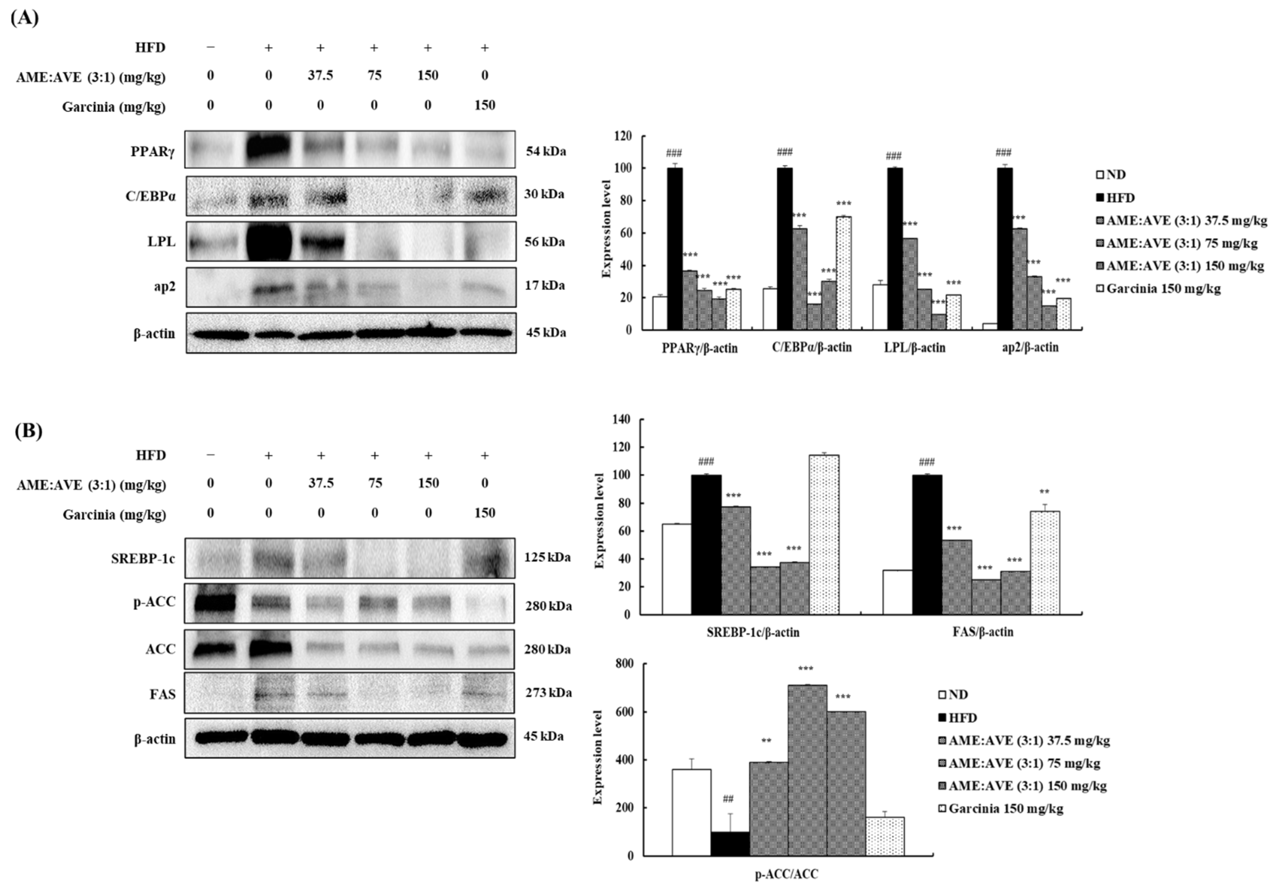
2.3. Effects of Mixtures of Atractylodes macrocephala and Amomum villosum Extracts (3:1) on Proteins in Adipogenesis and Lipogenesis Downstream in 3T3-L1 Adipocytes

Expression of key transcriptional factors involved in adipogenesis, such as PPARγ and C/EBPα, and transcriptional factor target genes, such as LPL and ap2, was also examined by western blotting. The results showed that levels of PPARγ, C/EBPα, LPL, and ap2 protein in MDI-induced 3T3-L1 adipocytes increased significantly compared with the control. However, treatment with mixtures of AME:AVE (3:1) at 0.1, 0.2, and 0.5 mg/mL inhibited the expression of PPARγ by 46.18, 60.35, and 66.73% (p < 0.01) and C/EBPα by 5.02, 19.99, and 64.49% (p < 0.01), respectively, and LPL in the range of 10.70–48.66% (p < 0.01) and ap2 in the range of 26.70–77.54% in a dose-dependent manner (p < 0.01).
To determine the mechanism of inhibition of lipid accumulation by mixtures of AME:AVE (3:1), we analyzed the expression of FAS. AME:AVE (3:1) treatment inhibited the expression of FAS dose-dependently compared with fully differentiated adipocytes where the percentage of inhibition ranged between 17.86% and 62.84% (p < 0.01). In addition, we investigated the expression of SREBP-1c, which is an important factor regulating lipid accumulation in adipocytes mediated via regulation of the expression of FAS, a lipogenic enzyme. The results showed that the mixtures of AME:AVE (3:1) suppressed the protein expression of SREBP-1c dose-dependently with an inhibition range of 12.06–57.30% (p < 0.01). ACC is a rate-limiting enzyme in the fatty acid synthesis pathway mediated via phosphorylation and inactivation. Phosphorylation of ACC was increased significantly (p < 0.01) (Figure 3).

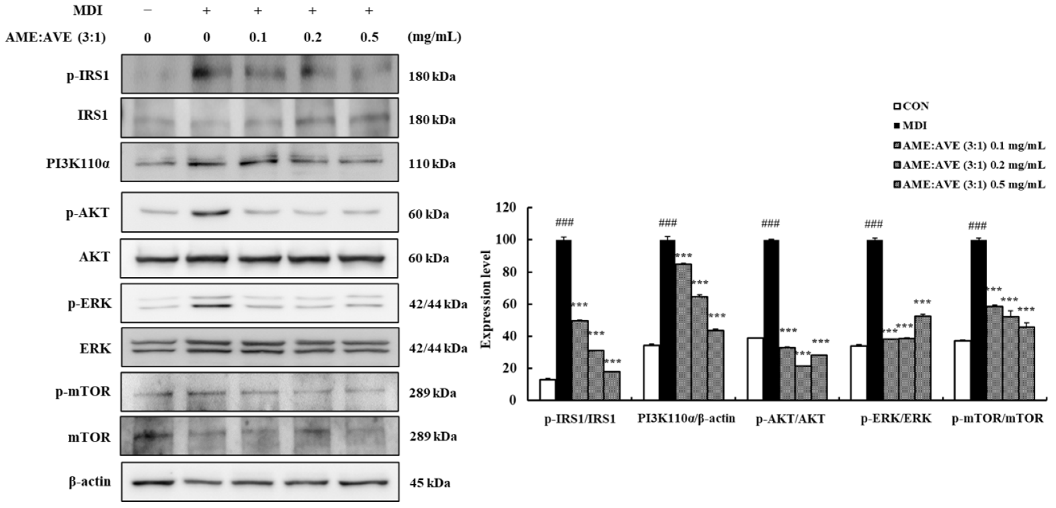
2.4. Effects of Atractylodes macrocephala and Amomum villosum Extracts (3:1) on Upstream Proteins in Adipogenesis and Lipogenesis in 3T3-L1 Adipocytes

To further investigate the mechanism of AME:AVE (3:1) in adipogenesis and lipogenesis, the expression of p-IRS1, PI3K110α, p-AKT, p-ERK, and p-mTOR was measured. MDI-induced 3T3-L1 adipocytes significantly increased the expression of activated p-IRS1, PI3K110α, p-AKT, p-ERK and p-mTOR compared with the control group. However, AME:AVE (3:1) concentrations of 0.1, 0.2, and 0.5 mg/mL markedly decreased the expression of p-IRS1, PI3K, p-AKT, p-ERK, and p-mTOR (p < 0.01) (Figure 4).

2.5. Effects of Atractylodes macrocephala and Amomum villosum Extracts (3:1) on Body and Organ Weights of High-Fat Diet-Induced Obesity in Mice

Mixtures of AME:AVE (3:1) inhibited adipogenesis/lipogenesis in 3T3-L1 cells. Based on these results, we investigated the effect of AME:AVE (3:1) mixture on obesity in HFD-induced obesity in mice. The body weight gain of the HFD group was signifificantly increased when compared with the ND group after 8 weeks of feeding. In contrast, administration of AME:AVE (3:1) mixtures 75 and 150 mg/kg resulted in a significant decrease in body weight gain of 6.00 ± 0.34 g, and 3.96 ± 0.46 g (p < 0.01), respectively. (Figure 5A). Food intake did not display signifificant group wise differences (Figure 5B).
The weight of liver, total fat, and retroperitoneal fat showed a significant decrease (p < 0.05) in the AME:AVE (3:1) 75 and 150 groups compared with the HFD group. The weight of epididymal and perirenal fat increased significantly in the HFD group compared with the ND group, and a tendency toward decrease was detected following exposure to AME:AVE (3:1); however, the epididymal fat showed significance only in the AME:AVE (3:1) 75 group (p < 0.05) (Figure 5C).

2.6. Effects of Mixtures of Atractylodes macrocephala and Amomum villosum Extracts (3:1) on Liver and Epididymal Fat Morphology in High-Fat Diet-Induced Obesity in Mice

Histological observations of liver and epididymal fats were based on 6 groups: ND; HFD; HFD + AME:AVE (3:1) 37.5, 75, and 150; HFD + G 150 groups. Sections of liver and epididymal fats were stained with H&E to analyze the lipid accumulation and the cellular morphology. As a result, it was confirmed that administration of AME:AVE (3:1) decreased hepatic steatosis, in a dose-dependent manner (Figure 6).
The adipocyte size of epididymal fat in the HFD group showed an increase compared with the ND group. In contrast, the adipocyte size in the HFD + AME:AVE (3:1) groups showed a significant decrease compared with the HFD group. In particular, the AME:AVE (3:1) 150 group showed excellent reduction in adipocyte size (Figure 6).

2.7. Effects of Atractylodes macrocephala and Amomum villosum Extracts (3:1) on Plasma Biochemical Parameters in High-Fat Diet-Induced Obesity in Mice

The plasma biochemical parameters, including glucose, T-CHO, TG, LDL-c, HDL-c/LDL-c ratio, ALT, AST, leptin, and adiponectin were confirmed. As shown in Table 1, glucose, T-CHO, TG, LDL-c, ALT, AST, and leptin levels were markedly increased and HDL-c/LDL-c ratio and adiponectin levels were decreased in the HFD group. However, the glucose levels in AME:AVE (3:1) groups were significantly decreased in a dose-dependent manner (p < 0.01). In addition, ALT and AST levels of AME:AVE (3:1) 75 and 150 groups were also significantly decreased (p < 0.05). T-CHO, TG, LDL-c, and leptin levels showed a tendency to decrease in the group treated with AME:AVE (3:1), whereas T-CHO and leptin levels showed significance only in the AME:AVE (3:1) 75 group. The HDL-c/LDL-c ratio and adiponectin levels tended to increase in the group exposed to AME:AVE (3:1).

2.8. Effects of Mixtures of Atractylodes macrocephala and Amomum villosum Extracts (3:1) on Adipogenesis- and Lipogenesis-Related Protein Levels in High-Fat Diet-Induced Obesity in Mice

Western blot analysis was used to confirm the levels of adipogenesis- and lipogenesis-related proteins. HFD group showed a significant increase in PPARγ, C/EBPα, LPL, and ap2 protein expression in epididymal fat. In contrast, treated with AME:AVE (3:1) significantly decreased the expression of PPARγ and C/EBPα PPARγ, C/EBPα, LPL, and ap2 in epididymal fat (p < 0.01) (Figure 7A). As shown in Figure 7B, the levels of SREBP-1c, ACC, and FAS protein were increased, while p-ACC level was decreased in the HFD group. Our results revealed that AME:AVE (3:1) treatment markedly inhibited this increase in epididymal fat and also increased the p-ACC level significantly (p < 0.01) (Figure 7B).

3. Discussion

According to the report of Song et al. [27], the extract of Atractylodes macrocephala significantly reduced the weight gain induced by a high-fat diet, and this effect was related to a reduction in hepatic fat accumulation. In addition, Lu et al. [28] reported that Amomum villosum inhibits increased fatty liver formation mediated via gut-liver axis.
Recently, the treatment with Atractylodes macrocephala and Amomum villosum extract (AME:AVE (3:1)) resulted in excellent anti-obesity effect [25]. The mixture at a ratio of 3:1 significantly inhibited OP9 adipocyte differentiation [24] and weight gain induced by high-fat diet [25,26]. In particular, Kim et al. [26] reported that the anti-obesity effects was confirmed in the DIO1 model administrated with AME:AVE (3:1) while inducing obesity. Based on these studies, the present study confirmed the anti-obesity effects and the underlying mechanisms of adipogenesis and lipogenesis in 3T3-L1 cells. Then, we confirmed the anti-obesity effects in the DIO2 model in which the AME:AVE (3:1) was administered with mice after inducing obesity.
Our results suggested that AME:AVE (3:1) treatment inhibited the differentiation and lipid accumulation of 3T3-L1 adipocytes. This result was further supported by in vivo experiments, showing that AME:AVE (3:1) treatment for 8 weeks significantly reduced the weight gain, and the weight of liver and retroperitoneal fats in HFD-induced obesity in mice. Additionally, AME:AVE (3:1) treatment did not affect the food intake in HFD-induced obesity in mice, indicating that the weight-loss effect of AME:AVE (3:1) was not due to a reduction in food intake. These results are consistent with previous reports suggesting that the mixture of AME:AVE (3:1) has anti-obesity effects.
Obesity is closely related to hyperplasia and hypertrophy of adipose cells [29,30] Adipocyte hypertrophy is caused by excessive accumulation of fat within the cells due to excessive energy intake. Since adipocyte proliferation occurs due to the complex interaction between proliferation and differentiation of preadipocytes [31], the 3T3-L1 preadipocyte MDI-induced differentiation system was utilized in this experiment. We demonstrated that AME:AVE (3:1) treatment suppressed lipid accumulation during 3T3-L1 adipocyte differentiation without cytotoxicity. Also, the treatment of HFD-fed mice with AME:AVE (3:1) resulted in a significant decrease in liver and total fat weight. In addition, histological data showed a decrease in adipocyte size in liver and epididymal fat in AME:AVE (3:1) groups, suggesting effects on lipid accumulation and adipocyte size in adipose tissue, which is likely related to reduced fat weight.
In obesity, fatty liver is induced by the accumulation of fat in the liver tissue [32]. Histological data of the liver showed that AME:AVE (3:1) treatment reduced the size and number of lipid droplets and restored tissue structure, which indicates that AME:AVE (3:1) treatment inhibits lipid accumulation in the liver. In our study, AME:AVE (3:1) treatment lowered the levels of plasma glucose and T-CHO and also tended to decrease TG and LDL-c levels. These results suggest that AME:AVE (3:1) treatment ameliorates hyperlipidemia in HFD-induced obesity in mice by reducing hepatic fat accumulation.
Excessive fat intake triggers oxidative stress, which leads to hepatic dysfunction and fatty degeneration via abnormal activation of ALT and AST [33]. In this study, AME:AVE (3:1) treatment reduced plasma ALT and AST levels in HFD-induced obesity in mice compared with the HFD group, and thereby ameliorated liver dysfunction induced by fat accumulation.
In obesity, the accumulation of visceral fat affects adipokines in adipose tissue including leptin and adiponectin. Leptin promotes energy consumption by inducing satiety in the central circuits of the hypothalamus, and adiponectin increases fat oxidation and inhibits lipid accumulation. It is known that the concentration of leptin increases and the concentration of adiponectin decreases in obesity [34]. Therefore, the concentrations of leptin and adiponectin in the serum were measured to evaluate the effect of exposure to AME:AVE (3:1) on serum adipokines in mice. Leptin showed a significant decrease in AME:AVE (3:1) 75 group and adiponectin levels showed a tendency to increase in the group treated with the herbal extracts.
To investigate the underlying anti-obesity mechanism associated with AME:AVE (3:1), we measured the levels of lipogenesis- and adipogenesis-related proteins in 3T3-L1 adipocytes and epididymal fats in HFD-induced obesity in mice.
Adipogenesis, which is indicated by preadipocyte differentiation into mature adipocytes, is causally linked to obesity. Regulation of adipocyte differentiation genes and proteins controls adipogenesis. Insulin signaling is a key signal underlying adipogenesis regulation mediated via activation of glucose uptake and inhibition of lipolysis in adipocytes [35,36]. IRS-1 belongs to a family of docking molecules that link the activation of IR, which activate downstream kinase cascades including PI3K and AKT [37]. Activation of IRS-1 is mediated via phosphorylation of serine residues and enhanced IRS-1-related PI3K activity [38]. In addition, insulin signaling is intimately linked to the nutrient-responsive mammalian target of rapamycin (mTOR) signaling pathway via activation of AKT [39]. Phosphorylation of IRS-1 and AKT, ERK, and mTOR was enhanced in 3T3-L1 adipocytes but inhibited by AME:AVE (3:1) treatment.
PPARγ is an important factor regulating adipocyte differentiation and adipogenesis. C/EBPα promotes adipocyte differentiation via interaction with PPARγ, which increases the expression of adipocyte-specific genes such as aP2 and LPL [40,41]. SREBP-1c is also a transcription factor that plays an important role in lipid metabolism by increasing the expression of several adipogenic genes, such as FAS, in adipose tissue and liver. PPARγ is activated via regulation of expression and production of endogenous PPARγ ligands [42,43]. Lipoprotein lipase is a major regulator of blood TG levels via breakdown of lipoprotein triglycerides. It plays an important role in transporting fat and hydrolyzed fat transporting lipoproteins [44]. The binding of these transcription factors triggers the expression of target genes and proteins responsible for TG synthesis and storage, leading to accelerated adipogenesis. ACC is a rate-limiting enzyme in the fatty acid synthesis via phosphorylation and inactivation [45]. We found that treatment with AME:AVE (3:1) reduced the expression of PPARγ, C/EBPα, aP2, and FAS proteins in 3T3-L1 adipocytes, and increased ACC phosphorylation. The same factors showed significant results following treatment with AME:AVE (3:1) in the epididymal fat of high-fat diet-induced mice. Our results suggest that AME:AVE (3:1) treatment modulates the insulin signaling pathway and suppresses the expression of adipogenesis and lipogenesis-related proteins, thereby resulting in anti-obesity effects.
In conclusion, our results showed that AME:AVE (3:1) treatment inhibited lipid accumulation in 3T3-L1 adipocytes, and reduced body weight gain, organ weight, adipocyte size, and regulated plasma biochemical levels in the HFD-mice model. In addition, it regulated the proteins related to adipogenesis and lipogenesis in 3T3-L1 adipocytes and HFD-induced obesity in mice, and the insulin regulatory pathway was confirmed in 3T3-L1 adipocytes (Figure 8). These findings indicated that AME:AVE (3:1) prevents the development of obesity and hyperlipidemia in 3T3-L1 adipocytes and HFD-induced obesity in mice, which provide valuable insight into the potential benefits of AME:AVE (3:1) in obesity. Moreover, it is considered that additional clinical studies are needed to suggest the possibility of application in the prevention of obesity and obesity-related diseases in humans.

4. Materials and Methods

4.1. Chemicals and Reagents

3-Isobutyl-1-methylxanthine (IBMX), dexamethasone, and insulin were obtained from Wako Pure Chemical Industries Ltd. (Osaka, Japan). Dulbecco’s modified Eagle’s medium (DMEM), bovine calf serum (BCS), and penicillin/streptomycin were purchased from Gibco BRL (Grand Island, NY, USA). Fetal bovine serum (FBS) was obtained from ATLAS Biologicals (Fort Collins, CO, USA). The following antibodies were ordered from Santa Cruz Biotechnology (Dallas, TX, USA): Peroxisome proliferator-activated receptor γ (PPARγ; #sc-7273), CCAAT/enhancer-binding protein α (C/EBPα; #sc-365318), lipoprotein lipase (LPL; #sc-373759), sterol regulatory element binding proteins (SREBP-1c; #sc-365513). Adipocyte fatty acid-binding protein 2 (ap2; #PA5-30591) was acquired from Invitrogen (Carlsbad, CA, USA). β-actin (#8457), phospho-acetyl-CoA carboxylase (p-ACC; #3661), ACC (#3676), fatty acid synthase (FAS; #3180), phospho-insulin receptor substrate 1 (p-IRS1; #2388), IRS1 (#2382), phosphoinositide 3-kinase 110α (PI3K110α; #4249), phospho-protein kinase B (p-AKT; #4060), AKT (#4691), phospho-extracellular signal-regulated kinase (p-ERK; #4377), ERK (#4695), phospho-mechanistic target of rapamycin (p-mTOR; #2974), mTOR (#2972) were obtained from Cell Signaling Technology (Danvers, MA, USA). Horseradish peroxidase (HRP)-linked anti-rabbit IgG (#SA002-500) and HRP-linked anti-mouse IgG (#SA001-500) were purchased from GenDEPOT (Barker, TX, USA).

4.2. Preparation of AME:AVE (3:1) Extracts

AME:AVE (3:1) is a mixture of the aqueous extracts of two herbs, namely Atractylodes macrocephala and Amomum villosum, in a 3:1 ratio. The AME:AVE (3:1) mixture manufacturing process was as follows: Atractylodes macrocephala and Amomum villosum were extracted using water. The resulting solutions were concentrated and dried to yield AME and AVE, respectively. Then, the mixture it is prepared by mixing AME and AVE in a ratio of 3:1. This manufacturing process was designed and developed by NUON Co., Ltd., (Seongnam, Korea), who manufactures and sells this product. To confirm the composition of AME:AVE (3:1) mixture, we performed high-performance liquid chromatography (HPLC) analysis. Atractylenolide III is a phytochemical marker for Atractylodes macrocephala raw material, while vanillic acid is phytochemical marker for Amomum villosum raw material, respectively. For in vitro studies, AME:AVE (3:1) was dissolved in dimethyl sulfoxide (DMSO) at concentration of 100, 200, and 500 mg/mL, respectively and then diluted in culture medium at concentration of 0.1, 0.2, and 0.5 mg/mL. respectively. Control is 3T3-L1 cells treated with culture medium containing only DMSO (final DMSO concentration 0.1%). For in vivo studies, AME:AVE (3:1) extracts were homogenized in 0.5 mL 0.5% carboxymethyl cellulose sodium salt (CMC-Na) at concentration of 37.5, 75, and 150 mg/kg, respectively and orally administered. The control mice group were given the same volume of 0.5% CMC-Na instead of the test solution.

4.3. Cell Culture

3T3-L1 preadipocytes (American Type Culture Collection, Rockville, MD, USA) cells were cultured in DMEM with 10% bovine calf serum (BCS), 1% penicillin/streptomycin and maintained in a humified atmosphere of 5% CO2 at 37 °C. To induce adipocyte differentiation, fully confluent preadipocytes were treated with MDI (3-isobutyl-1-methylxanthine, dexamethasone, and insulin) differentiation medium (DMEM, 10% fetal bovine serum (FBS), 0.5 mM IBMX, 5 μg/mL insulin, and 1 μM dexamethasone) with or without AME:AVE (3:1) (defined as Day 0). After two days of incubation, the cell medium was replaced by DMEM supplemented with 10% FBS and 5 μg/mL of insulin in the presence or absence of AME:AVE (3:1) (Day 2). After another two days, the medium was changed with DMEM containing 10% FBS (Day 4). 3T3-L1 preadipocytes were completely differentiated to mature adipocytes on Day 6.

4.4. MTT Assay for Cell Viability

3T3-L1 preadipocytes were seeded on 96-well plates and treated with various concentration of AME:AVE (3:1) for 24 h. Cells treated with 3-(4,5-dimethylthiazol-2-yl)-2,5-diphenyl tetrazolium bromide (MTT) solution (5 mg/mL) were incubated at 37 °C for 4 h. After removal of the supernatant, 100 μL of DMSO was added to dissolve formazan crystals, and the MTT-formazan product was measured using a microplate plate reader (Tecan, Mannedorf, Switzerland).

4.5. Oil-Red O Staining

Differentiated 3T3-L1 adipocytes were washed three times with PBS and fixed with 10% (v/v) formalin solution (Sigma-Aldrich, St. Louis, MO, USA) for 1 h. Fresh Oil-red O stock solution was mixed with distilled water in a ratio of 6:4 to obtain the working solution, followed by incubation for 1 h at room temperature (RT), and filtered. After fixation, cells were washed three times with distilled water and then stained with Oil-red O working solution at 25 °C for 2 h. Cells were rinsed three times with distilled water and photographed with an ECLIPSE Ts2 microscope (Nikon Corporation, Tokyo, Japan). The Oil-red O dye was eluted with isopropanol to determine the intracellular lipid content using a microplate reader (Tecan) at 520 nm.

4.6. Animals and Diets

This experiment was carried out in compliance with the Animal Experimental Ethics Regulations of INVIVO Co., Ltd. (Chungnam, Korea) (Approval No. IV-RA-04-2010-36). Experimental animals were acquired from Orient Bio Co., Ltd (Seongnam, Korea). Five-week-old male C57BL/6 mice (n = 48) in specific-pathogen free (SPF) state were used in the experiment after a week of acclimatization. The experimental diet during the acclimatization period included general solid feed (Purina Lab Rodent Chow #38057, Purina Co., Seoul Korea). The mice were housed in a controlled atmosphere (23 ± 1 °C at 50 ± 5% relative humidity) with a 12 h light/dark cycle. Feed and water were provided ad libitum.
After acclimatization, the mice were fed either the normal-fat diet (ND, n = 8) as a normal group or high-fat diet (HFD, n = 40, rodent diet with 60% kcal fat (#D12492, Research diets INC) as the HFD induced obese group for 5 weeks. The obesity animal model when the weight gain was about 20% compared with the normal group, the obesity was established. After obesity induction, the mice with obesity were further divided into 5 experimental groups (n = 8/group) and were matched by body weight. The following groups were studied for 8 weeks: ND group; HFD group; AME:AVE (3:1) 37.5 group (high-fat diet + AME:AVE (3:1) 37.5 mg/kg); AME:AVE (3:1) 75 group (high-fat diet + AME:AVE (3:1) 75 mg/kg); AME:AVE (3:1) 150 group (high-fat diet + AME:AVE (3:1) 150 mg/kg). Another HFD group received oral administration of 150 mg/kg of Garcinia cambogia extract (G 150) daily as a positive control group because of the weight loss effects of Garcinia cambogia extract [46,47,48].
Weight, diet and water intake were recorded weekly. In order to observe the external changes before autopsy, bodies were photographed in each group, and the animals were anesthetized via inhalation anesthesia before blood was drawn. After sacrificing the mice, the tissues were excised. Before tissue excision, photographs were obtained to confirm the accumulation of adipose tissue in the abdomen, and liver, epididymal fats, retroperitoneal fats, and perirenal fats were extracted. The excised tissues were photographed to observe external changes, and the weight of each organ was measured. The tissues were then divided and fixed in 10% neutral formalin, and frozen. Frozen tissues were stored at −80 °C until use.

4.7. Biochemical Analysis

After the blood was coagulated for 30 min, the serum was recovered by centrifugation at 3000 rpm for 10 min. The levels of total cholesterol (T-CHO), triglyceride (TG), glucose, low density lipoprotein cholesterol (LDL-C), high density lipoprotein cholesterol (HDL-C), alanine aminotransferase (ALT), and aspartate aminotransferase (AST) in the recovered serum were analyzed. The contents of leptin and adiponectin in the blood were analyzed using an ELISA kit (leptin, ab199082, Abcam, Cambridge, UK; adiponectin, MRP300, R&D Systems, Wiesbaden, Germany).

4.8. Histological Analysis

The fixed liver and epididymal fats were paraffinized, cut into slices, attached to slides, and analyzed via hematoxylin & eosin staining. The stained slides were photographed under a microscope, and the fat tissue area was measured using the Image J program developed at the National Institutes of Health (Bethesda, MD, USA).

4.9. Protein Extraction and Western Blot Analysis

3T3-L1 adipocytes were lysed in CelLytic buffer (Sigma-Aldrich) and then centrifuged at 12,000 rpm for 20 min at 4 °C. Epididymal fat was washed with cold PBS and RIPA Homogenize buffer containing 50 mM Tris-HCl pH 7.4, 150 mM NaCl, 1 mM EDTA, 1% Triton X-100, 1% sodium deoxycholate, 0.1% SDS, 1 mM PMSE, and 1% protease inhibitor. The homogenate was also centrifuged at 12,000 rpm for 20 min at 4 °C. After the above procedure, both cells and adipose tissue supernatant were collected. Protein concentration was measured via Bradford assay (Bio-Rad Laboratories, Hercules, CA, USA). Proteins were separated via SDS-PAGE and transferred to Immobilon-P membranes (Millipore, Bedford, MA, USA) and then membranes were cut to detect the location of each biomarker specific according to each antibody manufacturer’s datasheet. The transferred membrane was blocked with 5% skim milk dissolved in Tris-buffered saline containing 0.1% Tween-20 (TBS-T) for 1 h, and then incubated overnight at 4 °C with a specific primary antibody. The blots were then incubated in the corresponding horseradish peroxidase-conjugated anti-rabbit or anti-mouse immunoglobulin G for 1 h at room temperature (RT). Detection was performed with ECL solution (GenDEPOT), and band intensity was measured using a LuminoGraph (Atto, Tokyo, Japan). The band images were digitized using the Image J program and corrected by dividing each group result value by each β-actin value and there was no significant difference between every β-actin-to-β-actin levels.

4.10. Statistical Analysis

All data are presented as mean ± standard deviation, and statistical calculations were performed using the t-test and ANOVA (one-way analysis of variance) with Bonferroni’s multiple comparison tests. p < 0.05, p < 0.01, and p < 0.001 were used to indicate statistical significance [49,50,51].

Author Contributions

H.-L.K. and D.-E.M.: writing—original draft preparation; S.-K.L.: writing—review and editing; B.-K.C.: conceptualization; D.-R.L.: supervision. All authors have read and agreed to the published version of the manuscript.

Funding

This research was financially supported by the Ministry of Small and Medium-sized Enterprises (SMEs) and Startups (MSS), Korea, under the “Regional Specialized Industry Development Plus Program (R&D, 1425147709)” supervised by the Korea Institute for Advancement of Technology (KIAT).

Institutional Review Board Statement

The study was carried out in compliance with the Animal Experimental Ethics Regulations of INVIVO Co., Ltd. (Chungnam, Korea) (Approval No. IV-RA-04-2010-36).

Informed Consent Statement

Not applicable.

Data Availability Statement

Not applicable.

Conflicts of Interest

Authors H.-L.K. and D.-E.M. are researchers at NUON Co., Ltd. Author S.-K.L. is head researcher of NUON Co., Ltd. Author B.-K.C. is chief executive officer of NUON Co., Ltd. Author D.-R.L. is principal researcher and director of research institute of NUON Co., Ltd. The authors declare no conflict of interest.

Sample Availability

Samples of the compounds are not available from the authors.

References

  1. Giri, S.; Rattan, R.; Haq, E.; Khan, M.; Yasmin, R.; Won, J.S.; Key, L.; Singh, A.K.; Singh, I. AICAR inhibit adipocyte differentiation in 3T3L1 and restores metabolic alterations in diet-induced obesity mice model. Nutr. Metab. 2006, 3, 31–50. [Google Scholar] [CrossRef] [PubMed] [Green Version]
  2. Kim, S.K.; Kong, C.S. Anti-adipogenic effect of dioxinodehydoeckol via AMPK activation in 3T3-L1 adipocytes. Chem. Biol. 2010, 186, 24–29. [Google Scholar]
  3. Spiegelman, M.B.; Flier, J.S. Adipogenesis and obesity: Rounding out the big picture. Cell 1996, 87, 377–389. [Google Scholar] [CrossRef] [Green Version]
  4. Fujioka, K. Management of obesity as a chronic disease: Non- pharmacologic, pharmacologic, and surgical options. Obes. Res. 2002, 10, 116S–123S. [Google Scholar] [CrossRef] [Green Version]
  5. Kopelman, P.G. Obesity as a medical problem. Nature 2000, 404, 635–643. [Google Scholar] [CrossRef]
  6. Song, Y.; Park, H.J.; Kang, S.N.; Jang, S.-H.; Lee, S.-J.; Ko, Y.-G.; Kim, G.-S.; Cho, J.-H. Blueberry Peel Extracts Inhibit Adipogenesis in 3T3-L1 Cells and Reduce High-Fat Diet-Induced Obesity. PLoS ONE 2013, 8, e69925. [Google Scholar] [CrossRef] [Green Version]
  7. Jeon, T.; Hwang, S.G.; Hirai, S.; Matsui, T.; Yano, H.; Kawada, T.; Lim, B.O.; Park, D.K. Red yeast rice extracts suppress adipogenesis by down-regulating adipogenic transcription factors and gene expression in 3T3-L1 cells. Life Sci. 2004, 75, 3195–3203. [Google Scholar] [CrossRef]
  8. Hong, H.; Park, J.; Lumbera, W.L.; Hwang, S.G. Monascus ruber-fermented buckwheat (Red Yeast Buckwheat) suppresses adipo-genesis in 3T3-L1 cells. J. Med. Food 2017, 20, 352–359. [Google Scholar]
  9. Lee, D.-R.; Lee, Y.-S.; Choi, B.-K.; Lee, H.J.; Park, S.-B.; Kim, T.-M.; Oh, H.J.; Yang, S.H.; Suh, J.-W. Roots extracts of Adenophora triphylla var. japonica improve obesity in 3T3-L1 adipocytes and high-fat diet-induced obese mice. Asian Pac. J. Trop. Med. 2015, 8, 898–906. [Google Scholar] [CrossRef] [Green Version]
  10. Kim, H.L.; Lee, H.J.; Choi, B.K.; Park, S.B.; Woo, S.M.; Lee, D.R. Roots Extract of Adenophora triphylla var. japonica Inhibits Ad-ipogenesis in 3T3-L1 Cells through the Downregulation of IRS1. J. Physiol. Pathol. Korean Med. 2020, 34, 136–141. [Google Scholar] [CrossRef]
  11. Derosa, G.; Maffifioli, P. Anti-obesity drugs: A review about their effects and their safety. Expert Opin. Drug Saf. 2012, 11, 459–471. [Google Scholar] [CrossRef] [PubMed]
  12. Mayer, M.A.; Hocht, C.L.; Puyo, A.; Taira, C.A. Recent advances in obesity pharmacotheraphy. Curr. Clin. Pharmacol. 2009, 4, 53–61. [Google Scholar] [CrossRef] [PubMed]
  13. Yun, J.W. Possible anti-obesity therapeutics from nature–a review. Phytochemistry 2010, 71, 1625–1641. [Google Scholar] [CrossRef] [PubMed]
  14. China Pharmacopoeia Committee. Pharmacopoeia of the People’s Republic of China (Part 1); China Chemical Industry Press: Beijing, China, 2005; p. 58.
  15. Shan, Z.J. Check and Punctuate on Shennong’s Classic of Herbology; Wannan Medial College Press: Hefei, China, 1981; p. 48. [Google Scholar]
  16. Chen, Q.; Li, P.; Yang, H.; Zhu, J. Sensitive capillary GC-MS-SIM determination of Atractylenolide I and Atractylenolide III in At-ractylodes macrocephala. Anal. Lett. 2009, 42, 2547–2555. [Google Scholar] [CrossRef]
  17. Sun, X.; Jian, G. Simultaneous determination of atractylenolide II and III in Rhizoma Atractylodes macrocephalae and Chinese medicinal preparation by reverse-phase high-performance liquid chromatography. Lect. Notes Electr. Eng. 2014, 269, 1283–1289. [Google Scholar]
  18. Li, X.; Lin, J.; Han, W.; Mai, W.; Wang, L.; Li, Q.; Lin, M.; Bai, M.; Zhang, L.; Chen, D. Antioxidant Ability and Mechanism of Rhizoma Atractylodes macrocephala. Molecules 2012, 17, 13457–13472. [Google Scholar] [CrossRef] [Green Version]
  19. Li, C.-Q.; He, L.-C.; Dong, H.-Y.; Jin, J.-Q. Screening for the anti-inflammatory activity of fractions and compounds from Atractylodes macrocephala Koidz. J. Ethnopharmacol. 2007, 114, 212–217. [Google Scholar] [CrossRef]
  20. Huang, Q.S.; Li, H.Z.; Zhang, Y.L.; Qiu, S.L. The research on the effect of abstraction of Atractylodes on the ability of resisting mutation of human peripheral blood lymphocytes for Cr 6+. Prog. Mod. Biomed. 2006, 6, 20–21. [Google Scholar]
  21. Wen, Z.; Liu, B.; Zheng, Z.; You, X.; Pu, Y.; Li, Q. Preparation of liposomes entrapping essential oil from Atractylodes macrocephala Koidz by modified RESS technique. Chem. Eng. Res. Des. 2010, 88, 1102–1107. [Google Scholar] [CrossRef]
  22. Zhou, S. Cultivation of Amomum villosum in tropical forests. For. Ecol. Manag. 1993, 60, 157–162. [Google Scholar] [CrossRef]
  23. Huang, Q.; Duan, Z.; Yang, J.; Ma, X.; Zhan, R.; Xu, H.; Chen, W. SNP Typing for Germplasm Identification of Amomum villosum Lour. Based on DNA Barcoding Markers. PLoS ONE 2014, 9, e114940. [Google Scholar] [CrossRef] [Green Version]
  24. Kim, H.R.; Kwon, Y.K.; Choi, B.K.; Baek, D.G. Inhibitiory Effects of Mixture of Atractylodes macrocephala and Amomum villosum Extracts on Adipocyte Differentiation in OP9 Cells. J. Physiol. Pathol. Korean Med. 2020, 34, 24–29. [Google Scholar] [CrossRef]
  25. Kim, H.R.; Choi, B.K.; Jung, H.J. Anti-Obesity Effects of Mixture of Atractylodes macrocephala and Amomum villosum Extracts. J. Physiol. Pathol. Korean Med. 2019, 33, 282–287. [Google Scholar] [CrossRef]
  26. Kim, H.R.; Kwon, Y.K.; Choi, B.K.; Jung, H.J.; Baek, D.G. Effect of Mixture of Atractylodes macrocephala and Amomum villosum Extracts on Body Weight and Lipid Metabolism in High Fat Diet-Induced Obesity Model. J. Physiol. Pathol. Korean Med. 2020, 34, 75–80. [Google Scholar] [CrossRef]
  27. Song, M.Y.; Lim, S.K.; Wang, J.H.; Kim, H. The Root of Atractylodes macrocephala Koidzumi Prevents Obesity and Glucose Intol-erance and Increases Energy Metabolism in Mice. Int. J. Mol. Sci. 2018, 19, 278. [Google Scholar] [CrossRef] [Green Version]
  28. Lu, S.; Zhang, T.; Gu, W.; Yang, X.; Lu, J.; Zhao, R.; Yu, J. Volatile Oil of Amomum villosum Inhibits Nonalcoholic Fatty Liver Disease via the Gut-Liver Axis. BioMed Res. Int. 2018, 2018, 3589874. [Google Scholar] [CrossRef] [Green Version]
  29. Ducharme, N.A.; Bickel, P.E. Minireview: Lipid Droplets in Lipogenesis and Lipolysis. Endocrinology 2008, 149, 942–949. [Google Scholar] [CrossRef] [Green Version]
  30. Rosenbaum, M.; Leibel, R.L.; Hirsch, J. Obesity. N. Engl. J. Med. 1997, 337, 396–407. [Google Scholar] [CrossRef]
  31. Hsieh, Y.H.; Wang, S.Y. Lucidone from Linera erythrocarpa Makino fruits suppresses adipogenesis in 3T3-L1 cells and attenuates obesity and consequent metabolic disorders in high-fat diet C57BL/6 mice. Phytomedicine 2013, 20, 394–400. [Google Scholar] [CrossRef]
  32. Browning, J.D.; Horton, J.D. Molecular mediators of hepatic steatosis and liver injury. J. Clin. Investig. 2004, 114, 147–152. [Google Scholar] [CrossRef] [Green Version]
  33. Marchesini, G.; Moscatiello, S.; Di Domizio, S.; Forlani, G. Obesity-Associated Liver Disease. J. Clin. Endocrinol. Metab. 2008, 93, s74–s80. [Google Scholar] [CrossRef] [PubMed]
  34. Ohashi, K.; Shibata, R.; Murohara, T.; Ouchi, N. Role of anti-inflammatory adipokines in obesity-related diseases. Trends Endocrinol. Metab. 2014, 25, 348–355. [Google Scholar] [CrossRef] [PubMed]
  35. Miki, H.; Yamauchi, T.; Suzuki, R.Y.O.; Komeda, K.; Tsuchida, A.; Kubota, N.; Terauchi, Y.; Kamon, J.; Kaburagi, Y.; Matsui, J.; et al. Essential role of insulin receptor substrate 1 (IRS-1) and IRS-2 in adipocyte differentiation. Mol. Cell. Biol. 2001, 21, 2521–2532. [Google Scholar] [CrossRef] [PubMed] [Green Version]
  36. Rosen, E.D.; MacDougald, O.A. Adipocyte differentiation from the inside out. Nat. Rev. Mol. Cell Biol. 2006, 7, 885–896. [Google Scholar] [CrossRef]
  37. Petersen, K.F.; Shulman, G. Etiology of Insulin Resistance. Am. J. Med. 2006, 119, S10–S16. [Google Scholar] [CrossRef] [Green Version]
  38. Boucher, J.; Kleinridders, A.; Kahn, C.R. Insulin Receptor Signaling in Normal and Insulin-Resistant States. Cold Spring Harb. Perspect. Biol. 2014, 6, a009191. [Google Scholar] [CrossRef] [Green Version]
  39. Zoncu, R.; Efeyan, A.; Sabatini, D.M. mTOR: From growth signal integration to cancer, diabetes and ageing. Nat. Rev. Mol. Cell Biol. 2011, 12, 21–35. [Google Scholar] [CrossRef] [Green Version]
  40. Kim, J.B.; Spiegelman, B.M. ADD1/SREBP1 promotes adipocyte differentiation and gene expression linked to fatty acid metabolism. Genes Dev. 1996, 10, 1096–1107. [Google Scholar] [CrossRef] [Green Version]
  41. Farmer, S.R. Regulation of PPAR γ activity during adipogenesis. Int. J. Obes. 2005, 29, S13–S16. [Google Scholar] [CrossRef] [Green Version]
  42. Rosen, E.D.; Walkey, C.J.; Puigserver, P.; Spiegelman, B.M. Transcriptional regulation of adipogenesis. Genes Dev. 2000, 14, 1293–1307. [Google Scholar] [CrossRef]
  43. Park, J.; Rho, H.K.; Kim, K.H.; Choe, S.S.; Lee, Y.S.; Kim, J.B. Overexpression of Glucose-6-Phosphate Dehydrogenase Is Associated with Lipid Dysregulation and Insulin Resistance in Obesity. Mol. Cell. Biol. 2005, 25, 5146–5157. [Google Scholar] [CrossRef] [PubMed] [Green Version]
  44. Peterson, J.; Bihain, B.E.; Bengtsson-Olivecrona, G.; Deckelbaum, R.J.; Carpentier, Y.A.; Olivecrona, T. Fatty acid control of lipo-protein lipase: A link between energy metabolism and lipid transport. Proc. Natl. Acad. Sci. USA 1990, 87, 909–913. [Google Scholar] [CrossRef] [PubMed] [Green Version]
  45. Kim, K.-H. Regulation of Mammalian Acetyl-Coenzyme A Carboxylase. Annu. Rev. Nutr. 1997, 17, 77–99. [Google Scholar] [CrossRef] [PubMed]
  46. Nepali, S.; Kim, D.-K.; Lee, H.-Y.; Ki, H.-H.; Kim, B.-R.; Hwang, S.-W.; Park, M.; Kim, D.-K.; Lee, Y.-M. Euphorbia supina extract results in inhibition of high-fat-diet-induced obesity in mice. Int. J. Mol. Med. 2018, 41, 2952–2960. [Google Scholar] [CrossRef] [PubMed] [Green Version]
  47. Zheng, Y.; Choi, Y.H.; Lee, J.H.; Lee, S.Y.; Kang, I.J. Anti-Obesity Effect of Erigeron annuus (L.) Pers. Extract Containing Phenolic Acids. Foods 2021, 10, 1266. [Google Scholar] [CrossRef] [PubMed]
  48. Zheng, Y.; Lee, J.; Lee, E.-H.; In, G.; Kim, J.; Lee, M.-H.; Lee, O.-H.; Kang, I.-J. A Combination of Korean Red Ginseng Extract and Glycyrrhiza glabra L. Extract Enhances Their Individual Anti-Obesity Properties in 3T3-L1 Adipocytes and C57BL/6J Obese Mice. J. Med. Food 2020, 23, 215–223. [Google Scholar] [CrossRef] [Green Version]
  49. Campitelli, M.; Desiderio, A.; Cacace, G.; Nigro, C.; Prevenzano, I.; Leone, A.; de Simone, S.; Campiglia, P.; Formisano, P.; Raciti, G.A.; et al. Citrus aurantium L. Dry Extracts Ameliorate Adipocyte Differentiation of 3T3-L1 Cells Exposed to TNFα by Down-Regulating miR-155 Expression. Nutrients 2020, 12, 1587. [Google Scholar]
  50. Ilmiawati, C.; Fitri, F.; Rofinda, Z.D.; Reza, M. Green coffee extract modifies body weight, serum lipids and TNF-α in high-fat di-et-induced obese rats. BMC Res. Notes 2020, 13, 208. [Google Scholar] [CrossRef] [Green Version]
  51. Ji-Ping, L.; Ren-Chao, T.; Xiao-Meng, S.; Hao-Yue, Z.; Shuai, S.; Ai-Zhen, X.; Zheng-Tao, W.; Li, Y. Comparison of main chemical composition of Plantago asiatica L. and P. depressa Willd. seed extracts and their anti-obesity effects in high-fat diet-induced obese mice. Phytomedicine 2020, 81, 153362. [Google Scholar] [CrossRef]
Figure 1. Cytotoxic effects of the mixture of Atractylodes macrocephala and Amomum villosum extracts (3:1) in 3T3-L1 preadipocytes. No significant difference was found in viability compared with the control group not treated with the herbal mixture (0.1, 0.2, and 0.5 mg/mL). Data represent mean ± SD of three independent experiments. CON, only dimethyl sulfoxide (DMSO) treated control.
Molecules 27 00906 g001
Figure 2. Effects of Atractylodes macrocephala and Amomum villosum extracts (3:1) on adipocyte differentiation in 3T3-L1 preadipocytes. Confluent 3T3-L1 preadipocytes were differentiated into adipocytes in media supplemented with or without AME:AVE (3:1) for 6 days. Lipid accumulation was measured via Oil-Red O staining and absorbance. Data represent mean ± SD of three independent experiments. Scale bar = 500 μm. CON, only dimethyl sulfoxide (DMSO) treated no differentiation control; MDI, only 3-isobutyl-1-methylxanthine, dexamethasone, and insulin treated differentiation control. ## p < 0.01 vs. CON group. * p < 0.05, and ** p < 0.01 vs. MDI group.
Molecules 27 00906 g002
Figure 3. Effects of Atractylodes macrocephala and Amomum villosum extracts (3:1) on levels of downstream proteins associated with adipogenesis and lipogenesis in 3T3-L1 adipocytes. Western blot bands were analyzed using Image J software. β-actin was used as a control for normalization. Data represent mean ± SD of three independent experiments. CON, only dimethyl sulfoxide (DMSO) treated no differentiation control; MDI, only 3-isobutyl-1-methylxanthine, dexamethasone, and insulin treated differentiation control. ### p < 0.001 vs. CON group. ** p < 0.01, and *** p < 0.001 vs. MDI group.
Molecules 27 00906 g003
Figure 4. Effects of treatment with Atractylodes macrocephala and Amomum villosum extracts (3:1) on upstream expression of proteins related to adipogenesis and lipogenesis in 3T3-L1 adipocytes. Western blot bands were analyzed using Image J software. β-actin and total-form was used as a control for normalization. Data represent mean ± SD of three independent experiments. CON, only dimethyl sulfoxide (DMSO) treated no differentiation control; MDI, only 3-isobutyl-1-methylxanthine, dexamethasone, and insulin treated differentiation control. ### p < 0.001 vs. CON group. *** p < 0.001 vs. MDI group.
Molecules 27 00906 g004
Figure 5. Effects of Atractylodes macrocephala and Amomum villosum extracts (3:1) on body and organ weights in high-fat diet-induced obesity in mice. (A) Body weight gain. (B) Food intake. (C) Organ weight. ND, normal-diet group; HFD, high-fat diet group. # p < 0.05, and ### p < 0.001 vs. ND group. * p < 0.05, ** p < 0.01, and *** p < 0.001 vs. HFD group.
Molecules 27 00906 g005
Figure 6. Effects of Atractylodes macrocephala and Amomum villosum extracts (3:1) on liver and epididymal fat morphology in high-fat diet-induced obesity in mice. Liver and epididymal adipose tissues were stained with hematoxylin and eosin and observed under a microscope. Steatosis grade scores of liver sections and the size of adipocyte area in epididymal fats were analyzed graphically. Scale bar = 100 μm. ND, normal-diet group; HFD, high-fat diet group. ### p < 0.001 vs. ND group. * p < 0.05, ** p < 0.01 and *** p < 0.001 vs. HFD group.
Molecules 27 00906 g006
Figure 7. Effects of exposure to Atractylodes macrocephala and Amomum villosum extracts (3:1) on proteins related to adipogenesis and lipogenesis in high-fat diet-induced obesity in mice. (A) Adipogenesis-related protein levels. (B) Lipogenesis-related protein levels. Western blot bands were analyzed using Image J software. β-actin was used as a control for normalization. Data represent mean ± SD of three independent experiments. ND, normal-diet group; HFD, high-fat diet group. ## p < 0.01, and ### p < 0.001 vs. ND group. ** p < 0.01, and *** p < 0.001 vs. HFD group.
Molecules 27 00906 g007
Figure 8. The proposed anti-obesity effects mechanism of Atractylodes macrocephala and Amomum villosum extracts (3:1).
Molecules 27 00906 g008
Table 1. Effects of Atractylodes macrocephala and Amomum villosum extracts (3:1) on plasma biochemical levels in high-fat diet-induced obesity in mice.
IndexesNDHFDHFD
AME:AVE (3:1) 37.5AME:AVE (3:1) 75AME:AVE (3:1) 150G 150
Glucose (mg/dL)251.0 ± 15.1374.5 ± 13.9 ###305.9 ± 9.8 **325.3 ± 2.8 **268.6 ± 6.0 ***301.8 ± 8.7 **
T-CHO (mg/dL)97.5 ± 3.7205.5 ± 19.5 ###186.0 ± 5.1160.2 ± 3.1 *159.5 ± 4.0165.5 ± 4.9
TG (mg/dL)73.0 ± 2.698.3 ± 5.3 ##106.3 ± 3.292.5 ± 3.185.6 ± 4.483.5 ± 2.0 *
LDL-c (mg/dL)9.4 ± 0.237.7 ± 7.0 ##32.7 ± 0.829.3 ± 0.931.5 ± 1.331.9 ± 1.3
HDL-c/LDL-c11.3 ± 0.54.7 ± 0.4 ##4.9 ± 0.15.3 ± 0.24.9 ± 0.34.8 ± 0.3
ALT (U/L)29.3 ± 3.950.6 ± 6.9 ##40.6 ± 4.532.6 ± 1.4 *32.7 ± 2.6 *29.8 ± 0.9 *
AST (U/L)129.4 ± 8.6185.9 ± 20.2 #144.6 ± 9.3140.2 ± 3.6 *125.1 ± 8.5 *134.7± 2.9 *
Leptin (ng/mL)1.5 ± 0.148.8 ± 9.1 ###35.4 ± 3.126.0 ± 1.4 *26.7 ± 6.733.6 ± 3.7
Adiponectin (ng/mL)14.2 ± 1.013.9 ± 0.514.1 ± 0.614.4 ± 1.114.7 ± 0.312.0 ± 0.3
ND, normal-diet group; HFD, high-fat diet group. # p < 0.05, ## p < 0.01, and ### p < 0.001 vs. ND group. * p < 0.05, ** p < 0.01, and *** p < 0.001 vs. HFD group.
Publisher’s Note: MDPI stays neutral with regard to jurisdictional claims in published maps and institutional affiliations.

