Article

Production and Structural Diversification of Withanolides by Aeroponic Cultivation of Plants of Solanaceae: Cytotoxic and Other Withanolides from Aeroponically Grown Physalis coztomatl

1
Southwest Center for Natural Products Research, School of Natural Resources and the Environment, College of Agriculture and Life Sciences, University of Arizona, 250 E. Valencia Road, Tucson, AZ 85706, USA
2
State Key Laboratory of Drug Research, Shanghai Institute of Materia Medica, Chinese Academy of Sciences, 501 Haike Road, Zhangjiang Hi-Tech Park, Shanghai 201203, China
*
Author to whom correspondence should be addressed.
Submission received: 26 December 2021 / Revised: 21 January 2022 / Accepted: 26 January 2022 / Published: 28 January 2022
(This article belongs to the Special Issue Biological Activity of Plant Compounds and Extracts, 2nd Edition)

Abstract

:
Withanolides constitute one of the most interesting classes of natural products due to their diversity of structures and biological activities. Our recent studies on withanolides obtained from plants of Solanaceae including Withania somnifera and a number of Physalis species grown under environmentally controlled aeroponic conditions suggested that this technique is a convenient, reproducible, and superior method for their production and structural diversification. Investigation of aeroponically grown Physalis coztomatl afforded 29 withanolides compared to a total of 13 obtained previously from the wild-crafted plant and included 12 new withanolides, physacoztolides I−M (913), 15α-acetoxy-28-hydroxyphysachenolide C (14), 28-oxophysachenolide C (15), and 28-hydroxyphysachenolide C (16), 5α-chloro-6β-hydroxy-5,6-dihydrophysachenolide D (17), 15α-acetoxy-5α-chloro-6β-hydroxy-5,6-dihydrophysachenolide D (18), 28-hydroxy-5α-chloro-6β-hydroxy-5,6-dihydrophysachenolide D (19), physachenolide A-5-methyl ether (20), and 17 known withanolides 35, 8, and 2133. The structures of 920 were elucidated by the analysis of their spectroscopic data and the known withanolides 35, 8, and 2133 were identified by comparison of their spectroscopic data with those reported. Evaluation against a panel of prostate cancer (LNCaP, VCaP, DU-145, and PC-3) and renal carcinoma (ACHN) cell lines, and normal human foreskin fibroblast (WI-38) cells revealed that 8, 13, 15, and 1719 had potent and selective activity for prostate cancer cell lines. Facile conversion of the 5,6-chlorohydrin 17 to its 5,6-epoxide 8 in cell culture medium used for the bioassay suggested that the cytotoxic activities observed for 1719 may be due to in situ formation of their corresponding 5β,6β-epoxides, 8, 27, and 28.

Graphical Abstract

1. Introduction

Withanolides, a class of polyoxygenated steroidal lactones frequently encountered in plants of the family Solanaceae [1], are known to exhibit a variety of biological activities including cytotoxic, anti-feedant, insecticidal, trypanocidal, leishmanicidal, antimicrobial, anti-inflammatory, phytotoxic, cholinesterase inhibitory and immune-regulatory activities, and the effects on neurite outgrowth and synaptic reconstruction [2,3]. Despite these interesting and diverse biological activities, studies on withanolides have not proceeded beyond preliminary evaluation in cellular and biochemical assays, arguably due to their supply issues as is the case with many biologically active natural products (NPs), including Taxol® [4]. Traditionally, plant-based NPs are obtained from plant biomass produced by conventional cultivation in soil and/or wild-crafting. Both these methods are susceptible to unanticipated environmental catastrophes and also suffer from disadvantages as the former is labor intensive and is costly in land and water usage and the latter may lead to non-sustainable excessive harvesting causing ecological damage to their natural environment. To overcome these disadvantages, we have investigated the use of an environmentally controlled aeroponic cultivation technique for the production of biomass of some plants of Solanaceae and their constituent withanolides.
Possible application of soil-less aeroponic and hydroponic cultivation systems in controlled environments for research and commercial scale production of plant biomass has been recognized for nearly two decades [5]. Compared with the well-known hydroponic technique which utilizes a nutrient solution flowing over or in constant contact with the plant roots, the aeroponic cultivation technique constitutes a modified hydroponic technique in which the nutrient medium is intermittently sprayed on the roots which are suspended in air enclosed in an aeroponic chamber [6] (for details, see Supplementary Materials Figure S1). It has been suggested that out of the two techniques, aeroponic is the optimum technique for growing intact plants [7], especially because it allows control of root zone temperature, nutrition, moisture, and gas exchange while at the same time reducing disease occurrence and transmission [8]. It is known that cultivation of medicinal plants under aeroponic conditions provides opportunities for biomass production and improving the quality, purity, and consistency of the material produced, thus overcoming some of the major disadvantages of wild-harvesting and conventional soil and hydroponic cultivation techniques [9]. In addition, aeroponic cultivation under environmentally controlled conditions has been estimated to save the labor cost considerably, water usage by 98%, fertilizer usage by 60%, pesticide and herbicides usage by 100% and increase plant yield by 45% to 75% than either hydroponic or geoponic (soil-based) systems [10]. Although aeroponic systems have been used for the production of food crops [11,12,13,14] and medicinal plants [9], and its potential for improving production of high-value phyto-pharmaceuticals has been suggested [8], to the best of our knowledge this technique has not been exploited for the production of plant secondary metabolites prior to our recent report on the efficient production of a potential pro-drug of withaferin A (1), namely 2,3-dihydrowithaferin A-3β-O-sulfate (2) (Figure 1), by aeroponically grown Withania somnifera (Solanaceae) [15,16].
Studies with W. somnifera and another Solanaceae species, Physalis crassifolia, also suggested that the plant growth rate, yields of biomass and major withanolides, and the ability to produce structurally-diversified withanolides were improved when cultivated using the aeroponic technique compared to soil cultivation under identical controlled-environmental conditions. Thus, aeroponic cultivation of W. somnifera resulted in the production of two unusual withanolides, 3α-(uracil-1-yl)-2,3-dihydrowithaferin A and 3β-(adenin-9-yl)-2,3-dihydrowithaferin, in addition to withaferin A (1), 2,3-dihydrowithaferin A-3β-O-sulfate (2), and ten other known withanolides [17] (see Supplementary Data, Figure S2). Significantly, the aeroponic cultivation of P. crassifolia produced eleven new 17β-hydroxywithanolides (17-BHWs) [18] together with 15α-acetoxyphysachenolide D, 15α-acetoxy-28-hydroxyphysachenolide D, 18-acetoxy-17-epi-withanolide K, and physachenolide D encountered in the wild-crafted/soil-grown plant [19] (see Supplementary Data, Figure S3). We have also had notable success with the aeroponic technique in cultivating other plants of the Solanaceae, such as P. peruviana [20], P. philadelphica [21], P. acutifolia [22], and P. coztomatl (this study) and isolating and characterizing over 33 new withanolides, some with promising activities related to their potential use as anticancer agents. Depicted in Table 1 are some Solanaceae plant species grown using the aeroponic technique and comparison of the number of withanolides produced and the % yields of major withanolides (18, Figure 1) obtained from the biomass of the aeroponically grown plants with the wild-harvested and/or soil-cultivated plants.
We have previously demonstrated that unlike the most extensively studied cytotoxic withanolides including withaferin A (1) with a β-oriented side chain, 17β-hydroxywithanolides (17-BHWs) such as physachenolide C (8), with an α-oriented side chain, selectively inhibited prostate cancer (PC) cell lines at nanoMolar concentrations without affecting many other cancer cell lines and normal human fibroblast cells [18,19,20,21]. Our recent studies suggested that the 17-BHW, physachenolide C (8), was also capable of potentiating immunotherapy of renal carcinoma and melanoma, when used in combination with the immune adjuvants, tumor necrosis factor-α related apoptosis-inducing ligand (TRAIL) and the ds-RNA mimetic, poly I:C [29,30,31,32,33], respectively. Physachenolide C (7) was also shown to induce complete regression of established murine melanoma tumors via apoptosis and cell cycle arrest [34]. Thus, it was of interest to investigate withanolides belonging to different structural types for their potential anticancer activity. Herein we report the isolation and identification of 12 new (920) and 17 known (35, 8, and 2133) withanolides from aeroponically grown Physalis coztomatl Moc. and Sessé ex Dunal (Solanaceae) and in vitro evaluation of withanolides (35, and 833) against a panel of prostate cancer and renal carcinoma cell lines, and normal human fibroblast cells. Previous studies on P. coztomatl, a plant native to South America, has resulted in the isolation of 13 withanolides in two independent studies [27,28], including six 17-BHWs (3, 4, 25, 26, 30, and 32), all of which were also encountered in the biomass obtained from aeroponic cultivation of this plant.

2. Results and Discussion

2.1. Isolation and Structure Elucidation

A MeOH extract of the aerial parts of aeroponically grown P. coztomatl on fractionation by solvent–solvent partitioning, and column chromatograpy (CC) employing HP-20SS, C18 RP, and silica gel followed by purification using prep TLC and HPLC afforded withanolides 35, 8 (Figure 1), and 933 (Figure 2).
Compounds 9 and 10 were identified as withanolide glycosides from their characteristic NMR data and were named as physacostolides I and J, respectively. The molecular formula of 9 was determined to be C36H48O13 by a combination of its HRESIMS and NMR data, suggesting thirteen degrees of unsaturation. The 1H NMR spectrum of 9 (Table 2) showed three singlet methyl signals typical of withanolides [δH 1.22 (s), 1.28 (s), 1.85 (s)], a signal for an acetate group [δH 2.07 (s)], four olefinic signals [δH 5.57 (br s, H-6), 5.82 (br s, H-16), 5.83 (br d, J = 9.6 Hz, H-2), 6.77 (br d, J = 9.6 Hz, H-3)], three oxygenated methylenes [δH 4.59 (d, J = 10.4 Hz, H-18), 3.99 (d, J = 10.4 Hz, H-18), 4.46 (m) and 3.81 (m, Ha-6′); 3.82 (m, Hb-6′)], and an anomeric proton of a sugar moiety [δH 4.27 (d, J = 6.0 Hz)]. The 13C NMR spectrum of 9 (Table 2) displayed thirty-six carbon signals including six signals typical of a glucoside moiety (δC 102.5, 73.3, 75.8, 69.4, 76.4, and 61.2), three methyls (δC 12.3, 18.7, and 24.8), eight olefinic carbons (δC 151.1, 148.2, 145.7, 135.2, 127.8, 126.7, 124.9, and 123.4), two ester carbonyls (δC 165.9 and 171.4), and a conjugated ketone carbonyl (δC 204.4). The absence of a signal due to an oxygenatied carbon around 87 ppm in the 13C NMR spectrum indicated that C-17 is non-oxygenated [18]. Thus, 9 was suspected to contain a 16,17-double bond and this was confirmed by the HMBC correlations (see Supplementary Data, Figures S7 and S60) of H3-21 [δH 1.28 (s)]/C-17(δC 151.1), H3-21/C-20 (δC 74.4), and H3-21/C-22 (δC 80.8). The NMR chemical shifts (δH 1.85; δC 12.3) of one of the methyl group suggested that it was attached to an olefinic carbon. This was confirmed to be C-27 methyl group by the HMBC correlations of H3-27 [δH 1.85 (s)]/C-26 (δC 165.9), H3-27/C-25 (δC 123.4), and H3-27/C-24 (δC 148.2). The absence of a signal due to C-28 methyl group and the presence of an oxygenated CH2 group suggested the possible attachment of an O-glucosyl moiety to C-28 [18]. The HMBC correlations of H3-19 [δH 1.22 (s)]/C-1 (δC 204.4), H3-27 [δH 1.85 (s)]/C-26 (δC 165.9), H3-27/C-25 (δC 123.4), and H3-27/C-24 (δC 148.2) (see Supplementary Data, Figure S60) precluded oxygenation of C-19 and C-27 methyl groups. The identity of the sugar moiety was confirmed to be a d-glucose by the acid hydrolysis of 9 to afford a sugar with positive specific optical rotation. The ECD spectrum of 9 showed positive cotton effect at 256 nm (see Supplementary Data, Figure S59), suggesting the R configuration of C-22 [35,36]. Based on the foregoing data, the structure of physacoztolide I was determined as (20S,22R)-18-acetoxy-28β-d-O-glucopyranosyl-14α,20β-dihydroxy-1-oxo-witha-2,5,16,24-tetraenolide (9).
The molecular formula of physacostolide J (10) was determined to be C36H48O12 from its HRESIMS and NMR data. The 1H NMR data of 10 (Table 1) were similar to those of 9, and the difference in molecular formulae between 10 (C36H48O12) and 9 (C36H48O13) indicated that 10 may be a deoxygenated analogue of 9. The assignment of the 13C NMR spectrum (Table 2) by HSQC and HMBC data (see Supplementary Data, Figures S11 and S60) also revealed the similarities between 9 and 10. The major difference in the NMR data was found to be the absence of oxymethine group at δC 83.4, which was assigned to C-14 in 9. Instead, 10 showed the presence of a methine group (δC 57.6). This was confirmed by the up-field chemical shifts (ΔδC: −3.2 ppm for C-8, −5.7 ppm for C-13, and −9.3 ppm for C-15) of carbons located β to C-14 in 10 when compared with those of 9 (Table 2). Acid hydrolysis of 10 gave d-glucose. The ECD spectrum of 10 showed a positive cotton effect at 257 nm (see Supplementary Data, Figure S59), suggesting the R configuration of C-22 [35,36]. Thus, the structure of physacoztolide J was determined as (20S,22R)-18-acetoxy-28β-d-O-glucopyranosyl-20β-hydroxy-1-oxo-witha-2,5,16,24-tetraenolide (10).
The HRESIMS, 1H and 13C NMR data of physacoztolide K (11) were consistent with the molecular formula, C30H40O9. The 1H NMR data of 11 (Table 2) exhibited signals typical of a withanolide consisting of three olefinic protons [δH 5.57 (brs, H-6), 5.84 (dd, J = 10.0, 2.0 Hz, H-2), and 6.78 (ddd, J = 10.0, 4.8, 2.4 Hz, H-3)], suggesting the presence of 2,3-en-1-one and 5,6-double bond moieties similar to physachenolide D (3) [18]. The 1H NMR signals due to four methyls including an acetate group [δH 1.20 (s), 1.43 (s), 2.06 (s), and 2.07 (s)] suggested that two of the methyl groups of the withanolide skeleton are substituted. This was confirmed by the presence of two oxygenated methylene signals [δH 4.34 (2H, s), 3.98 (1H, d, J = 11.6 Hz) and 4.40 (1H, d, J = 11.6 Hz)]. The two low-field oxygenated methines [δH 4.23 (d, J = 7.8 Hz) and 4.35 (d, J = 7.8 Hz)] in the 1H NMR spectrum of 11 which coupled with each other suggested that C-23 is oxygenated. The 13C NMR spectrum of 11 (Table 2) exhibited signals for five oxygenated carbons including two methylenes, two methines, and one non-protonated carbon, implying that C-14 or C-17 in 11 are not oxygenated. The absence of any oxygen-bearing substituents at C-17 was confirmed by the HMBC correlation of H3-21 [δH 1.43 (s)]/C-17 (δC 49.3) (see Supplementary Data, Figure S60), and the triplet for H-17 [δH 2.70 (t, J = 9.2 Hz)] established the configuration of the side chain at C-17 to be β [37]. This was further supported by the NOE correlations of H3-21/H-17α and H3-21/H-12β (see Supplementary Data, Figure S61). The presence of oxygenated substituents at C-23 and C-27 of 11 was apparent from the HMBC correlations of H3-28 [δH 2.06 (s)]/C-23 (δC 66.9) and H2-27 [δH 4.34 (s)]/C-26 (δC 164.6) (see Supplementary Data, Figures S15 and S60). The large coupling constant observed for H-22/H-23 (J = 7.8 Hz) was consistent with 23β-hydroxyphysacoztolide E-type sub-structure [18], suggesting the orientation of OH-23 as β. The positive Cotton Effect at 256 nm in its ECD spectrum (see Supplementary Data, Figure S59) established the 22R configuration [35,36]. On the basis of the foregoing evidence, the structure of physacoztolide K was elucidated as (17R,20S,22R)-18-acetoxy-14α,20β,23β,27-tetrahydroxy-1-oxo-witha-2,5,24-trienolide (11).
The molecular formula of physacoztolide L (12) was determined to be C30H42O9 from its HRESIMS and NMR data. The 1H and 13C NMR data (Table 2) suggested that the ring E of 12 is saturated unlike the other withanolides found to co-occur in this plant which contained an unsaturated E-ring. The 1H NMR spectrum of 12 (Table 2) exhibited signals due to four methyls including an acetyl and a secondary methyl [δH 1.22 (s), 1.28 (d, J = 6.8 Hz), 1.40 (s,), and 2.14 (s,)], two oxygenated methylenes [δH 4.89 (d, J = 11.6 Hz)/3.70 (d, J = 11.6 Hz) and 3.83 (1H, dd, J = 11.2, 2.0 Hz)/3.64 (1H, dd, J = 11.2, 7.6 Hz)], three olefinic protons [δH 5.57 (d, J = 5.6 Hz, H-6), 5.85 (dd, J = 10.0, 2.0 Hz, H-2), and 6.76 (ddd, J = 10.0, 4.8, 2.4 Hz, H-3)], and two oxygenated methines [δH 4.06 (dd, J = 8.4, 8.0 Hz, H-23), 3.60 (d, J = 8.4 Hz, H-22)]. The coupling between the two oxygenated methines suggested possible hydroxylation at C-23 [18]. The 13C NMR spectrum of 12 (Table 2) displayed thirty carbon signals including an acetyl group (δC 169.7 and 21.2). The assignment of 13C NMR spectrum with the help of HSQC and HMBC data suggested that C-17 (δC 49.9) is not oxygenated like in 11, and ring E is saturated as indicated by the up-field shift of the carbonyl signal (δC 177.6) compared withanolides bearing an unsaturated E-ring δ-lactone [18]. The remaining 13C NMR signals [δC 75.9 (CH), 81.1 (CH), 51.0 (CH), and 37.0 (CH)] of the δ-lactone further confirmed that ring E is saturated. The presence of HMBC correlation of H3-27 [δH 1.28 (d, J = 6.8 Hz)]/C-26 (δC 177.6) (see Supplementary Data, Figures S21 and S60) confirmed that C-28 methyl is oxygenated to a CH2OH moiety. The 1H–1H COSY correlations of H-22/H-23, H-23/H-24, H-24/H2-28, and H3-27/H-25 (see Supplementary Data, Figures S22 and S60) together with the above data established the planer structure of ring E. The NOESY correlations of H3-21/H-23 and H-22/H23 (see Supplementary Data, Figures S23 and S61) suggested the cis configuration for H-22 and H-23, and hence the orientation of H-23 should be equatorial. The coupling constant (J = 8.0 Hz) between H-23 and H-24 was identical to that of 24,25-dihydro-23β,28-dihydroxywithanolide G [20], which established the trans configuration of H-23 and H-24. The irradiation of H3-27 showed an NOE with H-24, suggesting the trans configuration of CH3-27 and CH2OH-28. These data indicated that the gross structure of ring E of 12 is the same as that of 24,25-dihydro-23β,28-dihydroxywithanolide G, which was further supported by their almost identical 13C NMR chemical shifts for the carbons of the ring E moiety [20]. The absolute configuration of C-22 was determined as R by the positive Cotton effect at 256 nm in its ECD spectrum [28] (see Supplementary Data, Figure S59). The appearance of H-17 as a triplet [δH 2.73 (t, J = 9.7 Hz)] in its 1H NMR spectrum established the configuration of the side chain at C-17 as β [37]. Thus, the structure of physacoztolide L was identified as (17S,20R,22R,24S,25R)-18-acetoxy-14α,20β,23β,28-tetrahydroxy-1-oxo-witha-2,5-dienolide (12).
Withanolides 1315 were found to contain a 5β,6β-epoxide moiety as indicated by their 1H NMR spectra having a typical broad singlet or a doublet with a small coupling constant for H-6α [δH 3.23 (br s) for 13, 3.20 (d, J = 2.0 Hz) for 14, and 3.15 (br s) for 15] and from their 13C NMR signals for C-5 and C-6 [δC 63.4 (C-5) and 65.0 (C-6) for 13, 63.2 (C-5) and 65.0 (C-6) for 14, and 62.0 (C-5), and 63.9 (C-6) for 14]. Their ECD spectra (see Supplementary Data, Figure S59) were almost identical and showed positive cotton effects at 258 nm and 341 nm, suggesting the cis-linkage of rings A/B and the 22R configuration [35,36].
Based on its HRESIMS and NMR data, withanolide 13 was determined to have the molecular formula C30H40O10 indicating eleven degrees of unsaturation. The 1H NMR and 13C NMR data (Table 3) suggested that its structure is closely related to that of physachenolide C (8), the major withanolide of this plant. These NMR data, together with its molecular formula, indicated that 13 contained one oxygen atom more than that of physachenolide C (8). Comparison of the 1H and 13C NMR data (Table 3) of 13 with those of 8 [38] revealed that CH3-27 or CH3-28 in 8 was oxygenated to a CH2OH group [δH 4.36 (d, J = 13.6 Hz) and 4.24 (d, J = 13.6 Hz); δC 61.9] in 13. The presence of CH2OH-18 in 13 was confirmed by the HMBC correlations of H3-27 [δH 1.88 (s)]/C-26 (δC 169.1) and H2-28/C-23 (δC 30.3) (see Supplementary Data, Figures S27 and S60). The ECD spectrum of 13 showed a positive cotton effect at 256 nm (see Supplementary Data, Figure S59), suggesting the R configuration of C-22 [35,36]. Thus, withanolide 13 was identified as 28-hydroxyphysachenolide C [(20S,22R)-18-acetoxy-5β,6β-epoxy-14α,17β,20β,28-tetrahydroxy-1-oxo-witha-2,24-dienolide].
The molecular formula of 14 was established as C32H42O12 by its HRESIMS and NMR data. Careful analysis of 1H NMR and 13C NMR spectra of 14 (Table 3) suggested that it could be an acetoxy analogue of 28-hydroxyphysachenolide C (13) or an oxygenated analogue of 15α-acetoxyphysachenolide C (27) [29]. Comparison of the NMR data of 14 with those of 13 and 27 confirmed that the signals due to the rings A–D of 14 were identical with those of 27 [18], and the signals of the side chain including ring E of 14 were same as those of 13 suggesting that it could be 15α-acetoxy analogue of 28-hydroxyphysachenolide C. The structure was further supported by the HMBC correlations of H2-28 (δH 4.27 and 4.20)/C-23 (δC 30.3), H2-28/C-25 (δC 122.4), and H3-27 (δH 1.87)/C-26 (δC 169.0) (see Supplementary Data, Figures S31 and S60). The positive Cotton effect at 258 nm (see Supplementary Data, Figure S59) in its ECD spectrum established the R configuration for C-22 of 14 [35,36]. Thus, the structure of this withanolide was elucidated as 15α-acetoxy-28-hydroxyphysachenolide C [(20S,22R)-15α,18-diacetoxy-5β,6β-epoxy-14α,17β,20β,28-tetrahydroxy -1-oxo-witha-2,24-dienolide] (14).
The molecular formula of withanolide 15 was determined to be C30H38O10 based on its HRESIMS and NMR data, suggesting twelve degrees of unsaturation. The analysis of the 1H NMR and 13C NMR spectra (Table 3) with the help of HSQC and HMBC data revealed that the signals of rings A, B, C, and D of 15 were similar to those of 28-hydroxyphysachenolide C (13), except for those of ring E. Comparison of the molecular formula of 15 (C30H38O10) with that of 13 (C30H40O10) indicated that 15 contains two protons fewer than 13. The presence of a low-field proton signal at 10.30 ppm in the 1H NMR spectrum and a signal at 190.2 ppm in the 13C NMR spectrum of 15 suggested that the CH2OH group attached to the ring E of 13 has undergone oxidation to a CHO group in 15. The HMBC correlations of H3-27 [δH 2.35 (s)]/C-26 (δC 165.6) and H-28 [δH 10.30 (s)]/C-23 (δC 24.5) (see Supplementary Data, Figures S35 and S60) located this CHO to be at C-28. The positive Cotton effect at 258 nm (see Supplementary Data, Figure S59) in its ECD spectrum established the R configuration of C-22 [35,36]. The structure of 15 was thus established as 28-oxophysachenolide C [(20S,22R)-18-acetoxy-5β,6β-epoxy-14α,17β,20β-trihydroxy-1,28-dioxo-witha-2,24-dienolide].
Based on its HRESIMS and NMR data, withanolide 16 was determined to have the molecular formula C30H40O10. It was suspected to be a glucoside from its molecular formula, C34H46O11, and the presence of a signal due to an anomeric proton at δH 4.23 (d, J = 8.0 Hz) and the typical 13C NMR signals (δC 102.5, 73.3, 75.9, 69.8, 76.4, and 61.5) of the glucose moiety and was named physacoztolide M. The 1H NMR spectrum of 16 (Table 3) displayed signals due to four singlet methyl protons (δH 1.11, 1.18, 1.26, and 1.82), four olefinic protons [δH 5.79 (dd, J = 10.0, 2.0 Hz, H-2), 6.74 (ddd, J = 10.0, 4.8, 2.4 Hz, H-3), 5.57 (d, J = 5.6 Hz, H-6), and 5.78 (br s, H-16)], an oxygenated methine proton [δH 4.37 (t, J = 8.0 Hz, H-22)], and protons on an oxygenated methylene group [δH 4.42 (br s)]. The olefinic region of the 1H NMR spectrum of 16 was found to similar to that of physacoztolide I (9) (see above). The absence of a singlet methyl signal around 2.0 ppm in 16 suggested that it lacked the acetyl group present in 9. These data suggested 16 has a similar skeleton as that of 9 and contained three double bonds at 2(3), 5(6), and 16(17) positions, and the AcOCH2 at C-13 in 9 was replaced by a CH3 group in 16. The assignment of the 13C NMR spectrum of 16 (Table 3) with the help of the HSQC data (see Supplementary Data, Figure S38) and HMBC data (see Supplementary Data, Figures S39 and S60) and comparison of the 13C NMR data with those of 9 further confirmed that the AcO group at C-18 of 9 is replaced with a proton in 16. The presence of the double bonds at 2(3) and 5(6), and 16(17) in 16 was further confirmed by the HMBC correlations of H3-19/C-5, H3-19/C-1, H-4/C-2, H-4/C-6, and H3-18/C-17, H3-21/C-17, H-16/C-20, respectively (see Supplementary Data, Figure S60). The long-range HMBC correlation between the anomeric proton of the glucose moiety and C-28 (δC 67.7) located the O-glycosyl moiety at C-28. The presence of the d-glucose moiety in 16 was further confirmed by the acid hydrolysis and the positive [α]D obtained for the resulting sugar. The ECD spectrum of 16 showed a positive Cotton effect at 256 nm (see Supplementary Data, Figure S59) establishing the R configuration of C-22 [35,36]. Therefore, the structure of physacoztolide M was determined as (20S,22R)-28β-d-O-glucopyranosy-14α,20β-dihydroxy-1-oxo-witha-2,5,16,24-tetraenolide (16).
The HRESIMS data together with their 1H and 13C NMR spectra (Table 4) indicated that compounds 1719 are chlorinated withanolides. The 13C NMR chemical shifts of C-5 and C-6 of these [δC 79.5 (C-5) and 75.0 (C-6) for 17, 79.0 (C-5) and 74.6 (C-6) for 18, and 82.3 (C-5), and 75.6 (C-6) for 19] suggested that these are chlorohydrins containing 5-chloro-6-hydroxy substituents similar to withanolide C [39]. The 13C NMR data of 1719 also showed some resemblance to those of physachenolide A (21) [38] suggesting their structural relationships (Table 4). Based on its HRMS and NMR data, 17 was determined to have the molecular formula C30H41ClO9. The 1H NMR spectrum of 17 (Table 4) displayed signals due to five singlet methyl protons (δH 1.36, 1.41, 1.88, 1.92, and 2.07), two olefinic protons characteristic of the 2,3-en-1-one moiety [δH 5.89 (dd, J = 10.0, 2.4 Hz, H-2), 6.62 (br dd, J = 10.0, 3.6 Hz, H-3)], protons of two oxygenated methines [δH 4.06 (br s, H-6), 4.90 (t, J = 8.4 Hz, H-22)], and an oxygenated methylene [δH 4.43 (s, H2-18)]. The 13C NMR spectrum of 17 (Table 4) assigned with the help of HSQC (see Supplementary Data, Figure S60) and HMBC (see Supplementary Data, Figures S43 and S60) data indicated that it has a structure closely related to that of physachenolide A (21) [38], but with minor chemical shift differences in the carbon signals of ring A/B moieties, especially C-1, C-4, C-5, and C-6 (see Table 4). These differences were suspected to be due to the presence of different substituents at C-5 and C-6 of 17 (chlorine and hydroxy) compared to 21 (dihydroxy). The 13C NMR chemical shifts of C-5, and C-6 of 17 [δC 79.5 (C-5) and 75.0 (C-6)] are consistent with those of withanolide C [δC 80.4 (C-5) and 74.7 (C-6)] [39] and physanicandrolide C [δC 80.9 (C-5) and 74.5 (C-6)] [40], suggesting the presence of 5α-Cl, 6β-OH substituents in 17. The NOESY correlations of H-7β [δH 2.54 (m)]/H-6 [δH 4.06 (br s)] and H-7α [δH 1.52 (m)]/H-6 in 17 (see Supplementary Data, Figure S61) together with the appearance of H-6 as a broad singlet in its 1H NMR spectrum, confirmed the equatorial orientation of H-6 (i.e., β-orientation of 6-OH) as in physachenolide A (21) [38]. The ECD spectrum of 17 exhibited a positive Cotton effect at 252 nm and a negative cotton effect at 336 nm (see Supplementary Data, Figure S59) establishing the R configuration of C-22 [35,36], and trans-linkage of rings A/B (and hence α configuration of Cl-5) [39], respectively. Thus, the structure of 17 was elucidated as 5α-chloro-6β-hydroxy-5,6-dihydrophysachenolide D [(20S,22R)-18-acetoxy-5α-chloro-6β,14α,17β,20β-tetrahydroxy-1-oxo-witha-2,24-dienolide].
The molecular formula of compound 18 was determined to be C32H43ClO11 by the analysis of its HRESIMS and NMR data. The 1H NMR spectrum of 18 (Table 4) resembled that of 5α-chloro-6β-hydroxy-5,6-dihydrophysachenolide D (17), but exhibited an additional oxygenated methine signal [δH 5.20 (t, J = 8.8 Hz)], two acetyl methyl signals [δH 2.08 (s) and 2.09 (s)] besides the typical signals for H-2, H-3, H-22, H2-18, and the methyl groups. These data together with the difference in molecular formulae between 18 (C32H43ClO11) and 17 (C30H41ClO9) suggested that 18 may be an acetoxylated analogue of 17. The 13C NMR spectrum of 18 (Table 4), assigned with the help of HSQC (see Supplementary Data, Figure S47) and HMBC data (see Supplementary Data, Figures S48 and S60), also suggested structural similarities between 18 and 17 except for the carbon signals of ring D, especially C-17 and C-15. The up-field shift of C-17 [Δ = δ(10) − δ(9) = −3.5 ppm, γ-effect] and down-field shift of C-16 [Δ = δ(10) − δ(9) = 4.6 ppm, β-effect] placed the acetoxy substituent at C-15. The orientation of this OAc group was determined to be α by the large coupling constant for H-15 [δH 5.20 (t, J = 8.8 Hz)] in its 1H NMR spectrum, which is identical with that of 15α-acetoxyphysachenolide D (4) [27]. The NOESY correlations of H-7β [δH 2.61 (m)]/H-6 [δH 3.97 (br s)] and H-7α [δH 1.54 (m)]/H-6 (see Supplementary Data, Figure S49), together with the appearance of H-6 as a broad singlet in its 1H NMR spectrum, confirmed the orientation of OH-6 of 18 as β, same as that of 17. The ECD spectrum of 18 exhibited a positive Cotton effect at 252 nm and a negative cotton effect at 335 (see Supplementary Data, Figure S59) establishing the R configuration of C-22 and the trans-linkage of rings A/B [35,36]. On the basis of the foregoing evidence, the structure of withanolide 18 was elucidated as 15α-acetoxy-5α-chloro-6β-hydroxy-5,6-dihydrophysachenolide D [(20S,22R)-15α,18-diacetoxy-5α-chloro-6β,14α,17β,20β-tetrahydroxy-1-oxo-witha-2,24-dienolide].
The molecular formula of compound 19 was deduced to be C30H41ClO10 from its HRESIMS and NMR data. The 1H NMR spectrum of 19 (Table 4) displayed signals due protons of an α,β-unsaturated ketone moiety [δH 5.83 (dd, J = 10.0, 2.0 Hz, H-2), 6.75 (ddd, J = 10.0, 4.8, 2.0 Hz, H-3)], two oxygenated methylenes [δH 4.40 (s, H2-18), 4.39 (d, J = 14.0 Hz, H-28), 4.23 (d, J = 14.0 Hz, H-28)], two oxygenated methines [δH 3.93 (t, J = 2.9 Hz, H-6), 4.90 (m, H-22)], an acetate [2.12 (s)], and three singlet methyls [δH 1.38 (s), 1.40 (s), 1.89 (s)]. The 13C NMR spectrum of 19 (Table 4) assigned with the help of HSQC (see Supplementary Data, Figure S52) and HMBC data (see Supplementary Data, Figures S53 and S60) closely resembled that of 5α-chloro-6β-hydroxy-5,6-dihydrophysachenolide D (17), except for the signal assigned to C-28 which appeared at 62.0 ppm compared to that of 17 at 20.7 ppm. The above NMR data together with the difference in molecular formulae of 19 (C30H41ClO10) and 17 (C30H41ClO9) suggested that 19 could be a C-28 hydroxylated analogue of 17. The presence of OH-28 was confirmed by the HMBC correlations of H3-27 [δH 1.89 (s)]/C-26 [δC 169.2], H2-28/C-25 [δC 122.5], and H2-28/C-23 [δC 30.3] (see Supplementary Data, Figure S60). The small coupling constant of H-6 [δH 3.93 (t, J = 2.9 Hz) indicated the orientation of OH-6 to be β similar to those of 17 and 18. The ECD spectrum of 19 exhibited a positive Cotton effect at 255 nm and a negative Cotton effect at 336 nm (see Supplementary Data, Figure S59) establishing the R configuration of C-22 and the trans-linkage of rings A/B [35,36]. Based on foregoing evidence, the structure of this withanolide was elucidated as 28-hydroxy-5α-chloro-6β-hydroxy-5,6-dihydrophysachenolide D [(20S,22R)-18-acetoxy-5α-chloro-6β,14α,17β,20β,28-pentahydroxy-1-oxo-witha-2,24-dienolide] (19).
Withanolide 20 was determined to have the molecular formula, C31H44O10, based on its HRESIMS and NMR data. Its 1H NMR spectrum (Table 4) displayed signals typical of a withanolide, including those due to two olefinic protons of the 2,3-en-1-one moiety [δH 5.73 (dd, J = 10.0, 2.8 Hz, H-2), 6.48 (ddd, J = 10.0, 5.2, 2.0 Hz, H-3)], two oxygenated methines [δH 3.86 (brs, H-6), 4.84 (dd, J = 13.6, 3.2 Hz, H-22)], an oxygenated methylene [δH 4.37 (d, J = 11.2 Hz, H-18), 4.30 (d, J = 11.2 Hz, H-18)], an acetyl [δH 2.03 (s)], and four singlet methyls [δH 1.22 (s), 1.31 (s), 1.82 (s), 1.88 (s)]. A signal due to an OCH3 group [δH 2.93 (s)] rare in withanolides was also encountered, indicating that 20 is a methoxylated withanolide. Comparison of the 13C NMR data of 20 (Table 4) with those of physachenolide A (21) [38] revealed that the major differences are for C-4, C-5, and C-6 signals of ring A suggesting that one of the OH groups at C-5/C-6 of 21 has undergone methylation to a OCH3 group. It was also found that the 13C NMR signals due to C-4 and C-6 of 20 have shifted up-field by 7.6 and 5.2 ppm (β-effect), respectively, and C-5 has shifted down-field by 4.0 ppm (α-effect) compared to those of 21, locating this OCH3 substituent at C-5. The presence of CH3O-5 in 20 was further supported by the HMBC correlation of CH3O/C-5 (see Supplementary Data, Figure S60). The orientation of OH-6 was determined as β by the small coupling constant of H-6 similar those of 1719 and 21. The ECD spectrum of 20 exhibited a positive Cotton effect at 257 nm and a negative Cotton effect at 335 nm (see Supplementary Data, Figure S59) establishing the 22R configuration and the trans-linkage of rings A/B (and hence α-orientation of CH3O-5) [35,36]. The structure of 20 was thus elucidated as physachenolide-A-5-methyl ether [(20S,22R)-18-acetoxy-6β,14α,17β,20β-tetrahydroxy-5α-methoxy-1-oxo -witha-2,24-dienolide].
Comparison of the spectroscopic data with those reported led to the identification of the remaining seventeen withanolides as physachenolide A (21) [38], physachenolide D (3) [38], 15α-hydroxyphysachenolide D (22) [18], 15α-acetoxyphysachenolide D (4) [27], orizabolide (23) [41], 15α-acetoxy-28-hydroxyphysachenolide D (24) [19], physacoztolide G (25) [27], 28-O-β-d-glucopyranosyl-physachenolide D (26) [27], physachenolide C (8) [38], 15α-acetoxyphysachenolide C (27) [18], 15α-hydroxyphysachenolide C (28) [29], 18-deacetylphysachenolide C (29) [29], physacoztolide H (30) [27], withanolide E (5) [42], withaperuvin L (31) [43], physacoztolide D (32) [28], and 18-acetoxy-17-epi-withanolide K (33) [19]. This constitutes the first report of the natural occurrence of 15α-hydroxyphysachenolide C (28) and 18-deacetylphysachenolide C (29).
A small number of chlorinated withanolides have previously been encountered in plants of Solanaceae as minor metabolites and many of these occur as 5,6-chlorohydrins containing 5α-chloro-6β-hydroxy substituents [25,44,45,46,47,48]. It has been suggested that the chlorine atom present in these 5,6-chlorohydrins may originate from NaCl present in the plant [2]. However, the occurrence of corresponding 5β,6β-epoxides as major matabolites in their source plants (as in P. costomatl) suggests that 5,6-chlorohydrins of withanolides may be possible artifacts formed from their corresponding 5β,6β-epoxides during the extraction of these plants and/or during the isolation of withanolides. The possibility of formation of withanolide chlorohydrins during the isolation process has previously been suggested [49] for which a probable mechanism involving acid catalyzed opening of the 5β,6β-epoxy moiety to generate 5,6-chlorohydrins has been proposed [25]. To test this, we exposed the major withanolide of P. coztomatl, physachenolide C (8), to 0.5% methanolic HCl at 25 °C for 30 min (TLC control). The investigation of the crude product mixture by HPLC suggested that under these mildly acidic conditions, the 5β,6β-epoxide ring of physachenolide C (8) underwent an acid-catalyzed ring opening to afford the corresponding 5,6-chlorohydrin [5α-chloro-6β-hydroxyphysachenolide C (17)], 5α-methoxy-6β-hydroxy analogue [physachenolide A-5-methyl ether (19)] and 5α,6β-dihydroxy analogue [physachenolide A (21)] (see Supplementary Data, Figure S62), all of which were encountered in P. coztomatl. Additional experiments to investigate whether these withanolides are genuine plant metabolites or artifacts are currently in progress.

2.2. Biological Activities of Withanolides from P. coztomatl

We have previously discovered that some 17β-hydroxywithanolides, including physachenolide C (8), were capable of selectively inhibiting the proliferation of prostate cancer cells at nanoMolar concentrations without affecting many other cancer cells and normal human fibroblast cells [19]. In this study, withanolides 35 and 833 obtained from aeroponically grown P. crassifolia were evaluated for their cytotoxic activity against a panel of four human prostate cancer (PC) cell lines, LNCaP and VCaP (androgen-sensitive PC), DU-145 and PC-3 (androgen-independent PC), human renal adenocarcinoma (ACHN) cell line, and normal human fibroblast cells, WI-38. Of those tested, withanolides 8, 10, 13, 15, 17, and 18 showed >50% inhibition against at least one of the cancer cell lines at 5.0 μM concentration. Significantly, all those showing promising activity were 18-acetoxy-17β-hydroxywithanolides and these were then evaluated for their IC50s (concentrations required to inhibit 50% of the cells). The IC50 data obtained are depicted in Table 5.
It is noteworthy that 5α-chloro-6β-hydroxy-5,6-dihdrophysachenolide D (17) containing a trans-fused A/B-ring system exhibited cytotoxic activities very close to those of physachenolide C (8) bearing a cis-fused A/B-ring system, against all the cell lines tested (Table 5). This is somewhat surprising as it contradicts our previous finding that the cis-fused A/B-ring conformation (as in 8) is important for the cytotoxic activity of 17β-hydroxywithanolides [33]. This unexpected potent activity of 5α-chloro-6β-hydroxy-5,6-dihydrophysachenolide D (17) and other withanolide 5,6-chlorohydrins may be attributed to the possible conversion of these to their corresponding 5β,6β-epoxides in the cell culture medium. To test this, 17 was incubated with the cell culture medium (DMEM) used for the cytotoxicity assays with LNCaP and ACHN cell lines and under the conditions used for the assay (37 °C in a 5% CO2 incubator). The analysis of the incubation mixture by HPLC at intervals of 0 min, 5 min, 2 h, 8 h, and 24 h, suggested that its conversion to physachenolide C (8) is facile and almost complete in 24 h (Figure 3). Since the cytotoxicity assay involves incubation of the test compound for 72 h in the cell culture medium, it is very likely that the unexpected activity observed for 5α-chloro-6β-hydroxywithanolides is due to the conversion of these into their corresponding 5β,6β-epoxywithanolides.

3. Materials and Methods

3.1. General Methods and Materials

Optical rotations were measured at 25 °C with a JASCO Dip-370 digital polarimeter using MeOH as solvent. UV spectra were recorded in MeOH using a Shimadzu UV-1601 UV-Vis spectrometer. ECD spectra were measured with JASCO J-810 circular dichroism spectropolarimeter. 1D and 2D NMR spectra were recorded on a Bruker Avance III 400 NMR instrument at 400 MHz for 1H NMR and 100 MHz for 13C NMR. Chemical shift values (δ) are given in parts per million (ppm), and the coupling constants are in Hz. High-resolution MS were recorded on an Agilent G6224A TOF mass spectrometer. Normal phase column chromatography was performed using Baker silica gel 40 μm flash chromatography packing (J. T. Baker) and reversed-phase chromatography was carried out using BAKERBOND C18 40 μm preparative LC packing (J. T. Baker). Analytical and preparative thin-layer chromatography (TLC) were performed on pre-coated 0.20 mm thickness plates of silica gel 60 F254 (Merck) and RP-18 F254 (Merck). HPLC purifications were carried out using 10 mm × 250 mm Phenomenex Luna 5 μm C-18 column (3 mL/min flow rate) with a Waters Delta Prep system consisting of a PDA 996 detector. MM2 energy minimizations of possible conformations of compounds were performed using Chem3D 15.0 from Perkin Elmer Inc. (Waltham, MA, USA).
The cell culture media used for the bioassays are: RPMI medium with 10% FBS, 1% glutamax, and 100 U/mL penicillin, and 100 μg/mL streptomycin for PC-3 cells; EMEM medium with 10% FBS, 1% glutamax, and 100 U/mL penicillin, and 100 μg/mL streptomycin for DU-145 and WI-38 cells; DMEM medium with 10% FBS and 100 U/mL penicillin, and 100 μg/mL streptomycin for VCaP cells; RPMI medium with 5% FCS, 2 mM L-glutamine, 1× nonessential amino acids, 1 mM sodium pyruvate, 100 U/mL penicillin, 100 μg/mL streptomycin, 10mM HEPES, and 5 × 10⁻⁵ M 2-mercaptoethanol for LNCaP and ACHN cells.

3.2. Aeroponic Cultivation and Harvesting of P. coztomatl

The seeds of P. coztomatl obtained from Trade Wind Fruit (P.O. Box 1102, Windsor, CA 95492, USA) were germinated in 1.0 inch Grodan rock-wool cubes in a Barnstead Lab-Line growth chamber kept at 28 °C under 16 h of fluorescent lighting and maintaining 25–50% humidity. After ca. 4 weeks in the growth chamber, seedlings with an aerial length of ca. 5.0 cm were transplanted to aeroponic culture boxes for further growth, as described previously for Withania somnifera and Physalis crassifolia [17,18]. Aerial parts of aeroponically grown plants were harvested when fruits were almost mature (ca. 3 months under aeroponic growth conditions). Harvested plant materials were dried in the shade, powdered, and stored at 5 °C prior to extraction.

3.3. Extraction, Isolation and Identification of Withanolides

Dried and powdered aerial parts of P. coztomatl (200.0 g) were extracted with MeOH (3.0 L) in an ultrasonic bath at 25 °C for 2 h, and then allowed to stand for overnight. After filtration, the resulting filtrate was concentrated under reduced pressure at 40 °C to afford the crude extract (45.0 g). The crude extract (45.0 g) was subjected to solvent–solvent partitioning between hexanes and 80% aqueous MeOH, and the resulting 80% aqueous MeOH layer was diluted with H2O to give 50% aqueous MeOH solution, which was further extracted with CHCl3 to afford the CHCl3 extract. These were concentrated to afford hexanes (3.2 g) and CHCl3 extracts (3.5 g). The 50% aq. MeOH layer obtained above was passed through a column of HP-20SS (Supelco, 200.0 g), washed with MeOH and concentrated yielding the 50% aq. MeOH fraction (0.28 g), which showed a TLC profile similar to the CHCl3 fraction. Thus, the combined CHCl3 and 50% aq. MeOH fractions (3.78 g) was subjected to column chromatography (CC) on RP C18 (200.0 g) and eluted with 600.0 mL each of 50%, 60%, 70%, 80%, 90% aq. MeOH and finally with MeOH to afford eleven fractions, A–K: A (155.9 mg) eluted with 50% aq. MeOH; B (163.9 mg) with 50% aq. MeOH; C (289.0 mg) with 60% aq. MeOH; D (160.0 mg) with 60% aq. MeOH; E (384.5mg) with 70% aq. MeOH; F (65.6 mg) with 70% aq. MeOH; G (105.3 mg) with 80% aq. MeOH; H (223.0 mg) with 80% aq. MeOH; I (208.3 mg) with 90% aq. MeOH; J (1007.3 mg) with 90% aq. MeOH; K (450.1 mg) with MeOH. Fraction C on further fractionation by silica gel (25.0 g) CC and eluting with 200.0 mL each of 95:5, 90:10, and 80:20 CHCl3/MeOH, and further purification of the resulting fractions by RP C18 HPLC or prep TLC afforded 11 (2.5 mg, Rf = 0.7, SiO2 TLC, 9:1 EtOAc/MeOH), 17 (65.4 mg, Rf = 0.3, SiO2 TLC, 95:5 CHCl3/MeOH), 18 (27.7 mg, Rf = 0.4, SiO2 TLC, 95:5 CHCl3/MeOH), and 26 (11.3 mg, tR = 16.5 min, 55% aq. MeOH). Fraction D was fractionated by silica gel (25.0 g) CC and eluting with 200.0 mL each of 95:5, 90:10, and 80:20 CHCl3/MeOH. Further purification of the resulting fractions by RP C18 HPLC or prep TLC afforded 9 (6.0 mg, Rf = 0.4, SiO2 TLC, 8:2 EtOAc/MeOH), 11 (3.5 mg, Rf = 0.5, SiO2 TLC, 8:2 EtOAc/MeOH), 13 (7.0 mg, tR = 22.0 min, 55% aq. MeOH), 15 (2.6 mg, tR = 24.4 min, 55% aq. MeOH), 19 (3.4 mg, tR = 16.3 min, 55% aq. MeOH), 21 (2.8 mg, tR = 66.0 min, 47% aq. MeOH), 25 (18.9 mg, tR = 72.3 min, 47% aq. MeOH), 29 (3.3 mg, tR = 16.3 min, 55% aq. MeOH), and 32 (3.0 mg, tR = 17.8 min, 55% aq. MeOH). Fraction E was fractionated by silica gel (25.0 g) CC and eluting with 200 mL each of 95:5, 90:10, and 80:20 CHCl3/MeOH, followed by further purification by RP C18 HPLC or preparative TLC to afford an additional amount of 9 (3.4 mg, Rf = 0.5, SiO2 TLC, 94:6 EtOAc/MeOH) together with 10 (1.7 mg, Rf = 0.3, SiO2 TLC, 9:1 CHCl3/MeOH), 12 (4.5 mg, Rf = 0.6, SiO2 TLC, 9:1 CHCl3/MeOH), 16 (1.0 mg, Rf = 0.6, RP C18 TLC, 65% aq. MeOH), 20 (1.8 mg, tR = 41.2 min, 55% aq. MeOH), 21 (1.4 mg, tR = 21.0 min, 55% aq. MeOH), 23 (33.6 mg, Rf = 0.6, SiO2 TLC, 9:1 CHCl3/MeOH), 24 (4.1 mg, tR = 25.4 min, 55% aq. MeOH), 25 (4.1 mg, tR = 25.4 min, 55% aq. MeOH), 8 (93.8 mg, tR = 12.4 min, 65% aq. MeOH), 27 (35.7 mg, tR = 20.5 min, 60% aq. MeOH), 28 (2.1 mg, Rf = 0.3, SiO2 TLC, 95:5 CHCl3/MeOH), 30 (4.3 mg, tR = 31.1 min, 55% aq. MeOH), and 31 (2.6 mg, Rf = 0.3, SiO2 TLC, 94:6 EtOAc/MeOH). Fraction F was further fractionated by silica gel (20.0 g) CC and eluting with 200 mL each of 95:5 and 90:10 CHCl3/MeOH. Further purification of the fractions thus obtained by RP C18 HPLC yielded 14 (2.5 mg, tR = 20.1 min, 65% aq. MeOH), 22 (1.1 mg, tR = 22.9 min, 60% aq. MeOH), 4 (1.7 mg, tR = 17.8 min, 62% aq. MeOH), and 8 (3.1 mg, tR = 12.4 min, 65% aq. MeOH). Fraction G was further fractionated by silica gel (25.0 g) CC and eluting with 250 mL each of 98:2 and 96:4 CHCl3/MeOH. Purification of the resulting fractions by RP C18 HPLC afforded 3 (47.0 mg, tR = 28.0 min, 62% aq. MeOH), 4 (7.0 mg, tR = 28.0 min, 62% aq. MeOH) and 5 (2.0 mg, tR = 24.2 min, 62% aq. MeOH). Fraction H was fractionated by silica gel (25.0 g) CC and eluting with 250 mL each of 98:2, 96:4, 94:6, and 92:8 CHCl3/MeOH. The resulting sub-fractions were further purified by RP C18 HPLC to afford 3 (1.5 mg, Rf = 0.4, SiO2 TLC, 9:1 CHCl3/MeOH) and 33 (0.9 mg, tR = 16.7 min, 70% aq. MeOH).
Physacoztolide I (9): amorphous, colorless solid; [α] D 25 + 32.6 (c 0.34, MeOH); UV (MeOH) λmax (log ε) 224 (4.09) nm; ECD (MeOH) λmaxε) 338 (−2.36), 256 (4.25); 1H and 13C NMR data, see Table 2; positive HRESIMS m/z 711.2986 [M + Na]+ (calcd. for C36H48O13Na, 711.2993).
Physacoztolide J (10): amorphous, colorless solid; [α] D 25 + 46.4 (c 0.25, MeOH); UV (MeOH) λmax (log ε) 224 (4.21) nm; ECD (MeOH) λmaxε) 338 (−2.92), 257 (4.74); 1H and 13C NMR data, see Table 2; positive HRESIMS m/z 695.3037 [M + Na]+ (calcd. for C36H48O12Na, 695.3038).
Physacoztolide K (11): amorphous, colorless solid; [α] D 25 + 46.4 (c 0.25, MeOH); UV (MeOH) λmax (log ε) 224 (4.21) nm; ECD (MeOH) λmaxε) 338 (−2.92), 256 (2.38); 1H and 13C NMR data, see Table 2; positive HRESIMS m/z 567.2539 [M + Na]+ (calcd. for C30H40O9Na, 567.2570).
Physacoztolide L (12): amorphous, colorless solid; [α] D 25 − 4.0 (c 0.10, MeOH); UV (MeOH) λmax (log ε) 223 (3.77) nm; ECD (MeOH) λmaxε) 338 (−2.37), 256 (1.37); 1H and 13C NMR data, see Table 2; positive HRESIMS m/z 569.2727 [M + Na]+ (calcd. for C30H42O9Na, 569.2727).
28-Hydroxyphysachenolide C (13): amorphous, colorless solid; [α] D 25 + 97.8 (c 0.30, MeOH); UV (MeOH) λmax (log ε) 224 (4.09) nm; ECD (MeOH) λmaxε) 342 (1.29), 258 (2.59); 1H and 13C NMR data, see Table 3; positive HRESIMS m/z 583.2520 [M + Na]+ (calcd. for C30H40O10Na, 583.2519).
15α-Acetoxy-28-hydroxyphysachenolide C (14): amorphous, colorless solid; [α] D 25 + 116.1 (c 0.27, MeOH); UV (MeOH) λmax (log ε) 224 (4.16) nm; ECD (MeOH) λmaxε) 341 (1.25), 258 (2.86); 1H and 13C NMR data, see Table 3; positive HRESIMS m/z 641.2579 [M + Na]+ (calcd. for C32H42O12Na, 641.2574).
28-Oxophysachenolide C (15): amorphous, colorless solid; [α] D 25 + 107.2 (c 0.34, MeOH); UV (MeOH) λmax (log ε) 225 (4.13) nm; ECD (MeOH) λmaxε) 341 (1.37), 258 (3.06); 1H and 13C NMR data, see Table 3; positive HRESIMS m/z 581.2355 [M + Na]+ (calcd. for C30H38O10Na, 581.2363).
Physacoztolide M (16): amorphous, colorless solid; [α] D 25 + 46.4 (c 0.25, MeOH); UV (MeOH) λmax (log ε) 225 (4.11) nm; ECD (MeOH) λmaxε) 340 (−2.71), 256 (5.01); 1H and 13C NMR data, see Table 3; positive HRESIMS m/z 653.2937 [M + Na]+ (calcd. for C34H46O11Na, 653.2938).
5α-Chloro-6β-hydroxy-5,6-dihydrophysachenolide D (17): amorphous, colorless solid; [α] D 25 + 58.5 (c 0.34, MeOH); UV (MeOH) λmax (log ε) 227 (4.10) nm; ECD (MeOH) λmaxε) 336 (−1.24), 252 (3.82); 1H and 13C NMR data, see Table 4; positive HRESIMS m/z 603.2349 [M + Na]+ (calcd. for C30H41ClO9Na, 603.2337).
15α-Acetoxy-5α-chloro-6β-hydroxy-5,6-dihydrophysachenolide D (18): amorphous, colorless solid; [α] D 25 + 74.4 (c 0.17, MeOH); UV (MeOH) λmax (log ε) 227 (4.05) nm; ECD (MeOH) λmaxε) 335 (−1.18), 252 (3.41); 1H and 13C NMR data, see Table 4; positive HRESIMS m/z 661.2437 [M + Na]+ (calcd. for C32H43ClO11Na, 661.2392).
28-Hydroxy-5α-chloro-6β-hydroxy-5,6-dihydrophysachenolide D (19): amorphous, colorless solid; [α] D 25 + 52.7 (c 0.10, MeOH); UV (MeOH) λmax (log ε) 227 (4.05) nm; ECD (MeOH) λmaxε) 335 (−1.33), 255 (2.82); 1H and 13C NMR data, see Table 4; positive HRESIMS m/z 619.2292 [M +Na]+ (calcd. for C30H41ClO10Na, 619.2286).
Physachenolide A-5-methyl ether (20): amorphous, colorless solid; [α] D 25 + 59.8 (c 0.45, MeOH); UV (MeOH) λmax (log ε) 225 (4.00) nm; ECD (MeOH) λmaxε) 335 (−0.60), 257 (1.70); 1H and 13C NMR data, see Table 4; positive HRESIMS m/z 599.2836 [M + Na]+ (calcd. for C31H44O10Na, 599.2832).

3.4. Acid Hydrolysis of Glycosides 9, 10, and 16

To a solution of each glycoside (9, 10 or 16, 0.5 mg) in MeOH (0.5 mL) was added 2N HCl solution (0.5 mL). The mixture was heated at 100 °C. After 1 h (TLC control), the reaction mixtures were concentrated and the residues thus obtained were chromatographed over a column of silica gel (0.5 g) using CHCl3/MeOH (8:2) as the eluent. Fractions containing the sugar were collected based on their TLC profiles, concentrated, dissolved in water for qualitative measurement of [α]D.

3.5. Cytotoxicity Assay

A tetrazolium dye-based colorometric (MTT) assay was used for evaluating cytotoxicity of the compounds against cancer cell lines, LNCaP (androgen-sensitive prostate adenocarcinoma), PC-3 (androgen-insensitive prostate adenocarcinoma), DU-145 (androgen-insensitive prostate adenocarcinoma), VCaP (androgen-sensitive metastatic prostate cancer), and ACHN (renal carcinoma), and normal human lung fibroblast cells, WI-38. The cells were plated at 1000–4000 cells/well (depending on the cell growth rate) in 96-well flat-bottomed microplates. After incubation at 37 °C for 24 h in an atmosphere of 5% CO2, serial dilutions of compounds in DMSO were added to triplicate wells so that the final DMSO concentration in each well is <0.2%. Doxorubicin and DMSO were used as positive and negative controls, respectively. After incubation for 72 h at 37 °C in an atmosphere of 5% CO2, MTT solution (2 mg/mL, 25.0 μL) was added to each well, and continued to incubate for 3–4 h at 37 °C. The media were removed and 100 μL/well of DMSO was added before data acquisition using a microplate reader at 570 nm.

3.6. Conversion of 5α-Chloro-6β-hydroxy-5,6-dihydrophysachenolide D (17) to Physachenolide C (8)

A solution 5α-chloro-6β-hydroxy-5,6-dihydrophysachenolide D (17) (0.2 mg) in DMSO (1.0 μL) was added to the RPMI medium (1.0 mL) used for the cytotoxicity assays with LNCaP and ACHN cells (see General Methods and Materials). The solution was kept at 37 °C in a 5% CO2 incubator, and 100.0 μL samples were withdrawn for HPLC analysis at 0 min, 5 min, 2 h, 8 h, and 24 h. The HPLC analysis was carried out on an Agilent HP 1100 HPLC system with a Phenomenex Spherisorb 5 μ ODS (2) 80A, 250 mm × 4.6 mm HPLC column (flow rate: 0.7 mL/min; MeOH-H2O gradient solvent system by increasing MeOH from 40% to 100% in 30 min; UV detection at 230 nm). The product formed was identified as physachenolide C (8) by its retention time and the peak enhancement method.

4. Conclusions

Withanolides constitute one of the most interesting classes of natural products due to their diversity of structures and biological activities. The work reported here further supports our previous findings that the application of the aeroponic technique for cultivation of plants of Solanaceae is a convenient, reproducible, and superior method for production and structural diversification of withanolides. Investigation of aeroponically grown Physalis coztomatl afforded 29 withanolides including 12 new withanolides (920), and 17 known withanolides (35, 8, and 2133). Evaluation of these withanolides against a panel of prostate cancer (LNCaP, VCaP, DU-145, and PC-3) and renal carcinoma (ACHN) cell lines, and normal human foreskin fibroblast (WI-38) cells suggested that 8, 13, 15, and 1719 had potent and selective activity for prostate cancer cell lines. This work also resulted in the discovery that the potent cytotoxic activity of withanolide 5,6-chlorohydrins may be due to their facile conversion into the corresponding 5β,6β-epoxides in the cell culture medium used for the bioassay.

Supplementary Materials

The following supporting information can be downloaded online. Figure S1: Aeroponic cultivation of plants of Solanaceae; Figure S2: Aeroponic cultivation of Withania somnifera; Figure S3: Aeroponic cultivation of Physalis crassifolia; Figures S4–S58: 1D and 2D NMR spectra of withanolides 920; Figure S59: CD spectra of compounds 920; Figure S60: Key HMBC correlations of and key 1H–1H correlations of 11, 12, 19, and 20; Figure S61: Key NOESY correlations of 11, 12, 17, and 18; Figure S62: Investigation of products formed on exposure of physachenolide C (8) to mild acidic conditions by HPLC.

Author Contributions

Conceptualization, A.A.L.G.; methodology, Y.-M.X.; investigation, Y.-M.X., E.M.K.W., M.X.L., L.X. and W.W.; writing—original draft preparation, Y.-M.X.; writing—review and editing, A.A.L.G., E.M.K.W.; supervision, A.A.L.G.; funding acquisition, A.A.L.G. All authors have read and agreed to the published version of the manuscript.

Funding

This work was supported by a grant from the Arizona Biomedical Research Centre [grant number ADHS-16-162515] and the College of Agriculture and Life Sciences, University of Arizona.

Institutional Review Board Statement

Not applicable.

Informed Consent Statement

Not applicable.

Data Availability Statement

The data presented in this study are available on request from the corresponding author.

Acknowledgments

We thank Daniel P. Bunting for his help with germination and aeroponic cultivation of P. coztmatl used in this work.

Conflicts of Interest

The authors declare no conflict of interest. The funders had no role in the design of the study; in the collection, analyses, or interpretation of data; in the writing of the manuscript, or in the decision to publish the results.

Sample Availability

Samples of the major compounds isolated from P. coztomatl are available from the corresponding author.

References

  1. Glotter, E. Withanolides and related ergostane-type steroids. Nat. Prod. Rep. 1991, 8, 415–440. [Google Scholar] [CrossRef] [PubMed]
  2. Chen, L.-X.; He, H.; Qiu, F. Natural withanolides: An overview. Nat. Prod. Rep. 2011, 28, 705–740. [Google Scholar] [CrossRef] [PubMed]
  3. Misico, R.I.; Nicotra, V.E.; Oberti, J.C.; Barboza, G.; Gil, R.R.; Burton, G. Withanolides and related steroids. In Progress in the Chemistry of Organic Natural Products; Kinghorn, A.D., Falk, H., Kobayashi, J., Eds.; Springer: Wiena, Austria; New York, NY, USA, 2011; Volume 94, pp. 127–211. [Google Scholar]
  4. Cragg, G.M.; Schepartz, S.A.; Suffness, M.; Grever, M.R. The taxol supply crisis. New NCI policies for handling the large-scale production of novel natural product anticancer and anti-HIV agents. J. Nat. Prod. 1993, 56, 1657–1668. [Google Scholar] [CrossRef] [PubMed]
  5. Savage, A.J. Hydroponics Worldwide: State of the Art in Soilless Crop Production; International Center for Special Studies: Honolulu, HI, USA, 1985; p. 194. [Google Scholar]
  6. Lakhiar, I.A.; Gao, J.; Syed, T.N.; Chandio, F.A.; Buttar, N.A. Modern plant cultivation technologies in agriculture under controlled environment: A review on aeroponics. J. Plant Interact. 2018, 13, 338–352. [Google Scholar] [CrossRef]
  7. Zobel, R.W. Steady-state control and investigation of root system morphology. In Applications of Continuous and Steady-State Methods to Root Biology; Torrey, J.G., Winship, L.J., Eds.; Kluwer: Dordrecht, The Netherlands, 1989; pp. 165–172. [Google Scholar]
  8. Weathers, P.J.; Zobel, R.W. Aeroponics for the culture of organisms, tissues and cells. Biotech. Adv. 1992, 10, 93–115. [Google Scholar] [CrossRef]
  9. Hayden, A.L. Aeroponic and hydroponic systems for medicinal herb, rhizome, and root crops. Hort. Sci. 2006, 41, 536–538. [Google Scholar] [CrossRef] [Green Version]
  10. NASA Spinoff. Progressive plant growing has business blooming. In Environmental and Agricultural Resources; NASA Spinoff: New York, NY, USA, 2006; pp. 64–77. [Google Scholar]
  11. Truong, B.; Beunard, P. Etude de la croissance racinaire de six variétés de riz pluvial en culture aéroponique. Premiers resultats. L’Agronomie Tropicale 1978, 33, 231–236. [Google Scholar]
  12. Wagner, R.E.; Wilkinson, H.T. An aeroponics system for investigating disease development on soybean taproots infected with Phytophthora sojae. Plant Dis. 1992, 76, 610–614. [Google Scholar] [CrossRef]
  13. Barak, P.; Smith, J.D.; Krueger, A.R.; Peterson, L.A. Measurement of short-term nutrient uptake rates in cranberry by aeroponics. Plant Cell Environ. 1996, 19, 236–242. [Google Scholar] [CrossRef]
  14. Buckseth, T.; Sharma, A.K.; Pande, K.K.; Singh, B.P.; Muthuraj, R. Methods of pre-basic seed potato production with special reference to aeroponic—A review. Sci. Hortic. 2016, 204, 79–87. [Google Scholar] [CrossRef]
  15. Xu, Y.M.; Marron, M.T.; Seddon, E.; McLaughlin, S.P.; Ray, D.T.; Whitesell, L.; Gunatilaka, A.A.L. 2,3-Dihydrowithaferin A-3β-O-sulfate, a new potential prodrug of withaferin A from aeroponically grown Withania somnifera. Bioorg. Med. Chem. 2009, 17, 2210–2214. [Google Scholar] [CrossRef] [PubMed]
  16. Gunatilaka, A.A.L.; Xu, Y.; Wijeratne, E.M.K.; Whitesell, L.; Lindquist, S.L. Isolation and Preparation of Withaferin A Analogs for the Treatment of Proliferative, Neurodegenerative, Autoimmune and Inflammatory Diseases. International Patent No. WO 2010/030395 A3, 18 March 2010. [Google Scholar]
  17. Xu, Y.M.; Gao, S.; Bunting, D.P.; Gunatilaka, A.A.L. Unusual withanolides from aeroponically grown Withania somnifera. Phytochemistry 2011, 72, 518–522. [Google Scholar] [CrossRef] [PubMed]
  18. Xu, Y.M.; Bunting, D.P.; Liu, M.X.; Bandaranayake, H.A.; Gunatilaka, A.A.L. 17β-Hydroxy-18-acetoxywithanolides from aeroponically grown Physalis crassifolia and their potent and selective cytotoxicity for prostate cancer cells. J. Nat. Prod. 2016, 79, 821–830. [Google Scholar] [CrossRef] [PubMed]
  19. Xu, Y.M.; Liu, M.X.; Grunow, N.; Wijeratne, E.M.K.; Paine-Murrieta, G.; Felder, S.; Kris, R.M.; Gunatilaka, A.A.L. Discovery of potent 17β-hydroxywithanolides for castration-resistant prostate cancer by high-throughput screening of a natural products library for androgen-induced gene expression inhibitors. J. Med. Chem. 2015, 58, 6984–6993. [Google Scholar] [CrossRef]
  20. Xu, Y.M.; Wijeratne, E.M.K.; Babyak, A.L.; Marks, H.R.; Brooks, A.D.; Tewary, P.; Xuan, L.J.; Wang, W.Q.; Sayers, T.J.; Gunatilaka, A.A.L. Withanolides from aeroponically grown Physalis peruviana and their selective cytotoxicity to prostate cancer and renal carcinoma cells. J. Nat. Prod. 2017, 80, 1981–1991. [Google Scholar] [CrossRef]
  21. Xu, Y.M.; Wijeratne, E.M.K.; Brooks, A.D.; Tewary, P.; Xuan, L.J.; Wang, W.Q.; Sayers, T.J.; Gunatilaka, A.A.L. Cytotoxic and other withanolides from aeroponically grown Physalis philadelphica. Phytochemistry 2018, 152, 174–181. [Google Scholar] [CrossRef]
  22. Xu, G.B.; Xu, Y.M.; Wijeratne, E.M.K.; Ranjbar, F.; Liu, M.X.; Gunatilaka, A.A.L. Cytotoxic physalins from aeroponically grown Physalis acutifolia. J. Nat. Prod. 2021, 84, 187–194. [Google Scholar] [CrossRef]
  23. Wijeratne, E.M.; Xu, Y.M.; Scherz-Shouval, R.; Marron, M.T.; Rocha, D.D.; Liu, M.X.; Costa-Lotufo, L.V.; Santagata, S.; Lindquist, S.; Whitesell, L.; et al. Structure-activity relationships for withanolides as inducers of the cellular heat-shock response. J. Med. Chem. 2014, 57, 2851–2863. [Google Scholar] [CrossRef]
  24. Abraham, A.; Kirson, I.; Lavie, D.; Glotte, E. The withanolides of Withania somnifera chemotypes I and II. Phytochemistry 1975, 14, 189–194. [Google Scholar] [CrossRef]
  25. Lan, Y.; Chang, F.; Pan, M.; Wu, C.; Wu, S.; Chen, S.; Wang, S.; Wu, M.; Wu, Y. New cytotoxic withanolides from Physalis peruviana. Food Chem. 2009, 116, 462–469. [Google Scholar] [CrossRef]
  26. Maldonado, E.; Pérez-Castorena, A.L.; Garcés, C.; Martínez, M. Philadelphicalactones C and D and other cytotoxic compounds from Physalis philadelphica. Steroids 2011, 76, 724–728. [Google Scholar] [CrossRef] [PubMed]
  27. Zhang, H.; Cao, C.-M.; Gallagher, R.J.; Day, V.W.; Kindscher, K.; Timmermann, B.N. Withanolides from Physalis coztomatl. Phytochemistry 2015, 109, 147–153. [Google Scholar] [CrossRef] [PubMed]
  28. Pérez-Castorena, A.L.; Oropeza, R.F.; Vázquez, A.R.; Martínez, M.; Maldonado, E. Labdanes and withanolides from Physalis coztomatl. J. Nat. Prod. 2006, 69, 1029–1033. [Google Scholar] [CrossRef] [PubMed]
  29. Xu, Y.M.; Brooks, A.D.; Wijeratne, E.M.K.; Henrich, C.J.; Tewary, P.; Sayers, T.J.; Gunatilaka, A.A.L. 17β-Hydroxywithanolides as sensitizers of renal carcinoma cells to tumor necrosis factor-α related apoptosis inducing ligand (TRAIL) mediated apoptosis: Structure–activity relationships. J. Med. Chem. 2017, 60, 3039–3051. [Google Scholar] [CrossRef] [PubMed]
  30. Tewary, P.; Gunatilaka, A.A.L.; Sayers, T.J. Using natural products to promote caspase-8-dependent cancer cell death. Cancer Immunol. Immunother. 2017, 66, 223–231. [Google Scholar] [CrossRef] [PubMed]
  31. Tewary, P.; Brooks, A.D.; Xu, Y.M.; Wijeratne, E.M.K.; Gunatilaka, A.A.L.; Sayers, T.J. A specific 17-beta-hydroxywithanolide (LG-02) sensitizes cancer cells to apoptosis in response to TRAIL and TLR3 ligands. American Association for Cancer Research Annual Meeting, Washington, DC, USA, 1–5 April 2007; AACR: Philadelphia, PA, USA. Cancer Res. 2017, 77 (Suppl. 13), 2159. [Google Scholar]
  32. Tewary, P.; Brooks, A.D.; Xu, Y.M.; Wijeratne, E.M.K.; Babyak, A.L.; Back, T.C.; Chari, R.; Evans, C.N.; Henrich, C.J.; Meyer, T.J.; et al. Small-molecule natural product physachenolide C potentiates immunotherapy efficacy by targeting BET proteins. Cancer Res. 2021, 81, 3374–3386. [Google Scholar] [CrossRef]
  33. Wijeratne, E.M.K.; Xu, Y.M.; Liu, M.X.; Inacio, M.C.; Brooks, A.D.; Tewary, P.; Sayers, T.J.; Gunatilaka, A.A.L. Ring A/B-modified 17β-hydroxywithanolide analogues as antiproliferative agents for prostate cancer and potentiators of immunotherapy for renal carcinoma and melanoma. J. Nat. Prod. 2021, 84, 3029–3038. [Google Scholar] [CrossRef]
  34. Adams, A.C.; Macy, A.M.; Kang, P.; Castro-Ochoa, K.F.; Wijeratne, E.M.K.; Xu, Y.M.; Liu, M.X.; Charos, A.; Bosenberg, M.W.; Gunatilaka, A.A.L.; et al. Physachenolide C induces complete regression of established murine melanoma tumors via apoptosis and cell cycle arrest. Trans. Oncol. 2022, 15, 101259. [Google Scholar] [CrossRef]
  35. Moiseeva, G.P.; Vasina, O.E.; Abubakirov, N.K. Withasteroids of Physalis. X. Circular dichroism of withasteroids from plants of the genus Physalis. Chem. Nat. Compd. 1990, 26, 308–312. [Google Scholar] [CrossRef]
  36. Kuroyanagi, M.; Shibata, K.; Umehara, K. Cell differentiation inducing steroids from Withania somnifera L. (DUN.). Chem. Pharm. Bull. 1999, 47, 1646–1649. [Google Scholar] [CrossRef] [Green Version]
  37. Kasal, A.; Budesinsky, M.; Griffiths, W.J. Spectroscopic methods of steroid analysis. In Steroid Analysis, 2nd ed.; Makin, H.L.J., Gower, D.B., Eds.; Springer: Dordrecht, The Netherlands, 2010; pp. 27–161. [Google Scholar]
  38. Maldonado, E.; Torres, F.R.; Martínez, M.; Pérez-Castorena, A.L. 18-Acetoxywithanolides from Physalis chenopodifolia. Planta Med. 2004, 70, 59–64. [Google Scholar] [PubMed]
  39. Bessalle, R.; Lavie, D. Withanolide C, A chlorinated withanolide from Withania somnifera. Phytochemistry 1992, 31, 3648–3651. [Google Scholar] [CrossRef]
  40. Torres, F.R.; Pérez-Castorena, A.L.; Arredondo, L.; Toscano, R.A.; Nieto-Camacho, A.; Martínez, M.; Maldonado, E. Labdanes, withanolides, and other constituents from Physalis nicandroides. J. Nat. Prod. 2019, 82, 2489–2500. [Google Scholar] [CrossRef] [PubMed]
  41. Maldonado, E.; Gutiérrez, R.; Pérez-Castorena, A.L.; Martínez, M. Orizabolide, a new withanolide from Physalis orizabae. J. Mex. Chem. Soc. 2012, 56, 128–130. [Google Scholar] [CrossRef]
  42. Ozawa, M.; Morita, M.; Hirai, G.; Tamura, S.; Kawai, M.; Tsuchiya, A.; Oonuma, K.; Maruoka, K.; Sodeoka, M. Contribution of cage-shaped structure of physalins to their mode of action in inhibition of NF-κB activation. ACS Med. Chem. Lett. 2013, 4, 730–735. [Google Scholar] [CrossRef] [Green Version]
  43. Fang, S.-T.; Liu, J.-K.; Li, B. Ten new withanolides from Physalis peruviana. Steroids 2012, 77, 36–44. [Google Scholar] [CrossRef]
  44. Nittala, S.S.; Vande, V.V.; Frolow, F.; Lavie, D. Chlorinated withanolides from Withania somnifera and Acnistus breviflorus. Phytochemistry 1981, 20, 2547–2552. [Google Scholar] [CrossRef]
  45. Pramanick, S.; Roy, A.; Ghosh, S.; Majumder, H.K.; Mukhopadhyay, S. Withanolide Z, a new chlorinated withanolide from Withania somnifera. Planta Med. 2008, 74, 1745–1748. [Google Scholar] [CrossRef] [Green Version]
  46. Choudhary, M.I.; Hussain, S.; Yousuf, S.; Dar, A.; Mudassar; Rahman, A.-U. Chlorinated and diepoxy withanolides from Withania somnifera and their cytotoxic effects against human lung cancer cell line. Phytochemistry 2010, 71, 2205–2209. [Google Scholar] [CrossRef]
  47. Tong, X.; Zhang, H.; Timmermann, B.N. Chlorinated withanolides from Withania somnifera. Phytochem. Lett. 2011, 4, 411–414. [Google Scholar] [CrossRef] [PubMed] [Green Version]
  48. Dembitsky, V.M.; Gloriozova, T.A.; Poroikov, V.V. Chlorinated plant steroids and their biological activities. Int. J. Curr. Res. Biosci. Plant Biol. 2017, 4, 70–85. [Google Scholar] [CrossRef] [Green Version]
  49. Fajardo, V.; Podesta, F.; Shamma, M.; Freyer, A. New withanolides from Jaborosa magellanica. J. Nat. Prod. 1991, 54, 554–563. [Google Scholar] [CrossRef]
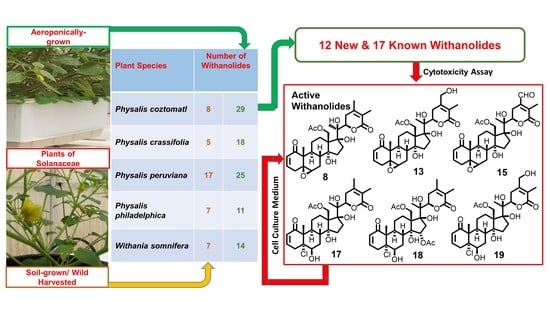
Figure 1. Major withanolides (18) encountered in some aeroponically grown plants of Solanaceae.
Molecules 27 00909 g001
Figure 2. Structures of withanolides 933 encountered in aeroponically grown P. coztomatl (for structures of other withanolides (35 and 8) encountered, see Figure 1).
Molecules 27 00909 g002
Figure 3. The HPLC analysis of 5α-chloro-6β-hydroxy-5,6-dihydrophysachenolide D (17) incubated with the cell culture medium (DMEM) showing facile and complete conversion of it to physachenolide C (8).
Molecules 27 00909 g003
Table 1. Comparison of the number of withanolides and % yields of major withanolides isolated from aeroponically grown and wild-crafted plants of some plants of Solanaceae.
PlantCultivation Method/SourceNumber of Withanolides Isolated aMajor Withanolides(% Yield) bRefs and Notes
Withania somniferaAeroponics14Withaferin A (1) (0.42)[15,17,23]
2,3-Dihydrowithaferin A-3β-O-sulfate (2) (0.51)
Wild-crafted (chemotype I)7Withaferin A (1) (0.23)[24]
Physalis crassifoliaAeroponics18Physachenolide D (3) (0.30)[18]
15α-Acetoxyphysachenolide D (4) (0.03)
Wild-crafted5Physachenolide D (3) (0.01)[19]
15α-Acetoxyphysachenolide D (4) (0.005)
Physalis peruvianaAeroponics25Withanolide E (5) (0.18)[20]
4β-Hydroxywithanolide E (6) (0.15)
Wild-crafted17Withanolide E (5) c[25]
4β-Hydroxywithanolide E (6) (0.008)
Physalis philadelphicaAeroponics11Ixocarpalactone B (7) (0.10)[21]
Wild-crafted7Ixocarpalactone B (7) (0.02)[26]
Physalis coztomatlAeroponics29Physachenolide C (8) (0.05)This study
Physachenolide D (3) (0.02)
Wild-crafted8Physachenolide C (8) d[27]
Physachenolide D (3) (0.02)
a The number of withanolides for wild-crafted/soil-grown plant refers to the referenced study reporting highest yield(s) of the major withanolide(s). b Since 2 is a prodrug of 1 [15], total % yield of withaferin A (1) in aeroponically-grown plant is 0.93. c Yield not reported. d Not encountered in wild-crafted plants [27,28].
Table 2. 1H and 13C NMR data for 912 in CDCl3.
Position9 a10 a1112
δHδCδHδCδHδCδHδC
1 204.4 204.7 204.6 203.9
25.83 (br d, 9.6)127.85.84 (br d, 10.0)127.85.84 (dd, 10.0, 2.0)127.75.85 (dd, 10.0, 2.0)127.9
36.77 (br d, 9.6)145.76.77 (ddd, 10.0, 4.8, 2.8)145.46.78 (ddd, 10.0, 4.8, 2.4)145.96.76 (ddd, 10.0, 4.8, 2.4)145.3
43.28 (m)33.53.28 (m)33.53.27 (br d, 21.2)33.53.27 (br d, 21.6)33.4
2.83 (m)2.83 (dd, 21.6, 4.8)2.83 (dd, 21.2, 2.4)2.82 (dd, 21.6, 4.8)
5 135.2 136.1 135.0 135.3
65.57 (br s)124.95.57 (d, 4.8)124.45.57 (br s)124.75.57 (d, 5.6)124.4
72.22 (m)25.52.00 (m)30.52.08 (m)25.32.10 (m)25.4
1.82 (m)1.62 (m)1.81 (m)1.81 (m)
81.85 (m)35.11.69 (m)31.91.82 (m)36.21.80 (m)36.4
92.37 (m)36.01.76 (m)43.42.07 (m)36.12.15 (m)36.1
10 50.8 50.6 50.6 50.8
112.31 (m)22.02.34 (m)23.32.15 (m)22.32.21 (m)22.0
1.51 (m)1.52 (m)1.34 (m)1.34 (m)
122.34 (m)24.82.64 (m)25.32.09 (m)27.21.96 (m)27.1
1.95 (m)2.55 (m)1.81 (m)1.91 (m)
13 56.3 50.6 50.5 50.6
14 83.41.76 (m)57.6 83.3 82.7
152.43 (m)41.22.09 (m)31.91.59 (dd, 12.2, 9.2)32.11.61 (m)32.4
2.17 (m)1.40 (m)1.39 (m)
165.82 (br s)126.75.74 (br s)128.32.00 (m)21.21.86, (m)20.7
1.88, (m)
17 151.1 153.02.70 (t, 9.2)49.32.73 (t, 9.7)49.9
184.59 (d, 10.4)67.34.56 (d, 11.2)66.64.40 (d, 11.6)62.94.89, (d, 11.6)62.9
3.99 (d, 10.4)4.02 (d, 11.2)3.98 (d, 11.6)3.70, (d, 11.6)
191.22 (s)18.71.23 (s)18.91.20 (s)18.91.22 (s)18.8
20 74.4 74.7 76.1 76.5
211.28 (s)24.81.30 (s)26.61.43 (s)23.41.40 (s)20.9
224.49 (m)80.84.45 (m)81.54.23 (d, 7.8)85.83.60 (d, 8.4)75.9
232.50–2.78 (m)25.12.38 (m)32.34.35 (d, 7.8)66.94.06 (dd, 8.4, 8.0)81.1
1.78 (m)
24 148.2 147.6 156.72.32 (m)51.0
25 123.4 123.5 124.52.29 (m)37.0
26 165.9 165.8 164.6 177.6
271.85 (s)12.31.86 (s)12.34.34 (s)57.31.28 (d, 6.8)14.1
284.46 (m)67.74.45 (m)68.02.06 (s)15.43.83 (dd, 11.2, 2.0)63.3
3.64, (dd, 11.2, 7.6)
OAc-182.07 (s)21.32.08 (s)21.32.07 (s)21.22.14 (s)21.2
171.4 171.8 170.7 169.7
Glc-1′4.27 (d, 6.0)102.54.30 (d, 7.2)102.5
Glc-2′3.38 (m)73.33.39 (m)73.4
Glc-3′3.26 (m)75.83.28 (m)75.8
Glc-4′3.55 (m)69.43.60 (m)69.8
Glc-5′3.47 (m)76.43.51 (m)76.4
Glc-6′3.81 (m)61.23.82 (brs)61.5
a CDCl3/CD3OD (100:1) was used as the solvent.
Table 3. 1H and 13C NMR data for 1316.
Position13 a14 a15 b16 c
δHδCδHδCδHδCδHδC
1 205.4 205.4 203.1 205.0
25.99 (dd, 10.0, 2.8)130.05.98 (dd, 10.0, 2.8)129.95.98 (dd, 10.0, 2.8)129.65.79 (dd, 10.0, 2.0)127.7
36.97 (ddd, 10.0, 6.0, 2.4)147.16.97 (ddd, 10.0, 6.0, 2.4)147.16.80 (ddd,10.0, 6.4, 2.0)144.16.74 (ddd, 10.0, 4.8, 2.4)145.9
42.96 (dt, 18.8, 2.4)33.82.96 (dt, 18.8, 2.4)33.82.93 (dt, 18.4, 2.4)32.83.23 (m)33.4
1.94 (m)1.94 (m)1.85 (m)2.79 (dd, 21.6, 4.8)
5 63.4 63.2 62.0 134.8
63.23 (br s)65.03.20 (d, 2.0)65.03.15 (br s)63.95.57 (d, 5.6)125.3
71.95 (m)27.72.05 (m)27.41.94 (m)26.32.20 (m)25.3
1.87 (m)1.93 (m)1.79 (m)
81.92 (m)35.52.65 (m)35.91.87 (m)34.21.87 (m)34.4
91.83 (m)38.71.89 (m)38.61.89 (m)36.82.28 (m)35.8
10 49.7 49.8 48.4 50.6
112.03 (m)24.52.05 (m)24.52.06 (m)22.82.19 (m)22.2
1.38 (m)1.40 (m)1.34 (m)1.55 (m)
122.15 (m)26.62.24 (m)26.62.18 (m)25.52.20 (m)28.3
1.89 (m)1.85 (m)1.68 (m)1.47 (m)
13 58.6 58.6 57.5 52.2
14 83.0 81.3 81.5 84.5
151.62 (m)33.85.06 (dd, 9.2, 8.4)77.51.66 (m)32.92.29 (m)39.7
1.55 (m)1.59 (m)2.14 (m)
162.56 (m)37.82.36 (m)43.82.64 (m)37.85.78 (br s)124.5
1.65 (m)2.24 (m)1.58 (m)
17 88.9 85.8 87.9 155.9
184.36 (d, 11.2)65.94.46 (d, 11.2)65.44.40 (d, 11.6)64.71.11 (s)22.3
4.30 (d, 11.2)4.32 (d, 11.2)4.28 (d, 11.6)
191.19 (s)15.31.20 (s)15.41.20 (s)14.81.18 (s)18.6
20 79.8 80.0 79.2 74.6
211.39 (s)19.01.35 (s)19.11.43 (s)19.01.26 (s)22.4
224.86 (dd, 13.6, 3.2)83.74.86 (dd, 13.6, 3.2)83.64.90 (dd, 13.6, 3.2)81.34.37 (t, 8.0)81.1
233.22 (dd, 18.8, 2.8)30.33.18 (dd, 20.4, 2.0)30.33.14 (m)24.52.63 (m)25.0
2.38 (m)2.37 (m)2.30, (m)
24 154.9 154.9 142.3 149.0
25 122.4 122.4 138.1 122.8
26 169.1 169.0 165.6 166.2
271.88 (s)12.11.87 (s)12.12.35 (br s)11.51.82 (s)11.9
284.36 (d, 13.6)61.94.37 (d, 14.0)61.810.30 (s)190.24.42 (br s)67.7
4.24 (d, 13.6)4.20 (d, 14.0)
OAc-182.13 (s)21.32.13 (s)21.32.15 (s)21.1
173.5 173.1 170.8
OAc-15 2.06 (s)21.4
172.6
Glc-1′ 4.23 (d, 8.0)102.5
Glc-2′ 3.26 (m)73.3
Glc-3′ 3.23 (m)75.9
Glc-4′ 3.39 (m)69.8
Glc-5′ 3.37 (m)76.4
Glc-6′ 3.79 (dd, 12.0, 2.8)61.5
3.71 (dd, 12.0, 4.4)
a CD3OD was used as the solvent. b CDCl3 was used as the solvent. c CDCl3/CD3OD (100:1) was used as the solvent.
Table 4. 1H and 13C NMR data for 1720 in CDCl3.
Position171819 a20 b21 b,c
δHδCδHδCδHδCδHδCδC
1 201.3 201.0 204.1 204.8205.3
25.89 (dd, 10.0, 2.4)128.65.89 (dd, 10.0, 2.4)128.55.83 (dd, 10.0, 2.0)129.25.73 (dd, 10.0, 2.8)129.0127.8
36.62 (br dd, 10.0, 3.6)141.26.63 (ddd, 10.0, 4.8, 2.4)141.56.75 (ddd, 10.0, 4.8, 2.0)144.16.48 (ddd, 10.0, 5.2, 2.0)139.6142.2
43.49 (br d, 20.0)37.23.53 (dt, 20.4, 2.4)37.23.56 (br d, 20.0)38.72.95 (dt, 18.8, 2.4)27.435.0
2.49 (m)2.47 (dd, 20.4, 8.0)2.49 (dd, 20.0, 4.8)2.28 (m)
5 79.5 79.0 82.3 81.377.3
64.06 (br s)75.03.97 (br s)74.63.93 (t, 2.9)75.63.86 (brs)68.473.6
72.54 (m)29.72.61 (m)28.82.38 (m)30.41.98 (m)29.428.5
1.52 (m)1.54 (m)1.54 (m)1.37 (m)
82.13 (m)34.62.82 (dt, 4.0, 12.0)34.82.26 (m)35.92.02 (m)33.933.4
92.76 (m)34.92.37 (dt, 4.0, 12.0)34.82.67 (m)36.42.61 (m)33.633.9
10 53.0 52.5 54.5 52.751.8
112.50 (m)22.42.54 (m)22.52.39 (m)24.02.35 (m)22.622.6
1.22 (m)1.24 (m)1.24 (m)1.18 (m)
122.40 (m)26.22.46 (m)26.12.28 (m)27.22.30 (m)26.126.1
1.81 (m)1.86 (m)1.94 (m)1.75 (m)
13 57.8 58.0 58.9 57.557.4
14 81.9 80.0 83.8 82.082.6
151.68 (m)32.95.20 (t, 8.8)75.81.58–1.71 (m)33.61.63 (m)32.632.4
1.59 (m)1.51 (m)
162.71(m)37.92.53 (m)42.52.60 m37.82.61 (m)37.437.0
1.55 m2.26 (m)1.68 m1.47 (m)
17 88.2 84.7 89.1 87.987.7
184.43 (s)65.54.79 (d, 11.6)64.94.40 (s)66.44.37 (d, 11.2)65.565.3
4.22 (d, 11.6)4.30 (d, 11.2)
191.36 (s)16.11.36 (s)16.51.38 (s)17.01.22 s15.415.5
20 78.9 79.4 79.9 78.278.1
211.41 (s)19.31.38 (s)19.21.40 (s)19.21.31 s18.418.1
224.90 (t, 8.4)79.74.91 (br d, 8.0)79.74.90 (m)84.04.84 (dd, 13.6, 3.2)80.881.0
232.53 (m)33.82.50 (m)33.82.40 (m)30.32.58 (m)33.733.6
2.44 (m)
24 149.7 149.9 154.9 150.6151.0
25 121.8 121.8 122.5 121.4121.2
26 165.7 165.7 169.2 167.2167.5
271.88 (s)12.41.88 (s)12.41.89 (s)12.11.82 s12.212.0
281.92 (s)20.71.92 (s)20.74.39 (d, 14.0)62.01.88 s20.620.5
4.23 (d, 14.0)
OAc-182.07 (s)21.32.09 (s)21.42.12 (s)21.32.03 s21.321.2
170.4 171.4 173.6 171.1171.2
OAc-15 2.08 (s)21.8
172.2
OMe 2.93 s49.6
a CD3OD was used as the solvent. b CDCl3/CD3OD (100:1) was used as the solvent. c 13C NMR data obtained in CDCl3/CD3OD (100:1) for physachenolide A (21) are included for the purpose of comparison as the reported 13C NMR data for 21 were for CDCl3/DMSO-d6 [38].
Table 5. Cytotoxicity data of withanolides from Physalis coztomatl against a panel of selected tumor cell lines and normal cells a.
CompoundCell Line b
LNCaPDU-145PC-3VCaPACHNWI-38
ActivitySI cActivitySI cActivitySI cActivitySI cActivitySI c
80.03 ± 0.0115.00.26 ± 0.011.70.06 ± 0.017.50.03 ± 0.0115.01.02 ± 0.200.40.45 ± 0.11
132.78 ± 0.66>1.8>5.0 2.90 ± 0.32>1.71.11 ± 0.19>4.5>5.0 >5.0
151.04 ± 0.183.62.67 ± 0.151.41.18 ± 0.213.20.82 ± 0.114.63.98 ± 0.100.93.77 ± 0.06
170.03 ± 0.0117.00.67 ± 0.080.80.09 ± 0.015.70.08 ± 0.016.41.73 ± 0.180.30.51 ± 0.03
180.64 ± 0.16>7.84.53 ± 0.55>1.10.86 ± 0.19>5.80.27 ± 0.08>18.5>5.0 >5.0
191.98 ± 0.44>2.5>5.0 2.67 ± 0.21>1.91.26 ± 0.224.0>5.0 >5.0
Doxorubicin0.11 ± 0.02 0.04 ± 0.01 0.34 ± 0.05 0.67 ± 0.06 0.05 ± 0.01 0.80 ± 0.08
a Results are expressed as IC50 values in μM. Doxorubicin and DMSO were used as positive and negative controls. b Key: LNCaP = androgen-sensitive human prostate adenocarcinoma; DU-145 = androgen-independent human prostate cancer; PC-3 = androgen-independent human prostate cancer; VCaP = androgen-sensitive human prostate cancer; ACHN = human renal adenocarcinoma; WI-38 = normal human fibroblast cells; c SI = Selectivity Index (against normal cells, WI-38).
Publisher’s Note: MDPI stays neutral with regard to jurisdictional claims in published maps and institutional affiliations.

