Article

Achieving Luminescence of Sr3Ga1.98In0.02Ge4O14:0.03Cr3+ via [In3+] Substitution [Ga3+] and Its Application to NIR pc-LED in Non-Destructive Testing

1
College of Science, China University of Petroleum (East China), Qingdao 266580, China
2
Hebei Key Laboratory of Optic-Electronic Information and Materials, College of Physics Science & Technology, Hebei University, Baoding 071002, China
*
Authors to whom correspondence should be addressed.
Submission received: 9 November 2023 / Revised: 8 December 2023 / Accepted: 10 December 2023 / Published: 13 December 2023
(This article belongs to the Special Issue Organic and Inorganic Luminescent Materials)

Abstract

:
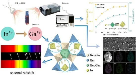
Cr3+-doped Sr3Ga2Ge4O14:0.03Cr3+ (SGGO:0.03Cr3+) phosphor was synthesized via a high-temperature solid-phase method. Considering the tunable structure of SGGO, Ga3+ ions in the matrix were substituted with In3+ ions at a certain concentration. The tuned phosphor produced a red-shifted emission spectrum, with its luminescence intensity at 423 K maintained at 63% of that at room temperature; moreover, the internal quantum efficiency increased to 65.60%, and the external quantum efficiency correspondingly increased to 21.94%. On this basis, SGIGO:0.03Cr3+ was encapsulated into a pc-LED, which was applied in non-destructive testing (NDT) experiments, successfully realizing the recognition of water and anhydrous ethanol, proving its potential application in the field of NDT.

Graphical Abstract

1. Introduction

In recent years, research on near-infrared light has continued to deepen due to its invisibility, high penetration, and non-destructive and other excellent characteristics; according to its specific band, near-infrared light is widely used in plant lighting, night vision lighting, non-destructive testing, medical testing, and other fields [1,2,3,4,5,6]. At present, halogen lamps and infrared lasers are the most common and representative traditional infrared light sources. However, halogen lamps have high power consumption, low efficiency, and short service life. Infrared lasers are large in size and high in cost, which limit their application in actual production work to a certain extent [7,8,9,10,11,12]. The emergence of near-infrared phosphor-converted light-emitting diodes (NIR pc-LEDs) has become a hot research topic due to their multiple advantages such as small size, energy savings, environmental protection, and simple preparation. which also increase the spectral coverage of LED devices over a larger area [13,14,15,16]. However, the emission wavelength coverage of LED chips is severely limited as their emission wavelength depends largely on the composition of the LED chip. To solve these problems, the industry has developed down-conversion phosphors, an important component of NIR pc-LEDs that affects the emission spectra of the light-emitting device, photoelectric efficiency, device lifetime, temperature characteristics, and many other critical aspects. For this reason, there is a need to develop high-performance NIR phosphors that are well matched to blue LED chips.
NIR phosphors are usually composed of activated ions and host compound components. Cr3+, Bi3+, Eu2+, and Mn2+ are typical activated ions that produce NIR phosphor emission peaks. Although the emission of Bi3+ and Eu2+ is led by the wavelength close to the deep red region, the half-peak width of NIR is still not wide. The application of Mn2+-doped oxide phosphors suffers from drawbacks such as a low color rendering index and high color temperature. These drawbacks limit their application in the NIR. To solve the above problems, Cr3+ has attracted much attention. It can simply modulate its emission by changing its local environment. The 3d3 electron leaps of Cr3+ are significantly affected by the crystal field. Depending on the strength of the crystal field environment, the typical luminescence of Cr3+ usually exhibits three features: spin-forbidden leaps 2E→4A2 are generated when Cr3+ is in a strong crystal field (Dq/B > 2.3), and a narrow-band emission is produced near 700 nm. When Cr3+ is in a weak crystal field (Dq/B < 2.3), a tunable broadband emission is generated by the spin-allowed jump 4T24A2; when in an intermediate crystal field (Dq/B ≈ 2.3), the spectrum exhibits both of the above phenomena, Hence, the luminescence performance of Cr3+ can be flexibly tuned using these properties. It has been found that the cation substitution strategy is an effective method to improve the spectra and enhance the phosphor temperature performance [17,18,19,20,21,22,23]. This is because the change in cation components in the matrix material affects the overall crystal field environment, and Cr3+ is highly susceptible to the influence of the crystal field environment in which it is embedded. So, the luminescence performance of Cr3+ changes accordingly after the change in cation components in the matrix. Qiu et al. used Al3+ to replace Sc3+ on the host material Gd3Sc2Ga3O12 so that the internal quantum efficiency of the emission of Cr3+ was significantly increased to nearly 100%, and the temperature stability after modulation was improved to 86% after modulation [24]. Zhang et al. utilized [Lu3+-Al3+] to replace [Ca2+-Si4+] in Ca2LuZr2Al3O12:Cr3+, which broadened the spectrum and improved the emission intensity by changing the crystal field and electroacoustic coupling effects [11]. In previous experiments, through doping different concentrations of Cr3+ ions and comparing their luminescent properties, two excellent phosphors, Sr3Ga2Ge4O14:0.015Cr3+ and Sr3Ga2Ge4O14:0.03Cr3+, were obtained. Considering that the fluorescent materials with larger half-peak full-widths and higher temperature stability are more preferred in practical applications, we selected Sr3Ga2Ge4O14:0.03Cr3+ with a half-peak full width of 280 nm for further study [25].
Although some Cr3+-activated oxide phosphors [1,3,21,25,26,27,28,29,30,31] have been reported, no detailed report on the Cr3+-activated Sr3Ga2Ge4O14–Sr3In2Ge4O14 solid–solution system has been published to date. In this study, the NIR phosphor Sr3Ga2−2xInxGe4O14:0.03Cr3+ (SGIGO:0.03Cr3+) with higher emission intensity and better temperature stability was obtained via In3+-Ga3+ cation substitution, and the luminescence performance of the phosphor was analyzed. Detailed characterization and analysis revealed that the enhancement in the material morphology led to a significant increase in the emission intensity of the spectrum, which was maintained at 63% of room temperature at 423 K. The spectral intensity of the fluorescent phosphor was found to be higher than that at room temperature. Finally, good application capability was demonstrated through liquid non-destructive identification experiments.

2. Results and Discussion

2.1. Phase Purity and Crystal Structure Analysis

For the cation modulation strategy, the theoretical value of the radius difference between the dopant ion and the substituted ion should be less than 30%, which is more promising. In Sr3Ga2Ge4O14, the radii of dopant ions and substituted ions are as follows: In3+ [CN = 6, R = 0.80].
D r = 100 × R m ( N ) R d ( N ) R m ( N )
where Dr is the radius difference between the doped and substituted ions; Rm and Rd denote the radii of the substituted and doped ions, respectively; and N denotes the coordination number. According to Equation (1), it can be calculated that Dr [In3+-Ga3+] = 3%. It is obvious that the strategy of substituting In3+ ions for Ga3+ ions to modulate the crystal field is theoretically very feasible, and the substitution process is demonstrated in Figure 1a.
A series of SG2−2xIxGO:0.03Cr3+ (x = 0–0.05) samples were synthesized in the experiment using high temperature solid-phase method and their XRD diffraction data were tested as exhibited in Figure 1b. The XRD data of the samples did not exhibit any impurity peaks when compared with the SGGO standard card PDF#84-0783 (MDI Jade, version 1251), which indicates that the synthesized samples were all pure phases, and the introduction of In3+ ions did not affect the original structure of the material. In addition, when the crystalline phase was transformed from SGGO:0.03Cr3+ to SG2−2xIxGO:0.03Cr3+, the main peaks produced a shift towards a small angle, which can be explained by the Bragg equation [2]:
2d sin θ =
where d represents the intergranular spacing, θ represents the diffraction angle, k is the reflection level for a fixed value, and λ is the wavelength of the X-rays. From the formula, it can be seen that the intergranular spacing d is inversely proportional to the diffraction angle θ, which means that the larger the intergranular spacing, the smaller the diffraction angle. Due to the larger radius of In3+ ions, the process of replacing Ga3+ by In3+ leads to lattice expansion, and the intergranular spacing d then becomes larger, so the diffraction angle θ becomes smaller, which explains why the diffraction peaks in XRD are shifted to a small angle. In order to obtain more accurate crystal structure information, Rietveld refinement was performed, and the calculation results were obtained by the GSAS program. This is exhibited in Figure S1a,b (see Supplementary Materials). The Rietveld refinement results for SGGO:0.03Cr3+ and SG1.98I0.02GO:0.03Cr3+ were within reasonable limits, proving the good crystallinity of the samples. The Rietveld refinement results for the remaining samples are exhibited in Figure S1c–f (see Supplementary Materials).
As seen from the Rietveld refinement results, the cell volume V and the volume of [Ge1/Ga1] O6 kept increasing with the increase in the doping concentration of the In3+ ions. The lattice parameters a/b and c exhibited the same trend, as shown in Figure 1c,d. This is due to the fact that the radius of In3+ ions is larger than that of Ga3+ and, when doped, the lattice expands as a result.
In order to further analyze the morphology and elemental distribution of the synthesized samples, SEM tests were carried out on the samples, as exhibited in Figure 1e. The SEM images show that the material is a particle with a diameter of about 10 μm. In Figure 1f, it can be seen that the elemental distribution is uniform, and the above results demonstrate the successful doping of In3+.

2.2. Luminescence Characterization and Mechanism of Sr3Ga1.98In0.02Ge4O14:0.03Cr3+

Figure 2a exhibits the emission spectra of SG2−2xIxGO:0.03Cr3+ (x = 0–0.05) monitored at room temperature, which highlights that a trend in the luminescence intensity of increasing and then decreasing with the increase in the In3+ ion concentration. The intensity reaches the maximum value at 2% In3+ ion concentration, which is about 1.84 times of the luminescence intensity when undoped with In3+. Throughout the substitution process, the half-peak width of the emission spectra was maintained at about 280 nm, as shown in Figure 2b. Figure 2c exhibits the normalized excitation and emission spectra, and it can be seen that the shape of the excitation spectra after In3+ ion doping did not exhibit any significant changes and still agreed with the typical excitation spectral shape of Cr3+. In addition, the spectra produced a red shift of 50 nm with the increase in the In3+ concentration, which was related to the electron cloud expansion effect and the change in the crystal field strength. It can be understood like this: The mass number of In is larger than that of Ga. Adding the atom with the larger mass number ensures a “larger lattice constant”. Thus, the electron orbital clouds in the cation atoms enable shielding or screening of the crystal-field strength, resulting in a smaller value of Dq. Therefore, it ensures a smaller emission energy of 4T2 (i.e., the red shift in the emission wavelength) [4,21,32]. Previous research by solid-state scientists showed that when x changes from 0 to 0.05, it does not cause a significant change in the band-gap energy [33].
The electron cloud extension effect refers to the phenomenon that the more covalent an ion, the more diffuse its electron orbitals. The interactions between electrons are reduced, resulting in a shift in the jump energy between the electron energy levels in the lower energy direction. This is usually expressed using the following equation [34]:
1 − β = hk
where β denotes the electron cloud expansion effect, and h and k are the electron cloud expansion factors, which are related to the elemental electronegativity. The smaller the electronegativity of an element, the stronger the covalency, and the more pronounced the electron cloud expansion effect of the compound. Its spectral line shifts to the long-wave direction [27,35,36,37,38]. For Ga3+ and In3+, whose electronegativity is 1.81 and 1.78, respectively, when In3+ replaces Ga3+, the electron cloud extension effect is enhanced, resulting in a red shift in the emission spectra of SG2−2xIxGO:0.03Cr3+.
Figure 2d exhibits the excitation spectrum of SG2−2xIxGO:0.03Cr3+ (x = 0–0.05) monitored at 800 nm, which was normalized in order to see the change in the excitation spectrum after In3+ doping more clearly. The three excitation bands belong to the typical 4A24T1(4P), 4A24T1(4F), and 4A24T2(4F) leaps of Cr3+, respectively. It can be seen that, with the increase in the doping concentration of In3+, the peak position of 4A24T1(4F) is basically unchanged, whereas that of 4A24T2(4F) is more affected and shifted. In the previous section, we mentioned that the broadband emission of SGGO:Cr3+ under optimal doping is attributed to the radiative jump from the excited state 4T2(F) to the ground state 4A2(F). When exactly determining the various excited-state energies from experimental optical spectra for calculating Equations (4)–(6), one must consider the strong phonon coupling nature of the 3d3 (Cr3+) electrons with the phosphor host lattices [39,40]. Such excited-state energies to be used in the Racah parameter calculations in Equations (4)–(6) cannot be simply obtained from the peak energy of each PL or PLE band. It has been demonstrated that the use of such emission or excitation “peak” energies, but not the “zero-phonon line (ZPL)” energies, in calculating Racah parameters (such as Dq, B, and C) usually results in unacceptable conclusions [41,42]. For simplicity, however, we considered the “peak” energy of the various excited states taken from the experimental optical spectra for calculating Equations (4)–(6) with a suitable assumption of a Racah parameter ratio of C/B [41]. This approach is the same as those reported in the previous literature, but leaves problems to be solved in the near future [42].
D q = E ( T 2 4 ) = E ( A 2 4 T 2 4 ) Δ S / 2
D q B = 15 ( Δ E / D q 8 ) ( Δ E / D q ) 2 10 ( Δ E / D q )
Δ E = E ( T 1 4 ) E ( T 2 4 ) = E ( A 2 4 T 1 4 ) E ( A 2 4 T 2 4 )

2.3. Temperature Stability and Quantum Efficiency Analysis of Sr3Ga1.98In0.02Ge4O14:0.03Cr3+

The variation of luminescence intensity with temperature may be affected by a variety of factors, such as lattice vibrations, changes in energy level transitions, and changes in material structure. Some luminescent materials undergo structural phase transitions or diffusion of lattice defects at high temperatures, leading to a decrease in luminescence intensity. In a previous study of Rb2GeF6:Mn4+, it was found that the luminescence intensity was enhanced with increasing temperature in the range of 420 K to 450 K, and gradually weakened as the temperature increased at T > 450 K, which was the cause of thermal burst [43]. Therefore, temperature stability is an important factor to determine whether the luminescent material can realize successful application. For this purpose, the variable temperature spectrum of SG1.98I0.02GO:0.03Cr3+ was tested, as shown in Figure S2 (see Supplementary Materials), and its luminescence intensity was maintained at 63% of that at room temperature at 423 K, which is a significant enhancement compared to that of the unmodulated SGGO:0.03Cr3+ (56%) in the previous chapter.
In general, a more backward emission position, a larger half-peak width, and a higher temperature stability often do not go hand in hand because a stronger electroacoustic coupling effect leads to a stronger temperature burst phenomenon [44]. The reason for the elevated temperature stability after modulation can be explained by the bit pattern coordinate plot as shown in Figure 3b.
There are generally two ways for electrons to return to the ground state from the excited state, one of which is through radiative leaps [45]. In the previous section through calculations, we know that the doping of In3+ ions reduces the crystal field strength, which can be seen in the T-S diagrams, where the position of the 4T2 energy level should correspondingly be lowered, as exhibited in Figure 3b, and therefore the energy radiated in the process of returning to the ground state becomes smaller; this also explains the spectral red shift.
The other way is that the electron reaches the intersection of the ground and excited states through lattice relaxation and returns to the ground state via radiation with fewer leaps [46,47]. The energy required in this process is the activation energy Ea. The activation energy of SG1.98I0.02GO:0.03Cr3+ can be seen in the slope in Figure 3c. It was calculated using Equation (7) to be 0.25 eV, which is increased by 0.03 eV compared to the undoped state. Therefore, the doping of In3+ increases the activation energy of the material and decreases the chances of electron-radiation-free leaps, which leads to an improvement in the temperature stability of the material.
I T = I 0 1 + c * exp E a k T
Quantum efficiency is the second important factor to be considered in practical applications. The quantum efficiency of the phosphor doped with In3+ ions was tested, as exhibited in Figure 3d. Due to the limitation of the instrument, the test range was between 400 and 850 nm, and the internal quantum efficiency of its S1 part was measured as 15.80%. Considering the spectrum of the S2 part, the actual quantum efficiency was calculated as 42.21%, which is 34% higher than that of the quantum efficiency when undoped with In3+ ions (IQE = 31.60%). Its external quantum efficiency is correspondingly increased to 21.94%. It can be seen that the doping of In3+ ions improved the multifaceted properties of SGGO:Cr3+.

2.4. Application Exploration

The main component of anhydrous ethanol is C2H6O, which mainly absorbs light in the band near 800 nm; the main component of water is H2O, which mainly absorbs light in the band near 900 nm. As shown in the schematic diagram in Figure 4a, a 430 nm blue InGaN chip with SG1.98I0.02GO:0.03Cr3+ is encapsulated in a pc-LED, and the prepared pc-LED is used as a light source to irradiate the side of the cuvette. Due to the different absorption wavelengths of the different groups of light, there is a corresponding lack of wavelengths in the light transmitted through the cuvette. The spectrometer on the other side can output the corresponding data for comparison with the standard spectrum so as to achieve the purpose of non-destructive testing. Figure 4b,c exhibit comparisons of the chip light with the standard spectrum after passing through water and anhydrous ethanol, respectively. It can be seen that water has obvious absorption at 900 nm, and anhydrous ethanol has obvious absorption at 800 nm, so different liquids can be recognized using different absorption ranges, which proves that the pc-LED has good application prospects in non-destructive testing.

3. Materials and Methods

3.1. Sample Preparation

A series of Sr3Ga2−2xInxGe4O14:0.03Cr3+ (x = 0, 0.2, 0.4, 0.8, 1.2, 1.6, 2.0) phosphors were successfully synthesized via the high-temperature solid-phase reaction method. Gd2O3 (99.9%, Aladdin, Riverside, CA, USA), ZnO (99%, Aladdin, USA), Ga2O3 (99.999%, Zhu Zhou Heng Ma, Zhuzhou, China), GeO2 (99.9999%, Zhu Zhou Heng Ma, China), Cr2O3 (99%, Kermel, Tianjin, China), and 3%H3BO3 (99.5%, Aladdin, USA) as a flux were applied as raw materials. The raw materials were weighed according to the stoichiometric ratio, and then all the weighed drugs were poured into an agate mortar and evenly ground for more than 30 min to make a homogeneous mixture. Then, the resulting powder was placed into a corundum crucible, whose main component was Al2O3, which was labeled with a good order, and then put into a high-temperature furnace. The temperature was raised at a rate of 5 K/min, starting from 273 K to 773 K, and held at a temperature for 30 min, and then the temperature was increased to 1173 K, and held at that temperature for 360 min, to complete the first sintering. After waiting for its natural reduction to room temperature, it was removed and ground for 30 min. Then, the second sintering was carried out by increasing the temperature at a rate of 5 K/min, starting from 273 K to 773 K, holding for 30 min, and then increasing to 1423 K, holding for 360 min to complete the sintering. Finally, the sintered samples were milled again to make a uniform and fine particle powder for subsequent characterization and testing.

3.2. Preparation of pc-LED

A certain amount of phosphor in proportion to the organic silica gel was mixed and stirred for more than 30 min. Afterwards, the mixed sample entered the defoaming process (air bubbles lead to chip light leakage). Mixed and defoamed samples were placed on the blue LED chip and then sent through the LED test system to determine device-related performance. Once the device met the necessary requirements, it was placed in the drying oven at 423 K for one hour. Afterwards, the device was cooled to room temperature for the next step of the experiment.

3.3. Sample Characterization

Measurements were carried out using an X-ray diffractometer (Bruker D8 Advance, München, Germany) at 40 kV and 40 mA and Cu-Ka (l = 1.54056 Å) irradiation in the 2θ range from 10° to 80° in 0.02° scanning steps. General Structure Analysis System (GSAS) software (version 1251) was used to optimize and analyze the structure. The morphology of the powder samples was investigated using a field-emission scanning electron microscope (FEI Nova Nano SEM 450, Peabody, MA, USA), and the elemental mapping results were obtained via energy-dispersive spectrometry (EDS) on the SEM equipment. Elemental composition was measured using energy dispersive spectroscopy and an X-ray spectrometer connected to the SEM. Fluorescence spectra of SGGO were tested using a transient steady-state fluorescence spectrometer (HORIBA FLuorolog-3, Kyoto, Japan) equipped with a variable-temperature liquid helium optical thermostat. Temperature emission spectra were measured from room temperature to 423 K in 283 K intervals using an external heater. The excitation wavelength was 431 nm. Measurements of electroluminescence properties were performed with a spectrometer (HORIBA FLuorolog-3, Kyoto, Japan) with an external DC power supply of 3 V and a current of 100 to 400 mA.

4. Conclusions

Based on the study of the luminescence properties of SGGO:0.03Cr3+, SGGO:0.03Cr3+, which has a wider half-peak width and a luminescence position more inclined to the long-wave direction, was selected as the object for further modulation study on the premise that it is more conducive to practical applications. Based on the cation substitution strategy, a series of Sr3Ga2−2xInxGe4O14:0.03Cr3+ (x = 0–0.05) near-infrared phosphors with wider half-peaks and stronger luminescence intensity were obtained by replacing Ga3+ ions with In3+ ions in the matrix at a certain concentration. The large-radius In3+ replaced Ga3+ ions affecting the crystal field strength, leading to spectral red shift, which was explained in detail by analyzing the electron cloud expansion effect, crystal field strength changes, and bit pattern coordinate diagram. In addition, the temperature stability and quantum efficiency after the modulation were significantly improved, which are more favorable for practical applications. Finally, the phosphor Sr3Ga1.98In0.02Ge4O14:0.03Cr3+ with the strongest emission intensity was selected to be encapsulated into a pc-LED, which was applied in NDT experiments to successfully realize the recognition of water and anhydrous ethanol, proving its application potential in the field of NDT.

Supplementary Materials

The following supporting information can be downloaded at: https://0-www-mdpi-com.brum.beds.ac.uk/article/10.3390/molecules28248059/s1, Figure S1: (a) Refined XRD pattern of SGGO:0.03Cr3+; (b) refined XRD pattern of SG1.98I0.02GO:0.03Cr3+; (c) refined XRD pattern of SG1.99I0.01GO:0.03Cr3+; (d) refined XRD pattern of SG1.97I0.03GO:0.03Cr3+; (e) refined XRD pattern of SG1.96I0.04GO:0.03Cr3+; (f) refined XRD pattern of SG1.95I0.05GO:0.03Cr3+; Figure S2: Three-dimensional temperature spectra of SG1.98I0.02GO:0.03Cr3+.

Author Contributions

Data curation, T.W. and B.G.; formal analysis, B.G. and J.L.; writing—original draft preparation, T.W. and J.L.; writing—review and editing, Z.W. and P.L.; supervision, Z.W. and P.L. All authors have read and agreed to the published version of the manuscript.

Funding

This research was funded by the National Natural Science Foundation of China (No. 61805285, No. 12074434) and the Shandong Provincial Natural Science Foundation (ZR2018PF015).

Institutional Review Board Statement

Not applicable.

Informed Consent Statement

Not applicable.

Data Availability Statement

Data are contained within the article and Supplementary Materials.

Conflicts of Interest

The authors declare no conflict of interest.

References

  1. Huang, D.; Zhu, H.; Deng, Z.; Yang, H.; Hu, J.; Liang, S.; Chen, D.; Ma, E.; Guo, W. A highly efficient and thermally stable broadband Cr3+-activated double borate phosphor for near-infrared light-emitting diodes. J. Mater. Chem. C 2021, 9, 164–172. [Google Scholar] [CrossRef]
  2. Miao, S.; Liang, Y.; Zhang, Y.; Chen, D.; Wang, X.-J. Broadband short-wave infrared light-emitting diodes based on Cr3+-doped LiScGeO4 phosphor. ACS Appl. Mater. Interfaces 2021, 13, 36011–36019. [Google Scholar] [CrossRef] [PubMed]
  3. Tan, T.; Wang, S.; Su, J.; Yuan, W.; Wu, H.; Pang, R.; Wang, J.; Li, C.; Zhang, H. Design of a novel near-infrared luminescence material Li2Mg3TiO6:Cr3+ with an ultrawide tuning range applied to near-infrared light-emitting diodes. ACS Sustain. Chem. Eng. 2022, 10, 3839–3850. [Google Scholar] [CrossRef]
  4. Zhong, J.; Zhuo, Y.; Du, F.; Zhang, H.; Zhao, W.; Brgoch, J. Efficient and Tunable Luminescence in Ga2−xInxO3:Cr3+ for Near-Infrared Imaging. ACS Appl. Mater. Interfaces 2021, 13, 31835–31842. [Google Scholar] [CrossRef] [PubMed]
  5. Zhao, F.; Cai, H.; Song, Z.; Liu, Q. Structural confinement for Cr3+ activators toward efficient near-infrared phosphors with suppressed concentration quenching. Chem. Mater. 2021, 33, 3621–3630. [Google Scholar] [CrossRef]
  6. Yang, J.; Liu, Y.; Zhao, Y.; Gong, Z.; Zhang, M.; Yan, D.; Zhu, H.; Liu, C.; Xu, C.; Zhang, H. Ratiometric afterglow nanothermometer for simultaneous in situ bioimaging and local tissue temperature sensing. Chem. Mater. 2017, 29, 8119–8131. [Google Scholar] [CrossRef]
  7. Li, Z.; Zhang, Y.; Wu, X.; Huang, L.; Li, D.; Fan, W.; Han, G. Direct aqueous-phase synthesis of sub-10 nm “luminous pearls” with enhanced in vivo renewable near-infrared persistent luminescence. J. Am. Chem. Soc. 2015, 137, 5304–5307. [Google Scholar] [CrossRef] [PubMed]
  8. Zhuang, Y.; Katayama, Y.; Ueda, J.; Tanabe, S. A brief review on red to near-infrared persistent luminescence in transition-metal-activated phosphors. Opt. Mater. 2014, 36, 1907–1912. [Google Scholar] [CrossRef]
  9. Chen, H.-W.; Zhu, R.-D.; He, J.; Duan, W.; Hu, W.; Lu, Y.-Q.; Li, M.-C.; Lee, S.-L.; Dong, Y.-J.; Wu, S.-T. Going beyond the limit of an LCD’s color gamut. Light Sci. Appl. 2017, 6, e17043. [Google Scholar] [CrossRef]
  10. Song, E.; Jiang, X.; Zhou, Y.; Lin, Z.; Ye, S.; Xia, Z.; Zhang, Q. Heavy Mn2+ doped MgAl2O4 phosphor for high-efficient near-infrared light-emitting diode and the night-vision application. Adv. Opt. Mater. 2019, 7, 1901105. [Google Scholar] [CrossRef]
  11. Zhang, L.; Zhang, S.; Hao, Z.; Zhang, X.; Pan, G.-h.; Luo, Y.; Wu, H.; Zhang, J. A high efficiency broad-band near-infrared Ca2LuZr2Al3O12:Cr3+ garnet phosphor for blue LED chips. J. Mater. Chem. C 2018, 6, 4967–4976. [Google Scholar] [CrossRef]
  12. Pan, L.; Lu, R.; Zhu, Q.; McGrath, J.M.; Tu, K. Measurement of moisture, soluble solids, sucrose content and mechanical properties in sugar beet using portable visible and near-infrared spectroscopy. Postharvest Biol. Technol. 2015, 102, 42–50. [Google Scholar] [CrossRef]
  13. Ye, M.; Gao, Z.; Li, Z.; Yuan, Y.; Yue, T. Rapid detection of volatile compounds in apple wines using FT-NIR spectroscopy. Food Chem. 2016, 190, 701–708. [Google Scholar] [CrossRef]
  14. Sarkar, S.; Le, P.; Geng, J.; Liu, Y.; Han, Z.; Zahid, M.U.; Nall, D.; Youn, Y.; Selvin, P.R.; Smith, A.M. Short-wave infrared quantum dots with compact sizes as molecular probes for fluorescence microscopy. J. Am. Chem. Soc. 2020, 142, 3449–3462. [Google Scholar] [CrossRef] [PubMed]
  15. Zabiliūtė, A.; Butkutė, S.; Žukauskas, A.; Vitta, P.; Kareiva, A. Sol-gel synthesized far-red chromium-doped garnet phosphors for phosphor-conversion light-emitting diodes that meet the photomorphogenetic needs of plants. Appl. Opt. 2014, 53, 907–914. [Google Scholar] [CrossRef] [PubMed]
  16. Guelpa, A.; Marini, F.; du Plessis, A.; Slabbert, R.; Manley, M. Verification of authenticity and fraud detection in South African honey using NIR spectroscopy. Food Control 2017, 73, 1388–1396. [Google Scholar] [CrossRef]
  17. Elzbieciak-Piecka, K.; Matuszewska, C.; Marciniak, L. Step by step designing of sensitive luminescent nanothermometers based on Cr3+, Nd3+ co-doped La3−xLuxAl5−yGayO12 nanocrystals. New J. Chem. 2019, 43, 12614–12622. [Google Scholar] [CrossRef]
  18. Bai, B.; Dang, P.; Zhu, Z.; Lian, H.; Lin, J. Broadband near-infrared emission of La3Ga5GeO14:Tb3+, Cr3+ phosphors: Energy transfer, persistent luminescence and application in NIR light-emitting diodes. J. Mater. Chem. C 2020, 8, 11760–11770. [Google Scholar] [CrossRef]
  19. Cui, J.; Li, P.; Cao, L.; Wang, X.; Yao, Y.; Zhang, M.; Zheng, M.; Yang, Z.; Suo, H.; Wang, Z. Achievement of broadband near-infrared phosphor Ca3Y2Ge3O12:Cr3+, Ce3+ via energy transfer for food analysis. J. Lumin. 2021, 237, 118170. [Google Scholar] [CrossRef]
  20. Gao, Z.; Liu, Y.; Ren, J.; Fang, Z.; Lu, X.; Lewis, E.; Farrell, G.; Yang, J.; Wang, P. Selective doping of Ni2+ in highly transparent glass-ceramics containing nano-spinels ZnGa2O4 and Zn1+xGa2−2xGexO4 for broadband near-infrared fiber amplifiers. Sci. Rep. 2017, 7, 1783. [Google Scholar] [CrossRef]
  21. Cao, L.; Li, P.; Cui, J.; Wang, X.; Yao, Y.; Zhang, M.; Zheng, M.; Yang, Z.; Suo, H.; Wang, Z. Achieving the potential multifunctional near-infrared materials Ca3In2−xGaxGe3O12:Cr3+ using a solid state method. RSC Adv. 2021, 11, 10043–10053. [Google Scholar] [CrossRef] [PubMed]
  22. Liu, G.; Molokeev, M.S.; Lei, B.; Xia, Z. Two-site Cr3+ occupation in the MgTa2O6:Cr3+ phosphor toward broad-band near-infrared emission for vessel visualization. J. Mater. Chem. C 2020, 8, 9322–9328. [Google Scholar] [CrossRef]
  23. Petermüller, B.; Petschnig, L.L.; Wurst, K.; Heymann, G.; Huppertz, H. Synthesis and Characterization of the New Strontium Borogermanate Sr3−x/2B2−xGe4+xO14 (x = 0.32). Inorg. Chem. 2014, 53, 9722–9728. [Google Scholar] [CrossRef] [PubMed]
  24. Basore, E.T.; Xiao, W.; Liu, X.; Wu, J.; Qiu, J. Broadband near-infrared garnet phosphors with near-unity internal quantum efficiency. Adv. Opt. Mater. 2020, 8, 2000296. [Google Scholar] [CrossRef]
  25. Li, J.; Li, P.; Wang, Y.; Shi, Y.; He, S.; Yang, Y.; Li, R.; Wei, G.; Suo, H.; Wang, Z. Two-stage ultra-broadband luminescence of Cr3+-doped multisite layered phosphor Sr3Ga2Ge4O14 and its application in pc-LEDs. Mater. Today Chem. 2022, 26, 101102. [Google Scholar] [CrossRef]
  26. Adachi, S. photoluminescence properties of Cr3+-activated oxide phosphors. ECS J. Solid State Sci. Technol. 2021, 10, 026001. [Google Scholar] [CrossRef]
  27. Chi, F.; Dai, W.; Liu, S.; Qiu, L.; Wei, X.; Chen, Y.; Yin, M. Luminescence properties of Cr3+-doped Al6Ge2O13 broadband near-infrared phosphor. Opt. Mater. 2022, 126, 112218. [Google Scholar] [CrossRef]
  28. Ma, C.; Wang, M.; Xue, B.; Lu, B. Structural features and photoluminescence behaviors of color-temperature tunable (Gd,Y)3Al5O12:Ce3+/Cr3+ phosphors with varying compositions. J. Non-Cryst. Solids 2022, 588, 121645. [Google Scholar] [CrossRef]
  29. Mao, N.; Liu, S.; Song, Z.; Yu, Y.; Liu, Q. A broadband near-infrared phosphor Ca3Y2Ge3O12:Cr3+ with garnet structure. J. Alloys Compd. 2021, 863, 158699. [Google Scholar] [CrossRef]
  30. Duong, L.; Tuan, N.; Quang, N.; Huy, P.T.; Nguyen, D.H. Synthesis and photoluminescence properties of deep-red-emitting cayalo4:Cr3+ phosphors. J. Electron. Mater. 2020, 49, 7464–7471. [Google Scholar] [CrossRef]
  31. Zhang, H.; Zhong, J.; Du, F.; Chen, L.; Zhang, X.; Mu, Z.; Zhao, W. Efficient and thermally stable broad-band near-infrared emission in a KAlP2O7:Cr3+ phosphor for nondestructive examination. ACS Appl. Mater. Interfaces 2022, 14, 11663–11671. [Google Scholar] [CrossRef]
  32. Zhao, F.; Cai, H.; Zhang, S.; Song, Z.; Liu, Q. Octahedron-dependent near-infrared luminescence in Cr3+-activated phosphors. Mater. Today Chem. 2022, 23, 100704. [Google Scholar] [CrossRef]
  33. Adachi, S. Properties of Semiconductor Alloys: Group-IV, III-V and II-VI Semiconductors; John Wiley & Sons: Hoboken, NJ, USA, 2009. [Google Scholar]
  34. Liu, H.; Zhao, F.; Cai, H.; Song, Z.; Liu, Q. Consequence of optimal bonding on cation ordering and enhanced near-infrared luminescence in Cr3+-doped pyroxene oxides. J. Mater. Chem. C 2022, 10, 9232–9240. [Google Scholar] [CrossRef]
  35. Zhou, Y.; Li, C.; Wang, Y. Crystal-field engineering control of an ultraviolet–visible-responsive near-infrared-emitting phosphor and its applications in plant growth, night vision, and NIR spectroscopy detection. Adv. Opt. Mater. 2022, 10, 2102246. [Google Scholar] [CrossRef]
  36. Xiao, H.; Zhang, J.; Zhang, L.; Wu, H.; Zhang, J. Cr3+ Activated Garnet Phosphor with Efficient Blue to Far-Red Conversion for pc-LED. Adv. Opt. Mater. 2021, 9, 2101134. [Google Scholar] [CrossRef]
  37. Wu, X.; Xu, D.; Li, W.; Wang, T.; Cao, L.; Meng, J. Synthesis and luminescence of novel near-infrared emitting BaZrSi3O9:Cr3+ phosphors. Luminescence 2017, 32, 1554–1560. [Google Scholar] [CrossRef] [PubMed]
  38. Liu, C.; Xia, Z.; Molokeev, M.S.; Liu, Q.; Guo, H. Synthesis, Crystal Structure, and Enhanced Luminescence of Garnet-Type Ca3Ga2Ge3O12:Cr3+ by Codoping Bi3+. J. Am. Ceram. Soc. 2015, 98, 1870–1876. [Google Scholar] [CrossRef]
  39. Tanabe, Y.; Sugano, S. On the absorption spectra of complex ions II. J. Phys. Soc. Jpn. 1954, 9, 766–779. [Google Scholar] [CrossRef]
  40. Adachi, S. New analysis model for the determination of racah and crystal-field splitting parameters: Verification and case studies. ECS J. Solid State Sci. Technol. 2020, 9, 046004. [Google Scholar] [CrossRef]
  41. Leśniewski, T. Evolution of the full energy structure of Mn4+ in fluoride phosphors under high pressure conditions. Phys. Chem. Chem. Phys. 2023, 25, 14449–14462. [Google Scholar] [CrossRef] [PubMed]
  42. Adachi, S. Mn4+ and Cr3+ ions in red and deep red-emitting phosphors: Spectral analysis and Racah parameter determination. J. Lumin. 2020, 223, 117217. [Google Scholar] [CrossRef]
  43. Adachi, S. Temperature dependence of luminescence intensity and decay time in Cr3+-activated oxide and fluoride phosphors. ECS J. Solid State Sci. Technol. 2022, 11, 066001. [Google Scholar] [CrossRef]
  44. Wen, D.; Liu, H.; Guo, Y.; Zeng, Q.; Wu, M.; Liu, R.S. Disorder–Order Conversion-Induced Enhancement of Thermal Stability of Pyroxene Near-Infrared Phosphors for Light-Emitting Diodes. Angew. Chem. Int. Ed. 2022, 61, e202204411. [Google Scholar] [CrossRef]
  45. Zhang, L.; Wang, D.; Hao, Z.; Zhang, X.; Pan, G.h.; Wu, H.; Zhang, J. Cr3+-doped broadband NIR garnet phosphor with enhanced luminescence and its application in NIR spectroscopy. Adv. Opt. Mater. 2019, 7, 1900185. [Google Scholar] [CrossRef]
  46. Xiang, J.; Zheng, J.; Zhao, X.; Zhou, X.; Chen, C.; Jin, M.; Guo, C. Synthesis of broadband NIR garnet phosphor Ca4ZrGe3O12:Cr3+, Yb3+ for NIR pc-LED applications. Mater. Chem. Front. 2022, 6, 440–449. [Google Scholar] [CrossRef]
  47. Lang, T.; Cai, M.; Fang, S.; Han, T.; He, S.; Wang, Q.; Ge, G.; Wang, J.; Guo, C.; Peng, L. Trade-off Lattice Site Occupancy Engineering Strategy for Near-Infrared Phosphors with Ultrabroad and Tunable Emission. Adv. Opt. Mater. 2022, 10, 2101633. [Google Scholar] [CrossRef]
Figure 1. (a) Schematic structure of In3+ replacing Ga3+ in SGGO. (b) XRD patterns of SGIGO:0.03Cr3+ and standard data of SGGO (PDF# 84-0783). (c) SG2−2xIxGO:0.03Cr3+ (x = 0–0.05) cell volume V and [Ge1/Ga1]O6 volume variations. (d) Variations in cell parameters a/b and c. (e) SEM mapping of SG1.98I0.02GO:0.03Cr3+. (f) EDS mapping of SG1.98I0.02GO:0.03Cr3+.
Molecules 28 08059 g001
Figure 2. (a) Emission spectra of SG2−2xIxGO:0.03Cr3+ (x = 0–0.05); (b) FWHM and emission intensity of SG2−2xIxGO:0.03Cr3+ (x = 0–0.05); (c) excitation and emission spectra of SG1.95I0.05GO:0.03Cr3+, and SGGO:0.03Cr3+ emission spectra of SGGO; (d) excitation spectra of SG2−2xIxGO:0.03Cr3+ (x = 0–0.05); (e) crystal field intensities Dq/B of SG2−2xIxGO:0.03Cr3+ (x = 0–0.05).
Molecules 28 08059 g002
Figure 3. (a) Variation in emission intensity with temperature for SG1.98I0.02GO:0.03Cr3+; (b) positional coordinate plot of SG1.98I0.02GO:0.03Cr3+; (c) activation energy plot of SG1.98I0.02GO:0.03Cr3+ using the Arrhenius equation; (d) internal quantum efficiency of SG1.98I0.02GO:0.03Cr3+, where the inset exhibits a schematic diagram of the tested and fitted ranges.
Molecules 28 08059 g003
Figure 4. (a) Schematic diagram of the liquid detection device; comparison of the spectra of the pc-LED after passing through (b) water and (c) anhydrous ethanol.
Molecules 28 08059 g004
Disclaimer/Publisher’s Note: The statements, opinions and data contained in all publications are solely those of the individual author(s) and contributor(s) and not of MDPI and/or the editor(s). MDPI and/or the editor(s) disclaim responsibility for any injury to people or property resulting from any ideas, methods, instructions or products referred to in the content.