
Identifying the Anti-MERS-CoV and Anti-HcoV-229E Potential Drugs from the Ginkgo biloba Leaves Extract and Its Eco-Friendly Synthesis of Silver Nanoparticles
 , ,
, ,  ,
,  and
and
Abstract
:1. Introduction
2. Results and Discussion
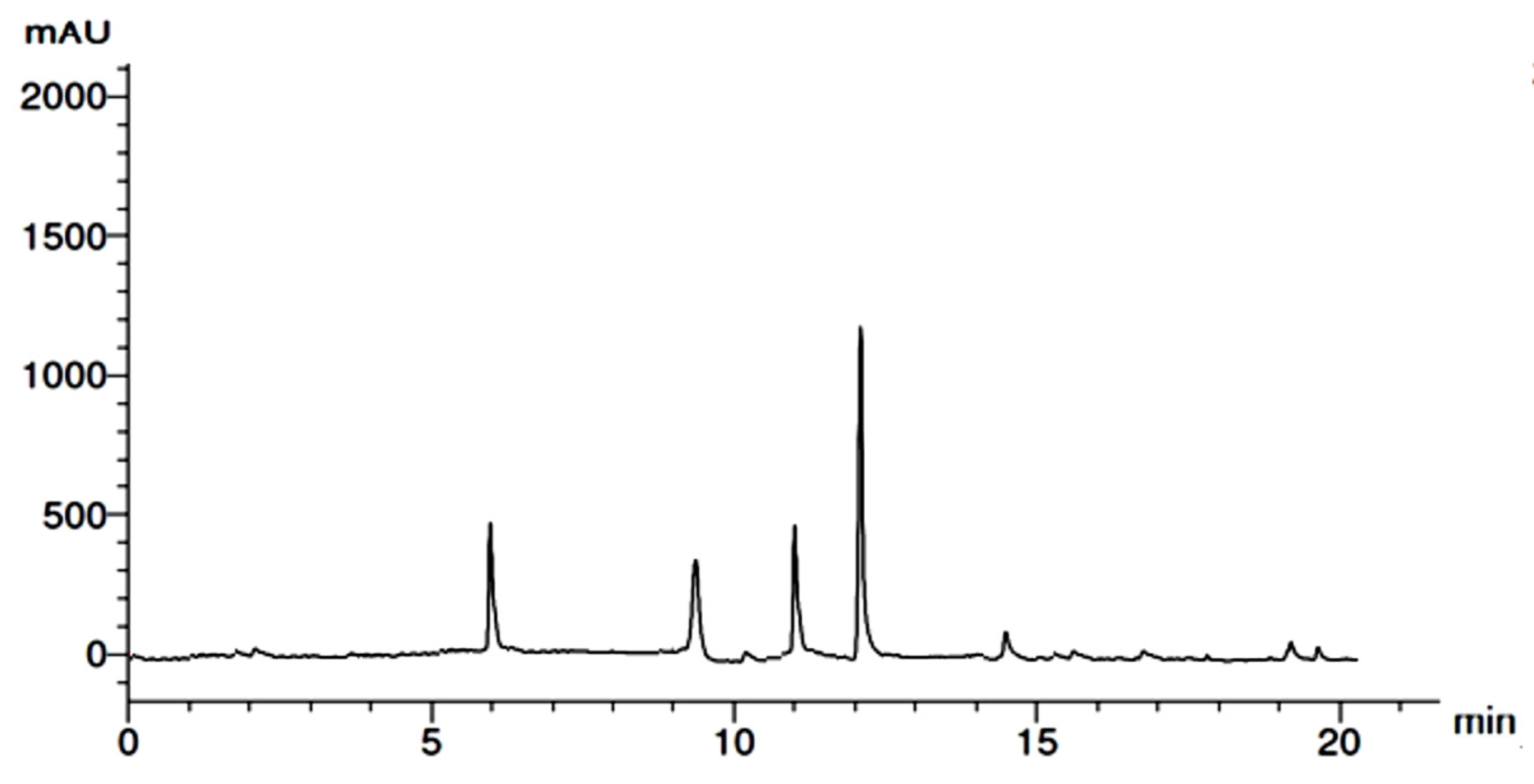
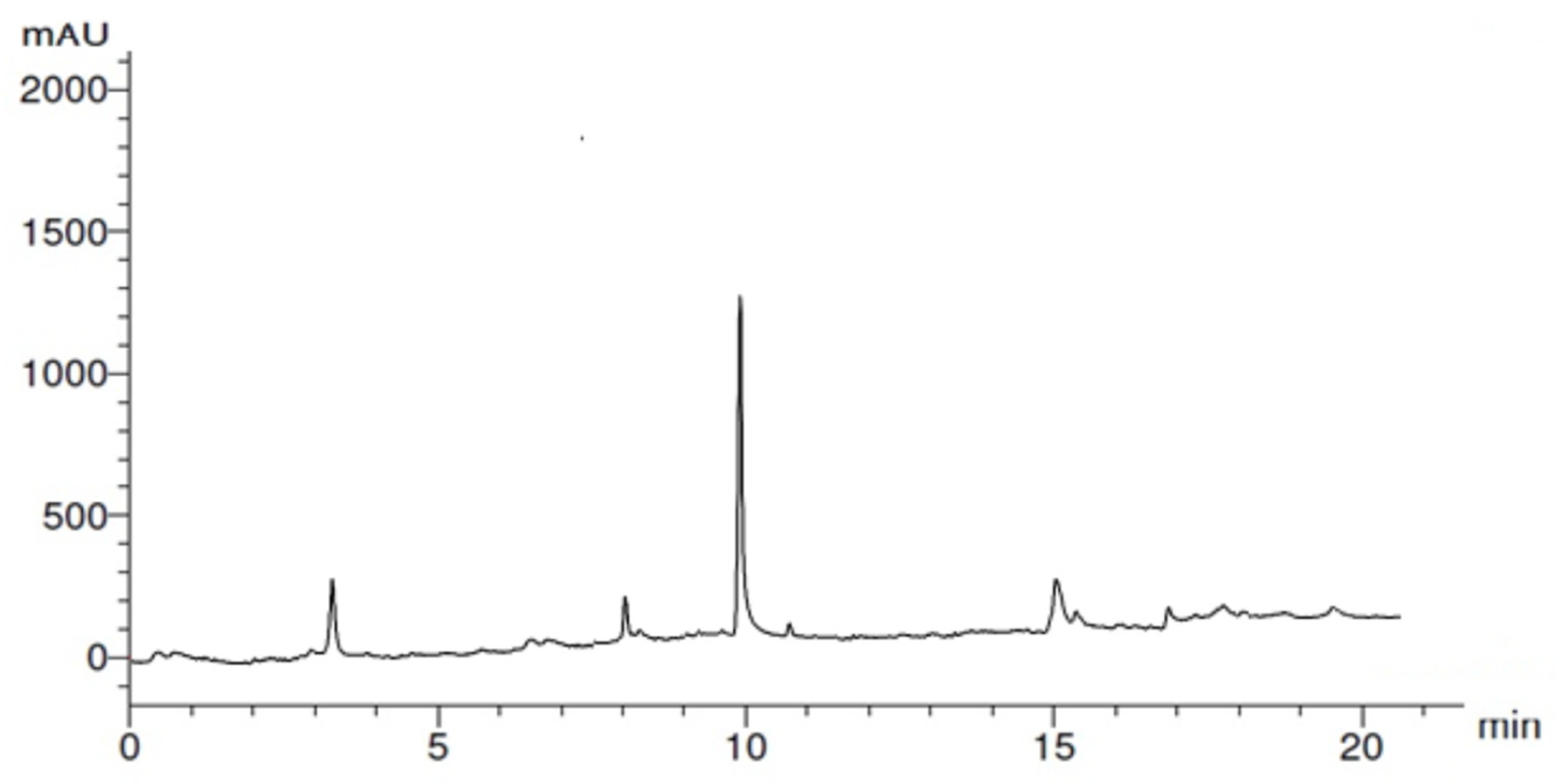
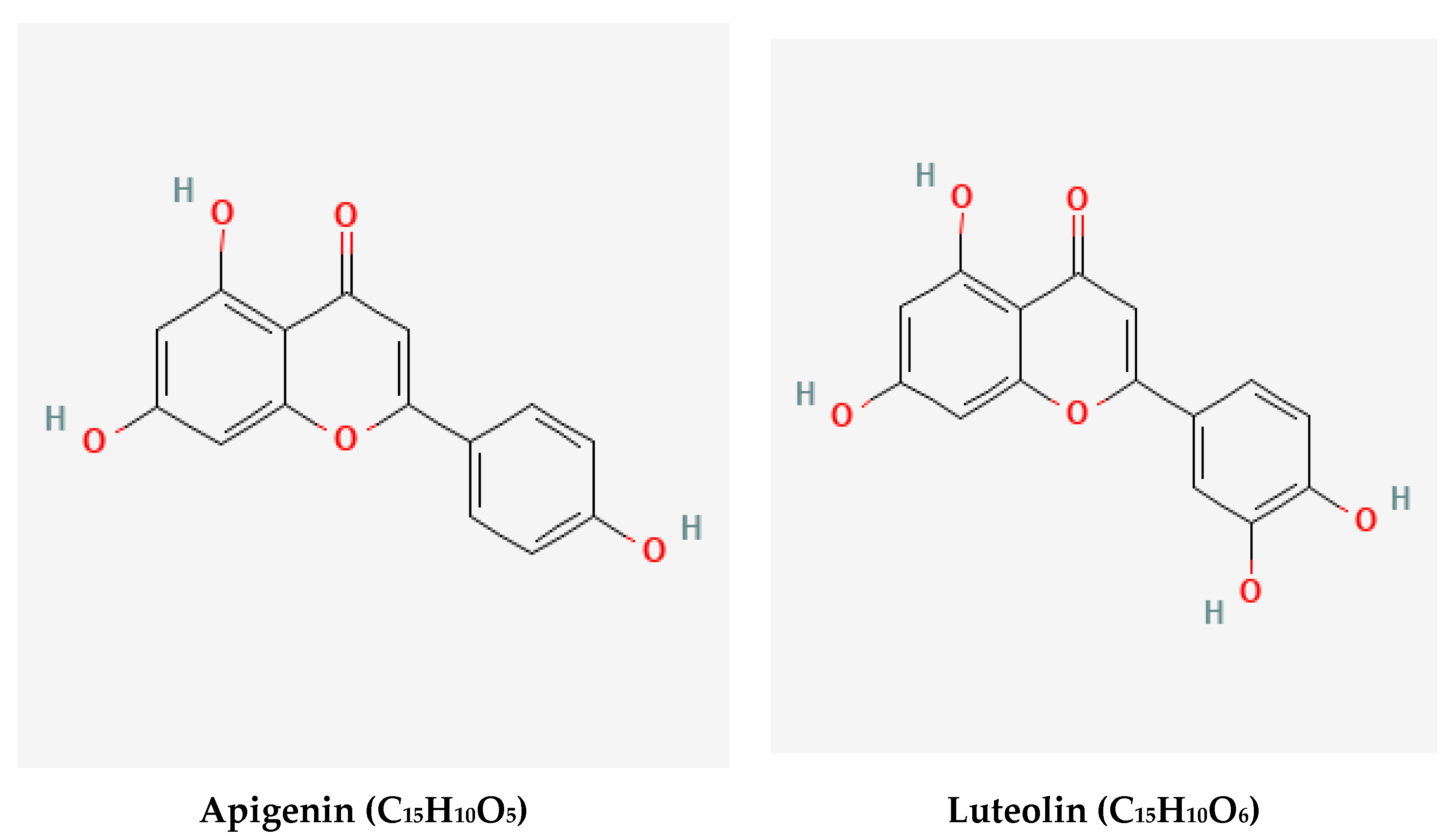
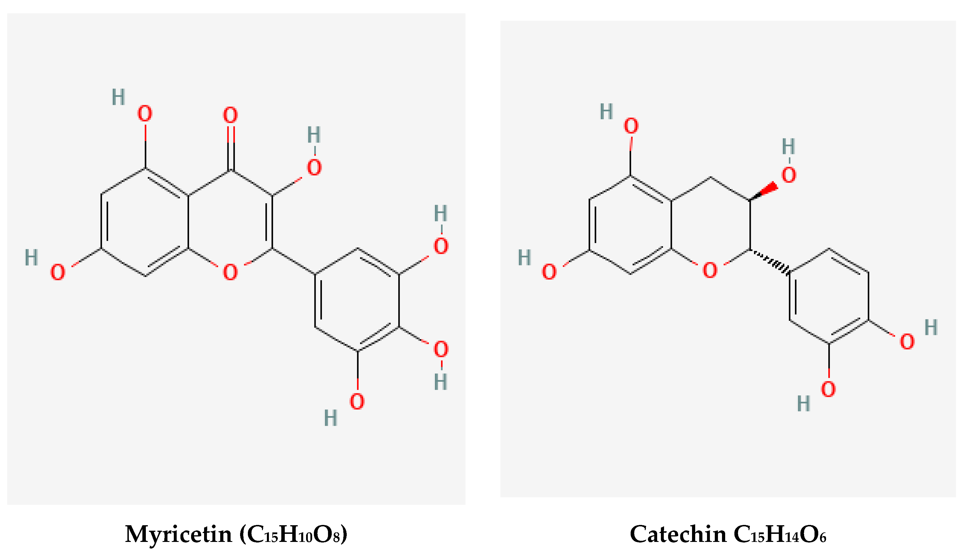
2.1. HPLC Analysis of Ginkgo Leaves Extract
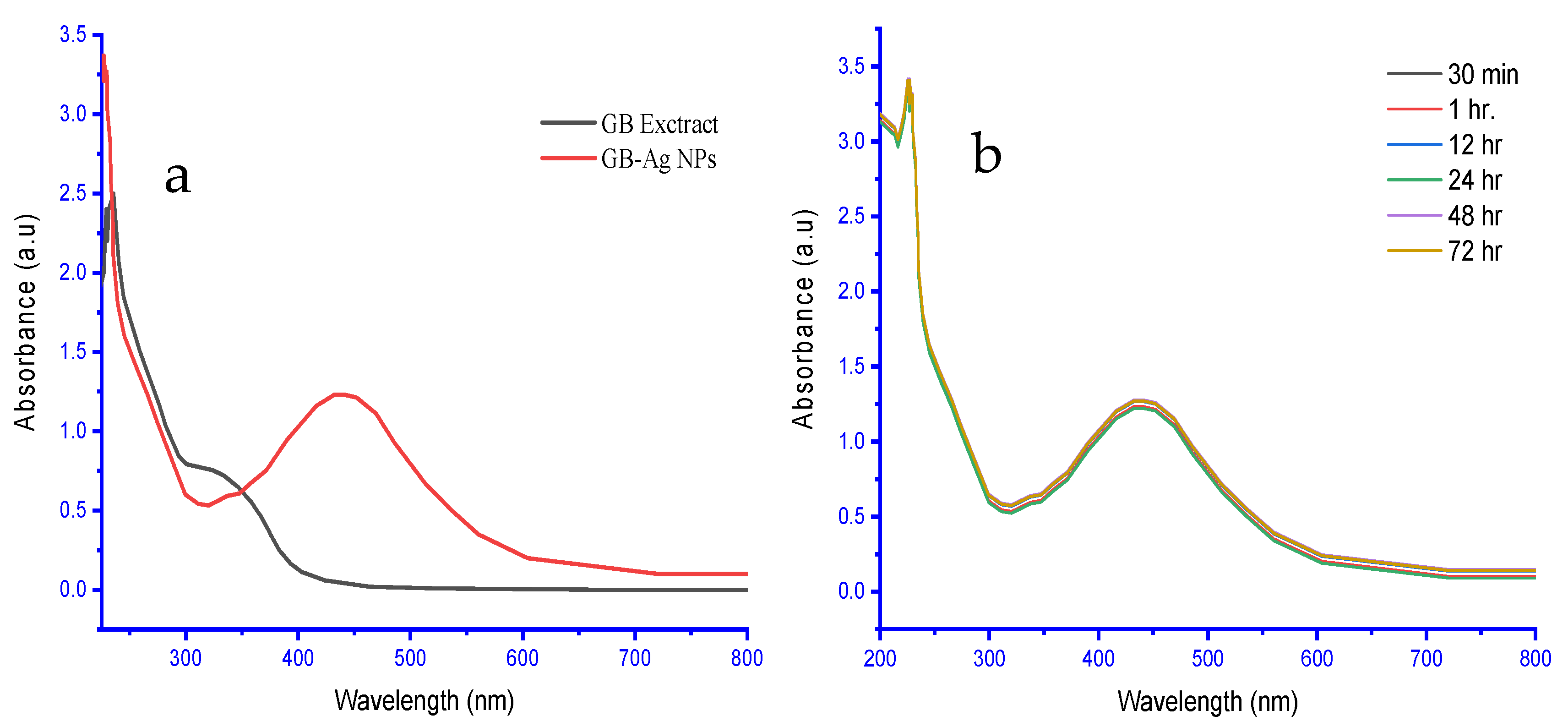
2.2. UV–Vis Analysis
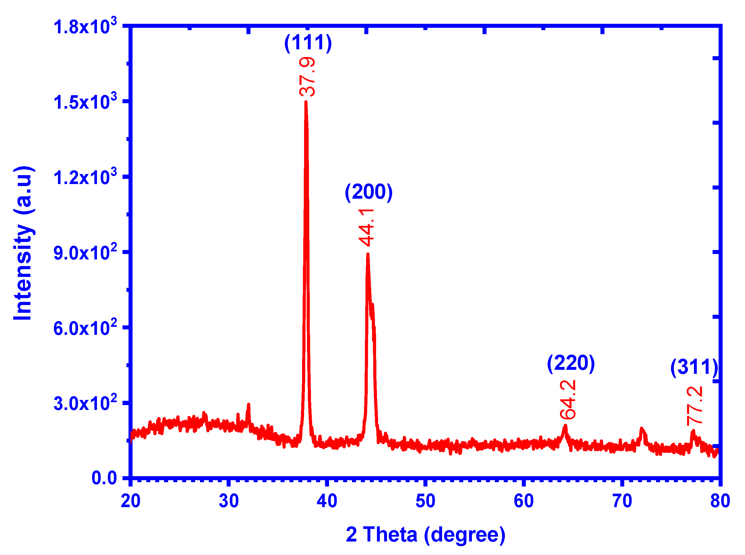
2.3. XRD Analysis
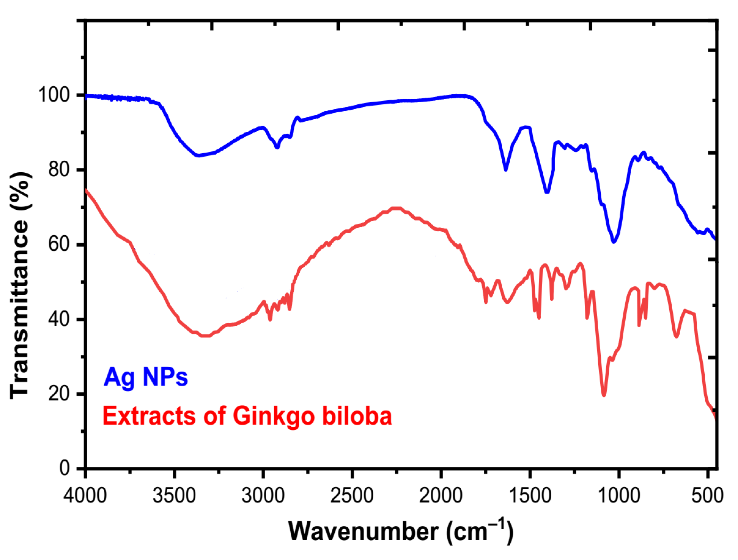
2.4. FT-IR Analysis
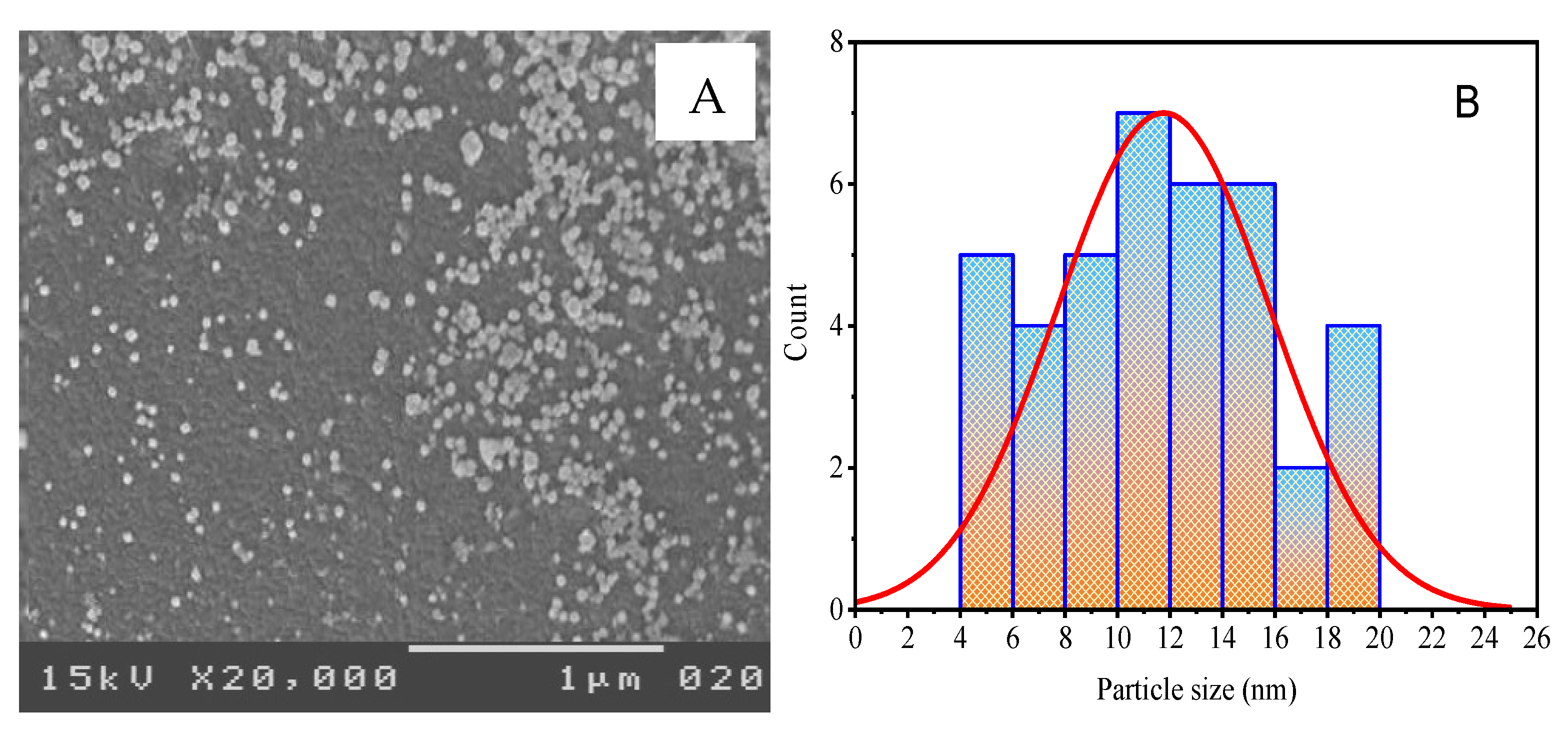
2.5. SEM Analysis
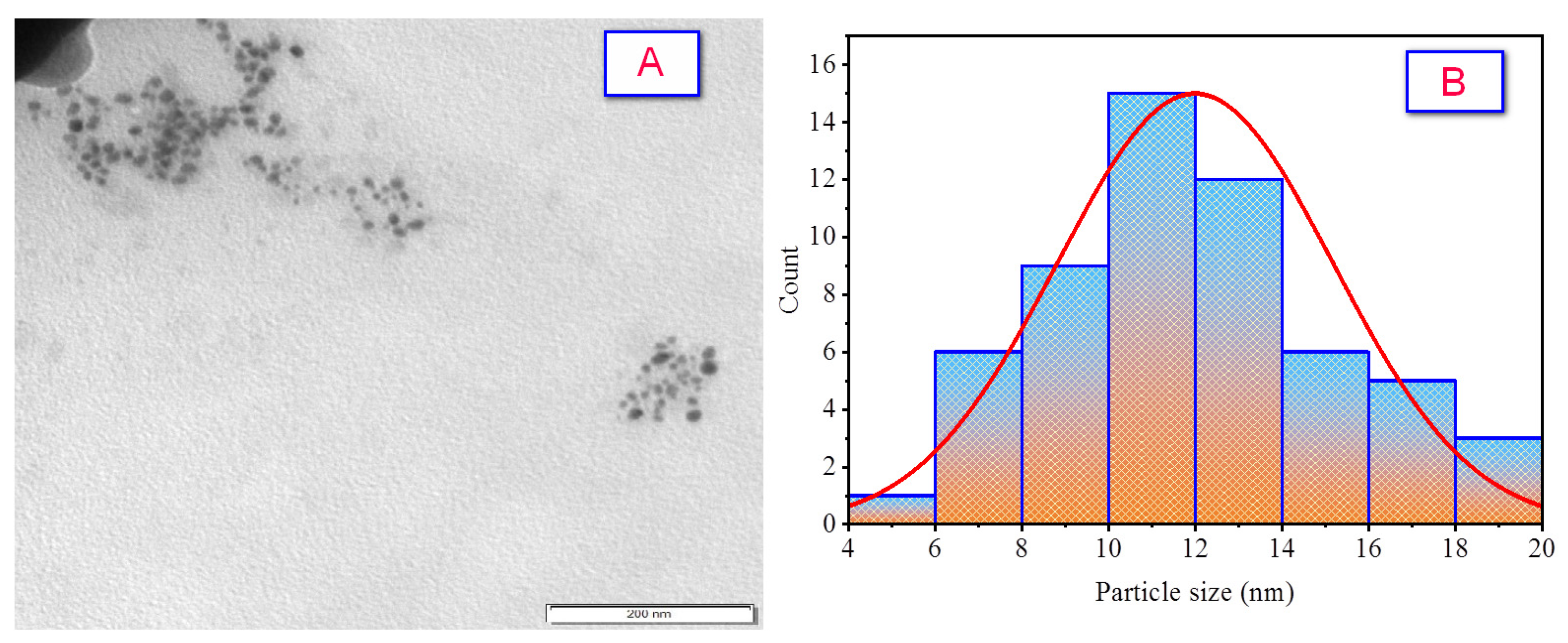
2.6. TEM Analysis
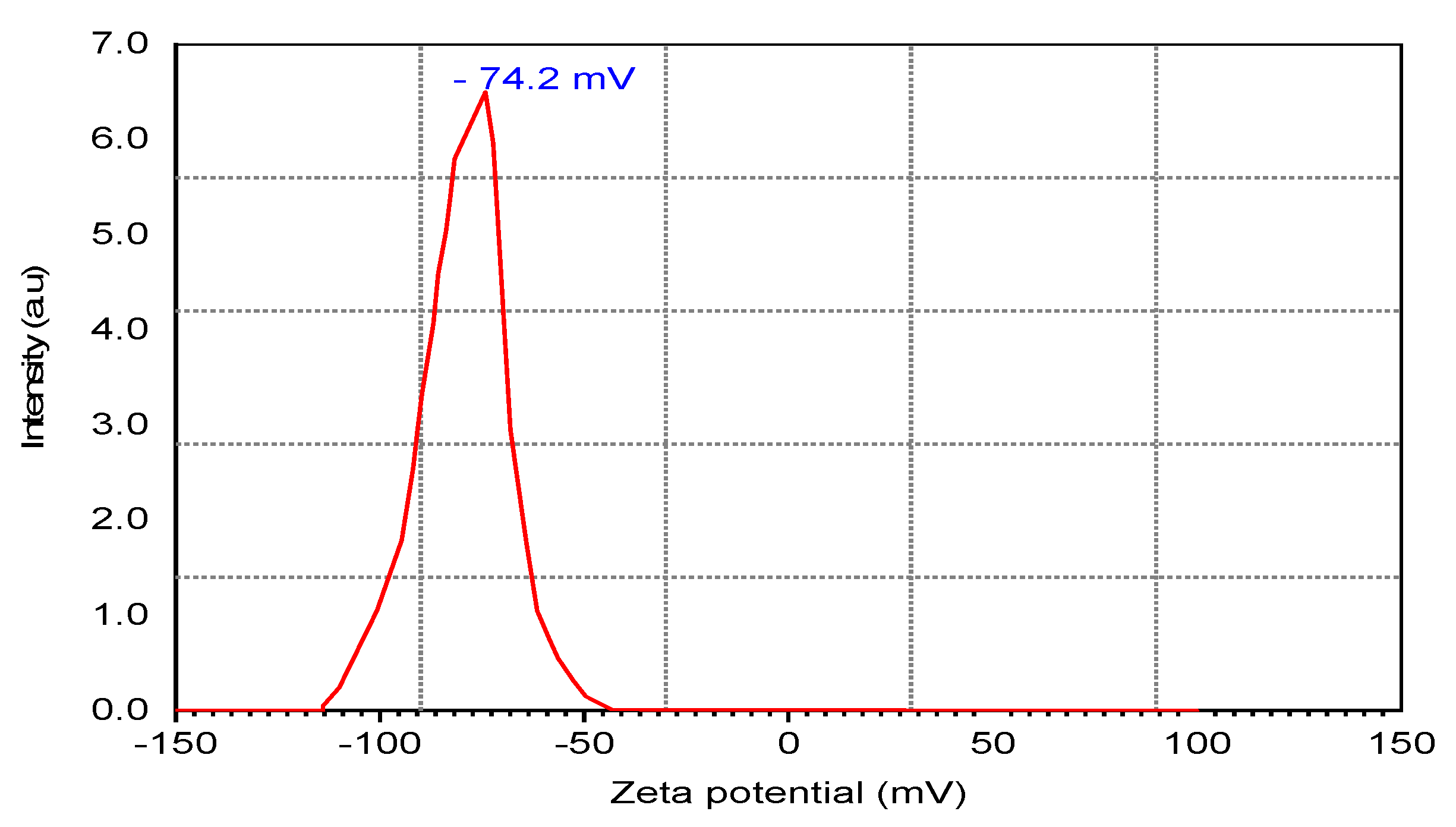
2.7. Zeta Potential
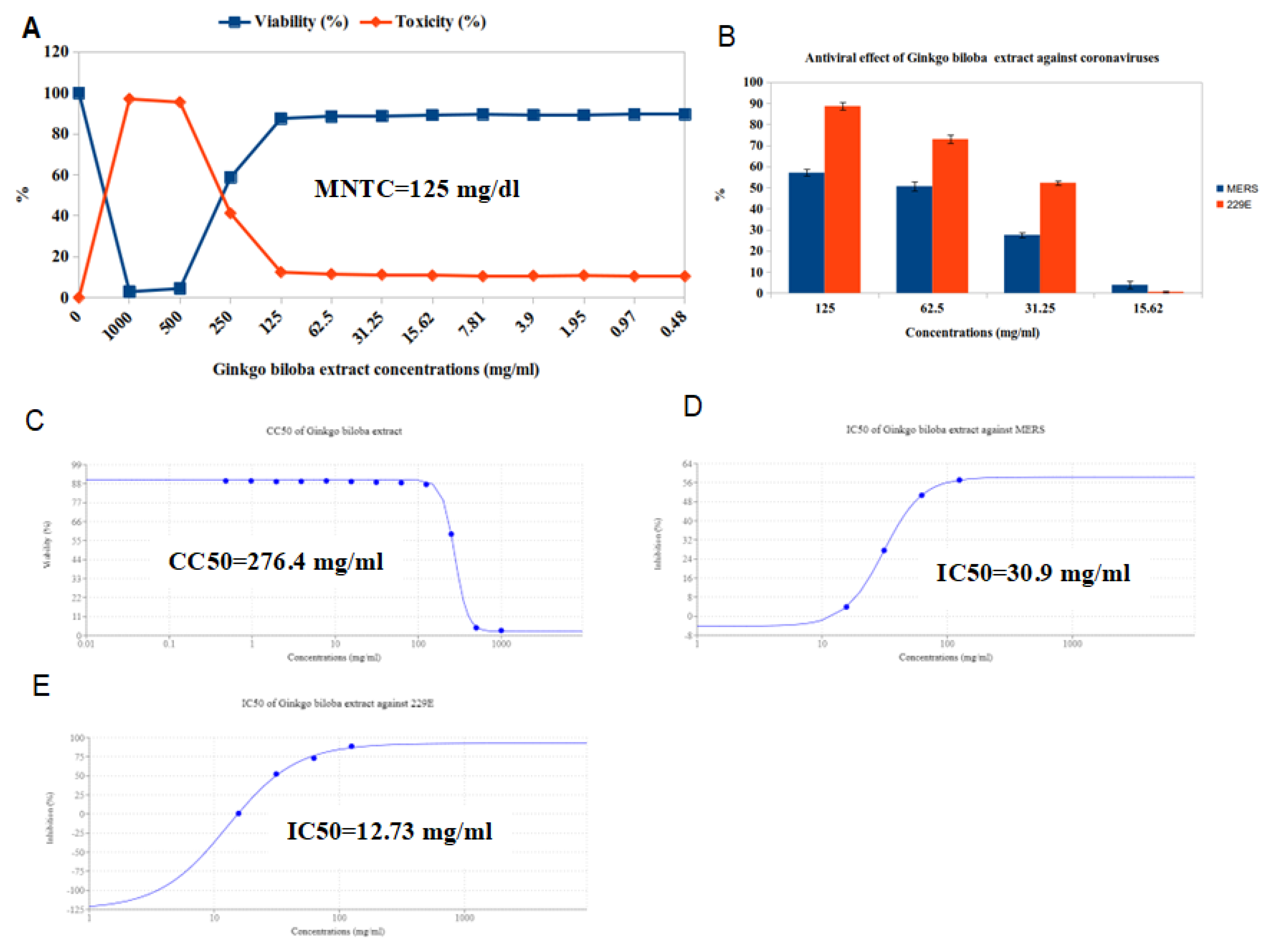
2.8. Anti-Coronaviruses Activity
3. Materials and Methods
3.1. Materials
3.2. The Active Compounds of Ginkgo Leaves Extract
3.3. Total Phenolics Content
3.4. Total Flavonoids Content
3.5. HPLC Analysis
3.6. The Biogenic Synthesis of Ag NPs
3.7. Characterization
3.8. Cytotoxicity of Ginkgo Biloba Extract and Ag NPs on VERO Cells and Viruses
3.9. MTT Assay Protocol
4. Conclusions
Author Contributions
Funding
Institutional Review Board Statement
Informed Consent Statement
Data Availability Statement
Conflicts of Interest
Sample Availability
References
- Van Beek, T.A.; Montoro, P. Chemical analysis and quality control of Ginkgo biloba leaves, extracts, and phytopharmaceuticals. J. Chromatogr. A 2009, 1216, 2002–2032. [Google Scholar] [CrossRef]
- Noor-E-Tabassum, U.; Das, R.; Lami, M.S.; Chakraborty, A.J.; Mitra, S.; Tallei, T.E.; Idroes, R.; Mohamed, A.A.-R.; Hossain, J.; Dhama, K.; et al. Ginkgo biloba: A Treasure of Functional Phytochemicals with Multimedicinal Applications. Evid. Based Complement. Altern. Med. 2022. [Google Scholar] [CrossRef]
- Chan, P.-C.; Xia, Q.; Fu, P. Ginkgo biloba leave extract: Biological, medicinal, and toxicological effects. J. Environ. Sci. Health Part C 2007, 25, 211–244. [Google Scholar] [CrossRef]
- Zha, J.; Dong, C.; Wang, X.; Zhang, X.; Xiao, X.; Yang, X. Green synthesis and characterization of monodisperse gold nanoparticles using Ginkgo biloba leaf extract. Optik 2017, 144, 511–521. [Google Scholar] [CrossRef]
- Nasrollahzadeh, M.; Sajadi, S.M. Green synthesis of copper nanoparticles using Ginkgo biloba L. leaf extract and their catalytic activity for the Huisgen [3 + 2] cycloaddition of azides and alkynes at room temperature. J. Colloid Interface Sci. 2015, 457, 141–147. [Google Scholar] [CrossRef]
- Gurunathan, S.; Han, J.W.; Park, J.H.; Eppakayala, V.; Kim, J.-H. Ginkgo biloba: A natural reducing agent for the synthesis of cytocompatible graphene. Int. J. Nanomed. 2014, 9, 363–377. [Google Scholar] [CrossRef]
- Ren, Y.-Y.; Yang, H.; Wang, T.; Wang, C. Green synthesis and antimicrobial activity of monodisperse silver nanoparticles synthesized using Ginkgo Biloba leaf extract. Phys. Lett. A 2016, 380, 3773–3777. [Google Scholar] [CrossRef]
- Abdulhadi, S.; Gouda, G.; Hamed, M.; Abu-Saied, M.; El-Mottaleb, M. Synthesis of zno nano powders using polyethylene glycol by the controlled microwave method. Bull. Pharmac. Sci. Assiut 2022, 45, 23–40. [Google Scholar] [CrossRef]
- Al-Hakkani, M.F.; Gouda, G.; Hassan, S. A review of green methods for phyto-fabrication of hematite (α-Fe2O3) nanoparticles and their characterization, properties, and applications. Heliyon 2021, 7, e05806. [Google Scholar] [CrossRef]
- Docea, A.O.; Calina, D.; Buga, A.M.; Zlatian, O.; Paoliello, M.; Mogosanu, G.D.; Streba, C.T.; Popescu, E.L.; Stoica, A.E.; Bîrcă, A.C.; et al. The Effect of Silver Nanoparticles on Antioxidant/Pro-Oxidant Balance in a Murine Model. Int. J. Mol. Sci. 2020, 21, 1233. [Google Scholar] [CrossRef] [Green Version]
- Dhanjal, D.S.; Mehra, P.; Bhardwaj, S.; Singh, R.; Sharma, P.; Nepovimova, E.; Chopra, C.; Kuca, K. Mycology-Nanotechnology Interface: Applications in Medicine and Cosmetology. Int. J. Nanomed. 2022, 17, 2505–2533. [Google Scholar] [CrossRef]
- Elnosary, M.; Aboelmagd, H.; Sofy, M.R.; Sofy, A.; Elshazly, E.H. Antiviral and Antibacterial Properties of synthesis Silver Nanoparticles with Nigella arvensis Aqueous Extract. Egypt. J. Chem. 2022, in press. [Google Scholar] [CrossRef]
- Liao, C.; Li, Y.; Tjong, S.C. Bactericidal and Cytotoxic Properties of Silver Nanoparticles. Int. J. Mol. Sci. 2019, 20, 449. [Google Scholar] [CrossRef]
- Boca, S.C.; Potara, M.; Gabudean, A.-M.; Juhem, A.; Baldeck, P.L.; Astilean, S. Chitosan-coated triangular silver nanoparticles as a novel class of biocompatible, highly effective photothermal transducers for in vitro cancer cell therapy. Cancer Lett. 2011, 311, 131–140. [Google Scholar] [CrossRef]
- Tian, J.; Wong, K.K.; Ho, C.M.; Lok, C.N.; Yu, W.Y.; Che, C.M.; Cliu, J.F.; Tam, P.K. Topical delivery of silver nanoparticles promotes wound healing. ChemMedChem 2007, 2, 129–136. [Google Scholar] [CrossRef]
- Liu, X.; Lee, P.-Y.; Ho, C.-M.; Lui, V.C.H.; Chen, Y.; Che, C.-M.; Tam, P.K.H.; Wong, K.K.Y. Silver Nanoparticles Mediate Differential Responses in Keratinocytes and Fibroblasts during Skin Wound Healing. ChemMedChem 2010, 5, 468–475. [Google Scholar] [CrossRef]
- Rigo, C.; Ferroni, L.; Tocco, I.; Roman, M.; Munivrana, I.; Gardin, C.; Cairns, W.R.L.; Vindigni, V.; Azzena, B.; Barbante, C.; et al. Active Silver Nanoparticles for Wound Healing. Int. J. Mol. Sci. 2013, 14, 4817–4840. [Google Scholar] [CrossRef]
- Pallavicini, P.; Arciola, C.R.; Bertoglio, F.; Curtosi, S.; Dacarro, G.; D’Agostino, A.; Ferrari, F.; Merli, D.; Milanese, C.; Rossi, S.; et al. Silver nanoparticles synthesized and coated with pectin: An ideal compromise for anti-bacterial and anti-biofilm action combined with wound-healing properties. J. Colloid Interface Sci. 2017, 498, 271–281. [Google Scholar] [CrossRef]
- Benyettou, F.; Rezgui, R.; Ravaux, F.; Jaber, T.; Blumer, K.; Jouiad, M.; Motte, L.; Olsen, J.-C.; Platas-Iglesias, C.; Magzoub, M.; et al. Synthesis of silver nanoparticles for the dual delivery of doxorubicin and alendronate to cancer cells. J. Mater. Chem. B 2015, 3, 7237–7245. [Google Scholar] [CrossRef]
- Qin, G.; Tang, S.; Li, S.; Lu, H.; Wang, Y.; Zhao, P.; Li, B.; Zhang, J.; Peng, L. Toxicological evaluation of silver nanoparticles and silver nitrate in rats following 28 days of repeated oral exposure. Environ. Toxicol. 2016, 32, 609–618. [Google Scholar] [CrossRef]
- Park, K. Toxicokinetic Differences and Toxicities of Silver Nanoparticles and Silver Ions in Rats After Single Oral Administration. J. Toxicol. Environ. Health Part A 2013, 76, 1246–1260. [Google Scholar] [CrossRef]
- Bergin, I.L.; Wilding, L.A.; Morishita, M.; Walacavage, K.; Ault, A.; Axson, J.L.; Stark, D.I.; Hashway, S.A.; Capracotta, S.S.; Leroueil, P.R.; et al. Effects of particle size and coating on toxicologic parameters, fecal elimination kinetics and tissue distribution of acutely ingested silver nanoparticles in a mouse model. Nanotoxicology 2016, 10, 352–360. [Google Scholar] [CrossRef]
- Gerba, C.P.; Betancourt, W.Q. Viral Aggregation: Impact on Virus Behavior in the Environment. Environ. Sci. Technol. 2017, 51, 7318–7325. [Google Scholar] [CrossRef]
- Rivera, A.; Messaoudi, I. Pathophysiology of Ebola Virus Infection: Current Challenges and Future Hopes. ACS Infect. Dis. 2015, 1, 186–197. [Google Scholar] [CrossRef]
- Colpitts, C.C.; Verrier, E.R.; Baumert, T.F. Targeting Viral Entry for Treatment of Hepatitis B and C Virus Infections. ACS Infect. Dis. 2015, 1, 420–427. [Google Scholar] [CrossRef]
- Debnath, S.K.; Srivastava, R.; Omri, A. Emerging therapeutics for the management of COVID 19. Expert Opin. Emerg. Drugs 2020, 25, 337–351. [Google Scholar] [CrossRef]
- ZeinEldin, R.A.; Ahmed, M.M.; Hassanein, W.S.; Elshafey, N.; Sofy, A.R.; Hamedo, H.A.; Elnosary, M.E. Diversity and Distribution Characteristics of Viruses from Soda Lakes. Genes 2023, 14, 323. [Google Scholar] [CrossRef]
- Zaki, A.M.; Van Boheemen, S.; Bestebroer, T.M.; Osterhaus, A.D.M.E.; Fouchier, R.A.M. Isolation of a Novel Coronavirus from a Man with Pneumonia in Saudi Arabia. N. Engl. J. Med. 2012, 367, 1814–1820. [Google Scholar] [CrossRef]
- Excler, J.-L.; Delvecchio, C.; Wiley, R.; Williams, M.; Yoon, I.-K.; Modjarrad, K.; Boujelal, M.; Moorthy, V.; Hersi, A.; Kim, J. Toward developing a preventive MERS-CoV vaccine—Report from a workshop organized by the Saudi Arabia Ministry of Health and the International Vaccine Institute, Riyadh, Saudi Arabia, 14–15 November 2015. Emerg. Infect. Dis. 2016, 22, e160229. [Google Scholar] [CrossRef]
- McNamara, P.; van Doorn, H.R. Respiratory viruses and atypical bacteria. In Manson’s Tropical Infectious Diseases; Elsevier: Amsterdam, The Netherlands, 2013; pp. 215–224. [Google Scholar] [CrossRef]
- Mori, Y.; Ono, T.; Miyahira, Y.; Nguyen, V.Q.; Matsui, T.; Ishihara, M. Antiviral activity of silver nanoparticle/chitosan composites against H1N1 influenza A virus. Nanoscale Res. Lett. 2013, 8, 93. [Google Scholar] [CrossRef]
- Lara, H.H.; Ayala-Nuñez, N.V.; Ixtepan-Turrent, L.; Rodriguez-Padilla, C. Mode of antiviral action of silver nanoparticles against HIV-1. J. Nanobiotechnol. 2010, 8, 1. [Google Scholar] [CrossRef]
- Huy, T.Q.; Thanh, N.T.H.; Thuy, N.T.; Van Chung, P.; Hung, P.N.; Le, A.-T. Cytotoxicity and antiviral activity of electrochemical—Synthesized silver nanoparticles against poliovirus. J. Virol. Methods 2017, 241, 52–57. [Google Scholar] [CrossRef] [PubMed]
- Jeremiah, S.S.; Miyakawa, K.; Morita, T.; Yamaoka, Y.; Ryo, A. Potent antiviral effect of silver nanoparticles on SARS-CoV-2. Biochem. Biophys. Res. Commun. 2020, 533, 195–200. [Google Scholar] [CrossRef]
- Xue, T.; Roy, R. Studying traditional Chinese medicine. Sciences 2003, 300, 740–741. [Google Scholar] [CrossRef] [PubMed]
- van Beek, T.A. Chemical analysis of Ginkgo biloba leaves and extracts. J. Chromatogr. A 2002, 967, 21–55. [Google Scholar] [CrossRef] [PubMed]
- Al-Hakkani, M.F.; Gouda, G.A.; Hassan, S.H.; Farghaly, O.A.; Mohamed, M.M. Fully investigation of rp-hplc analytical method validation parameters for determination of cefixime traces in the different pharmaceutical dosage forms and urine analysis. Acta Pharm. Sci. 2021, 59, 631. [Google Scholar] [CrossRef]
- Ražná, K.; Sawinska, Z.; Ivanišová, E.; Vukovic, N.; Terentjeva, M.; Stričík, M.; Kowalczewski, P.; Hlavačková, L.; Rovná, K.; Žiarovská, J. Properties of Ginkgo biloba L.: Antioxidant characterization, antimicrobial activities, and genomic microRNA based marker fingerprints. Int. J. Mol. Sci. 2020, 21, 3087. [Google Scholar] [CrossRef]
- Kobus, J.; Flaczyk, E.; Siger, A.; Nogala-Kałucka, M.; Korczak, J.; Pegg, R.B. Phenolic compounds and antioxidant activity of extracts of Ginkgo leaves. Eur. J. Lipid Sci. Technol. 2009, 111, 1150–1160. [Google Scholar] [CrossRef]
- Zhang, Q.; Chen, L.-J.; Ye, H.-Y.; Gao, L.; Hou, W.; Tang, M.; Yang, G.; Zhong, Z.; Yuan, Y.; Peng, A. Isolation and purification of ginkgo flavonol glycosides from Ginkgo biloba leaves by high-speed counter-current chromatography. J. Sep. Sci. 2007, 30, 2153–2159. [Google Scholar] [CrossRef]
- Qa’Dan, F.; Nahrstedt, A.; Schmidt, M.; Mansoor, K. Polyphenols from Ginkgo biloba. Sci. Pharm. 2010, 78, 897–907. [Google Scholar] [CrossRef]
- Al-Mokadem, A.Z.; Alnaggar, A.E.-A.M.; Mancy, A.G.; Sofy, A.R.; Sofy, M.R.; Mohamed, A.K.S.H.; Abou Ghazala, M.M.A.; El-Zabalawy, K.M.; Salem, N.F.G.; Elnosary, M.E.; et al. Foliar Application of Chitosan and Phosphorus Alleviate the Potato virus Y-Induced Resistance by Modulation of the Reactive Oxygen Species, Antioxidant Defense System Activity and Gene Expression in Potato. Agronomy 2022, 12, 3064. [Google Scholar] [CrossRef]
- Rimkiene, L.; Baranauskaite, J.; Marksa, M.; Jarukas, L.; Ivanauskas, L. Development and Evaluation of Ginkgo biloba L. Extract Loaded into Carboxymethyl Cellulose Sublingual Films. Appl. Sci. 2020, 11, 270. [Google Scholar] [CrossRef]
- Shui-Yuan, C.; Feng, X.; Yan, W. Advances in the study of flavonoids in Ginkgo biloba leaves. J. Med. Plants Res. 2009, 3, 1248–1252. [Google Scholar]
- Ellnain-Wojtaszek, M.; Zgórka, G. High-performance liquid chromatography and thin-layer chromatography of phenolic acids from Ginkgo biloba L. leaves collected within vegetative period. J. Liq. Chromatogr. Relat. Technol. 1999, 22, 1457–1471. [Google Scholar] [CrossRef]
- Meng, J.; Wang, B.; He, G.; Wang, Y.; Tang, X.; Wang, S.; Ma, Y.; Fu, C.; Chai, G.; Zhou, G. Metabolomics Integrated with Transcriptomics Reveals Redirection of the Phenylpropanoids Metabolic Flux in Ginkgo biloba. J. Agric. Food Chem. 2019, 67, 3284–3291. [Google Scholar] [CrossRef] [PubMed]
- Ban, C.; Park, J.-B.; Cho, S.; Kim, H.R.; Kim, Y.J.; Bae, H.; Kim, C.; Kang, H.; Jang, D.; Shin, Y.S.; et al. Characterization of Ginkgo biloba Leaf Flavonoids as Neuroexocytosis Regulators. Molecules 2020, 25, 1829. [Google Scholar] [CrossRef] [PubMed]
- Gruenwald, J.; Eckert, A.; Kressig, R.W. The Effects of Standardized Ginkgo Biloba Extracts (GBE) on Subjective Cognitive Decline (SCD) in Middle-Aged Adults: A Review. Adv. Aging Res. 2020, 9, 45–65. [Google Scholar] [CrossRef]
- Saxena, M.; Saxena, J.; Pradhan, A. Flavonoids and phenolic acids as antioxidants in plants and human health. Int. J. Pharm. Sci. Rev. Res. 2012, 16, 130–134. [Google Scholar]
- Anandalakshmi, K.; Venugobal, J.; Ramasamy, V. Characterization of silver nanoparticles by green synthesis method using Pedalium murex leaf extract and their antibacterial activity. Appl. Nanosci. 2015, 6, 399–408. [Google Scholar] [CrossRef]
- Kanwal, Z.; Raza, M.; Riaz, S.; Manzoor, S.; Tayyeb, A.; Sajid, I.; Naseem, S. Synthesis and characterization of silver nanoparticle-decorated cobalt nanocomposites (Co@AgNPs) and their density-dependent antibacterial activity. R. Soc. Open Sci. 2019, 6, 182135. [Google Scholar] [CrossRef]
- Panja, S.; Chaudhuri, I.; Khanra, K.; Bhattacharyya, N. Biological application of green silver nanoparticle synthesized from leaf extract of Rauvolfia serpentina Benth. Asian Pac. J. Trop. Dis. 2016, 6, 549–556. [Google Scholar] [CrossRef]
- Al-Hakkani, M.F.; Gouda, G.A.; Hassan, S.H.; Mohamed, M.M.; Nagiub, A.M. Cefixime wastewater management via bioengineered Hematite nanoparticles and the in-vitro synergetic potential multifunction activities of Cefixime@Hematite nanosystem. Surf. Interfaces 2022, 30, 101877. [Google Scholar] [CrossRef]
- Al-Hakkani, M.F.; Gouda, G.; Hassan, S.; Nagiub, A. Echinacea purpurea mediated hematite nanoparticles (α-HNPs) biofabrication, characterization, physicochemical properties, and its in-vitro biocompatibility evaluation. Surf. Interfaces 2021, 24, 101113. [Google Scholar] [CrossRef]
- Elshazly, E.H.; Mohamed, A.K.S.H.; Aboelmagd, H.A.; Gouda, G.A.; Abdallah, M.H.; Ewais, E.A.; Assiri, M.A.; Ali, G.A.M. Phytotoxicity and Antimicrobial Activity of Green Synthesized Silver Nanoparticles Using Nigella sativa Seeds on Wheat Seedlings. J. Chem. 2022, 2022, 9609559. [Google Scholar] [CrossRef]
- Patterson, A.L. The Scherrer Formula for X-Ray Particle Size Determination. Phys. Rev. 1939, 56, 978–982. [Google Scholar] [CrossRef]
- Wojtoniszak, M.; Chen, X.; Kalenczuk, R.; Wajda, A.; Łapczuk, J.; Kurzewski, M.; Drozdzik, M.; Chu, P.; Borowiak-Palen, E. Synthesis, dispersion, and cytocompatibility of graphene oxide and reduced graphene oxide B Biointerfaces. Colloids Surf. B Biointerfaces 2012, 89, 79–85. [Google Scholar] [CrossRef]
- Al-Farhan, B.S. Potentiometeric Study of New Schiff Base Complexes Bearing Morpholine in Ethanol-water Medium with some Metal Ions. Int. J. Electrochem. Sci. 2019, 14, 3350–3362. [Google Scholar] [CrossRef]
- Wang, W.; Kang, Q.; Liu, N.; Zhang, Q.; Zhang, Y.; Li, H.; Zhao, B.; Chen, Y.; Lan, Y.; Ma, Q.; et al. Enhanced dissolution rate and oral bioavailability of Ginkgo biloba extract by preparing solid dispersion via hot-melt extrusion. Fitoterapia 2015, 102, 189–197. [Google Scholar] [CrossRef]
- Gahlan, A.; Hassan, R.; Gouda, G.A.; Aly-Eldeen, M.A.-E.; Badawy, N.A. Development of zinc removal process from contaminated water using statistical approaches. Egypt. J. Chem. 2022, 65, 13. [Google Scholar] [CrossRef]
- Al-Saidi, H.M.; Gouda, G.; Abdel-Hakim, M.; Alsenani, N.; Alfarsi, A.; Mahross, M.; Farghaly, O.; Hosny, S. Synthesis and Characterization of Ni (II), Cu (II), Zn (II) and Azo Dye Based on 1, 10-o-Phenanthroline Binary Complexes: Corrosion Inhibition Properties and Computational Studies. Int. J. Electrochem. Sci. 2022, 17, 2. [Google Scholar]
- Osman, A.H.; Aly, A.A.M.; Elmottaleb, M.A.; Gouda, G.A.H. Photoreactivity and Thermogravimetry of Copper(II) Complexes of N-Salicylideneaniline and Its Derivatives. Bull. Korean Chem. Soc. 2004, 25, 45–50. [Google Scholar] [CrossRef]
- Sayed, A.S.; Abdelmottaleb, M.; Cheira, M.F.; Abdel-Aziz, G.; Gomaa, H.; Hassanein, T.F. Date seed as an efficient, eco-friendly, and cost-effective bio-adsorbent for removal of thorium ions from acidic solutions. Aswan Univ. J. Environ. Stud. 2020, 1, 106–124. [Google Scholar] [CrossRef]
- Melkamu, W.W.; Bitew, L.T. Green synthesis of silver nanoparticles using Hagenia abyssinica (Bruce) J.F. Gmel plant leaf extract and their antibacterial and antioxidant activities. Heliyon 2021, 7, e08459. [Google Scholar] [CrossRef]
- Aziz, S.B.; Hussein, G.; Brza, M.A.; Mohammed, S.J.; Abdulwahid, R.T.; Saeed, S.R.; Hassanzadeh, A. Fabrication of Interconnected Plasmonic Spherical Silver Nanoparticles with Enhanced Localized Surface Plasmon Resonance (LSPR) Peaks Using Quince Leaf Extract Solution. Nanomaterials 2019, 9, 1557. [Google Scholar] [CrossRef]
- Tarahovsky, Y.S.; Kim, Y.; Yagolnik, E.; Muzafarov, E. Flavonoid–membrane interactions: Involvement of flavonoid–metal complexes in raft signaling. Biochim. Biophys. Acta (BBA) Biomembr. 2014, 1838, 1235–1246. [Google Scholar] [CrossRef]
- Atmani, D.; Chaher, N.; Atmani, D.; Berboucha, M.; Debbache, N.; Boudaoud, H. Flavonoids in Human Health: From Structure to Biological Activity. Curr. Nutr. Food Sci. 2009, 5, 225–237. [Google Scholar] [CrossRef]
- Hosny, S.; Gouda, G.A.; Abu-El-Wafa, S.M. Novel Nano Copper Complexes of a New Schiff Base: Green Synthesis, a New Series of Solid Cr(II), Co(II), Cu(II), Pd(II) and Cd(II) Chelates, Characterization, DFT, DNA, Antitumor and Molecular Docking Studies. Appl. Organomet. Chem. 2022, 36, e6627. [Google Scholar] [CrossRef]
- Alkhalaf, M.I.; Hussein, R.H.; Hamza, A. Green synthesis of silver nanoparticles by Nigella sativa extract alleviates diabetic neuropathy through anti-inflammatory and antioxidant effects. Saudi J. Biol. Sci. 2020, 27, 2410–2419. [Google Scholar] [CrossRef]
- Al-Hakkani, M.F.; Gouda, G.A.; Hassan, S.H.A.; Saddik, M.S.; El-Mokhtar, M.A.; Ibrahim, M.A.; Mohamed, M.M.A.; Nagiub, A.M. Cefotaxime removal enhancement via bio-nanophotocatalyst α-Fe2O3 using photocatalytic degradation technique and its echo-biomedical applications. Sci. Rep. 2022, 12, 11881. [Google Scholar] [CrossRef] [PubMed]
- Al-Hakkani, M.F.; Hassan, S.; Saddik, M.; El-Mokhtar, M.; Al-Shelkamy, S. Bioengineering, characterization, and biological activities of C@Cu2O@ Cu nanocomposite based-mediated the Vicia faba seeds aqueous extract. J. Mater. Res. Technol. 2021, 14, 1998–2016. [Google Scholar] [CrossRef]
- Wang, Z.; Zhang, J.; Ren, T.; Dong, Z. Targeted metabolomic profiling of cardioprotective effect of Ginkgo biloba L. extract on myocardial ischemia in rats. Phytomedicine 2016, 23, 621–631. [Google Scholar] [CrossRef] [PubMed]
- Srikhao, N.; Kasemsiri, P.; Lorwanishpaisarn, N.; Okhawilai, M. Green synthesis of silver nanoparticles using sugarcane leaves extract for colorimetric detection of ammonia and hydrogen peroxide. Res. Chem. Intermed. 2021, 47, 1269–1283. [Google Scholar] [CrossRef]
- Rajendrachari, S.; Be, K. Biosynthesis of Silver Nanoparticles Using Leaves of Acacia Melanoxylon and their Application as Dopamine and Hydrogen Peroxide Sensors. Phys. Chem. Res. 2020, 8, 1–18. [Google Scholar] [CrossRef]
- Kathiravan, V. Green synthesis of silver nanoparticles using different volumes of Trichodesma indicum leaf extract and their antibacterial and photocatalytic activities. Res. Chem. Intermed. 2018, 44, 4999–5012. [Google Scholar] [CrossRef]
- Jayapriya, M.; Dhanasekaran, D.; Arulmozhi, M.; Nandhakumar, E.; Senthilkumar, N.; Sureshkumar, K. Green synthesis of silver nanoparticles using Piper longum catkin extract irradiated by sunlight: Antibacterial and catalytic activity. Res. Chem. Intermed. 2019, 45, 3617–3631. [Google Scholar] [CrossRef]
- Sofy, M.R.; Mancy, A.G.; Alnagger, A.E.M.; Refaey, E.E.; Mohmed, H.I.; Elnosary, M.E.; Sofy, A.R. A polishing the harmful effects of Broad Bean Mottle Virus infecting broad bean plants by enhancing the immunity using different potassium concentrations. Int. J. Notulae Botanicae Horti Agrobotanici Cluj-Napoca 2022, 50, 12654. [Google Scholar] [CrossRef]
- Karimi, N.; Chardoli, A.; Fattahi, A. Biosynthesis, Characterization, Antimicrobial and Cytotoxic Effects of Silver Nanoparticles Using Nigella arvensis Seed Extract. Iran. J. Pharm. Res. 2017, 16, 1167–1175. [Google Scholar] [CrossRef]
- Zhang, X.-F.; Liu, Z.-G.; Shen, W.; Gurunathan, S. Silver nanoparticles: Synthesis, characterization, properties, applications, and therapeutic approaches. Int. J. Mol. Sci. 2016, 17, 1534. [Google Scholar] [CrossRef]
- Elamawi, R.M.; Al-Harbi, R.E.; Hendi, A.A. Biosynthesis and characterization of silver nanoparticles using Trichoderma longibrachiatum and their effect on phytopathogenic fungi. Egypt. J. Biol. Pest Control 2018, 28, 28. [Google Scholar] [CrossRef]
- Xu, Z.; Feng, Q.; Wang, M.; Zhao, H.; Lin, Y.; Zhou, S. Green Biosynthesized Silver Nanoparticles with Aqueous Extracts of Ginkgo Biloba Induce Apoptosis via Mitochondrial Pathway in Cervical Cancer Cells. Front. Oncol. 2020, 10, 575415. [Google Scholar] [CrossRef]
- Khalir, W.K.A.W.M.; Shameli, K.; Jazayeri, S.D.; Othman, N.A.; Jusoh, N.W.C.; Hassan, N.M. Biosynthesized Silver Nanoparticles by Aqueous Stem Extract of Entada spiralis and Screening of Their Biomedical Activity. Front. Chem. 2020, 8, 620. [Google Scholar] [CrossRef]
- Valsalam, S.; Agastian, P.; Arasu, M.V.; Al-Dhabi, N.A.; Ghilan, A.-K.M.; Kaviyarasu, K.; Ravindran, B.; Chang, S.W.; Arokiyaraj, S. Rapid biosynthesis and characterization of silver nanoparticles from the leaf extract of Tropaeolum majus L. and its enhanced in-vitro antibacterial, antifungal, antioxidant and anticancer properties. J. Photochem. Photobiol. B Biol. 2018, 191, 65–74. [Google Scholar] [CrossRef]
- Awwad, A.M.; Salem, N.M. Green Synthesis of Silver Nanoparticles by Mulberry Leaves Extract. Nanosci. Nanotechnol. 2012, 2, 125–128. [Google Scholar] [CrossRef]
- Raman, N.; Sudharsan, S.; Veerakumar, V.; Pravin, N.; Vithiya, K. Pithecellobium dulce mediated extra-cellular green synthesis of larvicidal silver nanoparticles. Spectrochim. Acta Part A Mol. Biomol. Spectrosc. 2012, 96, 1031–1037. [Google Scholar] [CrossRef]
- Taghavizadeh Yazdi, M.E.; Hamidi, A.; Amiri, M.; Oskuee, R.K.; Hosseini, H.; Hashemzadeh, A.; Darroudi, M. Eco-friendly and plant-based synthesis of silver nanoparticles using Allium giganteum and investigation of its bactericidal, cytotoxicity, and photocatalytic effects. Mater. Technol. 2019, 34, 490–497. [Google Scholar] [CrossRef]
- Betle Broth, P. Phytofabrication and characterization of silver nanoparticles from Piper betle broth. Res. J. Nanosci. Nanotechnol. 2012, 2, 17–23. [Google Scholar]
- Rastogi, L.; Arunachalam, J. Sunlight based irradiation strategy for rapid green synthesis of highly stable silver nanoparticles using aqueous garlic (Allium sativum) extract and their antibacterial potential. Mater. Chem. Phys. 2011, 129, 558–563. [Google Scholar] [CrossRef]
- Manikandan, R.; Anjali, R.; Beulaja, M.; Prabhu, N.; Koodalingam, A.; Saiprasad, G.; Chitra, P.; Arumugam, M. Synthesis, characterization, anti-proliferative and wound healing activities of silver nanoparticles synthesized from Caulerpa scalpelliformis. Process. Biochem. 2019, 79, 135–141. [Google Scholar] [CrossRef]
- Okaiyeto, K.; Hoppe, H.; Okoh, A.I. Plant-Based Synthesis of Silver Nanoparticles Using Aqueous Leaf Extract of Salvia officinalis: Characterization and its Antiplasmodial Activity. J. Clust. Sci. 2020, 32, 101–109. [Google Scholar] [CrossRef]
- Rashid, S.; Azeem, M.; Khan, S.A.; Shah, M.M.; Ahmad, R. Characterization and synergistic antibacterial potential of green synthesized silver nanoparticles using aqueous root extracts of important medicinal plants of Pakistan. Colloids Surfaces B Biointerfaces 2019, 179, 317–325. [Google Scholar] [CrossRef]
- Mekky, A.E.; Farrag, A.A.; Sofy, A.R.; Hamed, A.A. Antibacterial and Antifungal Activity of Green-synthesized Silver Nanoparticles Using Spinacia oleracea leaves Extract. Egypt. J. Chem. 2021, 64, 5781–5792. [Google Scholar] [CrossRef]
- Elnosary, M.E.; Aboelmagd, H.A.; Habaka, M.A.; Salem, S.R.; El-Naggar, M.E. Synthesis of bee venom loaded chitosan nanoparticles for anti-MERS-COV and multi-drug resistance bacteria. Int. J. Biol. Macromol. 2023, 224, 871–880. [Google Scholar] [CrossRef] [PubMed]
- Sisay, M. 3CLpro inhibitors as a potential therapeutic option for COVID-19: Available evidence and ongoing clinical trials. Pharmacol. Res. 2020, 156, 104779. [Google Scholar] [CrossRef]
- Tao, Z.; Jin, W.; Ao, M.; Zhai, S.; Xu, H.; Yu, L. Evaluation of the anti-inflammatory properties of the active constituents in Ginkgo biloba for the treatment of pulmonary diseases. Food Funct. 2019, 10, 2209–2220. [Google Scholar] [CrossRef] [PubMed]
- Eisvand, F.; Razavi, B.M.; Hosseinzadeh, H. The effects of Ginkgo biloba on metabolic syndrome: A review. Phytotherapy Res. 2020, 34, 1798–1811. [Google Scholar] [CrossRef]
- Ryu, Y.B.; Jeong, H.; Kim, J.; Kim, Y.; Park, J.-Y.; Kim, D.; Naguyen, T.; Park, S.-J.; Chang, J.; Park, K. Biflavonoids from Torreya nucifera displaying SARS-CoV 3CLpro inhibition. Bioorganic Med. Chem. 2010, 18, 7940–7947. [Google Scholar] [CrossRef]
- Lü, J.-M.; Yan, S.; Jamaluddin, S.; Weakley, S.; Liang, Z.; Siwak, E.; Yao, Q.; Chen, C. Ginkgolic acid inhibits HIV protease activity and HIV infection in vitro. Med. Sci. Monit. Int. Med. J. Exp. Clin. Res. 2012, 18, BR293. [Google Scholar] [CrossRef]
- Borenstein, R.; Hanson, B.A.; Markosyan, R.M.; Gallo, E.S.; Narasipura, S.D.; Bhutta, M.; Shechter, O.; Lurain, N.S.; Cohen, F.S.; Al-Harthi, L.; et al. Ginkgolic acid inhibits fusion of enveloped viruses. Sci. Rep. 2020, 10, 4746. [Google Scholar] [CrossRef]
- Bosch, B.J.; van der Zee, R.; de Haan, C.A.; Rottier, P.J.M. The Coronavirus Spike Protein Is a Class I Virus Fusion Protein: Structural and Functional Characterization of the Fusion Core Complex. J. Virol. 2003, 77, 8801–8811. [Google Scholar] [CrossRef]
- Yang, X.; Ye, Y.; Wang, P.; Chen, J.; Guo, T. Study on anti-bacterium activities of extract of Ginkgo biloba leaves (EGbs) and Ginkgolic Acids (GAs). Food Sci. 2004, 25, 68–71. [Google Scholar]
- Ma, J.; Duan, W.; Han, S.; Lei, J.; Xu, Q.; Chen, X.; Jiang, Z.; Nan, L.; Li, J.; Chen, K.; et al. Ginkgolic acid suppresses the development of pancreatic cancer by inhibiting pathways driving lipogenesis. Oncotarget 2015, 6, 20993–21003. [Google Scholar] [CrossRef] [PubMed]
- Qiao, L.; Zheng, J.; Jin, X.; Wei, G.; Wang, G.; Sun, X.; Li, X. Ginkgolic acid inhibits the invasiveness of colon cancer cells through AMPK activation. Oncol. Lett. 2017, 14, 5831–5838. [Google Scholar] [CrossRef]
- Hua, Z.; Wu, C.; Fan, G.; Tang, Z.; Cao, F. The antibacterial activity and mechanism of ginkgolic acid C15:1. BMC Biotechnol. 2017, 17, 5. [Google Scholar] [CrossRef]
- Liu, H.; Li, J.; Lu, D.; Li, J.; Liu, M.; He, Y.; Williams, B.; Li, J.; Yang, T. Ginkgolic acid, a sumoylation inhibitor, promotes adipocyte commitment but suppresses adipocyte terminal differentiation of mouse bone marrow stromal cells. Sci. Rep. 2018, 8, 2545. [Google Scholar] [CrossRef] [Green Version]
- Ratan, Z.; Mashrur, F.; Chhoan, A.; Shahriar, S.; Haidere, M.; Runa, N.; Kim, S.; Kweon, D.; Hosseinzadeh, H.; Cho, J. Silver Nanoparticles as Potential Antiviral Agents. Pharmaceutics 2021, 13, 2034. [Google Scholar] [CrossRef]
- Gurunathan, S.; Qasim, M.; Choi, Y.; Do, J.; Park, C.; Hong, K.; Kim, J.-H.; Song, H. Antiviral potential of nanoparticles—Can nanoparticles fight against coronaviruses? Nanomaterials 2020, 10, 1645. [Google Scholar] [CrossRef] [PubMed]
- Mosselhy, D.A.; Virtanen, J.; Kant, R.; He, W.; Elbahri, M.; Sironen, T. COVID-19 pandemic: What about the safety of anti-coronavirus nanoparticles? Nanomaterials 2021, 11, 796. [Google Scholar] [CrossRef] [PubMed]
- Singleton, V.L.; Rossi, J. Colorimetry of total phenolics with phosphomolybdic-phosphotungstic acid reagents. Am. J. Enol. Vitic. 1965, 16, 144–158. [Google Scholar]
- Singleton, V.L.; Orthofer, R.; Lamuela-Raventós, R.M. Analysis of total phenols and other oxidation substrates and antioxidants by means of folin-ciocalteu reagent. Method Enzymol. 1999, 299, 152–178. [Google Scholar]
- Chang, C.-C.; Yang, M.-H.; Wen, H.-M.; Chern, J.-C. Estimation of total flavonoid content in propolis by two complementary colorimetric methods. J. Food Drug Anal. 2002, 10, 178–182. [Google Scholar]
- Lin, Y.-L.; Juan, I.-M.; Chen, Y.-L.; Liang, Y.-C.; Lin, J.-K. Composition of Polyphenols in Fresh Tea Leaves and Associations of Their Oxygen-Radical-Absorbing Capacity with Antiproliferative Actions in Fibroblast Cells. J. Agric. Food Chem. 1996, 44, 1387–1394. [Google Scholar] [CrossRef]
- Kuntić, V.; Pejić, N.; Ivković, B.; Vujić, Z.; Ilić, K.; Mićić, S.; Vukojević, V. Isocratic RP-HPLC method for rutin determination in solid oral dosage forms. J. Pharm. Biomed. Anal. 2007, 43, 718–721. [Google Scholar] [CrossRef] [PubMed]
- Sethi, P. Activity of Turbinaria ornata (turner) J. Agade against blue tongue virus (Btv). IOSR J. Pharm. 2016, 6, 93–95. [Google Scholar] [CrossRef]
- Andrighetti-Fröhner, C.R.; Antonio, R.V.; Creczynski-Pasa, T.B.; Barardi, C.R.M.; Simões, C.M.O. Cytotoxicity and potential antiviral evaluation of violacein produced by Chromobacterium violaceum. Memórias Inst. Oswaldo Cruz 2003, 98, 843–848. [Google Scholar] [CrossRef] [PubMed]
- Al-Saidi, H.M.; Gouda, G.; Farghaly, O. Potentiometric study of a new Schiff base and its metal ion complexes: Preparation, characterization and biological activity. Int. J. Electrochem. Sci. 2020, 15, 10785–10801. [Google Scholar] [CrossRef]
- Pauwels, R.; Balzarini, J.; Baba, M.; Snoeck, R.; Schols, D.; Herdewijn, P.; Desmyter, J.; De Clercq, E. Rapid and automated tetrazolium-based colorimetric assay for the detection of anti-HIV compounds. J. Virol. Methods 1988, 20, 309–321. [Google Scholar] [CrossRef]
- AAT Bioquest AAT Inc. Quest Graph™ IC50 Calculator; AAt Bioquest Inc.: Pleasanton, CA, USA, 2020. [Google Scholar]


| Sample | Contents (mg/g) | Methanol (70%) | Acetone (70%) | Ethanol (70%) | Water |
|---|---|---|---|---|---|
| GB leaves | Total flavonoids | 49.56 ± 2.82 | 54.59 ± 1.99 | 39.16 ± 2.12 | 27.55 ± 2.13 |
| Total phenolics | 53.28 ± 3.94 | 61.22 ± 3.19 | 33.38 ± 2.04 | 39.17 ± 2.47 |
| RT | Compound | Concentration (μg/mL) |
|---|---|---|
| 6.0 | Apegenin | 5.03 |
| 9.0 | Luteolin | 3.02 |
| 11.0 | Myricetin | 3.88 |
| 12.0 | Catechin | 11.69 |
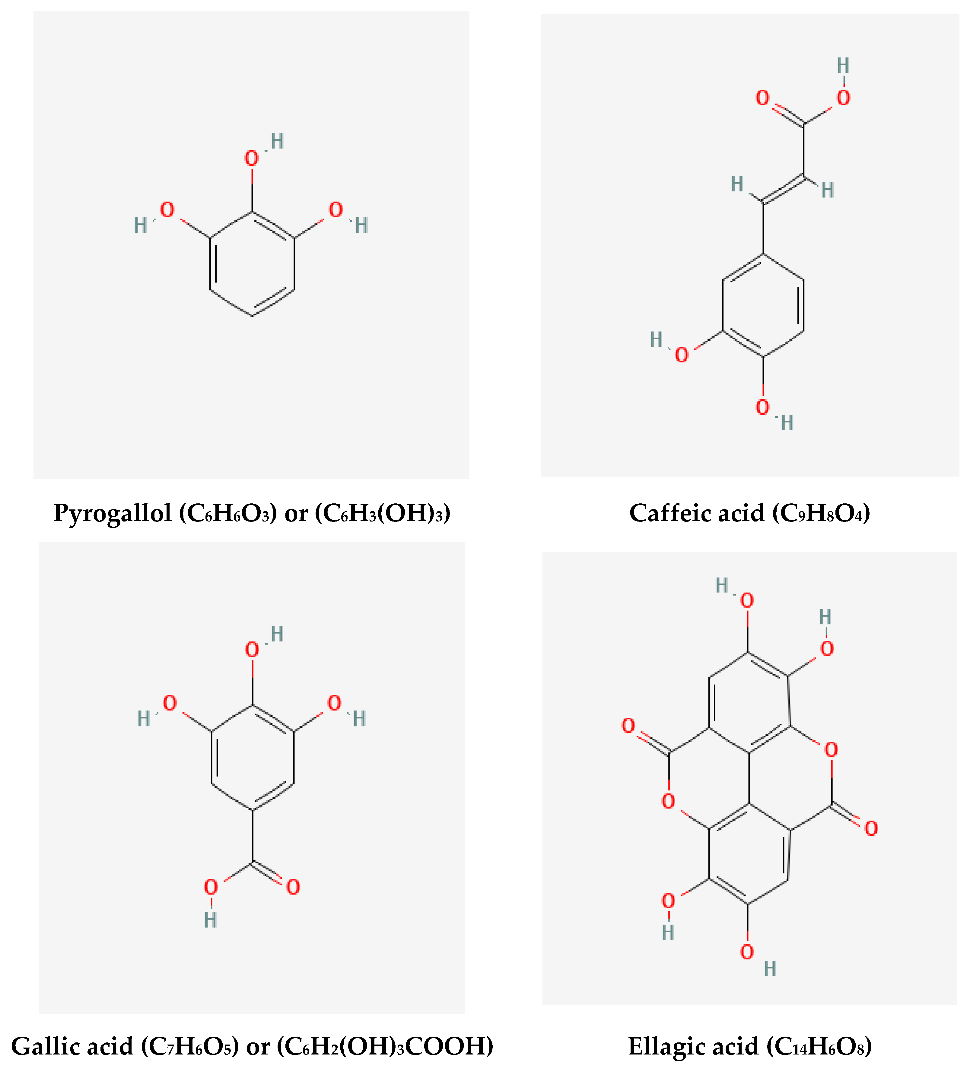
| RT | Compound | Concentration (μg/mL) |
|---|---|---|
| 3.0 | Pyrogallol | 1.88 |
| 8.0 | Caffeic acid | 0.45 |
| 10.0 | Gallic acid | 15.27 |
| 15.0 | Ellagic acid | 2.42 |
| Plant | Particle Size (nm) | Shape | Reference |
|---|---|---|---|
| Citrullus lanatus fruit rind | 17.96 ± 0.16 nm | Spherical | [77] |
| Pedalium murex leaf | 14 nm | Cubic | [50] |
| Nigella arvensis seed | 8.5 nm | Spherical | [78] |
| Bacillus species | 10 nm | Spherical, rod, and octagonal | [79] |
| Trichoderma | 10 nm | Spherical | [80] |
| longibrachiatum | 10–16 nm | Spherical | [7] |
| Ginkgo biloba leaf | 40.2 ± 1.2 nm | Spherical or oval | [81] |
| Entada spiralis | 18.49 ± 4.23 nm | Spherical | [82] |
| Tropaeolum majus leaf | 35–55 nm | Round | [83] |
| Morus nigra leaves | 23 nm | Cubic | [84] |
| Pithecellobium dulce leaves | 62 nm | Spherical rods | [85] |
| Allium giganteum shoots | 12 nm | Spherical | [86] |
| Piper betle leaves | 3–37 nm | Spherical | [87] |
| Allium sativaum root | 7.3 nm | Spherical | [88] |
| Annona reticulate leaves | 6–8 nm | - | [89] |
| Salvia officinalis leaf | 41 nm | Spherical | [90] |
| Rumex dantatus root | 25–70 nm | - | [91] |
| Spinacia oleracea leaves | 15 ± 5 nm | Cubic | [92] |
| Ginkgo biloba leaves | 11.99 ± 3.18 | Spherical | This study |
| Treatment Compounds | Virus | MTNC | CC 50 | IC 50 | SI. |
|---|---|---|---|---|---|
| GB leaves extract (mg/mL) | MERS | 30.9 | 8.94 | ||
| 229E | 125 | 276.49 | 12.7 | 21.76 | |
| Ag NPs (µg/mL) | MERS | 9.3 | 4.39 | ||
| 229E | 15.62 | 40.85 | 7.01 | 5.82 |
Disclaimer/Publisher’s Note: The statements, opinions and data contained in all publications are solely those of the individual author(s) and contributor(s) and not of MDPI and/or the editor(s). MDPI and/or the editor(s) disclaim responsibility for any injury to people or property resulting from any ideas, methods, instructions or products referred to in the content. |
© 2023 by the authors. Licensee MDPI, Basel, Switzerland. This article is an open access article distributed under the terms and conditions of the Creative Commons Attribution (CC BY) license (https://creativecommons.org/licenses/by/4.0/).
Share and Cite
Elshazly, E.H.; Nasr, A.; Elnosary, M.E.; Gouda, G.A.; Mohamed, H.; Song, Y. Identifying the Anti-MERS-CoV and Anti-HcoV-229E Potential Drugs from the Ginkgo biloba Leaves Extract and Its Eco-Friendly Synthesis of Silver Nanoparticles. Molecules 2023, 28, 1375. https://0-doi-org.brum.beds.ac.uk/10.3390/molecules28031375
Elshazly EH, Nasr A, Elnosary ME, Gouda GA, Mohamed H, Song Y. Identifying the Anti-MERS-CoV and Anti-HcoV-229E Potential Drugs from the Ginkgo biloba Leaves Extract and Its Eco-Friendly Synthesis of Silver Nanoparticles. Molecules. 2023; 28(3):1375. https://0-doi-org.brum.beds.ac.uk/10.3390/molecules28031375
Chicago/Turabian StyleElshazly, Ezzat H., Alyaa Nasr, Mohamed E. Elnosary, Gamal A. Gouda, Hassan Mohamed, and Yuanda Song. 2023. "Identifying the Anti-MERS-CoV and Anti-HcoV-229E Potential Drugs from the Ginkgo biloba Leaves Extract and Its Eco-Friendly Synthesis of Silver Nanoparticles" Molecules 28, no. 3: 1375. https://0-doi-org.brum.beds.ac.uk/10.3390/molecules28031375