Review

The Actin Depolymerizing Factor (ADF)/Cofilin Signaling Pathway and DNA Damage Responses in Cancer

1
Department of Biomedical Imaging and Radiological Sciences, National Yang-Ming University, Taipei 112, Taiwan
2
Division of Radiation Oncology, Taipei City Hospital RenAi Branch, Taipei 106, Taiwan
3
Biophotonics & Molecular Imaging Research Center (BMIRC), National Yang-Ming University, Taipei 112, Taiwan
*
Author to whom correspondence should be addressed.
These authors contributed equally to this work.
Int. J. Mol. Sci. 2015, 16(2), 4095-4120; https://0-doi-org.brum.beds.ac.uk/10.3390/ijms16024095
Submission received: 22 December 2014 / Revised: 26 January 2015 / Accepted: 9 February 2015 / Published: 13 February 2015
(This article belongs to the Special Issue DNA Damage and Repair in Degenerative Diseases 2014)

Abstract

:
The actin depolymerizing factor (ADF)/cofilin protein family is essential for actin dynamics, cell division, chemotaxis and tumor metastasis. Cofilin-1 (CFL-1) is a primary non-muscle isoform of the ADF/cofilin protein family accelerating the actin filamental turnover in vitro and in vivo. In response to environmental stimulation, CFL-1 enters the nucleus to regulate the actin dynamics. Although the purpose of this cytoplasm-nucleus transition remains unclear, it is speculated that the interaction between CFL-1 and DNA may influence various biological responses, including DNA damage repair. In this review, we will discuss the possible involvement of CFL-1 in DNA damage responses (DDR) induced by ionizing radiation (IR), and the implications for cancer radiotherapy.

1. Introduction

According to the World Health Organization (WHO), cancer is a leading cause of morbidity and mortality globally. The occurrence of cancer in part correlates to the accumulation of DNA damage and loss or deterioration of normal genomic control [1]. A variety of strategies have been used to treat human cancers, but the efficacy of these approaches depends on the types of tumor cells [2,3,4]. An evidence-based analysis indicates that the utilization rate of radiotherapy is close to 52% compared to other therapies [5]. Although several novel biomedical techniques and new drugs have been developed to conquer cancers, radiotherapy remains an important primary or adjuvant method for cancer treatment.
In adherent cells, actin filaments are important for cell attachment and spreading on the substratum, and this process is required for cell proliferation. The significance of actin filaments is to convey the extracellular signals and form an appropriate shape for G1 phase progression [6,7,8,9,10,11]. Destabilization of the actin cytoskeleton using actin-targeting toxins (ATT) has been reported to cause reversible G1 phase arrest [12], although long-term treatment of these toxins would lead to apoptosis [13,14,15]. ATT was developed for chemotherapy, and considered in combination with radiation for cancer treatment [16,17,18]. However, the efficacy of different ATTs on the enhancement of radiosensitivity remains controversial. For instance, the G1 phase is sub-sensitive to ionizing radiation compared to G2/M phase arrest. Therefore, ATT-mediated accumulation of cells in the G1 phase may not be beneficial for increased radiosensitivity, except for the additional damage incurred by these cells. It is also possible that different types of ATT may trigger different molecular signaling pathways to respond differently to ionizing radiation. Clarification of these signaling pathways triggered by ATT may explain the conflict radiation responses induced by these compounds.
Regulation of the actin cytoskeleton is dependent on a variety of actin-associated proteins (AAPs). These AAPs accelerate the actin dynamics by polymerization and depolymerization of monomeric actin, as well as promotion of nucleotide exchange of ADP-bound actin. The members of the actin depolymerizing factor protein family are known to sever and/or depolymerize actin filaments in vitro and in vivo [19,20,21]. It is believed that these actions would replenish the actin pools and the quantities of actin filaments for actin cytoskeletal turnover. Accumulated reports have also shown that cofilin-1 (CFL-1), the primary non-muscle isoform of the ADF/cofilin protein family may also be involved in signaling transduction, nuclear entry of actin, and actin rod formation in the nucleus [22,23,24]. Our previous and recent studies also found that over-expression of CFL-1 could delay DNA repair after irradiation [25]. Interestingly, certain ATTs that differently affect CFL-1 activity also display distinct radiation response. In this review, we will discuss the role of CFL-1, actin cytoskeleton and other AAPs on DDR, and the implication of actin targeting for cancer radiotherapy.

2. The Actin Depolymerizing Factor (ADF)/Cofilin Signaling Pathways and Actin Dynamics

In the past three decades, studies of the ADF/cofilin family in various organisms have demonstrated that this molecule plays a crucial role in regulating actin dynamics, which affects locomotion, migration, and cell viability. The ADF protein was first identified from embryonic chick brain by Bamburg et al. [26], and related isoforms were subsequently found in a variety of organisms via functional assay and sequence similarity [27,28]. The first mammalian ADF protein was isolated from bovine brain extracts by Berl et al. [29], and subsequently found in the porcine brain extracts and kidney (also called destrin) [30,31]. In 1984, Maekawa et al. purified cofilin from porcine brain, and it is subsequently characterized to bind to actin subunits on F-actin in a 1:1 ratio, and it is named through the formed cofiliamentous structure with actin [30,32,33]. CFL-1 is classified as the non-muscle isoform of the ADF/cofilin protein family, and its full cDNA sequence was first cloned to deduce the amino acid sequence [34]. Although ADF and CFL-1 share 70% of sequence identity and they functionally overlap, CFL-1 is the major non-muscle isoform of ADF/cofilin in various cell types [35]. On the other hand, deletion of the CFL-1 gene leads to lethality in mouse, yeast, fruit fly, and blastomere of Xenopus, suggesting that the functions of CFL-1 and ADF are not redundant [19,36,37]. Several important characteristics of CFL-1 will be discussed below.

2.1. Biophysics and Biochemistry of Cofilin-1 (CFL-1)

The fundamental function of CFL-1 is to accelerate the turnover of actin filaments by depolymerizing or severing the actin filaments. Interestingly, this action can enhance the actin polymerization for cell motility and other physiological behaviors. The depolymerization activity of CFL-1 can enhance actin dissociation from the pointed ends of actin filaments. The pointed ends of actin filaments contain ADP-bound actin subunits bound by CFL-1. Binding of CFL-1 opens the nucleotide-binding cleft of actin subunits on the filament, increases the average distance of adjacent actin subunits in the long-axis filaments and weakens the mutual interaction of actin subunits [38]. Therefore, the ADP-actin pool will be replenished by depolymerized actin that is subsequently converted to ATP-actin for the next round of polymerization. For the severing activity of CFL-1, the pointed end of actin filaments is occupied by CFL-1 to render a mechanical discontinuity of filaments [21]. The free barbed ends will increase by the severing activity of CFL-1, and they are the new sites for actin polymerization to enhance the actin dynamics [39]. Additionally, different concentrations of CFL-1 will also determine its action on nucleation or severing actin filaments [40]. Real-time fluorescent microscopic observation has shown that low CFL-1 concentration promotes severing of actin filaments, while high CFL-1 concentration decreases the extent of actin filamental severing [40,41]. When the CFL-1/actin ratio is high, actin-interacting protein 1 (Aip1) can bind to the CFL-1-actin filament and promote depolymerization of actin filaments to generate monomeric actins [42,43]. Additionally, CFL-1 binding to actin filaments is cooperative, but it actually does not affect the off-rate of actin filaments [44].
The function of CFL-1 on the turnover of actin filaments does not only rely only on the protein itself but also the environmental conditions. For example, growth factor-stimulated cell migration induces dramatic actin reorganization at the leading edge of cells, and this is associated with activation of CFL-1 via protein dephosphorylation and dissociation from phosphatidylinositol-4,5-bisphosphate (PtdIns(4,5)P2) [22]. Phosphorylation of the third serine residue (ser-3) on CFL-1 is believed to inactivate itself on actin depolymerization because the affinity between phosphorylated CFL-1 and actin is greatly reduced [45]. The ratio of phosphorylated CFL-1 to total CFL-1 is approximately 20%–50% in nontransformed mammalian cells [46,47]. CFL-1 can be phosphorylated by serine/threonine kinases including LIM kinase 1 (LIMK1), LIM kinase 2 (LIMK2) and testicular protein kinase 1/2 (TESK1/2) [48]. On the other hand, slingshot (SSH) phosphatase and chronophin can specifically dephosphorylate CFL-1 on ser-3. A recent report also proposes that tumor suppressor PTEN phosphates can directly dephosphorylate CFL-1 when prostaglandin E(2) (PGE(2)) is used to inhibit the phagocytosis of fungi, although it is not evident in mammalian cells [49]. In addition to protein phosphorylation, CFL-1 activity is also ablated by intracellular pH that is regulated by a sodium-proton exchanger on the cell membrane. CFL-1 will dissociate from cortactin or phosphatidylinositol 4,5-bisphosphate (PIP2) at higher pH, and the inhibitory effect on CFL-1 by these molecules will be removed [49]. Moreover, the subcellular localization of CFL-1, and regulation of actin dynamics by other AAPs such as profilin, tropomyosin and Aip1 will also directly or indirectly affect the activity of CFL-1 [39].

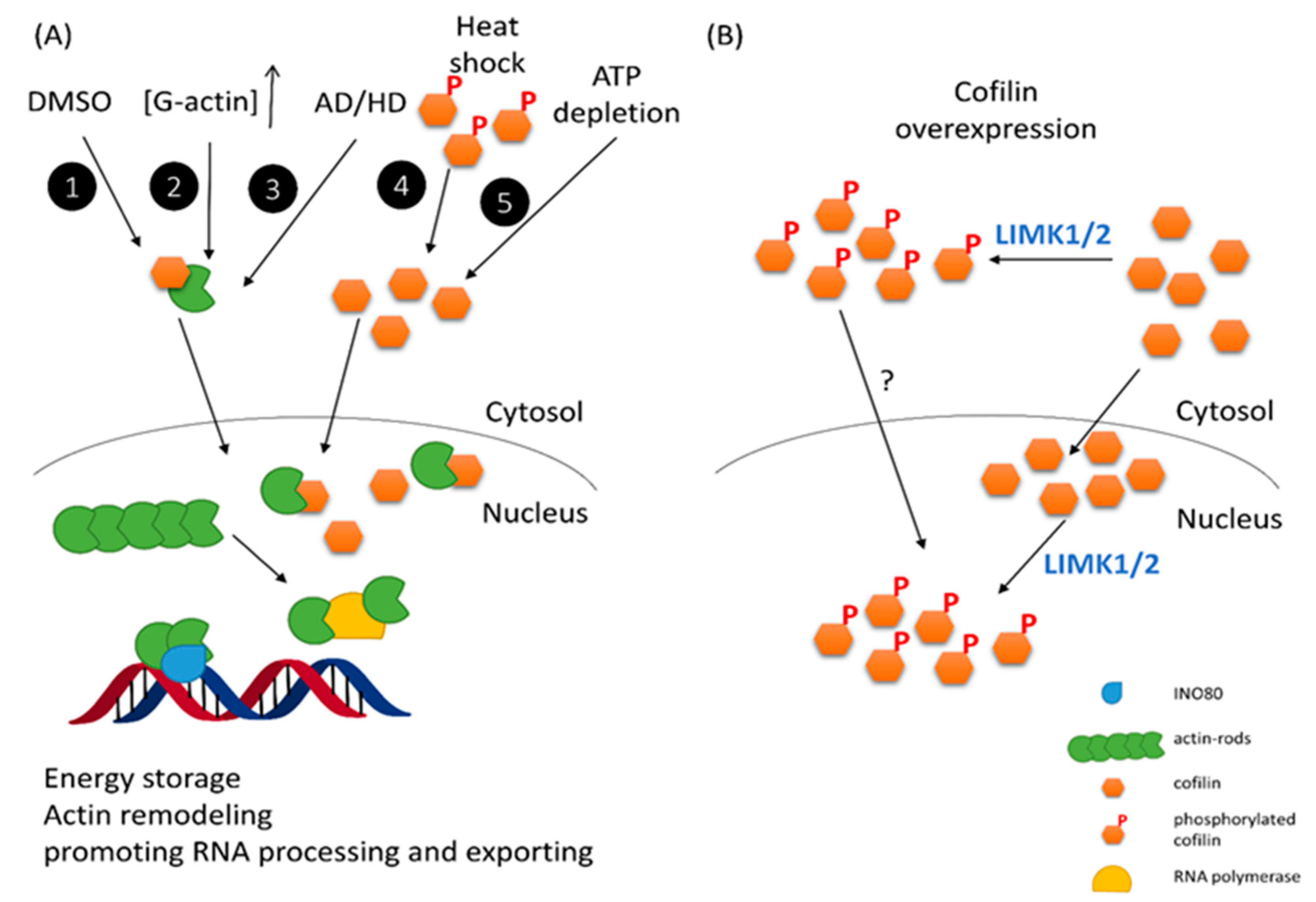
2.2. The Putative Role of CFL-1 in the Cell Nucleus

As one of the AAPs, CFL-1 is very different from others because it contains nuclear localization signals (NLS) in the protein sequence. This endows a particular ability of CFL-1 to bind and carry depolymerized actin to nucleus [50,51]. The NLS fragment of CFL-1 has been demonstrated to be functional when it is linked to nonnuclear proteins expressed in myotubes [51]. Recently, it was found that a conserved bipartite NLS, that is, two basic rich regions separated by seven amino acids (21-RKSSTPEEVKKRKK-34) played the functional role in mediating the nuclear localization of CFL-1 through the classic importin α/β interaction pathways [51,52]. When cells are faced with heat shock, ATP-depletion and dimethyl sulfoxide (DMSO) treatment, cytochalasin D or high cytosolic G-actin concentration, CFL-1 can translocate actin into nucleus and form actin rod-like structure [51,53,54,55,56]. Although the biological function of nuclear translocation of cofilin/actin is unclear, it may reduce the consumption of cellular energy because the ATP utilization in actin dynamics is tremendous. Sequestration of actins by cofilin in the nucleus would reduce actin dynamics in stressful condition and save cellular energy [57]. Although the dephosphorylated form of cofilin is essential for nuclear entry under these conditions, Nagaoka et al. have proposed that exogenous phosphorylated cofilin (S3D) is able to diffuse into the cell nucleus [58]. Our recent report also agrees that phosphorylatable, wild-type CFL-1 can be detected in cell nuclei by the tetracycline inducible gene over-expression system [25]. Of interest, a recent report showed that phosphorylated cofilin can transit from the cytoplasm to the nucleus in the laminar formation of the chicken optic tectum, indicating that phospho-cofilin plays a role in neural development biology [59]. On the other hand, it has been proposed that CFL-1 is phosphorylated in the nucleus by nuclear LIMK1 and LIMK2 [60,61]. Phosphorylated CFL-1 will be inactivated to depolymerize actin filaments, and nuclear actin rods are formed. A recent report has indicated that fluid shear stress can activate nuclear LIMK1/2 to phosphorylate cofilin in the nucleus and result in actin realignment for endothelial barrier integrity [62]. Moreover, the cell cycle regulator p57kip2 can bind to LIMK1, but not LIMK2, and translocate to the nucleus to reorganize actin fibers [63]. These mechanisms account for the importance of activated LIMK1/2 on the formation of phospho-CFL-1 in the cell nucleus. A summary of nuclear entry of cofilin via various stresses is represented in Figure 1. Taken together, the cytoplasm-nucleus transition of cofilin, either phosphorylated or unphosphorylated, is likely to play an essential role in various biological processes, and thus worthy of further investigation.
Although actin itself does not contain an NLS, it is well known that actin is important for chromatin remodeling by binding to INO80-associated chromatin modifying complexes [64]. Nuclear actin is also important for gene transcription by binding to RNA polymerase, promoting RNA processing and exporting mRNA to the cytoplasm [65]. It appears that ADF/cofilin may be an auxiliary molecule that transports actin to the cell nucleus for gene expression. The cytoplasmic ADF/cofilin may also influence gene expression via a nuclear actin independent pathway. For instance, ADF/cofilin-mediated actin turnover can promote the release of actin-bound nuclear transcription cofactors, such as the glucocorticoid receptor (GR) and serum response factor (SRF)-associated cofactor. Moreover, it has been reported that actin depolymerizing factor 9 (ADF9) of Arabidopsis controls the gene expression and muticellular development of plants [66]. We have performed a microarray analysis for mRNA and microRNA expression before and after induction of exogenous CFL-1 in human lung cancer cells, and we found many genes involved in cell cycle progression, amino acid metabolism, tumor suppression and even DNA damage response (DDR) are affected by over-expressed CFL-1 (unpublished data). Because CFL-1 does not contain a DNA binding domain, it is believed that the effects of CFL-1 on gene transcription would be indirect.
Figure 1. Nuclear entry of Cofilin-1 (CFL-1) via various stresses. (A) CFL-1 can bind G-actin and translocate to the nucleus to form actin rods by different exogenous and endogenous stresses. 1. 10% DMSO treatment; 2. Increase of G-actin concentration; 3. Neural degenerative diseases; 4. Heat shock stress; 5. ATP depletion. This action may avoid energy expenditure and promote chromatin remodeling via actin; (B) Enforced expression of CFL-1 can also occur in the nucleus. In this situation, phosphorylated CFL-1 is also detectable in nucleus, while the mechanism of nuclear entry of phospho-CFL-1 remains unclear. Because LIM kinase 1 (LIMK1) and LMK2 are responsible for CFL-1 phosphorylation in various cell types, the subcellular location of these kinases would determine the mechanisms of nuclear accumulation of phosphorylated CFL-1 and regulate actin dynamics in the cytosol and nucleus.
Ijms 16 04095 g001
Nuclear entry of ADF/cofilin is usually induced by environmental stress. As mentioned, DMSO has long been known to trigger the nuclear entry of cofilin-actin complexes to form actin rods [54]. Additionally, heat-shock stress can induce nuclear translocation of CFL-1 through the NLS [24,52]. Formation of cofilin/actin rods in the nuclei and cytosol has also been detected in neural degenerative diseases including Huntington’s disease (HD) and Alzheimer’s disease (AD), respectively [67,68]. In neurodegenerative diseases, actin cytoskeletal regulation through cofilin is believed to be critical for the pathological etiology of these aging or stress related disorders [69]. Therefore, nuclear translocation of CFL-1 may play an important role in degenerative diseases. Whether this biological behavior of CFL-1 is to form specific actin-rod structures in the nucleus or to influence chromatin remodeling for gene expression is of interest for further study.

3. Genotoxicity and DNA Damage Response (DDR)

DNA damage can be induced by various environmental stresses, including ionizing radiation (IR), oxidative stress, ultraviolet (UV) light, and even polycyclic aromatic hydrocarbons in cigarettes. These genotoxicities cause different types of DNA damage followed by initiation of corresponding DNA repair systems. Additionally, DNA damage also triggers cell cycle checkpoints and initiates signaling pathways leading to apoptosis, senescence and even autophagy [70,71]. The elegant intracellular signaling networks activated by genotoxicity to determine the fate of insulted cells, such as cell cycle arrest, premature senescence, and apoptosis, is collectively termed the DNA damage responses (DDR). Because of the cell heterogeneity, the DDR in each single cell of one population may be different. For instance, the survival fraction is commonly used to determine the cell tolerance to genotoxicity, and this is one example of the evidence of cell heterogeneity [72,73]. Ionizing radiation induced cell death is usually caused by unrepairable DNA damage. A functional DNA repair system would increase viability of irradiated cells, although it may leave errors in the DNA sequence and lead to mutation. Here we primarily focus on the ionizing radiation induced DDR and the potent effect on clinical radiotherapy [74].

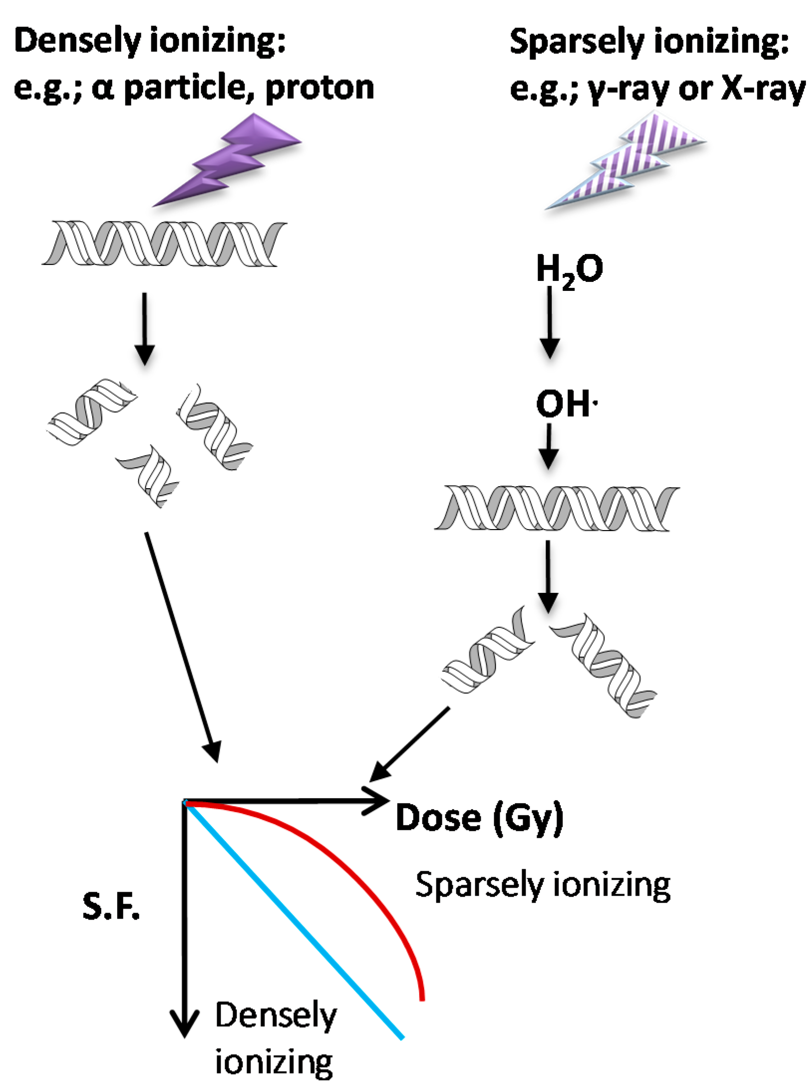
3.1. Types of Ionizing Radiation on Cell Survival and DDR

Ionizing radiation is generated by either high-energy photons or particles that can eject electrons or break the nucleus of the atom. Thus, the incident energy is to “ionize” charged particles that are originally bound in the atom, including secondary electrons and recoiled protons. X-ray and γ-rays belong to electromagnetic radiation with photon properties, while α particles and β particles belong to particulate radiation that can carry either a positive or negative charge. X-rays and γ-rays are regarded sparse types of radiation because they can easily penetrate through the object and only deposit little energy in it. On the other hand, α particles and protons are dense types of radiation that have difficulty to penetrate the object but can deposit large amounts of energy in it. X-rays and γ-rays can only generate ionized fast electron, which has the mass 1/2000 and 1/8000 of proton and α particle, respectively. Therefore, it is easy to understand that particulate radiation usually causes higher biological damage than electromagnetic radiation. X-rays and γ-rays generate fast electrons that can interact with water to generate free radicals that is the source of oxidative stress to damaged DNA. This is a so-called indirect effect. On the contrary, protons and α particles can directly deposit their energy on DNA even though they can also generate oxidative stress, and this is called a direct effect. Both effects are sufficient to break the covalent bonds of DNA and result in DNA double strand breaks (DSB), but the levels are more severe by protons or α particles than by X-rays and γ-rays under the same absorbed dosage. The levels of DSB are reflected through measuring the survival fractions which represent the tolerance of cells to different types of radiation. The lower survival fraction caused by particulate radiation is related to complex DNA damage that is difficult to be repaired. The survival curve is drawn according to the survival fractions corresponding to increased radiation dose. In a semi-log plot of survival curve, α particles or protons usually exhibit a linear curve that stands for exponential killing, but X-rays and γ-rays will give a “shoulder” at lower doses before the appearance of the exponential killing curve. The “shoulder” region of the survival curve indicates that at low dose radiation, cells would repair effectively after irradiation by X-rays and γ-rays. However, there is no, or a very narrow, shoulder of survival curve observed using α particles or proton irradiation under the same dose. A scheme represents the effects of radiation types on DDR and survival curves are shown in Figure 2.
Figure 2. Effects of different radiation types on DNA damage and cell survivals. Particulate radiations by α-particles or protons belong to densely ionizing radiation type because they can deposit most energy on their tracks. On the other hand, X-rays and γ-rays possess high penetration ability but leave little energy on the traveling track, so that they are sparsely ionizing radiation. Moreover, sparsely ionizing radiation mainly ionizes water to produce free radicals, which are highly electrophilic and prone to capture electrons from biological components. When DNA locates on the track of these two different radiation types, it displays different levels of damage. Densely ionizing radiation causes more double strand breaks than sparsely ionizing radiation, so that DNA repair will be accordingly less efficient. Under the same dosage, the survival fractions of particulate radiation irradiated cells will be lower than that of X-ray or γ-ray irradiated cells.
Ijms 16 04095 g002

3.2. DNA Repair Mechanisms Following Ionizing Radiation (IR)

DNA double-strand breaks (DSB) are the major type of DNA damage after exposure to ionizing radiation. DSB damage needs to be repaired to maintain genomic stability and survival. In mammalian cells, the DNA repair mechanisms include nonhomologous end joining (NHEJ) and homologous recombination repair (HRR), in which different DNA DSB repair proteins are involved. The key molecules for NHEJ include DNA end binding proteins Ku70/Ku80 heterodimer, which in turn binds and enhances DNA-dependent protein kinase (DNA-PKcs) activity [75,76,77,78]. Artemis protein, an exonuclease involved in V(D)J recombination for immunological responses, can gain endonuclease activity after joining a complex with DNA-PKcs to process the broken end of DNA after IR. Finally, the DNA ends are filled in and connected through DNA polymerase λ/μ and PNK/XRCC4/DNA ligase IV/XLF complex, respectively. This error-prone repair system mainly occurs in G0, G1 and early S phase because no replicative template is required for repairing DSB [79]. For HRR, E. coli recombinase RecA-homologous protein Rad51 and its interacting partner Rad52 are important for single-strand DNA binding, strand exchange, and annealing using the sister chromatid as the repair template [80,81,82,83]. The strand exchange or invasion is assisted by BRCA1/2 tumor suppressor proteins, and Rad54 is a helicase that can hydrolyze ATP to unwind the double-strand DNA in template. After completion of repair, the tangled DNA structures, so called Holiday junctions, are resolved by MMS4/MUS81 hetermodimer with endonuclease activity. Overall, this is an error-free repair system that is believed to be involved in late S and the G2 phase of the cell cycle. Deficiency of Ku70 or Ku80, or inhibition of Rad51 levels can lead to enhancement of radiosensitivity [78,84,85,86]. Moreover, over-expression of Rad52 confers resistance to ionizing radiation in mammalian cells [87]. Therefore, cell radiosensitivity is strongly associated with DNA repair capacity [86,88,89,90]. Increased cell radiosensitivity by repressing DNA repair capacity is one of the important strategies for design of radiosensitizers.

3.3. The Role of the Mre11-Rad50-NBS1 (MRN) Complex in Sensing IR-Induced DNA Damage

Although NHEJ and HRR utilize different molecules to directly execute the re-joining of broken DNA ends, they have the DSB sensor in common. The MRN complex comprised by Mre11 (meiotic recombination 11), Rad50 and NBS1 (Nijmegen breakage syndrome 1) is reported to recognize the sites of DSB to trigger both NHEJ and HRR [91]. The MRN complex is also known to maintain the integrity of telomeres of chromosomes in eucaryotic cells [92]. Mre11 can specifically bind to DSB termini and perform single-strand DNA (ssDNA) endonuclease and 3' to 5' double-strand DNA (dsDNA) exonuclease activities [93]. Rad50 can bind to Mre11 and form a core tetramer complex in vivo [94]. Rad50 can unwind dsDNA in DSB ends by hydrolyzing ATP [95]. NBS1 does not contain enzymatic activity, but is important for transport oft he MRN complex into the nucleus [96]. It also mediates the DNA repair response by promoting protein-protein interaction at DSB sites. Importantly, both NBS1 and Rad50 can be phosphorylated by Ataxia telangiectasia mutated (ATM) serine/theronine kinase for intra-S phase checkpoint and repair in response to IR [97,98,99]. However, ATM mediated phosphorylation of the MRN complex does not guide the MRN complex to DSB termini. On the contrary, this phosphorylation is required to recruit ATM to DSB termini for further activity. Binding of the MRN complex to DSB sites is important for both classical NHEJ (C-NHEJ) and alternative NHEJ (A-NHEJ, without XRCC4, Ku, and DNA-PKcs activity) because knockdown of Mre11 can impair both pathways [100]. Furthermore, the MRN complex is also essential for processing DSB termini for Rad51/Rad52 binding in HRR.

3.4. Ignition of DDR by Ataxia Telangiectasia Mutated (ATM) Kinase

ATM is a member of phosphatidylinosital 3-kinase related kinase (PIKKs) superfamily that also includes ATR (ATM- and Rad3-related), DNA-PKcs, mTOR (target of rapamycin), SMG1 (suppressor with morphological effect on genitalia), and TRRAP (transformation/transcription domain-associated protein). Recruitment of ATM to DSB is dependent on MRN complex, although this DSB sensor may be not required for C-NHEJ under low dose or low-LET irradiation [101,102]. ATM has been regarded a transducer following the MRN complex that functions as a sensor of DSB [103,104]. In addition to the DSB site, genotoxicity-induced chromatin relaxation around DSB is also important for activation of ATM. The chromatin surrounding DSB will be modified by PARP-1 (poly (ADP-ribose) polymerase 1) that introduces poly(ADP-ribose) chains (PAR chains) to histone H1 and histone H2B [105,106]. Several chromatin remodeling molecules including PcG (polycomb group), NuRD (nucleosome remodeling deacetylase), ALC1 (amplified in liver cancer 1), and HP1/-KAP-1 (H3-trimethyl K9 binding protein 1-KRAB associated protein 1) will interact with PARylated chromatin encompassing DSB for further processes [107,108,109,110]. These chromatin relaxation procedures mainly promote posttranslational modification of DSB and histones through ubiquitination, stabilization of chromatin structure, and unpacking of heterochromatic DSB [109,110,111,112]. ATM is also PARylated and activated by PARP1 after IR-induced DSB. Inhibition of PARP1 reduces ATM activity and DNA repair capacity, and it has been considered for design of cancer radiotherapeutic strategy [113,114].
Over 700 protein targets have been identified as phosphorylated by ATM and/or ATR after genotoxic treatment according to large-scale proteomic screening and analysis [115,116,117]. ATM itself is the substrate of this protein, and serine 1981, 367 and 1893 (ser1981, ser367, ser1893) are the autophosphorylation residues after IR-induced DSB [118]. As mentioned, the MRN complex is important for recruitment of ATM to the DSB sites. The lack of a MRN complex leads to the reduction of ser1981 phosphorylation and activity of ATM. The phosphorylation kinetics of ser1893 is slower than that of ser1981 of ATM. ATM phosphorylation on ser1893, unlike ser1981, fully depends on the MRN complex. This may explain the different phosphorylation kinetics between these two serines on ATM [118]. Activated ATM at DSB termini will further phosphorylate H2AX (γ-H2AX), NBS1, BRCA1 (breast cancer type 1 susceptibility protein), MDC1 (mediator of DNA damage checkpoint 1) and SMC1 (structural maintenance of chromosome 1) to promote DNA repair [119,120]. It is noteworthy that MDC1 can bind both γ-H2AX and NBS1 to cooperatively increase ATM activity for formation of γ-H2AX over megabases surrounding DSB sites [121,122,123]. 53BP1 is a scaffold for DSB responsive factors, and can be phosphorylated by ATM to increase the substrate-specificity of ATM [124,125,126]. When ATM is not bound to DSB sites, it can phosphorylate p53 and Chk1/2 to influence cell cycle checkpoint and apoptosis. Therefore, ATM is a critical center for DDR in response to genotoxicity.
Recruitment and autophosphorylation of ATM at the DSB site after IR damage requires further activation to maintain the ATM activity for substrate phosphorylation. That is, ATM needs to be further modified by other mechanisms in addition to phosphorylation. The histone acetyltransferase (HAT) Tip60 is recruited and bound to ATM by the MRN complex in response to DSB [127,128]. Tip60 can acetylate ATM at lys3016 and is required for ATM-mediated phosphorylation on H2AX, p53 and Chk2 [118]. Tip60 is further activated by c-Abl tyrosine kinase at the DSB site [128,129]. Additionally, Tip60 is strictly controlled by ATF2 (Activating Transcription Factor 2), a bifunctional transcription factor that can promote Tip60 ubiquitination via E3 ubiquitin ligase Cul3 and proteasomal degradation to influence global DSB repair [130]. Knockdown of ATF2 can stabilize Tip60 and lead to activation of ATM and increase cell survival after IR damage [131]. On the other hand, ATM can phosphorylate ATF2 on ser490/ser498 and confer the transcription-independent activity of ATF2 that can bind to γ-H2AX and MRN complex for S phase checkpoint [130]. Under this condition, inhibition of ATF2 leads to loss of S phase checkpoint and reduction of DNA repair capacity as well as ATM activity. Interestingly, Ser490A/ser498A phospho-mutant ATF2 cannot influence Tip60 stability after over-expression. This implies that ATM will negatively regulate its own activity by phosphorylating ATF2 to ablate Tip60, which may occur after DNA repair is completed. The interplay between c-Abl-Tip60 and ATF2 would be essential to regulate ATM activity after they are recruited to DSB following IR [129,132,133]. A scheme of DNA damage-induced molecular responses for DSB repair is shown in Figure 3.
Figure 3. DNA damage-induced molecular responses for DNA repair. In response to ionizing radiation, DNA double strand breaks (DSB) and chromatin relaxation surrounding DSB sites will occur. The MRN complex can sense and bind to DSB sites and quickly recruit ATM kinase for autophosphorylation and paraphosphorylation to various molecules, including NBS1 and Rad50 in the MRN complex. In addition, chromatin relaxation will activate PARP1 to execute PARylation on chromatin associated molecules and ATM kinase. Activated ATM needs to be maintained by acetylation via HAT Tip60, which is negatively regulated by the ATF2-Cul3 protein degradation pathway. Although c-Abl kinase can activate Tip60 at the DSB site, how DSB interacts with c-Abl is unclear. Additionally, whether MRN complex will regulate Tip60 to sustain ATM activity is unknown. ATM also regulates ATF2 activity, and it is likely to be an autoregulatory mechanism of ATM activity. Activated ATM can phosphorylate SMC1, NBS1, BRCA1, MDC1, 53BP1, and H2AX to localize DSB sites for further DSB repair mechanisms, including HRR and NHEJ.
Ijms 16 04095 g003

4. Actin Dynamics, ADF/cofilin and DDR

Although genotoxicity is meant to disrupt DNA structures, accumulated literature suggest that the genotoxicity induced DDR is not only associated with DNA breaking itself but also the reorganization of the actin cytoskeleton [134,135,136]. The expression and activity of AAPs are of interest to be investigated because their activity and/or subcellular distribution are affected by genotoxicity. These effects largely influence DDR, including apoptosis and cell cycle progression by interacting with other DNA damage induced molecules, such as the p53 tumor suppressor protein. Notably, destabilization of the actin cytoskeleton using ATTs or deliberate manipulation of AAP expression can also modulate genotoxicity-induced DDR. In this section, how actin dynamics and AAPs play a role in response to DNA damage will be discussed.

4.1. Actin Response Following DNA Damage

DNA damage can induce actin reorganization that influences apoptosis and cell cycle arrest subsequently. For instance, use of synchrotron radiation X-ray scattering, actin in solutions of purified calf spleen actin will polymerize without a lag phase [137]. Induction of transient actin polymerization and later actin depolymerization in HL-60 cells is important for ultraviolet (UV) irradiation or etoposide-induced apoptosis [138]. Of interest, cytolethal distending toxins (CDTs), a protein toxin generated by Gram-negative bacteria, are able to induce DNA damage and promote the formation of actin stress fibers [139]. However, the formed stress fibers are due to RhoA GTPase activation, which is mediated by a guanine nucleotide exchange factor (GEF), so-called neuroepithelioma transforming gene 1 (Net1), that also promotes the p38 mitogen-activated protein kinase (MAPK) pathway to extend cell survival [139]. This effect partially suggests that chronic exposure of genotoxicity will lead to genomic instability and mutation. Actin polymerization induced by DNA damage is also associated with p53. It has been reported that LIMK2b, a potent tumor suppressor, is induced by p53 to modulate actin dynamics for executing G2/M arrest through cofilin phosphorylation after DNA damage [140]. On the other hand, p53 mediated transaction of RhoC-LIMK2 leads to inactivation of cofilin, which reduces the actin depolymerization and leads to an increase of actin stress fibers. Under this condition, enhanced actin cytoskeletal formation promotes cell survival, and the efficacy of DNA damage-related therapy will be compromised [136]. The combination of a LIMK2 inhibitor and genotoxic therapy has potential for cancer treatment because it is impossible to inhibit p53 to prevent radio-chemoresistance of cancer cells. It appears that different splicing variants of LIMK2 transactivated by p53 would lead to distinct cell fates, and it requires careful investigation to clarify the effects of actin cytoskeletal remodeling mediated by the p53-LIMK2 pathway.
In another aspect, DNA damage-induced actin polymerization also negatively regulates p53 function by localizing p53 in the cytoplasm [135]. This effect may delay p53 mediated apoptotic processes and allow DNA repair to be processed before apoptosis. Indeed, it has been reported that polymerized actin is required for DSB repair [141]. Binding of p53 to actin filaments is calcium dependent, and the interaction between these two entities is enhanced by DNA damage [142]. On the other hand, p53 mediated gene transcription is also dependent on actin polymerization. A G-actin binding protein, so-called junction-mediated and regulatory protein (JMY) is released from polymerized actin following DNA damage. The free JMY containing a bipartite NLS enters the nucleus and interacts with p53 to enhance its transcriptional activity for DDR [134,143]. The potent actin cytoskeletal responses to DDR are summarized in Figure 4.
Figure 4. Regulatory mechanisms of actin dynamics in response to DNA damage. (A) Radiation-induced actin polymerization triggers the release of JMY from G-actin. JMY then enters the nucleus and binds to p53 for p53-dependent apoptosis; (B) When DNA damage occurs, polymerized actins may allow p53 binding to actin filaments and retain p53 in the cytoplasm. DNA repair would dominate p53-mediated apoptosis under this condition; and (C) DNA damage-mediated actin polymerization would be caused by p53 induction of the Rho/RCOK signaling pathway, followed by activation of LIMK2 to phosphorylate and inactivate cofilin. The actin depolymerization rate will be reduced to maintain polymerized actins.
Ijms 16 04095 g004

4.2. DDR Following Destabilization of the Actin Cytoskeleton

Since DNA damage promotes actin polymerization for subsequent biological responses, it is speculated that destabilization of the actin cytoskeleton will influence DDR [141]. Because the actin cytoskeleton is a dynamic structure, forced polymerization or depolymerization of actin will lead to destabilization of the actin cytoskeleton. Additionally, insufficient energy flux will impair nucleotide exchange of monomeric actin, a rate-limiting step of actin dynamics, to destabilize the actin cytoskeleton. Actin targeting toxins (ATTs) are extracted from fungi or sea sponges, and the biochemical functions of actin filaments are different among these ATTs (Table 1). Cytochalasin D (CD) has been reported to disrupt actin microfilaments and activate p53, which causes G1-to-S phase arrest and apoptosis [15]. It is unclear whether CD-induced p53 activity is the primary or secondary effect of microfilamental disruption since p53 up-regulation is largely dependent on DNA damage. On the other hand, disruption of actin filaments by cytochalasin B (CB), CD, and latrunculin B (LB) can induce cell cycle regulator p21CIP1/WAF in p53-null cells [144]. Interestingly, actin inhibitors-induced p21CIP1/WAF can be detected before p53 activation in p53-wild-type cells, suggesting that disruption of actin filaments by actin inhibitors would affect cell growth without DNA damage. Disruption of actin filaments using Rho inhibitors or actin inhibitors also increases p21CIP1/WAF protein stability, which has been reported to be caused by activation of the c-Jun N-terminal kinase (JNK) stress activated protein kinase (SAPK) pathway [145]. Additionally, disruption of the actin cytoskeleton can activate the JNK pathway via the mammalian Ste20-like (MST) kinase, which can stabilize the p21CIP1/WAF via a JNK dependent phosphorylation on Thr57 [146]. MST kinase actually is required for stabilization of p21CIP1/WAF after disruption of the actin cytoskeleton. Therefore, it is of interest to further investigate whether disruption of the actin cytoskeleton increased p21CIP1/WAF stability via the MST-JNK pathway is also independent of p53.
Table 1. The sources and functions of various actin inhibitors.
Actin InhibitorsSource/HostFunction
Cytochalasin B (CB)Helminthosporium dematioideum/fungiBlocking monomer add-on at the fast-growing end of actin filament
Cytochalasin D (CD)Zygosporium mansonii/fungiBlocking monomer add-on at the fast-growing end of actin filament, 10-fold more potent than CB
Latrunculin ALatrunculia magnifica/Red Sea SpongeFormation of a 1:1 complex with monomeric G-actin (Kd = 200 nM)
Misakinolid A (Bistheonellide A)Theonella sp./marine spongeInhibits actin polymerization by forming a 1:2 complex with G-actin
Mycalolide BMycale sp./marine spongeSevers F-actin and forms a 1:1 complex with G-actin to sequester it; it also suppresses actin-activated myosin Mg2+-ATPase activity
Swinholide ATheonella swinhoei/marine spongeSequestering actin dimers with a binding stoichiometry of 1:1, and rapidly severing F-actin
JasplakinolideJaspis johnstoni/marine spongeA potent inducer of actin polymerization and stabilization in vitro, cell-permeable
PhalloidinAmanita phalloides/fungiA potent and specific F-actin binding agent; Inhibitor of F- to G-actin conversion, cell non-permeable
Morphological change is one of the most important characteristics of apoptosis induced by DNA damage. Disruption of actin microfilaments or microtubules has been reported to accelerate actinomycin D (AD)-induced DNA damage and apoptosis, but forced stabilization of these cytoskeletons has no such effects [147]. Pretreatment of cells with latrunculin or CD followed by ionizing radiation shows that the NHEJ-related proteins Ku70/Ku80 are reduced to bind to the DNA ends caused by IR [141]. Jasplakinolide (JP), an actin stabilizing reagent, has been reported to exhibit an additive effect to radiation-treated prostate cancer cell lines [148]. We have used the colony formation assay, a gold standard of radiobiological research to investigate the effects of CB and latrunculin A (LA) on radiosensitivity. The data revealed that LA but not CB could enhance radiosensitivity [25]. Cellular γ-H2AX is induced by radiation combining CB but not LA, suggesting that LA-mediated destabilization of the actin cytoskeleton would interfere with H2AX phosphorylation induced by IR [25]. Additionally, LA and CD have been reported to increase the level of ser-3 phosphorylated cofilin [149], whereas we found that CB did not show this ability. Whether actin inhibitors influence the expression of certain AAPs to influence DDR is of interest to further investigate.

4.3. AAPs and DDR

Actin dynamics are ablated by a variety of AAPs. Although AAPs regulate actin organization in the cytoplasm, accumulated literature has demonstrated that several AAPs can shuttle between the cytoplasm and the nucleus to promote the formation of nuclear actin polymers and/or affect gene transcription. Moreover, they are important for the DNA repair system. Human actin-related proteins 5 (hArp5) predominantly localizes in the nucleus and associates with chromatin remodeling molecules INO80 to promote DNA repair through accumulation of γ-H2AX [150,151,152]. JMY also exists in both the cytoplasm and nucleus, while its level in the nucleus will increase following DNA damage [153]. Increase of JMY accumulation in the nucleus is actin dependent and required for p53 activation [143]. Filament A (FLNA), alternatively named actin-binding protein 280 (ABP-280), cross-links cortical actin filaments into a firm 3D structure. Interestingly, FLNA also binds DNA repair proteins BRCA1/2 for HRR [154,155,156]. Inhibition of FLNA can increase the radiosensitivity and chemosensitivity to IR and cisplatin, respectively [80]. The G-actin sequestering protein thymosine β4 (Tβ4) can bind to Ku80 to regulate the expression of plasminogen activator inhibitor type 1 [157], though it is unclear whether this interaction is essential for DNA repair. Although the actin nucleator, neural Wiskott–Aldrich syndrome protein (N-WASP), has not been reported to be related to DDR, its activator, so-called non-catalytic region of tyrosine kinase adaptor protein 1 (NCK1) can translocate to the nucleus to activate p53 [158]. Nuclear NCK1 also associates with the suppressor of cytokine signaling 7 (SOCS7) and G-actin to influence actin cytoskeletal reorganization in response to DNA damage [158].

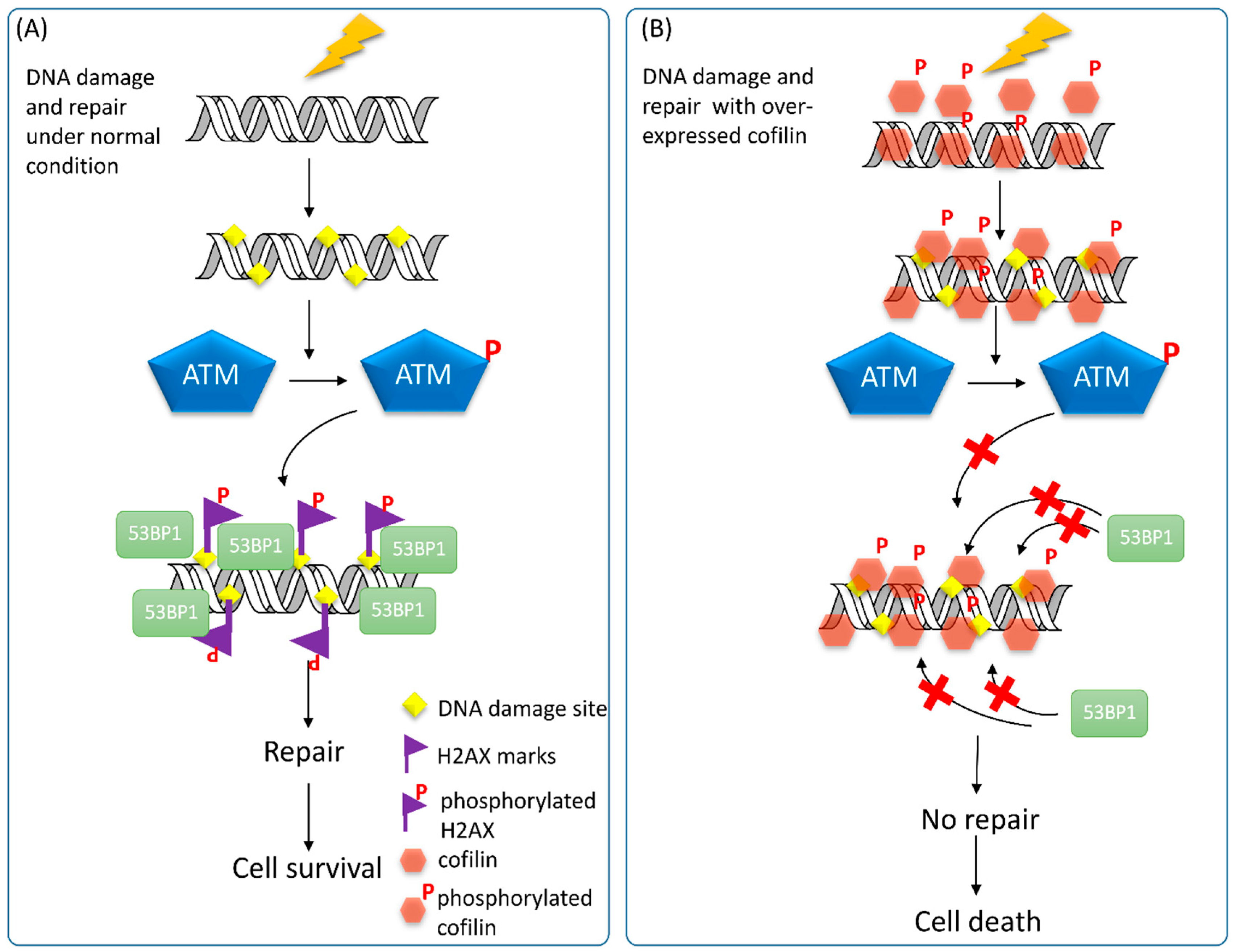
4.4. ADF/Cofilin and DDR

The effects of CFL-1 on DNA repair and radiosensitivity have been previously established. Over-expression of wild-type, S3A and S3D mutant CFL-1 all lead to enhanced radiosensitivity, but ρ-activated kinase (ROCK) inhibitor Y27632 does not increase radiosensitivity of treated cells [25]. Interestingly, γ-H2AX could not be detected in CFL-1 over-expressing cells after irradiation, whereas the ATM activity was not affected under this condition. Therefore, we hypothesize that over-expressed CFL-1 may enter the nucleus and hamper recognition of H2AX by ATM kinase, and this phenomenon would lead to failure of DNA repair initiation (Figure 5). As mentioned, the actin inhibitor LA but not CB can induce both cofilin phosphorylation and enhanced radiosensitivity that is associated with impaired formation of γ-H2AX [25]. This observation implies that the phospho-state of CFL-1 is involved in regulating the cellular radiosensivity. Because the phosphorylation of cofiln-1 is directly controlled by the Rho-LIMK pathway and SSHL1 phosphatase, it will be better to elucidate the role of phosphorylated CFL-1 on DNA repair and radiosensitivity by manipulating the expression of the LIMK and SSHL1 gene. On the other hand, Yan et al. reported that in radioresistant astrocytoma, CFL-1 is one of the proteins up-regulated in patients [159]. However, we have previously found that the CFL-1 level of human A549 lung adenocarcinoma is higher than that of H1299 cells [160], which exhibit stronger radioresistance than A549 cells [161]. Our recent data using in vivo isolated breast cancer stem cells also exhibit lower CFL-1 levels and higher radioresistance than parental breast cancer cells (manuscript in preparation). Therefore, the association of CFL-1 and radiation-induced DDR may be cell- or tissue-dependent. A global survey of CFL-1 expression on different types of cancer sources would be essential to design the therapeutic strategy to utilize the cofilin signaling pathway for cancer radiotherapy.
Figure 5. The putative effects of over-expressed CFL-1 on DNA repair. (A) Under normal condition, IR-induced DNA damage will trigger the activation of the ATM kinase, which phosphorylates H2AX encompassing DNA damage sites. 53BP1 will recognize γ-H2AX and recruit additional DNA repair molecules to fix the damaged DNA and lead to cell survival; and (B) Over-expression of wild type CFL-1 (phosphorylatable) may enter and locate around DNA. After IR, the ATM kinase remains to be activated, but cannot phosphorylate H2AX. Recruitment of 53BP1 to the γ-H2AX sites fails, so that the DNA repair capacity is impaired and results in cell death.
Ijms 16 04095 g005

5. Conclusions

The cross-talk between the cytoplasm and nucleus is increasingly important in upcoming bio-research phases. The actin cytoskeleton and associated proteins, originally support cellular mechanical functions in the cytoplasm, and have been regarded as critical messengers and cofactors to modulate the extracellular and intracellular signaling to the nucleus, a sanctuary of eukaryotic cells to orchestrate cell responses. Although DDR is believed to be associated with the dynamic architecture of the cytoskeleton, the role of different AAPs on regulation of DDR following genotoxicity remain largely unknown. As a primary regulator of actin dynamics and an essential gene to ablate cell viability, CFL-1-mediated DDR is of interest to be investigated. At a minimum, several conflicting results from different research groups need to be clarified in the future. It is expected that these studies would define the role of actin-targeting agents for radiotherapy.

Acknowledgments

This work was supported by the Ministry of Science and Technology (102-2628-B-010-012-MY3), Department of Health, Taipei City Government (10401-62-020), and a grant from the Ministry of Education, The Aim for the Top University Project.

Author Contributions

Chun-Yuan Chang was responsible for figure preparation and writing of text; Jyh-Der Leu contributed to revision and figure refinement; Yi-Jang Lee was responsible for manuscript preparation by writing the text and modifying the concepts of figures.

Conflicts of Interest

The authors declare no conflict of interest.

References

  1. Rew, D.A. Cancer—A degenerative disorder? Eur. J. Surg. Oncol. 1998, 24, 362–366. [Google Scholar] [CrossRef]
  2. Fallowfield, L.J.; Hall, A.; Maguire, G.P.; Baum, M. Psychological outcomes of different treatment policies in women with early breast cancer outside a clinical trial. BMJ 1990, 301, 575–580. [Google Scholar] [CrossRef] [PubMed]
  3. Borghaei, H.; Smith, M.R.; Campbell, K.S. Immunotherapy of cancer. Eur. J. Pharmacol. 2009, 625, 41–54. [Google Scholar] [CrossRef] [PubMed]
  4. Henderson, I.C. Chemotherapy of breast cancer. A general overview. Cancer 1983, 51, 2553–2559. [Google Scholar] [CrossRef] [PubMed]
  5. Delaney, G.; Jacob, S.; Featherstone, C.; Barton, M. The role of radiotherapy in cancer treatment: Estimating optimal utilization from a review of evidence-based clinical guidelines. Cancer 2005, 104, 1129–1137. [Google Scholar] [CrossRef] [PubMed]
  6. Assoian, R.K.; Zhu, X. Cell anchorage and the cytoskeleton as partners in growth factor dependent cell cycle progression. Curr. Opin. Cell Biol. 1997, 9, 93–98. [Google Scholar] [CrossRef] [PubMed]
  7. Huang, S.; Ingber, D.E. The structural and mechanical complexity of cell-growth control. Nat. Cell Biol. 1999, 1, E131–E138. [Google Scholar] [CrossRef] [PubMed]
  8. Reshetnikova, G.; Barkan, R.; Popov, B.; Nikolsky, N.; Chang, L.S. Disruption of the actin cytoskeleton leads to inhibition of mitogen-induced cyclin E expression, CDK2 phosphorylation, and nuclear accumulation of the retinoblastoma protein-related p107 protein. Exp. Cell Res. 2000, 259, 35–53. [Google Scholar] [CrossRef] [PubMed]
  9. Huang, S.; Ingber, D.E. A discrete cell cycle checkpoint in late G1 that is cytoskeleton-dependent and map kinase (ERK)-independent. Exp. Cell Res. 2002, 275, 255–264. [Google Scholar] [CrossRef] [PubMed]
  10. Iwig, M.; Czeslick, E.; Muller, A.; Gruner, M.; Spindler, M.; Glaesser, D. Growth regulation by cell shape alteration and organization of the cytoskeleton. Eur. J. Cell Biol. 1995, 67, 145–157. [Google Scholar] [PubMed]
  11. Bohmer, R.M.; Scharf, E.; Assoian, R.K. Cytoskeletal integrity is required throughout the mitogen stimulation phase of the cell cycle and mediates the anchorage-dependent expression of cyclin D1. Mol. Biol. Cell 1996, 7, 101–111. [Google Scholar] [CrossRef] [PubMed]
  12. Lohez, O.D.; Reynaud, C.; Borel, F.; Andreassen, P.R.; Margolis, R.L. Arrest of mammalian fibroblasts in G1 in response to actin inhibition is dependent on retinoblastoma pocket proteins but not on p53. J. Cell Biol. 2003, 161, 67–77. [Google Scholar] [CrossRef] [PubMed]
  13. Hwang, J.; Yi, M.; Zhang, X.; Xu, Y.; Jung, J.H.; Kim, D.K. Cytochalasin B induces apoptosis through the mitochondrial apoptotic pathway in HeLa human cervical carcinoma cells. Oncol. Rep. 2013, 30, 1929–1935. [Google Scholar] [PubMed]
  14. Kulms, D.; Dussmann, H.; Poppelmann, B.; Stander, S.; Schwarz, A.; Schwarz, T. Apoptosis induced by disruption of the actin cytoskeleton is mediated via activation of CD95 (Fas/Apo-1). Cell Death Differ. 2002, 9, 598–608. [Google Scholar] [CrossRef] [PubMed]
  15. Rubtsova, S.N.; Kondratov, R.V.; Kopnin, P.B.; Chumakov, P.M.; Kopnin, B.P.; Vasiliev, J.M. Disruption of actin microfilaments by cytochalasin d leads to activation of p53. FEBS Lett. 1998, 430, 353–357. [Google Scholar] [CrossRef] [PubMed]
  16. Trendowski, M.; Mitchell, J.M.; Corsette, C.M.; Acquafondata, C.; Fondy, T.P. Chemotherapy in vivo against m109 murine lung carcinoma with cytochalasin B by localized, systemic, and liposomal administration. Investig. New Drugs 2015. [Google Scholar] [CrossRef]
  17. Bousquet, P.F.; Paulsen, L.A.; Fondy, C.; Lipski, K.M.; Loucy, K.J.; Fondy, T.P. Effects of cytochalasin B in culture and in vivo on murine madison 109 lung carcinoma and on b16 melanoma. Cancer Res. 1990, 50, 1431–1439. [Google Scholar] [PubMed]
  18. Court, J.B.; Davies, G.; Davies, H.E.; Burn, C. Variation in radiosensitivity due to cell age and split-dose recovery in polykaryons induced by cytochalasin. Int. J. Radiat. Biol. 1995, 68, 647–654. [Google Scholar] [CrossRef] [PubMed]
  19. Bamburg, J.R. Proteins of the ADF/cofilin family: Essential regulators of actin dynamics. Annu. Rev. Cell Dev. Biol. 1999, 15, 185–230. [Google Scholar] [CrossRef] [PubMed]
  20. Carlier, M.F.; Ressad, F.; Pantaloni, D. Control of actin dynamics in cell motility. Role of ADF/cofilin. J. Biol. Chem. 1999, 274, 33827–33830. [Google Scholar] [CrossRef] [PubMed]
  21. Elam, W.A.; Kang, H.; de la Cruz, E.M. Biophysics of actin filament severing by cofilin. FEBS Lett. 2013, 587, 1215–1219. [Google Scholar] [CrossRef] [PubMed]
  22. Van Rheenen, J.; Song, X.; van Roosmalen, W.; Cammer, M.; Chen, X.; Desmarais, V.; Yip, S.C.; Backer, J.M.; Eddy, R.J.; Condeelis, J.S. EGF-induced PIP2 hydrolysis releases and activates cofilin locally in carcinoma cells. J. Cell Biol. 2007, 179, 1247–1259. [Google Scholar] [CrossRef] [PubMed]
  23. Han, L.; Stope, M.B.; de Jesus, M.L.; Oude Weernink, P.A.; Urban, M.; Wieland, T.; Rosskopf, D.; Mizuno, K.; Jakobs, K.H.; Schmidt, M. Direct stimulation of receptor-controlled phospholipase D1 by phospho-cofilin. EMBO J. 2007, 26, 4189–4202. [Google Scholar] [CrossRef] [PubMed]
  24. Munsie, L.N.; Desmond, C.R.; Truant, R. Cofilin nuclear-cytoplasmic shuttling affects cofilin-actin rod formation during stress. J. Cell Sci. 2012, 125, 3977–3988. [Google Scholar] [CrossRef] [PubMed]
  25. Leu, J.D.; Chiu, Y.W.; Lo, C.C.; Chiang, P.H.; Chiu, S.J.; Tsai, C.H.; Hwang, J.J.; Chen, R.C.; Gorbunova, V.; Lee, Y.J. Enhanced cellular radiosensitivity induced by cofilin-1 over-expression is associated with reduced DNA repair capacity. Int. J. Radiat. Biol. 2013, 89, 433–444. [Google Scholar] [CrossRef] [PubMed]
  26. Bamburg, J.R.; Harris, H.E.; Weeds, A.G. Partial purification and characterization of an actin depolymerizing factor from brain. FEBS Lett. 1980, 121, 178–182. [Google Scholar] [CrossRef] [PubMed]
  27. Mabuchi, I. Purification from starfish eggs of a protein that depolymerizes actin. J. Biochem. 1981, 89, 1341–1344. [Google Scholar] [PubMed]
  28. Hosoya, H.; Mabuchi, I.; Sakai, H. Actin modulating proteins in the sea urchin egg. I. Analysis of G-actin-binding proteins by DNase I-affinity chromatography and purification of a 17,000 molecular weight component. J. Biochem. 1982, 92, 1853–1862. [Google Scholar] [PubMed]
  29. Berl, S.; Chou, M.; Mytilineou, C. Actin-stimulated myosin Mg2+-atpase inhibition by brain protein. J. Neurochem. 1983, 40, 1397–1405. [Google Scholar] [CrossRef] [PubMed]
  30. Maekawa, S.; Nishida, E.; Ohta, Y.; Sakai, H. Isolation of low molecular weight actin-binding proteins from porcine brain. J. Biochem. 1984, 95, 377–385. [Google Scholar] [PubMed]
  31. Nishida, E.; Muneyuki, E.; Maekawa, S.; Ohta, Y.; Sakai, H. An actin-depolymerizing protein (destrin) from porcine kidney. Its action on F-actin containing or lacking tropomyosin. Biochemistry 1985, 24, 6624–6630. [Google Scholar] [CrossRef] [PubMed]
  32. McGough, A.; Pope, B.; Chiu, W.; Weeds, A. Cofilin changes the twist of F-actin: Implications for actin filament dynamics and cellular function. J. Cell Biol. 1997, 138, 771–781. [Google Scholar] [CrossRef] [PubMed]
  33. Nishida, E.; Maekawa, S.; Sakai, H. Cofilin, a protein in porcine brain that binds to actin filaments and inhibits their interactions with myosin and tropomyosin. Biochemistry 1984, 23, 5307–5313. [Google Scholar] [CrossRef] [PubMed]
  34. Matsuzaki, F.; Matsumoto, S.; Yahara, I.; Yonezawa, N.; Nishida, E.; Sakai, H. Cloning and characterization of porcine brain cofilin cDNA. Cofilin contains the nuclear transport signal sequence. J. Biol. Chem. 1988, 263, 11564–11568. [Google Scholar] [PubMed]
  35. Hotulainen, P.; Paunola, E.; Vartiainen, M.K.; Lappalainen, P. Actin-depolymerizing factor and cofilin-1 play overlapping roles in promoting rapid F-actin depolymerization in mammalian nonmuscle cells. Mol. Biol. Cell 2005, 16, 649–664. [Google Scholar] [CrossRef] [PubMed]
  36. Gurniak, C.B.; Perlas, E.; Witke, W. The actin depolymerizing factor n-cofilin is essential for neural tube morphogenesis and neural crest cell migration. Dev. Biol. 2005, 278, 231–241. [Google Scholar] [CrossRef] [PubMed]
  37. Bamburg, J.R.; McGough, A.; Ono, S. Putting a new twist on actin: ADF/cofilins modulate actin dynamics. Trends Cell Biol. 1999, 9, 364–370. [Google Scholar] [CrossRef] [PubMed]
  38. Fan, J.; Saunders, M.G.; Haddadian, E.J.; Freed, K.F.; De La Cruz, E.M.; Voth, G.A. Molecular origins of cofilin-linked changes in actin filament mechanics. J. Mol. Biol. 2013, 425, 1225–1240. [Google Scholar] [CrossRef] [PubMed]
  39. Bravo-Cordero, J.J.; Magalhaes, M.A.; Eddy, R.J.; Hodgson, L.; Condeelis, J. Functions of cofilin in cell locomotion and invasion. Nat. Rev. Mol. Cell Biol. 2013, 14, 405–415. [Google Scholar] [CrossRef] [PubMed]
  40. Andrianantoandro, E.; Pollard, T.D. Mechanism of actin filament turnover by severing and nucleation at different concentrations of ADF/cofilin. Mol. Cell 2006, 24, 13–23. [Google Scholar] [CrossRef] [PubMed]
  41. Pavlov, D.; Muhlrad, A.; Cooper, J.; Wear, M.; Reisler, E. Actin filament severing by cofilin. J. Mol. Biol. 2007, 365, 1350–1358. [Google Scholar] [CrossRef] [PubMed]
  42. Bamburg, J.R.; Bernstein, B.W. Roles of ADF/cofilin in actin polymerization and beyond. F1000 Biol. Rep. 2010, 2, 62. [Google Scholar] [PubMed]
  43. Okreglak, V.; Drubin, D.G. Loss of Aip1 reveals a role in maintaining the actin monomer pool and an in vivo oligomer assembly pathway. J. Cell Biol. 2010, 188, 769–777. [Google Scholar] [CrossRef] [PubMed]
  44. Hayakawa, K.; Sakakibara, S.; Sokabe, M.; Tatsumi, H. Single-molecule imaging and kinetic analysis of cooperative cofilin-actin filament interactions. Proc. Natl. Acad. Sci. USA 2014, 111, 9810–9815. [Google Scholar] [CrossRef] [PubMed]
  45. Moriyama, K.; Iida, K.; Yahara, I. Phosphorylation of Ser-3 of cofilin regulates its essential function on actin. Genes Cells 1996, 1, 73–86. [Google Scholar] [CrossRef] [PubMed]
  46. Gohla, A.; Bokoch, G.M. 14-3-3 regulates actin dynamics by stabilizing phosphorylated cofilin. Curr. Biol. 2002, 12, 1704–1710. [Google Scholar] [CrossRef] [PubMed]
  47. Meberg, P.J.; Ono, S.; Minamide, L.S.; Takahashi, M.; Bamburg, J.R. Actin depolymerizing factor and cofilin phosphorylation dynamics: Response to signals that regulate neurite extension. Cell Motil. Cytoskelet. 1998, 39, 172–190. [Google Scholar] [CrossRef]
  48. Van Troys, M.; Huyck, L.; Leyman, S.; Dhaese, S.; Vandekerkhove, J.; Ampe, C. Ins and outs of ADF/cofilin activity and regulation. Eur. J. Cell Biol. 2008, 87, 649–667. [Google Scholar] [CrossRef] [PubMed]
  49. Serezani, C.H.; Kane, S.; Medeiros, A.I.; Cornett, A.M.; Kim, S.H.; Marques, M.M.; Lee, S.P.; Lewis, C.; Bourdonnay, E.; Ballinger, M.N.; et al. PTEN directly activates the actin depolymerization factor cofilin-1 during PGE2-mediated inhibition of phagocytosis of fungi. Sci. Signal. 2012, 5, ra12. [Google Scholar] [PubMed]
  50. Pendleton, A.; Pope, B.; Weeds, A.; Koffer, A. Latrunculin B or ATP depletion induces cofilin-dependent translocation of actin into nuclei of mast cells. J. Biol. Chem. 2003, 278, 14394–14400. [Google Scholar] [CrossRef] [PubMed]
  51. Abe, H.; Nagaoka, R.; Obinata, T. Cytoplasmic localization and nuclear transport of cofilin in cultured myotubes. Exp. Cell Res. 1993, 206, 1–10. [Google Scholar] [CrossRef] [PubMed]
  52. Iida, K.; Matsumoto, S.; Yahara, I. The KKRKK sequence is involved in heat shock-induced nuclear translocation of the 18-kDa actin-binding protein, cofilin. Cell Struct. Funct. 1992, 17, 39–46. [Google Scholar] [CrossRef] [PubMed]
  53. Nishida, E.; Iida, K.; Yonezawa, N.; Koyasu, S.; Yahara, I.; Sakai, H. Cofilin is a component of intranuclear and cytoplasmic actin rods induced in cultured cells. Proc. Natl. Acad. Sci. USA 1987, 84, 5262–5266. [Google Scholar] [CrossRef] [PubMed]
  54. Ohta, Y.; Nishida, E.; Sakai, H.; Miyamoto, E. Dephosphorylation of cofilin accompanies heat shock-induced nuclear accumulation of cofilin. J. Biol. Chem. 1989, 264, 16143–16148. [Google Scholar] [PubMed]
  55. Nebl, G.; Meuer, S.C.; Samstag, Y. Dephosphorylation of serine 3 regulates nuclear translocation of cofilin. J. Biol. Chem. 1996, 271, 26276–26280. [Google Scholar] [CrossRef] [PubMed]
  56. Jiang, C.J.; Weeds, A.G.; Hussey, P.J. The maize actin-depolymerizing factor, ZmADF3, redistributes to the growing tip of elongating root hairs and can be induced to translocate into the nucleus with actin. Plant J. 1997, 12, 1035–1043. [Google Scholar] [CrossRef] [PubMed]
  57. Yahara, I.; Aizawa, H.; Moriyama, K.; Iida, K.; Yonezawa, N.; Nishida, E.; Hatanaka, H.; Inagaki, F. A role of cofilin/destrin in reorganization of actin cytoskeleton in response to stresses and cell stimuli. Cell Struct. Funct. 1996, 21, 421–424. [Google Scholar] [CrossRef] [PubMed]
  58. Nagaoka, R.; Abe, H.; Obinata, T. Site-directed mutagenesis of the phosphorylation site of cofilin: Its role in cofilin-actin interaction and cytoplasmic localization. Cell Motil. Cytoskelet. 1996, 35, 200–209. [Google Scholar] [CrossRef]
  59. Li, L.; Zhang, W.; Chai, X.; Zhang, Q.; Xie, J.; Chen, S.; Zhao, S. Neuronal maturation and laminar formation in the chicken optic tectum are accompanied by the transition of phosphorylated cofilin from cytoplasm to nucleus. Gene Expr. Patterns 2014, 16, 75–85. [Google Scholar] [CrossRef] [PubMed]
  60. Yang, N.; Mizuno, K. Nuclear export of LIM-kinase 1, mediated by two leucine-rich nuclear-export signals within the PDZ domain. Biochem. J. 1999, 338, 793–798. [Google Scholar] [CrossRef] [PubMed]
  61. Stanyon, C.A.; Bernard, O. LIM-kinase1. Int. J. Biochem. Cell Biol. 1999, 31, 389–394. [Google Scholar] [CrossRef] [PubMed]
  62. Slee, J.B.; Lowe-Krentz, L.J. Actin realignment and cofilin regulation are essential for barrier integrity during shear stress. J. Cell. Biochem. 2013, 114, 782–795. [Google Scholar] [CrossRef] [PubMed]
  63. Yokoo, T.; Toyoshima, H.; Miura, M.; Wang, Y.; Iida, K.T.; Suzuki, H.; Sone, H.; Shimano, H.; Gotoda, T.; Nishimori, S.; et al. p57Kip2 regulates actin dynamics by binding and translocating LIM-kinase 1 to the nucleus. J. Biol. Chem. 2003, 278, 52919–52923. [Google Scholar] [CrossRef] [PubMed]
  64. Kapoor, P.; Shen, X. Mechanisms of nuclear actin in chromatin-remodeling complexes. Trends Cell Biol. 2014, 24, 238–246. [Google Scholar] [CrossRef] [PubMed]
  65. Philimonenko, V.V.; Zhao, J.; Iben, S.; Dingova, H.; Kysela, K.; Kahle, M.; Zentgraf, H.; Hofmann, W.A.; de Lanerolle, P.; Hozak, P.; et al. Nuclear actin and myosin I are required for RNA polymerase I transcription. Nat. Cell Biol. 2004, 6, 1165–1172. [Google Scholar] [CrossRef] [PubMed]
  66. Burgos-Rivera, B.; Ruzicka, D.R.; Deal, R.B.; McKinney, E.C.; King-Reid, L.; Meagher, R.B. Actin depolymerizing factor 9 controls development and gene expression in arabidopsis. Plant Mol. Biol. 2008, 68, 619–632. [Google Scholar] [CrossRef] [PubMed]
  67. Minamide, L.S.; Striegl, A.M.; Boyle, J.A.; Meberg, P.J.; Bamburg, J.R. Neurodegenerative stimuli induce persistent ADF/cofilin-actin rods that disrupt distal neurite function. Nat. Cell Biol. 2000, 2, 628–636. [Google Scholar] [CrossRef] [PubMed]
  68. Maloney, M.T.; Bamburg, J.R. Cofilin-mediated neurodegeneration in Alzheimerʼs disease and other amyloidopathies. Mol. Neurobiol. 2007, 35, 21–44. [Google Scholar] [CrossRef] [PubMed]
  69. Bamburg, J.R.; Bernstein, B.W.; Davis, R.C.; Flynn, K.C.; Goldsbury, C.; Jensen, J.R.; Maloney, M.T.; Marsden, I.T.; Minamide, L.S.; Pak, C.W.; et al. ADF/cofilin-actin rods in neurodegenerative diseases. Curr. Alzheimer Res. 2010, 7, 241–250. [Google Scholar] [CrossRef] [PubMed]
  70. Rodriguez-Rocha, H.; Garcia-Garcia, A.; Panayiotidis, M.I.; Franco, R. DNA damage and autophagy. Mutat. Res. 2011, 711, 158–166. [Google Scholar] [CrossRef] [PubMed]
  71. Roos, W.P.; Kaina, B. DNA damage-induced cell death: From specific DNA lesions to the DNA damage response and apoptosis. Cancer Lett. 2013, 332, 237–248. [Google Scholar] [CrossRef] [PubMed]
  72. Jackson, S.P.; Bartek, J. The DNA-damage response in human biology and disease. Nature 2009, 461, 1071–1078. [Google Scholar] [CrossRef] [PubMed]
  73. Ciccia, A.; Elledge, S.J. The DNA damage response: Making it safe to play with knives. Mol. Cell 2010, 40, 179–204. [Google Scholar] [CrossRef] [PubMed]
  74. Fertil, B.; Malaise, E.P. Intrinsic radiosensitivity of human cell lines is correlated with radioresponsiveness of human tumors: Analysis of 101 published survival curves. Int. J. Radiat. Oncol. Biol. Phys. 1985, 11, 1699–1707. [Google Scholar] [CrossRef] [PubMed]
  75. Chan, D.W.; Ye, R.; Veillette, C.J.; Lees-Miller, S.P. DNA-dependent protein kinase phosphorylation sites in Ku 70/80 heterodimer. Biochemistry 1999, 38, 1819–1828. [Google Scholar] [CrossRef] [PubMed]
  76. Lee, S.H.; Kim, C.H. DNA-dependent protein kinase complex: A multifunctional protein in DNA repair and damage checkpoint. Mol. Cells 2002, 13, 159–166. [Google Scholar] [PubMed]
  77. Smith, G.C.; Divecha, N.; Lakin, N.D.; Jackson, S.P. DNA-dependent protein kinase and related proteins. Biochem. Soc. Symp. 1999, 64, 91–104. [Google Scholar] [PubMed]
  78. Jin, S.; Weaver, D.T. Double-strand break repair by Ku70 requires heterodimerization with Ku80 and DNA binding functions. EMBO J. 1997, 16, 6874–6885. [Google Scholar] [CrossRef] [PubMed]
  79. Ma, Y.; Schwarz, K.; Lieber, M.R. The artemis:DNA-PKcs endonuclease cleaves DNA loops, flaps, and gaps. DNA Repair 2005, 4, 845–851. [Google Scholar] [CrossRef] [PubMed]
  80. Meng, X.; Yuan, Y.; Maestas, A.; Shen, Z. Recovery from DNA damage-induced G2 arrest requires actin-binding protein filamin-A/actin-binding protein 280. J. Biol. Chem. 2004, 279, 6098–6105. [Google Scholar] [CrossRef] [PubMed]
  81. Sung, P. Catalysis of ATP-dependent homologous DNA pairing and strand exchange by yeast RAD51 protein. Science 1994, 265, 1241–1243. [Google Scholar] [CrossRef] [PubMed]
  82. Mortensen, U.H.; Bendixen, C.; Sunjevaric, I.; Rothstein, R. DNA strand annealing is promoted by the yeast RAD52 protein. Proc. Natl. Acad. Sci. USA 1996, 93, 10729–10734. [Google Scholar] [CrossRef] [PubMed]
  83. New, J.H.; Kowalczykowski, S.C. RAD52 protein has a second stimulatory role in DNA strand exchange that complements replication protein-A function. J. Biol. Chem. 2002, 277, 26171–26176. [Google Scholar] [CrossRef] [PubMed]
  84. Nussenzweig, A.; Sokol, K.; Burgman, P.; Li, L.; Li, G.C. Hypersensitivity of Ku80-deficient cell lines and mice to DNA damage: The effects of ionizing radiation on growth, survival, and development. Proc. Natl. Acad. Sci. USA 1997, 94, 13588–13593. [Google Scholar] [CrossRef] [PubMed]
  85. Sakakura, C.; Sweeney, E.A.; Shirahama, T.; Igarashi, Y.; Hakomori, S.; Tsujimoto, H.; Imanishi, T.; Ohgaki, M.; Yamazaki, J.; Hagiwara, A.; et al. Overexpression of bax enhances the radiation sensitivity in human breast cancer cells. Surg. Today 1997, 27, 90–93. [Google Scholar] [CrossRef] [PubMed]
  86. Taki, T.; Ohnishi, T.; Yamamoto, A.; Hiraga, S.; Arita, N.; Izumoto, S.; Hayakawa, T.; Morita, T. Antisense inhibition of the RAD51 enhances radiosensitivity. Biochem. Biophys. Res. Commun. 1996, 223, 434–438. [Google Scholar] [CrossRef] [PubMed]
  87. Park, M.S. Expression of human RAD52 confers resistance to ionizing radiation in mammalian cells. J. Biol. Chem. 1995, 270, 15467–15470. [Google Scholar] [CrossRef] [PubMed]
  88. Kraakman-van der Zwet, M.; Overkamp, W.J.; van Lange, R.E.; Essers, J.; van Duijn-Goedhart, A.; Wiggers, I.; Swaminathan, S.; van Buul, P.P.; Errami, A.; Tan, R.T.; et al. Brca2 (XRCC11) deficiency results in radioresistant DNA synthesis and a higher frequency of spontaneous deletions. Mol. Cell. Biol. 2002, 22, 669–679. [Google Scholar] [CrossRef] [PubMed]
  89. Collis, S.J.; Tighe, A.; Scott, S.D.; Roberts, S.A.; Hendry, J.H.; Margison, G.P. Ribozyme minigene-mediated RAD51 down-regulation increases radiosensitivity of human prostate cancer cells. Nucleic Acids Res. 2001, 29, 1534–1538. [Google Scholar] [CrossRef] [PubMed]
  90. Lambert, S.; Lopez, B.S. Inactivation of the RAD51 recombination pathway stimulates UV-induced mutagenesis in mammalian cells. Oncogene 2002, 21, 4065–4069. [Google Scholar] [CrossRef] [PubMed]
  91. Chen, L.; Nievera, C.J.; Lee, A.Y.; Wu, X. Cell cycle-dependent complex formation of BRCA1.CtIP.MRN is important for DNA double-strand break repair. J. Biol. Chem. 2008, 283, 7713–7720. [Google Scholar] [CrossRef] [PubMed]
  92. Lamarche, B.J.; Orazio, N.I.; Weitzman, M.D. The MRN complex in double-strand break repair and telomere maintenance. FEBS Lett. 2010, 584, 3682–3695. [Google Scholar] [CrossRef] [PubMed]
  93. Trujillo, K.M.; Yuan, S.S.; Lee, E.Y.; Sung, P. Nuclease activities in a complex of human recombination and DNA repair factors RAD50, Mre11, and p95. J. Biol. Chem. 1998, 273, 21447–21450. [Google Scholar] [CrossRef] [PubMed]
  94. He, J.; Shi, L.Z.; Truong, L.N.; Lu, C.S.; Razavian, N.; Li, Y.; Negrete, A.; Shiloach, J.; Berns, M.W.; Wu, X. RAD50 zinc hook is important for the Mre11 complex to bind chromosomal DNA double-stranded breaks and initiate various DNA damage responses. J. Biol. Chem. 2012, 287, 31747–31756. [Google Scholar] [CrossRef] [PubMed]
  95. Raymond, W.E.; Kleckner, N. RAD50 protein of s.Cerevisiae exhibits ATP-dependent DNA binding. Nucleic Acids Res. 1993, 21, 3851–3856. [Google Scholar] [CrossRef] [PubMed]
  96. Cerosaletti, K.; Wright, J.; Concannon, P. Active role for nibrin in the kinetics of ATM activation. Mol. Cell. Biol. 2006, 26, 1691–1699. [Google Scholar] [CrossRef] [PubMed]
  97. Lim, D.S.; Kim, S.T.; Xu, B.; Maser, R.S.; Lin, J.; Petrini, J.H.; Kastan, M.B. ATM phosphorylates p95/nbs1 in an S-phase checkpoint pathway. Nature 2000, 404, 613–617. [Google Scholar] [CrossRef] [PubMed]
  98. Wu, X.; Ranganathan, V.; Weisman, D.S.; Heine, W.F.; Ciccone, D.N.; OʼNeill, T.B.; Crick, K.E.; Pierce, K.A.; Lane, W.S.; Rathbun, G.; et al. Atm phosphorylation of nijmegen breakage syndrome protein is required in a DNA damage response. Nature 2000, 405, 477–482. [Google Scholar] [CrossRef] [PubMed]
  99. Gatei, M.; Jakob, B.; Chen, P.; Kijas, A.W.; Becherel, O.J.; Gueven, N.; Birrell, G.; Lee, J.H.; Paull, T.T.; Lerenthal, Y.; et al. ATM protein-dependent phosphorylation of RAD50 protein regulates DNA repair and cell cycle control. J. Biol. Chem. 2011, 286, 31542–31556. [Google Scholar] [CrossRef] [PubMed]
  100. Xie, A.; Kwok, A.; Scully, R. Role of mammalian Mre11 in classical and alternative nonhomologous end joining. Nat. Struct. Mol. Biol. 2009, 16, 814–818. [Google Scholar] [CrossRef] [PubMed]
  101. Lavin, M.F. ATM and the Mre11 complex combine to recognize and signal DNA double-strand breaks. Oncogene 2007, 26, 7749–7758. [Google Scholar] [CrossRef] [PubMed]
  102. Uziel, T.; Lerenthal, Y.; Moyal, L.; Andegeko, Y.; Mittelman, L.; Shiloh, Y. Requirement of the Mrn complex for ATM activation by DNA damage. EMBO J. 2003, 22, 5612–5621. [Google Scholar] [CrossRef] [PubMed]
  103. Polo, S.E.; Jackson, S.P. Dynamics of DNA damage response proteins at DNA breaks: A focus on protein modifications. Genes Dev. 2011, 25, 409–433. [Google Scholar] [CrossRef] [PubMed]
  104. Panier, S.; Durocher, D. Push back to respond better: Regulatory inhibition of the DNA double-strand break response. Nat. Rev. Mol. Cell Biol. 2013, 14, 661–672. [Google Scholar] [CrossRef] [PubMed]
  105. Adamietz, P.; Rudolph, A. ADP-ribosylation of nuclear proteins in vivo. Identification of histone H2B as a major acceptor for mono- and poly(ADP-ribose) in dimethyl sulfate-treated hepatoma Ah 7974 cells. J. Biol. Chem. 1984, 259, 6841–6846. [Google Scholar] [PubMed]
  106. Fontan-Lozano, A.; Suarez-Pereira, I.; Horrillo, A.; del-Pozo-Martin, Y.; Hmadcha, A.; Carrion, A.M. Histone H1 poly[ADP]-ribosylation regulates the chromatin alterations required for learning consolidation. J. Neurosci. 2010, 30, 13305–13313. [Google Scholar] [CrossRef] [PubMed]
  107. Campbell, S.; Ismail, I.H.; Young, L.C.; Poirier, G.G.; Hendzel, M.J. Polycomb repressive complex 2 contributes to DNA double-strand break repair. Cell Cycle 2013, 12, 2675–2683. [Google Scholar] [CrossRef] [PubMed]
  108. Smeenk, G.; Wiegant, W.W.; Vrolijk, H.; Solari, A.P.; Pastink, A.; van Attikum, H. The NuRD chromatin-remodeling complex regulates signaling and repair of DNA damage. J. Cell Biol. 2010, 190, 741–749. [Google Scholar] [CrossRef] [PubMed]
  109. Ahel, D.; Horejsi, Z.; Wiechens, N.; Polo, S.E.; Garcia-Wilson, E.; Ahel, I.; Flynn, H.; Skehel, M.; West, S.C.; Jackson, S.P.; et al. Poly(ADP-ribose)-dependent regulation of DNA repair by the chromatin remodeling enzyme ALC1. Science 2009, 325, 1240–1243. [Google Scholar] [CrossRef] [PubMed]
  110. Price, B.D.; DʼAndrea, A.D. Chromatin remodeling at DNA double-strand breaks. Cell 2013, 152, 1344–1354. [Google Scholar] [CrossRef] [PubMed]
  111. Chou, D.M.; Adamson, B.; Dephoure, N.E.; Tan, X.; Nottke, A.C.; Hurov, K.E.; Gygi, S.P.; Colaiacovo, M.P.; Elledge, S.J. A chromatin localization screen reveals poly (ADP ribose)-regulated recruitment of the repressive polycomb and NuRD complexes to sites of DNA damage. Proc. Natl. Acad. Sci. USA 2010, 107, 18475–18480. [Google Scholar] [CrossRef] [PubMed]
  112. Gottschalk, A.J.; Timinszky, G.; Kong, S.E.; Jin, J.; Cai, Y.; Swanson, S.K.; Washburn, M.P.; Florens, L.; Ladurner, A.G.; Conaway, J.W.; et al. Poly(ADP-ribosyl)ation directs recruitment and activation of an ATP-dependent chromatin remodeler. Proc. Natl. Acad. Sci. USA 2009, 106, 13770–13774. [Google Scholar] [PubMed]
  113. Haince, J.F.; Kozlov, S.; Dawson, V.L.; Dawson, T.M.; Hendzel, M.J.; Lavin, M.F.; Poirier, G.G. Ataxia telangiectasia mutated (ATM) signaling network is modulated by a novel poly(ADP-ribose)-dependent pathway in the early response to DNA-damaging agents. J. Biol. Chem. 2007, 282, 16441–16453. [Google Scholar] [CrossRef] [PubMed]
  114. Haince, J.F.; McDonald, D.; Rodrigue, A.; Dery, U.; Masson, J.Y.; Hendzel, M.J.; Poirier, G.G. PARP1-dependent kinetics of recruitment of Mre11 and NBS1 proteins to multiple DNA damage sites. J. Biol. Chem. 2008, 283, 1197–1208. [Google Scholar] [CrossRef] [PubMed]
  115. Matsuoka, S.; Ballif, B.A.; Smogorzewska, A.; McDonald, E.R., 3rd; Hurov, K.E.; Luo, J.; Bakalarski, C.E.; Zhao, Z.; Solimini, N.; Lerenthal, Y.; et al. ATM and ATR substrate analysis reveals extensive protein networks responsive to DNA damage. Science 2007, 316, 1160–1166. [Google Scholar] [CrossRef] [PubMed]
  116. Bensimon, A.; Schmidt, A.; Ziv, Y.; Elkon, R.; Wang, S.Y.; Chen, D.J.; Aebersold, R.; Shiloh, Y. ATM-dependent and -independent dynamics of the nuclear phosphoproteome after DNA damage. Sci. Signal. 2010, 3, rs3. [Google Scholar] [PubMed]
  117. Bennetzen, M.V.; Larsen, D.H.; Bunkenborg, J.; Bartek, J.; Lukas, J.; Andersen, J.S. Site-specific phosphorylation dynamics of the nuclear proteome during the DNA damage response. Mol. Cell. Proteomics 2010, 9, 1314–1323. [Google Scholar] [CrossRef] [PubMed]
  118. Kozlov, S.V.; Graham, M.E.; Peng, C.; Chen, P.; Robinson, P.J.; Lavin, M.F. Involvement of novel autophosphorylation sites in ATM activation. EMBO J. 2006, 25, 3504–3514. [Google Scholar] [CrossRef] [PubMed]
  119. Burma, S.; Chen, B.P.; Murphy, M.; Kurimasa, A.; Chen, D.J. ATM phosphorylates histone H2AX in response to DNA double-strand breaks. J. Biol. Chem. 2001, 276, 42462–42467. [Google Scholar] [CrossRef] [PubMed]
  120. Stiff, T.; O'Driscoll, M.; Rief, N.; Iwabuchi, K.; Lobrich, M.; Jeggo, P.A. ATM and DNA-PK function redundantly to phosphorylate H2AX after exposure to ionizing radiation. Cancer Res. 2004, 64, 2390–2396. [Google Scholar] [CrossRef] [PubMed]
  121. Mendez-Acuna, L.; Di Tomaso, M.V.; Palitti, F.; Martinez-Lopez, W. Histone post-translational modifications in DNA damage response. Cytogenet. Genome Res. 2010, 128, 28–36. [Google Scholar] [CrossRef] [PubMed]
  122. Shiloh, Y.; Ziv, Y. The ATM protein kinase: Regulating the cellular response to genotoxic stress, and more. Nat. Rev. Mol. Cell Biol. 2013, 14, 197–210. [Google Scholar] [CrossRef] [PubMed]
  123. Liu, C.; Srihari, S.; Cao, K.A.; Chenevix-Trench, G.; Simpson, P.T.; Ragan, M.A.; Khanna, K.K. A fine-scale dissection of the DNA double-strand break repair machinery and its implications for breast cancer therapy. Nucleic Acids Res. 2014, 42, 6106–6127. [Google Scholar] [CrossRef] [PubMed]
  124. Rappold, I.; Iwabuchi, K.; Date, T.; Chen, J. Tumor suppressor p53 binding protein 1 (53BP1) is involved in DNA damage-signaling pathways. J. Cell Biol. 2001, 153, 613–620. [Google Scholar] [CrossRef] [PubMed]
  125. Adams, M.M.; Carpenter, P.B. Tying the loose ends together in DNA double strand break repair with 53BP1. Cell Div. 2006, 1, 19. [Google Scholar] [CrossRef] [PubMed]
  126. Panier, S.; Boulton, S.J. Double-strand break repair: 53BP1 comes into focus. Nat. Rev. Mol. Cell Biol. 2014, 15, 7–18. [Google Scholar] [CrossRef] [PubMed]
  127. Sun, Y.; Jiang, X.; Chen, S.; Fernandes, N.; Price, B.D. A role for the Tip60 histone acetyltransferase in the acetylation and activation of ATM. Proc. Natl. Acad. Sci. USA 2005, 102, 13182–13187. [Google Scholar] [CrossRef] [PubMed]
  128. Sun, Y.; Xu, Y.; Roy, K.; Price, B.D. DNA damage-induced acetylation of lysine 3016 of ATM activates atm kinase activity. Mol. Cell. Biol. 2007, 27, 8502–8509. [Google Scholar] [CrossRef] [PubMed]
  129. Jiang, Z.; Kamath, R.; Jin, S.; Balasubramani, M.; Pandita, T.K.; Rajasekaran, B. Tip60-mediated acetylation activates transcription independent apoptotic activity of Abl. Mol. Cancer 2011, 10, 88. [Google Scholar] [CrossRef] [PubMed]
  130. Bhoumik, A.; Takahashi, S.; Breitweiser, W.; Shiloh, Y.; Jones, N.; Ronai, Z. ATM-dependent phosphorylation of ATF2 is required for the DNA damage response. Mol. Cell 2005, 18, 577–587. [Google Scholar] [CrossRef] [PubMed]
  131. Thompson, L.H. Recognition, signaling, and repair of DNA double-strand breaks produced by ionizing radiation in mammalian cells: The molecular choreography. Mutat. Res. 2012, 751, 158–246. [Google Scholar] [CrossRef] [PubMed]
  132. Baskaran, R.; Wood, L.D.; Whitaker, L.L.; Canman, C.E.; Morgan, S.E.; Xu, Y.; Barlow, C.; Baltimore, D.; Wynshaw-Boris, A.; Kastan, M.B.; et al. Ataxia telangiectasia mutant protein activates c-Abl tyrosine kinase in response to ionizing radiation. Nature 1997, 387, 516–519. [Google Scholar] [CrossRef] [PubMed]
  133. Shafman, T.; Khanna, K.K.; Kedar, P.; Spring, K.; Kozlov, S.; Yen, T.; Hobson, K.; Gatei, M.; Zhang, N.; Watters, D.; et al. Interaction between atm protein and c-Abl in response to DNA damage. Nature 1997, 387, 520–523. [Google Scholar] [CrossRef] [PubMed]
  134. Weston, L.; Coutts, A.S.; La Thangue, N.B. Actin nucleators in the nucleus: An emerging theme. J. Cell Sci. 2012, 125, 3519–3527. [Google Scholar] [CrossRef] [PubMed]
  135. Wang, L.; Wang, M.; Wang, S.; Qi, T.; Guo, L.; Li, J.; Qi, W.; Ampah, K.K.; Ba, X.; Zeng, X. Actin polymerization negatively regulates p53 function by impairing its nuclear import in response to DNA damage. PLoS One 2013, 8, e60179. [Google Scholar] [CrossRef] [PubMed]
  136. Croft, D.R.; Crighton, D.; Samuel, M.S.; Lourenco, F.C.; Munro, J.; Wood, J.; Bensaad, K.; Vousden, K.H.; Sansom, O.J.; Ryan, K.M.; et al. p53-mediated transcriptional regulation and activation of the actin cytoskeleton regulatory RhoC to LIMK2 signaling pathway promotes cell survival. Cell Res. 2011, 21, 666–682. [Google Scholar] [CrossRef] [PubMed]
  137. Sayers, Z.; Koch, M.H.; Bordas, J.; Lindberg, U. Time-resolved X-ray scattering study of actin polymerization from profilactin. Eur. Biophys. J. 1985, 13, 99–108. [Google Scholar] [PubMed]
  138. Levee, M.G.; Dabrowska, M.I.; Lelli, J.L., Jr.; Hinshaw, D.B. Actin polymerization and depolymerization during apoptosis in HL-60 cells. Am. J. Physiol. 1996, 271, C1981–C1992. [Google Scholar] [PubMed]
  139. Guerra, L.; Carr, H.S.; Richter-Dahlfors, A.; Masucci, M.G.; Thelestam, M.; Frost, J.A.; Frisan, T. A bacterial cytotoxin identifies the RhoA exchange factor Net1 as a key effector in the response to DNA damage. PLoS One 2008, 3, e2254. [Google Scholar] [CrossRef] [PubMed]
  140. Hsu, F.F.; Lin, T.Y.; Chen, J.Y.; Shieh, S.Y. p53-mediated transactivation of LIMK2b links actin dynamics to cell cycle checkpoint control. Oncogene 2010, 29, 2864–2876. [Google Scholar] [CrossRef] [PubMed]
  141. Andrin, C.; McDonald, D.; Attwood, K.M.; Rodrigue, A.; Ghosh, S.; Mirzayans, R.; Masson, J.Y.; Dellaire, G.; Hendzel, M.J. A requirement for polymerized actin in DNA double-strand break repair. Nucleus 2012, 3, 384–395. [Google Scholar] [CrossRef] [PubMed]
  142. Metcalfe, S.; Weeds, A.; Okorokov, A.L.; Milner, J.; Cockman, M.; Pope, B. Wild-type p53 protein shows calcium-dependent binding to F-actin. Oncogene 1999, 18, 2351–2355. [Google Scholar] [CrossRef] [PubMed]
  143. Zuchero, J.B.; Belin, B.; Mullins, R.D. Actin binding to WH2 domains regulates nuclear import of the multifunctional actin regulator JMY. Mol. Biol. Cell 2012, 23, 853–863. [Google Scholar] [CrossRef] [PubMed]
  144. Lee, Y.J.; Tsai, C.H.; Hwang, J.J.; Chiu, S.J.; Sheu, T.J.; Keng, P.C. Involvement of a p53-independent and post-transcriptional up-regulation for p21WAF/CIP1 following destabilization of the actin cytoskeleton. Int. J. Oncol. 2009, 34, 581–589. [Google Scholar] [PubMed]
  145. Coleman, M.L.; Densham, R.M.; Croft, D.R.; Olson, M.F. Stability of p21WAF1/CIP1 CDK inhibitor protein is responsive to RhoA-mediated regulation of the actin cytoskeleton. Oncogene 2006, 25, 2708–2716. [Google Scholar] [CrossRef] [PubMed]
  146. Densham, R.M.; O'Neill, E.; Munro, J.; Konig, I.; Anderson, K.; Kolch, W.; Olson, M.F. MST kinases monitor actin cytoskeletal integrity and signal via c-Jun N-terminal kinase stress-activated kinase to regulate p21WAF1/CIP1 stability. Mol. Cell. Biol. 2009, 29, 6380–6390. [Google Scholar] [CrossRef] [PubMed]
  147. Yamazaki, Y.; Dang, Y.; Shang, X.; Tsuruga, M.; Fujita, Y.; Tanaka, H.; Zhou, D.; Kawasaki, K.; Oka, S. Acceleration of DNA damage-induced apoptosis in leukemia cells by interfering with actin system. Exp. Hematol. 2000, 28, 1491. [Google Scholar] [CrossRef]
  148. Takeuchi, H.; Ara, G.; Sausville, E.A.; Teicher, B. Jasplakinolide: Interaction with radiation and hyperthermia in human prostate carcinoma and lewis lung carcinoma. Cancer Chemother. Pharmacol. 1998, 42, 491–496. [Google Scholar] [CrossRef] [PubMed]
  149. Jovceva, E.; Larsen, M.R.; Waterfield, M.D.; Baum, B.; Timms, J.F. Dynamic cofilin phosphorylation in the control of lamellipodial actin homeostasis. J. Cell Sci. 2007, 120, 1888–1897. [Google Scholar] [CrossRef] [PubMed] [Green Version]
  150. Kitayama, K.; Kamo, M.; Oma, Y.; Matsuda, R.; Uchida, T.; Ikura, T.; Tashiro, S.; Ohyama, T.; Winsor, B.; Harata, M. The human actin-related protein hArp5: Nucleo-cytoplasmic shuttling and involvement in DNA repair. Exp. Cell Res. 2009, 315, 206–217. [Google Scholar] [CrossRef] [PubMed]
  151. Van Attikum, H.; Fritsch, O.; Hohn, B.; Gasser, S.M. Recruitment of the INO80 complex by H2A phosphorylation links ATP-dependent chromatin remodeling with DNA double-strand break repair. Cell 2004, 119, 777–788. [Google Scholar] [CrossRef] [PubMed]
  152. Morrison, A.J.; Highland, J.; Krogan, N.J.; Arbel-Eden, A.; Greenblatt, J.F.; Haber, J.E.; Shen, X. INO80 and γ-H2AX interaction links ATP-dependent chromatin remodeling to DNA damage repair. Cell 2004, 119, 767–775. [Google Scholar] [CrossRef] [PubMed]
  153. Coutts, A.S.; Boulahbel, H.; Graham, A.; la Thangue, N.B. Mdm2 targets the p53 transcription cofactor JMY for degradation. EMBO Rep. 2007, 8, 84–90. [Google Scholar] [CrossRef] [PubMed]
  154. Velkova, A.; Carvalho, M.A.; Johnson, J.O.; Tavtigian, S.V.; Monteiro, A.N. Identification of filamin A as a BRCA1-interacting protein required for efficient DNA repair. Cell Cycle 2010, 9, 1421–1433. [Google Scholar] [CrossRef] [PubMed]
  155. Yue, J.; Wang, Q.; Lu, H.; Brenneman, M.; Fan, F.; Shen, Z. The cytoskeleton protein filamin-A is required for an efficient recombinational DNA double strand break repair. Cancer Res. 2009, 69, 7978–7985. [Google Scholar] [CrossRef] [PubMed]
  156. Yuan, Y.; Shen, Z. Interaction with BRCA2 suggests a role for filamin-1 (hsFLNa) in DNA damage response. J. Biol. Chem. 2001, 276, 48318–48324. [Google Scholar] [PubMed]
  157. Sosne, G.; Qiu, P.; Goldstein, A.L.; Wheater, M. Biological activities of thymosin β4 defined by active sites in short peptide sequences. FASEB J. 2010, 24, 2144–2151. [Google Scholar] [CrossRef] [PubMed]
  158. Kremer, B.E.; Adang, L.A.; Macara, I.G. Septins regulate actin organization and cell-cycle arrest through nuclear accumulation of NCK mediated by SOCS7. Cell 2007, 130, 837–850. [Google Scholar] [CrossRef] [PubMed]
  159. Yan, H.; Yang, K.; Xiao, H.; Zou, Y.J.; Zhang, W.B.; Liu, H.Y. Over-expression of cofilin-1 and phosphoglycerate kinase 1 in astrocytomas involved in pathogenesis of radioresistance. CNS Neurosci. Ther. 2012, 18, 729–736. [Google Scholar] [CrossRef] [PubMed]
  160. Tsai, C.H.; Chiu, S.J.; Liu, C.C.; Sheu, T.J.; Hsieh, C.H.; Keng, P.C.; Lee, Y.J. Regulated expression of cofilin and the consequent regulation of p27(kip1) are essential for G1 phase progression. Cell Cycle 2009, 8, 2365–2374. [Google Scholar] [CrossRef] [PubMed]
  161. Loriot, Y.; Mordant, P.; Dorvault, N.; de la motte Rouge, T.; Bourhis, J.; Soria, J.C.; Deutsch, E. BMS-690514, a VEGFR and EGFR tyrosine kinase inhibitor, shows anti-tumoural activity on non-small-cell lung cancer xenografts and induces sequence-dependent synergistic effect with radiation. Br. J. Cancer 2010, 103, 347–353. [Google Scholar] [CrossRef] [PubMed]

