
Hepatoma-Derived Growth Factor: Its Possible Involvement in the Progression of Hepatocellular Carcinoma

Abstract
:1. Introduction
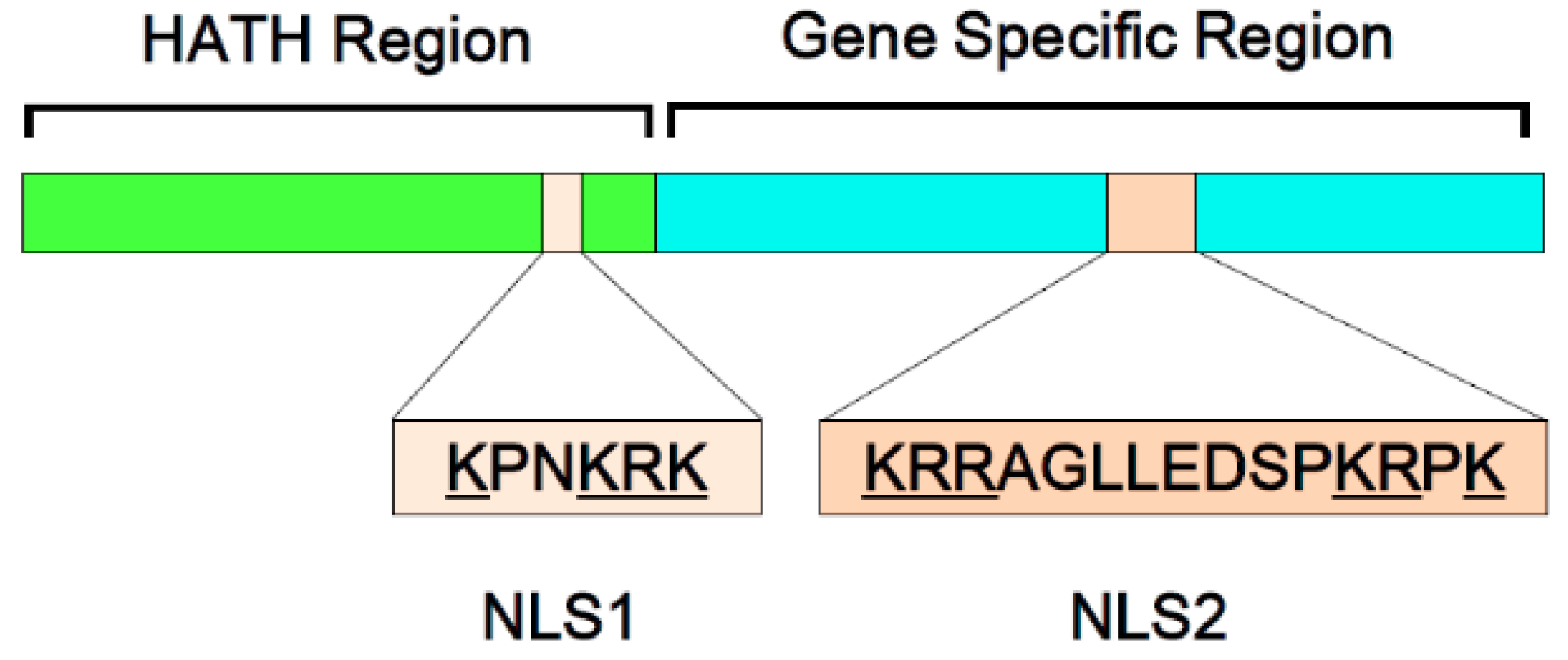
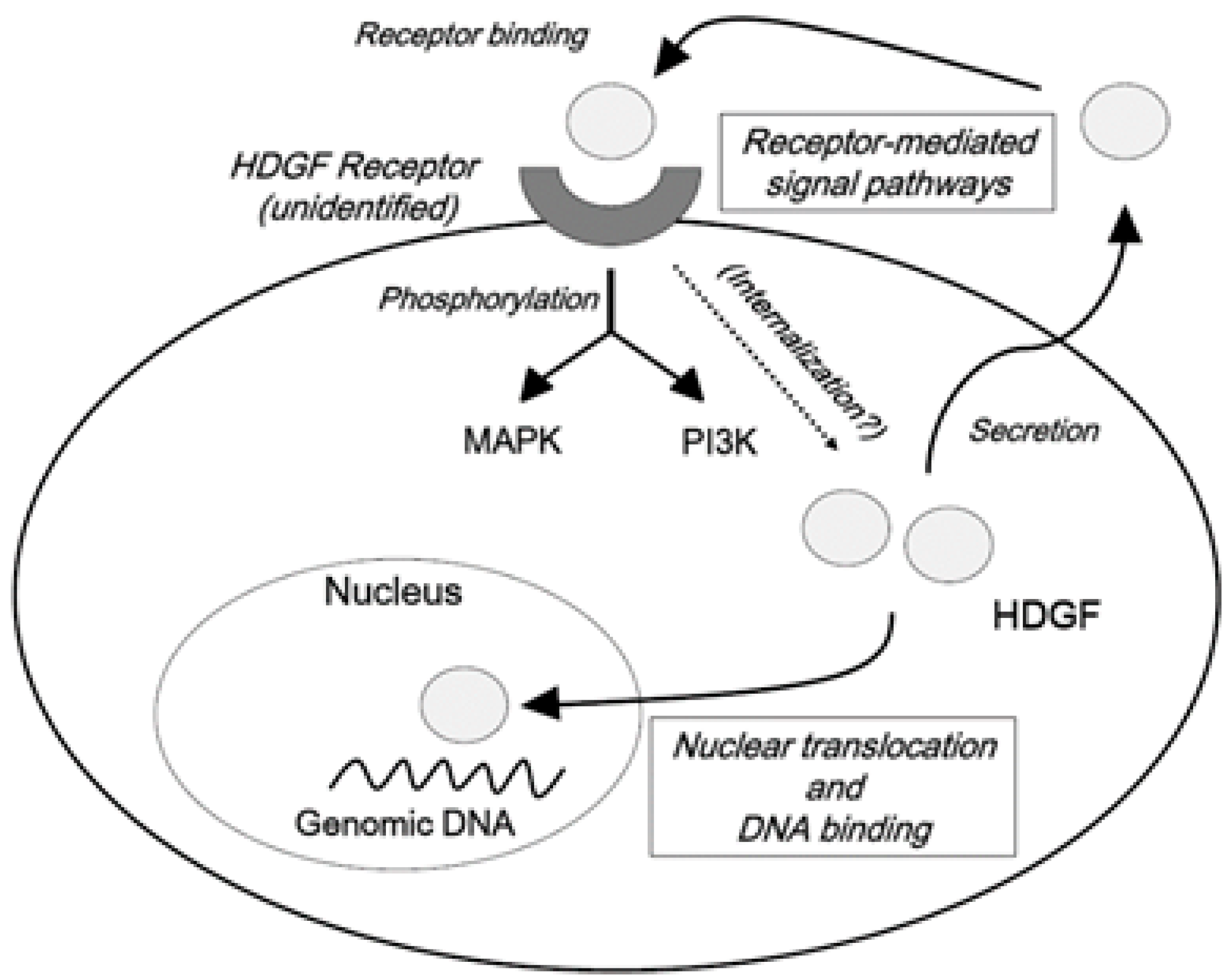
2. HDGF as a Novel Unique Growth Factor


3. HDGF as an Angiogenic Factor
4. HDGF as a Possible Anti-Apoptotic Factor
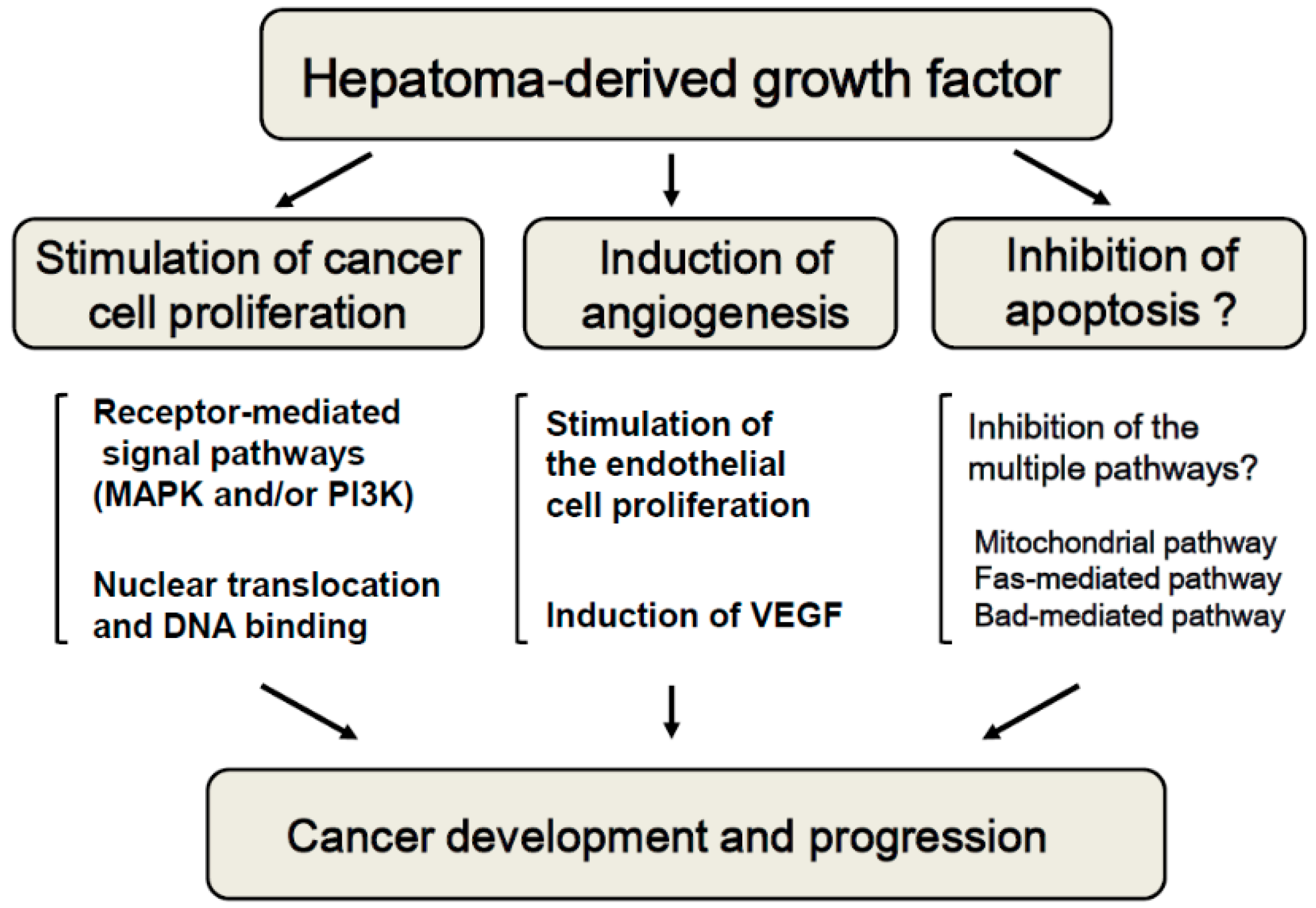
5. HDGF as a Growth-Promoting Factor for HCC
6. Future Perspectives

7. Conclusions
Acknowledgments
Author Contributions
Conflicts of Interest
Abbreviations
| HCC | hepatocellular carcinoma |
| VEGF | vascular endothelial growth factor |
| HDGF | hepatoma-derived growth factor |
| HRP | HDGF-related protein |
| HATH | homologous to the amino terminus of HDGF |
| MAPK | mitogen-activated protein kinase |
| PI3K | Phosphatidylinositol-3 kinase |
| NLS | nuclear localization signal |
| NSCLC | non-small cell lung cancer |
| miR | microRNA |
References
- El-Serag, H.B.; Kanwal, F. Epidemiology of hepatocellular carcinoma in the United States: Where are we? Where do we go? Hepatology 2014, 60, 1767–1775. [Google Scholar] [CrossRef] [PubMed]
- Ben Ari, Z.; Weitzman, E.; Safran, M. Oncogenic viruses and hepatocellular carcinoma. Clin. Liver Dis. 2015, 19, 341–360. [Google Scholar] [CrossRef] [PubMed]
- Bruix, J.; Sherman, M. Management of hepatocellular carcinoma: An update. Hepatology 2011, 53, 1020–1022. [Google Scholar] [CrossRef] [PubMed]
- European Association for the Study of the Liver; European Organisation for Research and Treatment of Cancer. EASL—EORTC clinical practice guidelines: Management of hepatocellular carcinoma. J. Hepatol. 2012, 56, 908–943. [Google Scholar]
- Deng, G.L.; Zeng, S.; Shen, H. Chemotherapy and target therapy for hepatocellular carcinoma: New advances and challenges. World J. Hepatol. 2015, 7, 787–798. [Google Scholar] [CrossRef] [PubMed]
- Llovet, J.M.; Ricci, S.; Mazzaferro, V.; Hilgard, P.; Gane, E.; Blanc, J.F.; de Oliveira, A.C.; Santoro, A.; Raoul, J.L.; Forner, A.; et al. Sorafenib in advanced hepatocellular carcinoma. N. Engl. J. Med. 2008, 359, 378–90. [Google Scholar] [CrossRef] [PubMed]
- Cheng, A.L.; Kang, Y.K.; Chen, Z.; Tsao, C.J.; Qin, S.; Kim, J.S.; Luo, R.; Feng, J.; Ye, S.; Yang, T.S.; et al. Efficacy and safety of sorafenib in patients in the Asia-Pacific region with advanced hepatocellular carcinoma: A phase III randomised, double-blind, placebo-controlled trial. Lancet Oncol. 2009, 10, 25–34. [Google Scholar] [CrossRef]
- Nakamura, H.; Izumoto, Y.; Kambe, H.; Kuroda, T.; Mori, T.; Kawamura, K.; Yamamoto, H.; Kishimoto, T. Molecular cloning of complementary DNA for a novel human hepatoma-derived growth factor. Its homology with high mobility group-1 protein. J. Biol. Chem. 1994, 269, 25143–25149. [Google Scholar] [PubMed]
- Nakamura, H.; Enomoto, H. Hepatoma-derived growth factor in carcinogenesis and cancer progression. Curr. Drug Ther. 2011, 6, 278–285. [Google Scholar] [CrossRef]
- Kishima, Y.; Yamamoto, H.; Izumoto, Y.; Yoshida, K.; Enomoto, H.; Yamamoto, M.; Kuroda, T.; Ito, H.; Yoshizaki, K.; Nakamura, H. Hepatoma-derived growth factor stimulates cell growth after translocation to the nucleus by nuclear localization signals. J. Biol. Chem. 2002, 277, 10315–10322. [Google Scholar] [CrossRef] [PubMed]
- Kishima, Y.; Yoshida, K.; Enomoto, H.; Yamamoto, M.; Kuroda, T.; Okuda, Y.; Uyama, H.; Nakamura, H. Antisense oligonucleotides of hepatoma-derived growth factor (HDGF) suppress the proliferation of hepatoma cells. Hepatogastroenterology 2002, 49, 1639–1644. [Google Scholar] [PubMed]
- Yoshida, K.; Nakamura, H.; Okuda, Y.; Enomoto, H.; Kishima, Y.; Uyama, H.; Ito, H.; Hirasawa, T.; Inagaki, S.; Kawase, I. Expression of hepatoma-derived growth factor in hepatocarcinogenesis. J. Gastroenterol. Hepatol. 2003, 18, 1293–1301. [Google Scholar] [CrossRef] [PubMed]
- Ren, H.; Tang, X.; Lee, J.J.; Feng, L.; Everett, A.D.; Hong, W.K.; Khuri, F.R.; Mao, L. Expression of hepatoma-derived growth factor is a strong prognostic predictor for patients with early-stage non-small-cell lung cancer. J. Clin. Oncol. 2004, 22, 3230–3237. [Google Scholar] [CrossRef] [PubMed]
- Guo, S.; Liu, H.D.; Liu, Y.F.; Liu, L.; Sun, Q.; Cui, X.J. Hepatoma-derived growth factor: A novel prognostic biomarker in intrahepatic cholangiocarcinoma. Tumour Biol. 2015, 36, 353–364. [Google Scholar] [CrossRef] [PubMed]
- Yoshida, K.; Tomita, Y.; Okuda, Y.; Yamamoto, S.; Enomoto, H.; Uyama, H.; Ito, H.; Hoshida, Y.; Aozasa, K.; Nagano, H.; et al. Hepatoma-derived growth factor is a novel prognostic factor for hepatocellular carcinoma. Ann. Surg. Oncol. 2006, 13, 159–167. [Google Scholar] [CrossRef] [PubMed]
- Bao, C.; Wang, J.; Ma, W.; Wang, X.; Cheng, Y. HDGF: A novel jack-of-all-trades in cancer. Future Oncol. 2014, 10, 2675–2685. [Google Scholar] [CrossRef] [PubMed]
- Bao, C.H.; Liu, K.; Wang, X.T.; Ma, W.; Wang, J.B.; Wang, C.; Jia, Y.B.; Wang, N.N.; Tan, B.X.; Song, Q.X.; et al. Prognostic role of hepatoma-derived growth factor in solid tumors of Eastern Asia: A systematic review and meta-analysis. Asian Pac. J. Cancer Prev. 2015, 16, 1803–1811. [Google Scholar] [PubMed]
- Tsai, C.C.; Huang, S.C.; Tai, M.H.; Chien, C.C.; Huang, C.C.; Hsu, Y.C. Hepatoma-derived growth factor upregulation is correlated with prognostic factors of early-stage cervical adenocarcinoma. Int. J. Mol. Sci. 2014, 15, 21492–21504. [Google Scholar] [CrossRef] [PubMed]
- Tao, F.; Ye, M.F.; Sun, A.J.; Lv, J.Q.; Xu, G.G.; Jing, Y.M.; Wang, W. Prognostic significance of nuclear hepatoma-derived growth factor expression in gallbladder cancer. World J. Gastroenterol. 2014, 20, 9564–9569. [Google Scholar] [PubMed]
- Izumoto, Y.; Kuroda, T.; Harada, H.; Kishimoto, T.; Nakamura, H. Hepatoma-derived growth factor belongs to a gene family in mice showing significant homology in the amino terminus. Biochem. Biophys. Res. Commun. 1997, 238, 26–32. [Google Scholar] [CrossRef] [PubMed]
- Ikegame, K.; Yamamoto, M.; Kishima, Y.; Enomoto, H.; Yoshida, K.; Suemura, M.; Kishimoto, T.; Nakamura, H. A new member of a hepatoma-derived growth factor gene family can translocate to the nucleus. Biochem. Biophys. Res. Commun. 1999, 266, 81–87. [Google Scholar] [CrossRef] [PubMed]
- Dietz, F.; Franken, S.; Yoshida, K.; Nakamura, H.; Kappler, J.; Gieselmann, V. The family of hepatoma-derived growth factor proteins: Characterization of a new member HRP-4 and classification of its subfamilies. Biochem. J. 2002, 366, 491–500. [Google Scholar] [CrossRef] [PubMed]
- Ge, H.; Si, Y.; Roeder, R.G. Isolation of cDNAs encoding novel transcription coactivators p52 and p75 reveals an alternate regulatory mechanism of transcriptional activation. EMBO J. 1998, 17, 6723–6729. [Google Scholar] [CrossRef] [PubMed]
- Enomoto, H.; Nakamura, H.; Nishiguchi, S. The Role of Hepatoma-derived Growth Factor (HDGF) in cancer development and progression. Curr. Res. Cancer 2011, 5, 11–25. [Google Scholar]
- Thakar, K.; Kröcher, T.; Savant, S.; Gollnast, D.; Kelm, S.; Dietz, F. Secretion of hepatoma-derived growth factor is regulated by N-terminal processing. Biol. Chem. 2010, 391, 1401–1410. [Google Scholar] [CrossRef] [PubMed]
- Jhaveri, N.; Chen, T.C.; Hofman, F.M. Tumor vasculature and glioma stem cells: Contributions to glioma progression. Cancer Lett. 2014. [Google Scholar] [CrossRef] [PubMed]
- Kao, Y.H.; Chen, C.L.; Jawan, B.; Chung, Y.H.; Sun, C.K.; Kuo, S.M.; Hu, T.H.; Lin, Y.C.; Chan, H.H.; Cheng, K.H.; et al. Upregulation of hepatoma-derived growth factor is involved in murine hepatic fibrogenesis. J. Hepatol. 2010, 52, 96–105. [Google Scholar] [CrossRef] [PubMed]
- Gómez, E.; Correia-Álvarez, E.; Caamaño, J.N.; Díez, C.; Carrocera, S.; Peynot, N.; Martín, D.; Giraud-Delville, C.; Duranthon, V.; Sandra, O.; et al. Hepatoma-derived growth factor: From the bovine uterus to the in vitro embryo culture. Reproduction 2014, 148, 353–365. [Google Scholar] [CrossRef] [PubMed]
- Lepourcelet, M.; Tou, L.; Cai, L.; Sawada, J.; Lazar, A.J.; Glickman, J.N.; Williamson, J.A.; Everett, A.D.; Redston, M.; Fox, E.A.; et al. Insights into developmental mechanisms and cancers in the mammalian intestine derived from serial analysis of gene expression and study of the hepatoma-derived growth factor (HDGF). Development 2005, 132, 415–427. [Google Scholar] [CrossRef] [PubMed]
- Everett, A.D.; Yang, J.; Rahman, M.; Dulloor, P.; Brautigan, D.L. Mitotic phosphorylation activates hepatoma-derived growth factor as a mitogen. BMC Cell Biol. 2011, 12. [Google Scholar] [CrossRef] [PubMed]
- Mori, M.; Morishita, H.; Nakamura, H.; Matsuoka, H.; Yoshida, K.; Kishima, Y.; Zhou, Z.; Kida, H.; Funakoshi, T.; Goya, S.; et al. Hepatoma-derived growth factor is involved in lung remodeling by stimulating epithelial growth. Am. J. Respir. Cell Mol. Biol. 2004, 30, 459–469. [Google Scholar] [CrossRef] [PubMed]
- Mao, J.; Xu, Z.; Fang, Y.; Wang, H.; Xu, J.; Ye, J.; Zheng, S.; Zhu, Y. Hepatoma-derived growth factor involved in the carcinogenesis of gastric epithelial cells through promotion of cell proliferation by Erk1/2 activation. Cancer Sci. 2008, 99, 2120–2127. [Google Scholar] [CrossRef] [PubMed]
- Everett, A.D.; Narron, J.V.; Stoops, T.; Nakamura, H.; Tucker, A. Hepatoma-derived growth factor is a pulmonary endothelial cell-expressed angiogenic factor. Am. J. Respir. Cell Mol. Biol. 2004, 286, L1194–L1201. [Google Scholar] [CrossRef] [PubMed]
- Kung, M.L.; Tsai, H.E.; Hu, T.H.; Kuo, H.M.; Liu, L.F.; Chen, S.C.; Lin, P.R.; Ma, Y.L.; Wang, E.M.; Liu, G.S.; et al. Hepatoma-derived growth factor stimulates podosome rosettes formation in NIH/3T3 cells through the activation of phosphatidylinositol 3-kinase/Akt pathway. Biochem. Biophys. Res. Commun. 2012, 425, 169–176. [Google Scholar] [CrossRef] [PubMed]
- Abouzied, M.M.; El-Tahir, H.M.; Prenner, L.; Häberlein, H.; Gieselmann, V.; Franken, S. Hepatoma-derived growth factor. Significance of amino acid residues 81–100 in cell surface interaction and proliferative activity. J. Biol. Chem. 2005, 280, 10945–10954. [Google Scholar] [CrossRef] [PubMed]
- Stec, I.; Nagl, S.B.; van Ommen, G.J.B.; den Dunnen, J.T. The PWWP domain: A potential protein-protein interaction domain in nuclear proteins influencing differentiation? FEBS Lett. 2000, 473, 1–5. [Google Scholar] [CrossRef]
- Qiu, C.; Sawada, K.; Zhang, X.; Cheng, X. The PWWP domain of mammalian DNA methyltransferase Dnmt3b defines a new family of DNA-binding folds. Nat. Struct. Biol. 2002, 9, 217–224. [Google Scholar] [CrossRef] [PubMed]
- Yang, J.; Everett, A.D. Hepatoma-derived growth factor binds DNA through the N-terminal PWWP domain. BMC Mol. Biol. 2007, 8, 101. [Google Scholar] [CrossRef] [PubMed]
- Everett, A.D.; Lobe, D.R.; Matsumura, M.E.; Nakamura, H.; McNamara, C.A. Hepatoma-derived growth factor stimulates smooth muscle cell growth and is expressed in vascular development. J. Clin. Investig. 2000, 105, 567–575. [Google Scholar] [CrossRef] [PubMed]
- Okuda, Y.; Nakamura, H.; Yoshida, K.; Enomoto, H.; Uyama, H.; Hirotani, T.; Funamoto, M.; Ito, H.; Everett, A.D.; Hada, T.; et al. Hepatoma-derived growth factor induces tumorigenesis in vivo through both direct angiogenic activity and induction of vascular endothelial growth factor. Cancer Sci. 2003, 94, 1034–1041. [Google Scholar] [CrossRef] [PubMed]
- Lee, K.H.; Choi, E.Y.; Kim, M.K.; Lee, S.H.; Jang, B.I.; Kim, T.N.; Kim, S.W.; Kim, S.W.; Song, S.K.; Kim, J.R.; et al. Hepatoma-derived growth factor regulates the bad-mediated apoptotic pathway and induction of vascular endothelial growth factor in stomach cancer cells. Oncol. Res. 2010, 19, 67–76. [Google Scholar] [CrossRef] [PubMed]
- Mohammad, R.M.; Muqbil, I.; Lowe, L.; Yedjou, C.; Hsu, H.Y.; Lin, L.T.; Siegelin, M.D.; Fimognari, C.; Kumar, N.B.; Dou, Q.P.; et al. Broad targeting of resistance to apoptosis in cancer. Semin. Cancer Biol. 2015. [Google Scholar] [CrossRef] [PubMed]
- Bao, C.H.; Wang, X.T.; Ma, W.; Wang, N.N.; Un Nesa, E.; Wang, J.B.; Wang, C.; Jia, Y.B.; Wang, K.; Tian, H.; et al. Irradiated fibroblasts promote epithelial-mesenchymal transition and HDGF expression of esophageal squamous cell carcinoma. Biochem. Biophys. Res. Commun. 2015, 458, 441–447. [Google Scholar] [CrossRef] [PubMed]
- Clermont, F.; Gonzalez, N.S.; Communi, D.; Franken, S.; Dumont, J.E.; Robaye, B. HDGF is dephosphorylated during the early steps of endothelial cell apoptosis in a caspase-dependent way. J. Cell. Biochem. 2008, 104, 1161–1171. [Google Scholar] [CrossRef] [PubMed]
- Machuy, N.; Thiede, B.; Rajalingam, K.; Dimmler, C.; Thieck, O.; Meyer, T.F.; Rudel, T. A global approach combining proteome analysis and phenotypic screening with RNA interference yields novel apoptosis regulators. Mol. Cell. Proteomics 2005, 4, 44–55. [Google Scholar] [CrossRef] [PubMed]
- Zhou, Z.; Yamamoto, Y.; Sugai, F.; Yoshida, K.; Kishima, Y.; Sumi, H.; Nakamura, H.; Sakoda, S. Hepatoma-derived growth factor is a neurotrophic factor harbored in the nucleus. J. Biol. Chem. 2004, 279, 27320–27326. [Google Scholar] [CrossRef] [PubMed]
- Borders, A.S.; Hersh, M.A.; Getchell, M.L.; van Rooijen, N.; Cohen, D.A.; Stromberg, A.J.; Getchell, T.V. Macrophage-mediated neuroprotection and neurogenesis in the olfactory epithelium. Physiol. Genomics 2007, 31, 531–543. [Google Scholar] [CrossRef] [PubMed]
- Tsang, T.Y.; Tang, W.Y.; Tsang, W.P.; Kong, S.K.; Kwok, T.T. Downregulation of hepatoma-derived growth factor activates the Bad-mediated apoptotic pathway in human cancer cells. Apoptosis 2008, 13, 1135–1147. [Google Scholar] [CrossRef] [PubMed]
- Liao, F.; Dong, W.; Fan, L. Apoptosis of human colorectal carcinoma cells is induced by blocking hepatoma-derived growth factor. Med. Oncol. 2010, 27, 1219–1226. [Google Scholar] [CrossRef] [PubMed]
- Liao, F.; Liu, M.; Lv, L.; Dong, W. Hepatoma-derived growth factor promotes the resistance to anti-tumor effects of nordihydroguaiaretic acid in colorectal cancer cells. Eur. J. Pharmacol. 2010, 645, 55–62. [Google Scholar] [CrossRef] [PubMed]
- Tsang, T.Y.; Tang, W.Y.; Tsang, W.P.; Co, N.N.; Kong, S.K.; Kwok, T.T. Mechanistic study on growth suppression and apoptosis induction by targeting hepatoma-derived growth factor in human hepatocellular carcinoma HepG2 cells. Cell. Physiol. Biochem. 2009, 24, 253–262. [Google Scholar] [CrossRef] [PubMed]
- Liu, W.; Nakamura, H.; Deng, H.; Enomoto, H.; Yamamoto, T.; Iwata, Y.; Koh, N.; Saito, M.; Imanishi, H.; Shimomura, S.; et al. A higher expression of hepatoma-derived growth factor in hepatocelular carcinoma cells and more tumor growth in vivo. Trends Cancer Res. 2009, 5, 29–36. [Google Scholar]
- Yamamoto, T.; Nakamura, H.; Liu, W.; Cao, K.; Yoshikawa, S.; Enomoto, H.; Iwata, Y.; Koh, N.; Saito, M.; Imanishi, H.; et al. Involvement of hepatoma-derived growth factor in the growth inhibition of hepatocellular carcinoma cells by vitamin K2. J. Gastroenterol. 2009, 44, 228–235. [Google Scholar] [CrossRef] [PubMed]
- Hu, T.H.; Huang, C.C.; Liu, L.F.; Lin, P.R.; Liu, S.Y.; Chang, H.W.; Changchien, C.S.; Lee, C.M.; Chuang, J.H.; Tai, M.H. Expression of hepatoma-derived growth factor in hepatocellular carcinoma. Cancer 2003, 98, 1444–1456. [Google Scholar] [CrossRef] [PubMed]
- Zhou, Y.; Zhou, N.; Fang, W.; Huo, J. Overexpressed HDGF as an independent prognostic factor is involved in poor prognosis in Chinese patients with liver cancer. Diagn. Pathol. 2010, 5. [Google Scholar] [CrossRef] [PubMed]
- Ren, H.; Chu, Z.; Mao, L. Antibodies targeting hepatoma-derived growth factor as a novel strategy in treating lung cancer. Mol. Cancer Ther. 2009, 8, 1106–1112. [Google Scholar] [CrossRef] [PubMed]
- Meng, J.; Xie, W.; Cao, L.; Hu, C.; Zhe, Z. shRNA targeting HDGF suppressed cell growth and invasion of squamous cell lung cancer. Acta Biochim. Biophys. Sin. 2010, 42, 52–57. [Google Scholar] [CrossRef] [PubMed]
- Zhao, J.; Ma, M.Z.; Ren, H.; Liu, Z.; Edelman, M.J.; Pan, H.; Mao, L. Anti-HDGF targets cancer and cancer stromal stem cells resistant to chemotherapy. Clin. Cancer Res. 2013, 19, 3567–3576. [Google Scholar] [CrossRef] [PubMed]
- Li, M.; Shen, J.; Wu, X.; Zhang, B.; Zhang, R.; Weng, H.; Ding, Q.; Tan, Z.; Gao, G.; Mu, J.; et al. Downregulated expression of hepatoma-derived growth factor (HDGF) reduces gallbladder cancer cell proliferation and invasion. Med. Oncol. 2013, 30. [Google Scholar] [CrossRef] [PubMed]
- Tsai, H.E.; Liu, G.S.; Kung, M.L.; Liu, L.F.; Wu, J.C.; Tang, C.H.; Huang, C.H.; Chen, S.C.; Lam, H.C.; Wu, C.S.; et al. Downregulation of hepatoma-derived growth factor contributes to retarded lung metastasis via inhibition of epithelial-mesenchymal transition by systemic POMC gene delivery in melanoma. Mol. Cancer Ther. 2013, 12, 1016–1025. [Google Scholar] [CrossRef] [PubMed]
- Enomoto, H.; Nakamura, H.; Liu, W.; Iwata, Y.; Nishikawa, H.; Takata, R.; Yoh, K.; Hasegawa, K.; Ishii, A.; Takashima, T.; et al. Reduced expression of hepatoma-derived growth factor inhibits the proliferation of a hepatocellular carcinoma cell line, SK-HEP-1, in vitro and in vivo. 2015; In preparation. [Google Scholar]
- Sasaki, Y.; Negishi, H.; Idogawa, M.; Yokota, I.; Koyama, R.; Kusano, M.; Suzuki, H.; Fujita, M.; Maruyama, R.; Toyota, M.; et al. p53 negatively regulates the hepatoma growth factor HDGF. Cancer Res. 2011, 71, 7038–7047. [Google Scholar] [CrossRef] [PubMed]
- Shih, T.C.; Tien, Y.J.; Wen, C.J.; Yeh, T.S.; Yu, M.C.; Huang, C.H.; Lee, Y.S.; Yen, T.C.; Hsieh, S.Y. MicroRNA-214 downregulation contributes to tumor angiogenesis by inducing secretion of the hepatoma-derived growth factor in human hepatoma. J. Hepatol. 2012, 57, 584–591. [Google Scholar] [CrossRef] [PubMed]
- Guo, H.; Li, W.; Zheng, T.; Liu, Z. miR-195 Targets HDGF to inhibit proliferation and invasion of NSCLC cells. Tumour Biol. 2014, 35, 8861–8866. [Google Scholar] [CrossRef] [PubMed]
- Zhao, W.Y.; Wang, Y.; An, Z.J.; Shi, C.G.; Zhu, G.A.; Wang, B.; Lu, M.Y.; Pan, C.K.; Chen, P. Downregulation of miR-497 promotes tumor growth and angiogenesis by targeting HDGF in non-small cell lung cancer. Biochem. Biophys. Res. Commun. 2013, 435, 466–471. [Google Scholar] [CrossRef] [PubMed]
- Chen, B.; Huang, T.; Jiang, J.; Lv, L.; Li, H.; Xia, S. miR-141 suppresses proliferation and motility of gastric cancer cells by targeting HDGF. Mol. Cell. Biochem. 2014, 388, 211–218. [Google Scholar] [CrossRef] [PubMed]
- Chen, S.C.; Hu, T.H.; Huang, C.C.; Kung, M.L.; Chu, T.H.; Yi, L.N.; Huang, S.T.; Chan, H.H.; Chuang, J.H.; Liu, L.F.; et al. Hepatoma-derived growth factor/nucleolin axis as a novel oncogenic pathway in liver carcinogenesis. Oncotarget 2015, in press. [Google Scholar]
© 2015 by the authors; licensee MDPI, Basel, Switzerland. This article is an open access article distributed under the terms and conditions of the Creative Commons Attribution license (http://creativecommons.org/licenses/by/4.0/).
Share and Cite
Enomoto, H.; Nakamura, H.; Liu, W.; Nishiguchi, S. Hepatoma-Derived Growth Factor: Its Possible Involvement in the Progression of Hepatocellular Carcinoma. Int. J. Mol. Sci. 2015, 16, 14086-14097. https://0-doi-org.brum.beds.ac.uk/10.3390/ijms160614086
Enomoto H, Nakamura H, Liu W, Nishiguchi S. Hepatoma-Derived Growth Factor: Its Possible Involvement in the Progression of Hepatocellular Carcinoma. International Journal of Molecular Sciences. 2015; 16(6):14086-14097. https://0-doi-org.brum.beds.ac.uk/10.3390/ijms160614086
Chicago/Turabian StyleEnomoto, Hirayuki, Hideji Nakamura, Weidong Liu, and Shuhei Nishiguchi. 2015. "Hepatoma-Derived Growth Factor: Its Possible Involvement in the Progression of Hepatocellular Carcinoma" International Journal of Molecular Sciences 16, no. 6: 14086-14097. https://0-doi-org.brum.beds.ac.uk/10.3390/ijms160614086