Article

Inhibition of SDF-1α/CXCR4 Signalling in Subchondral Bone Attenuates Post-Traumatic Osteoarthritis

Department of Orthopedics, Tongji Hospital, Tongji Medical College, Huazhong University of Science and Technology, 1095 Jiefang Avenue, Wuhan 430030, China
*
Authors to whom correspondence should be addressed.
Int. J. Mol. Sci. 2016, 17(6), 943; https://0-doi-org.brum.beds.ac.uk/10.3390/ijms17060943
Submission received: 2 April 2016 / Revised: 6 June 2016 / Accepted: 7 June 2016 / Published: 16 June 2016
(This article belongs to the Section Molecular Pathology, Diagnostics, and Therapeutics)

Abstract

:
Previous studies showed that SDF-1α is a catabolic factor that can infiltrate cartilage, decrease proteoglycan content, and increase MMP-13 activity. Inhibiting the SDF-1α/CXCR4 signalling pathway can attenuate the pathogenesis of osteoarthritis (OA). Recent studies have also shown that SDF-1α enhances chondrocyte proliferation and maturation. These results appear to be contradictory. In the current study, we used a destabilisation OA animal model to investigate the effects of SDF-1α/CXCR4 signalling in the tibial subchondral bone and the OA pathological process. Post-traumatic osteoarthritis (PTOA) mice models were prepared by transecting the anterior cruciate ligament (ACLT), or a sham surgery was performed, in a total of 30 mice. Mice were treated with phosphate buffer saline (PBS) or AMD3100 (an inhibitor of CXCR4) and sacrificed at 30 days post ACLT or sham surgery. Tibial subchondral bone status was quantified by micro-computed tomography (μCT). Knee-joint histology was analysed to examine the articular cartilage and joint degeneration. The levels of SDF-1α and collagen type I c-telopeptidefragments (CTX-I) were quantified by ELISA. Bone marrow mononuclear cells (BMMCs) were used to clarify the effects of SDF-1α on osteoclast formation and activity in vivo. μCT analysis revealed significant loss of trabecular bone from tibial subchondral bone post-ACLT, which was effectively prevented by AMD3100. AMD3100 could partially prevent bone loss and articular cartilage degeneration. Serum biomarkers revealed an increase in SDF-1α and bone resorption, which were also reduced by AMD3100. SDF-1α can promote osteoclast formation and the expression oftartrate resistant acid phosphatase (TRAP), cathepsin K (CK), and matrix metalloproteinase (MMP)-9 in osteoclasts by activating the MAPK pathway, including ERK and p38, but not JNK. In conclusion, inhibition of SDF-1α/CXCR4signalling was able to prevent trabecular bone loss and attenuated cartilage degeneration in PTOA mice.

Graphical Abstract

1. Introduction

Osteoarthritis (OA) is the most common joint disease, afflicting mainly the hips and knees, characterised by joint pain and loss of function. Currently, the pathogenesis of OA remains largely unclear, so pharmacological therapy for OA is largely ineffective, and joint replacement is the only treatment for end-stage disease [1]. Increasing evidence suggests that OA is a disease of the whole joint [1,2]; not only the cartilage, synovium, and ligaments, but also the bone and bone marrow, especially in PTOA, which represents ~12% of symptomatic OA cases. In PTOA patients, articular chondrocytes and subchondral bone cells perceive acute stress, whereas subchondral bone changes can usually be observed before articular cartilage degeneration [2,3,4,5].
Increasing evidence suggests that subchondral bone turnover and its interactions with the articular cartilage are more important than previously thought. The relationship between high subchondral bone turnover and articular cartilage degeneration may provide a new opportunity to slow down or prevent PTOA progression [2,6,7,8,9].
Recently, researchers have observed that inhibition of hypoxia-inducible factor–2α (HIF–2α) and transforming growth factor-β1 (TGF-β1) signalling in subchondral bone attenuates PTOA [10,11,12]. This illustrates the contribution of the subchondral bone to articular cartilage degeneration and hypertrophy. Thus, the subchondral bone should be regarded as a new target in OA.
The role of SDF-1α in the pathogenesis of OA has attracted more attention in recent years [13]. SDF-1α is related to several characteristic bone and cartilage changes [14,15,16]. The SDF-1α level is increased in OA synovial fluid, and synovectomy significantly reduces the concentrations of SDF-1α and MMPs in serum [17]. SDF-1 can increase articular cartilage MMP-13 expression in vitro, which induces matrix degradation [15]. Several groups have reported data suggesting that the SDF-1/CXCR4 signalling pathway may be a catabolic regulator of osteoarthritic cartilage destruction. However, SDF-1α appears to regulate BMP-2-induced chondrogenic differentiation of mesenchymal stem cells and enhances chondrocyte proliferation and maturation [18,19,20]. Moreover, it induces the migration of bone marrow MSCs to articular lesion sites [21].
Therefore, at present, the effects of SDF-1α on cartilage are inconclusive. Other researchers have reported that SDF-1α can upregulate bone resorption by human osteoclasts [22,23,24]. Also, targeted disruption of the SDF-1α/CXCR4 axis inhibits bone loss [25,26].
In this study, we investigated the effect of SDF-1α in an ACLT OA mouse model [10,27]. We quantified the effect of inhibition of SDF-1α /CXCR4 signalling on subchondral bone turnover level and cartilage degeneration. We hypothesised that inhibition of SDF-1α/CXCR4 signalling in the subchondral bone would attenuate the pathological changes and then avoid degeneration of articular cartilage in the ACLT mouse model.

2. Results

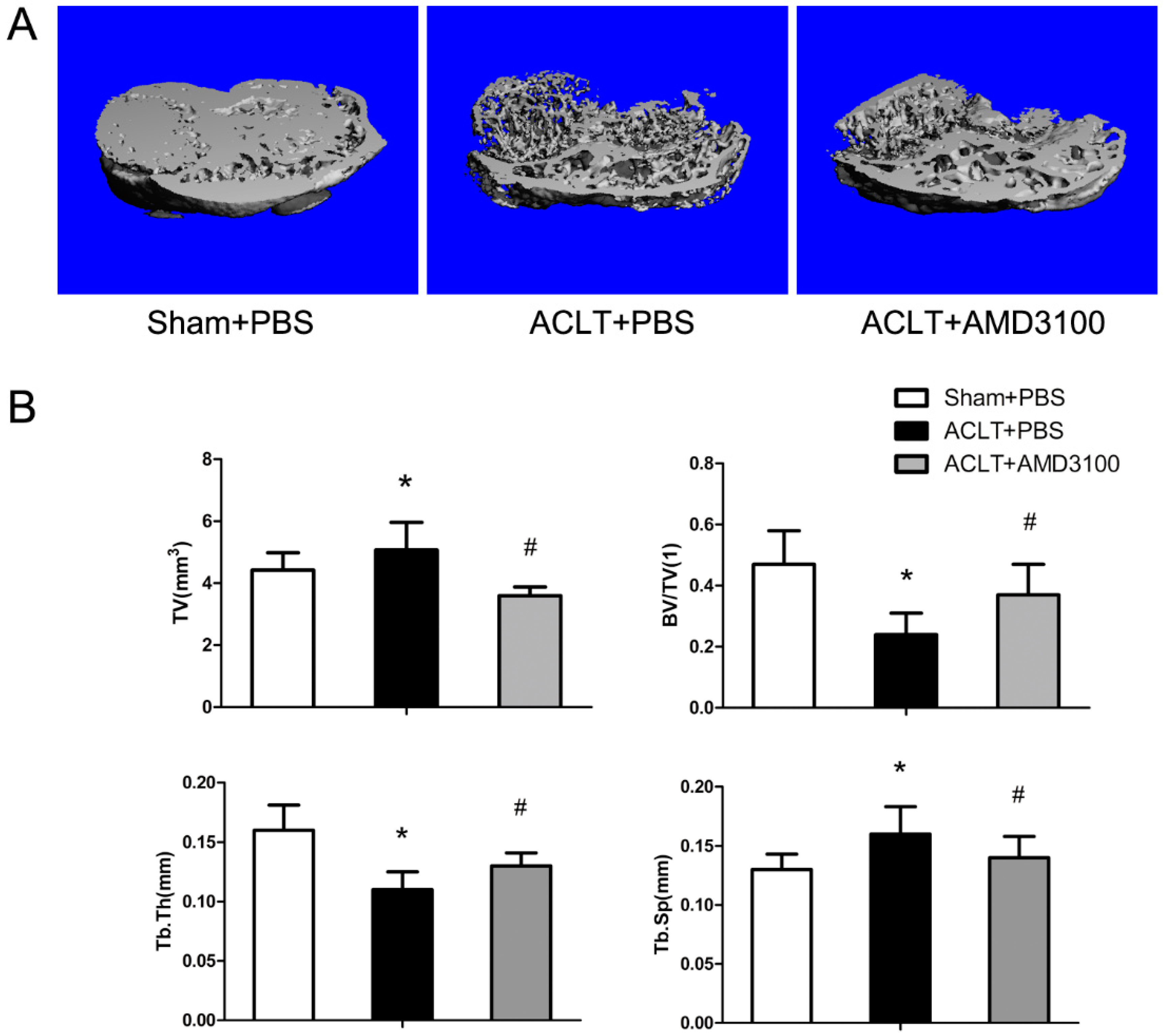
2.1. Tibial Subchondral Bone Analysis via μCTimaging

We examined the tibial subchondral bone changes in the ACLT OA animals. The 3D μCT in the tibial subchondral bone showed dramatic changes between the sham and ACLT groups. The total subchondral bone tissue volume (TV) increased by more than 13.6% in ACLT mice compared with the sham-operated controls. Moreover, the trabecular separation (Tb. Sp) was also increased significantly, 18.8%, when compared with sham-operated controls. Trabecular bone volume/tissue volume (BV/TV) and Trabecular thickness (Tb. Th) in ACLT mice decreased, by 48.9% and 37.5%, respectively. These effects demonstrated uncoupled bone remodelling in the tibial subchondral bone; bone resorption was increased and resulted in bone loss in the PTOA animals. Treatment with AMD3100 in ACLT mice dramatically attenuated tibial subchondral bone loss, compared with ACLT mice treated with vehicle (Figure 1).

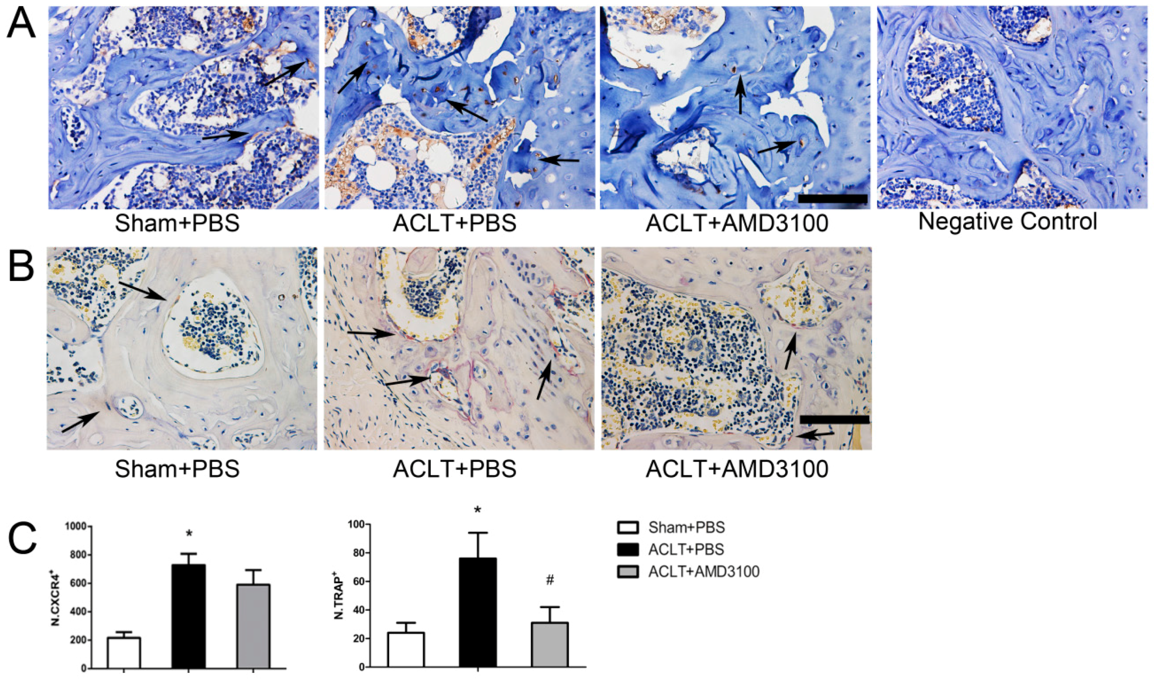
2.2. Elevated Active SDF-1α and Bone Resorption in Subchondral Bone

Immunohistochemistry indicated CXCR4 expression in subchondral bone. The number of CXCR4-positive cells increased by 2.6 times in ACLT mice compared with the sham-operated group at 30 days, and we conducted a quantitative estimation (Figure 2A). We next examined osteoclast differentiation in ACLT mice compared with the sham-operated controls; ACLT mice displayed an increased number of tartrate-resistant acid phosphatase (TRAP)-positive multinucleated cells in tibial subchondral bone. When treated with AMD3100, TRAP-positive multinucleated cells were reduced in ACLT mice (Figure 2B). These observations suggest that SDF-1α plays a role via binding to CXCR4 in the tibial subchondral bone. Osteoclast differentiation was increased in tibial subchondral bone, and AMD3100 functioned as a strong inhibitor of osteoclastogenesis.

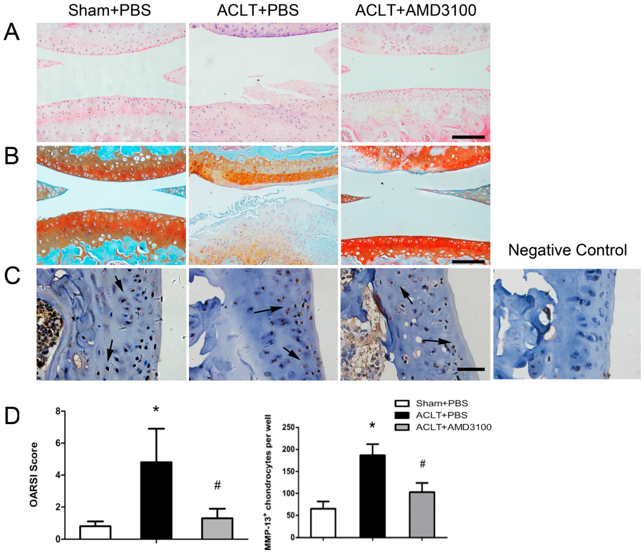
2.3. Inhibition of SDF-1α Signalling in Subchondral Bone Attenuates Cartilage Degeneration

We confirmed the dramatic change in tibial subchondral bone in ACLT mice versus sham-operated mice. Proteoglycan loss in cartilage in ACLT mice was assessed by Safranin O-Fast Green staining (Figure 3B). These results were further confirmed by H&E-stained bone sections, and ACLT mice exhibited increased expression of MMP13 in articular chondrocytes compared with sham-operated mice (Figure 3A,C). We observed obvious damage to the articular cartilage in ACLT mice at 30 days post-surgery, and OARSI scores confirmed the effects (Figure 3D). Treatment with AMD3100 significantly inhibited the changes as measured. Notably, inhibition of SDF-1α attenuated the degeneration of articular cartilage in PTOA mice, and it had similar effects in reducing the elevated concentrations of MMP13 in articular chondrocytes compared with the ACLT/PBS group. The OARSI score also indicated a protective effect of AMD3100 on articular cartilage.

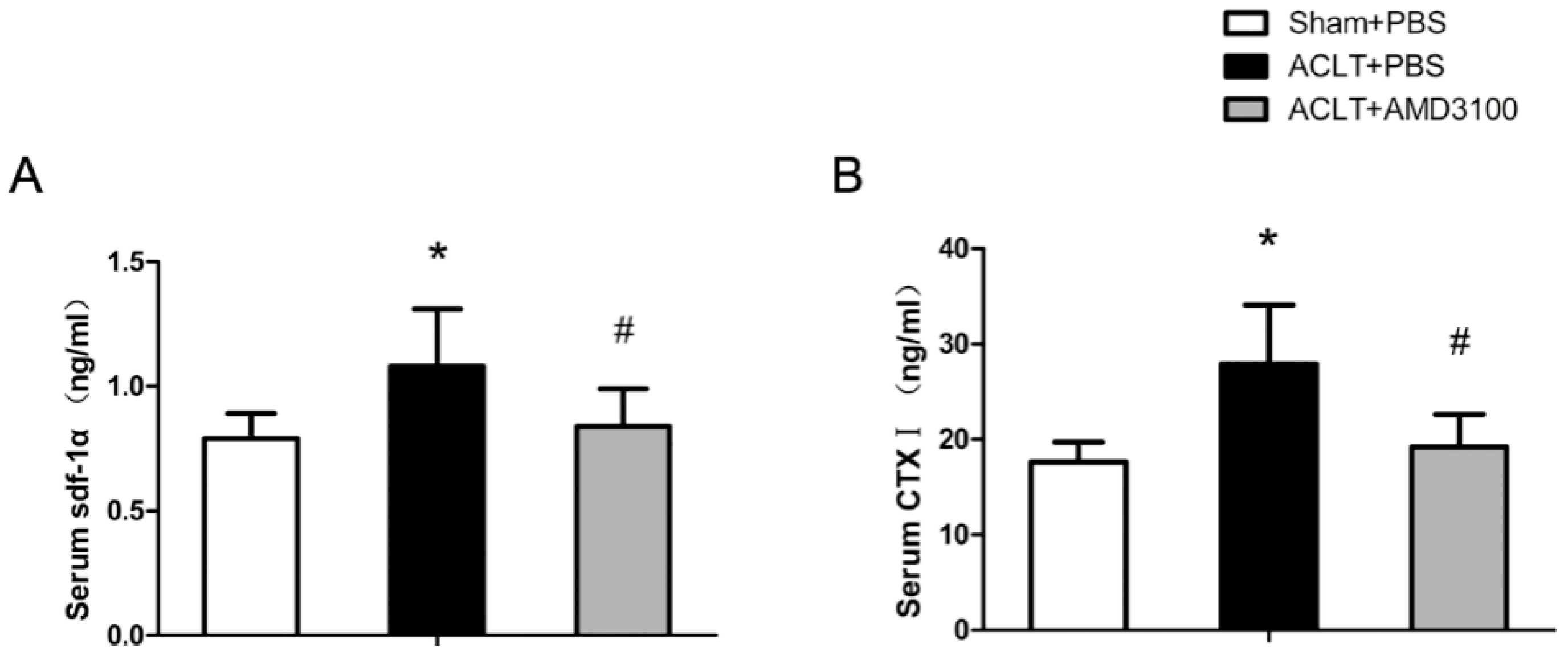
2.4. SDF-1α and CTX-I Concentrations in Serum

The levels of serum SDF-1α increased by 36.7% in ACLT mice at 30 days post-surgery compared with sham mice; this difference was statistically significant. AMD3100 treatment resulted in lower SDF-1α serum levels, by 22.2%, than the ACLT/PBS group. These results demonstrated that serum SDF-1α increased in the PTOA model, and that PTOA was relieved when treated with AMD3100 and serum SDF-1α dropped. Serum CTX-I levels were found to increase significantly, by 58.5%, in ACLT mice compared with sham mice. Moreover, in the ACLT/AMD3100 group, serum CTX-I was reduced versus the ACLT/PBS group. These results suggest that the destabilised OA animal model showed elevated SDF-1α concentrations in the serum and active bone resorption, and the trend was similar in subchondral bone and blood (Figure 4).

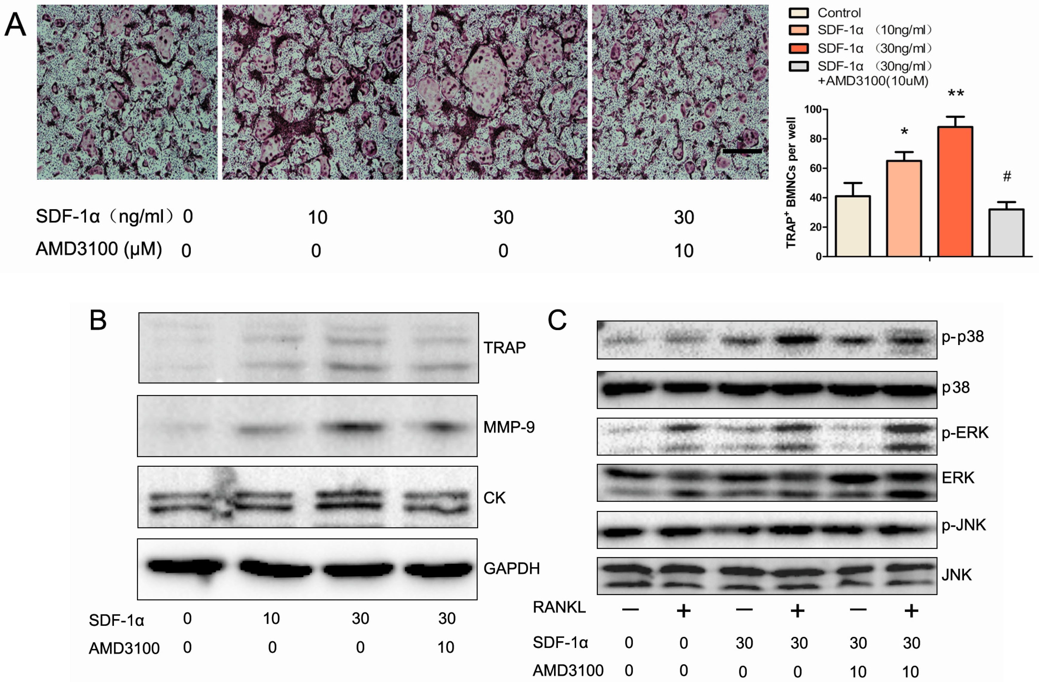
2.5. Effects of SDF-1α/CXCR4 Signalling on Osteoclast Differentiation in Vitro

We detected changes in osteoclast differentiation in tibial subchondral bone, so we next examined osteoclast differentiation in vitro. BMMCs were treated with different concentrations of SDF-1α for 6 days. SDF-1α promoted osteoclast formation in a dose-dependent manner, and the maximum effect occurred at 30 ng/mL. When BMMCs were treated with SDF-1α and, AMD3100 the promoting effect disappeared. These data demonstrated that SDF-1α promoted osteoclast formation in vivo and in vitro, and that AMD3100could neutralise the effect (Figure 5A).

2.6. Effects of SDF-1α/CXCR4 Signalling on Multiple Pathways Involved in Osteoclast Differentiation

Several osteoclast-specific proteins affect osteoclastogenesis, such as TRAP, MMP9, and cathepsin K. We further investigated the effects of SDF-1α on their expression. We found that treatment with SDF-1α strikingly elevated the expression of TRAP, MMP-9, and CK proteins, and that AMD3100 8HCl could neutralise the effect (Figure 5B). We also measured the activity of the MAPKs pathways in SDF-1α-treated BMMCs, which play an essential role in osteoclastogenesis. Western blotting data showed that phosphorylated p38 (p-p38) and phosphorylated ERK (p-ERK) demonstrated pronounced increases on RANKL stimulation, and SDF-1α treatment aggravated the response to RANKL, the expression of p-JNK were no significant difference. AMD3100 could diminish the effect on p-p38, but the change in p-ERK was insignificant (Figure 5C). These data showed that SDF-1α could promote the function of osteoclasts through ERK and p38 pathways.

3. Discussion

In this study, we used a destabilised OA animal model by transecting the ACL of the right leg in mice. Within 30 days of ACLT, we observed a significant loss of trabecular bone and articular cartilage degeneration in the operated knees. In the subchondral bone, CXCR4 was upregulated, which could heighten the response to SDF-1. We also observed increased serum levels of SDF-1α in ACLT mice. These results demonstrated that SDF-1α/CXCR4 was activated in PTOA. Moreover, the osteoclast number was increased in subchondral bone, and we detected elevated serum levels of CTX-I in ACLT mice, indicative of a rise in bone resorption. We also confirmed that inhibition of SDF-1α/CXCR4 in the subchondral bone can prevent loss of trabecular bone, and attenuate cartilage degeneration. We provide in vitro evidence that SDF-1α promoted the differentiation of osteoclasts. At the subcellular level, SDF-1α activated p38 and Erk1/2 MAP kinases, which mediate the SDF-1α-mediated induction of TRAP, MMP9, and cathepsin K protein expression. These data illustrate a developmental role for the SDF-1α/CXCR4 axis in OA.
In this study, we observed that cartilage degeneration was attenuated by AMD3100. Thomas et al. reported similar results [13]. However, a recent study showed that adding exogenous SDF-1α protein can enhance the chondrogenic differentiation induced by BMP2, while blocking the SDF-1α/CXCR4 pathway emerged the inhibitory effect, and another study indicated that SDF-1α functioned to promote chondrocyte proliferation and maturation [18,19,20,28,29]. These data suggest that the role of SDF-1α in cartilage homeostasis was still controversial and the effect of attenuated cartilage degeneration may have no direct impact on articular cartilage. In our study, we detected that SDF-1α/CXCR4 was activated in subchondral bone, and that AMD3100 could prevent loss of trabecular bone. It has been proven that cartilage deterioration can be initiated or at least accelerated by subchondral bone change, so we had reason to believe that relief of cartilage degeneration would be achieved by the prevention of subchondral bone changes [2,4,5,30]. Our findings first revealed that SDF-1α have a destructive effect on subchondral bone rather than articular cartilage.
Recently, evidence from human studies has shown that bone turnover in the subchondral region increases in OA [8]. We revealed similar results in the ACLT mice. The total subchondral bone TV increased more than 13.6%, and Tb. Pf increased 18.8%, indicating that bone resorption was strengthened. BV/TV and Tb. Th in ACLT mice decreased dramatically, and bone resorption markers, such as CTX-I, were found to increase significantly. These data showed uncoupled bone remodelling in the tibial subchondral bone. There was increased bone resorption, rather than changes in bone formation. Increased osteoclast formation and activity played an important role in this.
Here, we reported for the first time that inhibition of SDF-1α prevented osteoclastogenesis, both in vitro and in vivo. The decrease in CTX-I in the AMD3100 group confirmed the ability of an SDF-1α inhibitor in bone resorption, an important mechanism to attenuate OA. Previous studies also reported that antiresorptive pharmaceutical treatments, such as alendronate, may prevent trabecular bone loss and articular cartilage degeneration [31]. However, the effects of antiresorptive drugs remain controversial.
Elevated levels of SDF-1α in synovial fluid and serum have been observed in OA patients, and our data also demonstrate increased serum levels of SDF-1α in ACLT mice. A previous study found that SDF-1α may contribute to synovitis, and anti-inflammatories can reduce SDF-1α expression in bursal cells [32]. These studies suggest that SDF-1α may be associated with the progression of inflammation. Wei, F. et al. found that blockage of the SDF-1α/CXCR4 pathway reduced the level of IL-1 in a guinea pig OA model, and Hsien-Te Chen et al. reported that SDF-1α promoted IL-6 production in human synovial fibroblasts [31,33]. These results suggest that SDF-1α can induce several inflammatory factors. These findings may provide a better understanding of the mechanisms of SDF-1α in promoting bone resorption and osteoclastogenesis.
Previous studies have found positive regulation between SDF-1α and MAPK signalling. SDF-1α has been shown to promote MAPK activation in chondrocytes, to induce the proliferation and maturation of chondrocytes. MAPKs activation also plays an important role in osteoclastogenesis. Our results showed that SDF-1α activated both p-p38 and p-Erk1/2 pathways, which are potent inducers of activator protein-1 (AP-1) activity, increasing the expression of c-fos and c-jun. AP-1 is essential for the expression of TRAP, MMP-9, and CK. Moreover, AMD3100 can neutralise the effect of SDF-1α in MAPK signalling [23,34].
The ACLT mice model used in this study is somewhat limited because it produces very severe OA at 30 days post-operation. This is a universal limitation for all of the destabilisation OA animal models. However, representing a traumatic form of OA, the ACLT mice model is reasonable as a mimic of the pathogenesis of human OA, including cartilage degradation, subchondral bone sclerosis, and osteophyte formation, which are important for investigating OA pathogenesis and potential therapies. Moreover, we focus on the relationship between the subchondral bone and cartilage metabolism, so ACLT model is very applicable [10,35]. In our study, AMD3100 was given at 2 days post-operation, so the pharmacological agent may act before the formation of OA, AMD3100 was delivered at a rate of 180 μg/day that corresponds to steady serum level of 0.3 μg/mL, proven efficient in different studies [13]. Our study supports the use of AMD3100 for slowing or preventing the onset of PTOA following injury.The treatment of AMD3100 combined with granulocyte-colony stimulated factor had been approved by FDA for patients with multiple myeloma and non-Hodgkin’s lymphoma, which proved that AMD3100 is safe and valid. In our study, AMD3100 showed an effect in preventing the pathological progress of PTOA, and future clinical studies should explore this opportunity in orthopedics [36,37].

4. Materials and Methods

4.1. Reagents and Antibodies

Recombinant mouse SDF-1α, recombinant soluble mouse M-CSF and RANKL were obtained from PeproTech (Rocky Hill, NJ, USA). AMD3100 8HCl was purchased from Selleck Chemicals (Houston, TX, USA). Rabbit polyclonal antibodies specific for MMP13, MMP-9, cathepsin K, CXCR4, and tartrate-resistant acid phosphatase (TRAP) were purchased from Abcam (Cambridge, MA, USA). Rabbit antibodies against p-Erk1/2, Erk1/2, p-JNK, JNK, p-p38, and p38 MAP kinase were obtained from Cell Signaling Technology (Beverly, MA, USA). The secondary goat or rabbit IgGs were obtained from Santa Cruz Biotechnology (Santa Cruz, CA, USA). Anti-GAPDH and RIPA were obtained from Boster (Wuhan, China). Cell culture media and supplements were purchased from Invitrogen (Carlsbad, CA, USA).

4.2. Animals and Experimental Groups

C57BL/6J mice (30 males, 2 months old) were purchased from the Experimental Animal Centre of Tongji Medical College (Wuhan, China). We anaesthetised 8-week-old male mice with chloral hydrate, and then transected the anterior cruciate ligament on the right knee, which caused joint instability and induced PTOA. Sham operations were done on the right knees of control mice. Mice were randomised into three groups: Group 1 (n = 10) animals underwent sham operations and were treated with PBS via a constant infusion osmotic mini-pump (model 1004, 0.11 μL/h, Alzet, Palo Alto, CA, USA), Group 2 (n = 10) animals underwent ACLT surgery with vehicle treatment, and Group 3 (n = 10) animals underwent ACLT surgery and were treated withAMD3100 via a pump (AMD3100 was delivered at a dose of 180 μg/day). Mice were euthanised 30 days post-surgery. Approval was obtained from the Institutional Animal Care and Use Committee (IACUC) at Tongji Hospital (TJ-A20150802; September 2015).

4.3. Micro-Computed Tomography (μCT) Imaging

After removing soft tissues, the whole right knee joints were analysed using μCT (μ-CT50 Scanco Medical, Bassersdorf, Switzerland).Scans were obtained at 100 kV and 98 μA; the resolution was set to 10.5 μm. We used the built-in software to reconstruct and analyse images. We chose the tibial subchondral bone for 3D reconstruction and structural parameters analysis. 3D structural parameters analysed included the total tissue volume (TV), bone volume/tissue volume (BV/TV), trabecular thickness (Tb. Th.), and trabecular separation (Tb. Sp.).

4.4. Histochemistry and Immunohistochemistry

After μCT analysis, knee joints were fixed in 4% buffered formalin for 2 days at room temperature, decalcified for 3 weeks using 10% EDTA solution at 4 °C, and then processed for paraffin wax embedding. The tissues were cut into 4-μm sections in a sagittal orientation. H&E and Safranin O-Fast Green staining were performed. We performed TRAP staining following a standard protocol (Sigma-Aldrich, St. Louis, MO, USA). Immunohistochemistry was performed using a DAB Histostain-SP Kit; the sections were incubated with specific antibodies against MMP-13 and CXCR4 overnight at 4 °C. Histological measurements and images were observed under a digital microscope (Nikon ECLIPSE Ti-S, Nikon, Tokyo, Japan). OARSI osteoarthritis cartilage histopathology assessment system was chosen to assess the cartilage degeneration severity [38]. Numbers of positively stained cells were counted in the subchondral bone area per specimen.

4.5. Serum Biochemistry

Blood was collected from each mouse immediately prior to sacrifice by a retro-orbital puncture. Serum levels of SDF-1α were measured using ELISA kits following the manufacturer’s instructions. Serum levels of C-terminal telopeptide of type I collagen (CTX-I), a biomarker of bone resorption, were measured with ELISA kits (Bangyi Biotech Co., Shanghai, China).

4.6. Cell Cultures

Bone marrow mononuclear cells (BMMCs) were isolated from the whole femoral and tibial bone marrow of 6-week-old C57BL/6 mice. Briefly, bone marrow containing mononuclear cells were flushed out of the femoral and tibial by a 10-mL injector with α-MEM (supplemented with 10% fetal bovine serum, 100 IU/mL penicillin, 100 IU/mL streptomycin and 30 ng/mL M-CSF) and cultured at 37 °C with 5% CO2. After 24 h, floating cells were collected and then supplemented with M-CSF [39]. After 3 days, adherent cells were plated at a density of 2 × 10⁵ cells/well in 6-well plates or 96-well plates. To induce osteoclast formation, M-CSF (30 ng/mL) and RANKL (100 ng/mL) were added to the medium immediately. After 6 days, cells were used for TRAP staining and osteoclast function assays.

4.7. Western Blotting

Cellular lysates were prepared using the protein extraction reagent RIPA, 20 μg of total cellular protein per sample was loaded on 10% Bis-Tris gels following the manufacturer’s protocols (Boster), and then transferred to PVDF membranes (Millipore, Billerica, MA, USA). The membranes were blocked with 5% BSA in Tris-buffered saline (TBS) with 0.1% Tween-20 (TBST) for 1 h at room temperature and then probed with rabbit primary antibodies against MMP-9, cathepsin K, TRAP, p-Erk1/2, Erk1/2, p-JNK, JNK, p-p38, and p38 MAPK overnight at 4 °C, and the blots were incubated with IgG-horseradish peroxidase (HRP)-labelled secondary antibodies for 1 h at room temperature. Immunoreactivity was detected with enhanced chemiluminescence (ECL), and densitometry was performed using the Quantity One Software (Bio-Rad Laboratories Inc., Munich, Germany).

4.8. Statistical Analysis

Data are presented as means ± standard deviation for at least three independent experiments. Statistical analyses included a two-tailed paired t-test to assess differences between two groups. Between-group comparisons for all analyses were performed with analysis of variance (ANOVA). Significance was set at p < 0.05 for all tests.

5. Conclusions

In summary, this study suggests that SDF-1α promoted the differentiation of osteoclasts, and the inhibition of SDF-1α prevents osteoclastogenesis, both in vitro and in vivo. AMD3100 is able to prevent trabecular bone loss and attenuate cartilage degeneration in PTOA mice. These findings further highlight the importance of preventing subchondral bone trabecular bone loss in OA. These data suggest a potential therapeutic approach.

Acknowledgments

The authors thank our research assistant Jian Liu for contributing to μCT Imaging. We also thank Yuting Wang for drawing the graphical abstract and English edition. This research was supported by grants from the National Nature Science Foundation of China (NO. 81472082 and NO. 81171696).

Author Contributions

Anmin Chen and Qing Yang designed the experiments, revised the manuscript; Yonghui Dong conducted the main experiments and prepared the manuscript; Hui Liu and Xuejun Zhang analyzed the data. Fei Xu, Liang Qin and Peng Cheng contributed clarifications and guidance on the manuscript; Fengjing Guo and Hui Huang helped with manuscript revision. All authors read and approved the manuscript.

Conflicts of Interest

The authors declare no conflict of interest.

References

  1. Glyn-Jones, S.; Palmer, A.J.R.; Agricola, R.; Price, A.J.; Vincent, T.L.; Weinans, H.; Carr, A.J. Osteoarthritis. Lancet 2015, 386, 376–387. [Google Scholar] [CrossRef]
  2. Lories, R.J.; Luyten, F.P. The bone-cartilage unit in osteoarthritis. Nat. Rev. Rheumatol. 2011, 7, 43–49. [Google Scholar] [CrossRef] [PubMed]
  3. Felson, D.T.; Neogi, T. Osteoarthritis: Is it a disease of cartilage or of bone? Arthritis Rheum. 2004, 50, 341–344. [Google Scholar] [CrossRef] [PubMed]
  4. Mansell, J.P.; Collins, C.; Bailey, A.J. Bone, not cartilage, should be the major focus in osteoarthritis. Nat. Clin. Pract. Rheumatol. 2007, 3, 306–307. [Google Scholar] [CrossRef] [PubMed]
  5. Hayami, T.; Pickarski, M.; Zhuo, Y.; Wesolowski, G.A.; Rodan, G.A.; Duong, L.T. Characterization of articular cartilage and subchondral bone changes in the rat anterior cruciate ligament transection and meniscectomized models of osteoarthritis. Bone 2006, 38, 234–243. [Google Scholar] [CrossRef] [PubMed]
  6. Khorasani, M.S.; Diko, S.; Hsia, A.W.; Anderson, M.J.; Genetos, D.C.; Haudenschild, D.R.; Christiansen, B.A. Effect of alendronate on post-traumatic osteoarthritis induced by anterior cruciate ligament rupture in mice. Arthritis Res. Ther. 2015, 17, 30. [Google Scholar] [CrossRef] [PubMed]
  7. Karsdal, M.A.; Bay-Jensen, A.C.; Lories, R.J.; Abramson, S.; Spector, T.; Pastoureau, P.; Christiansen, C.; Attur, M.; Henriksen, K.; Goldring, S.R.; et al. The coupling of bone and cartilage turnover in osteoarthritis: Opportunities for bone antiresorptives and anabolics as potential treatments? Ann. Rheum. Dis. 2014, 73, 336–348. [Google Scholar] [CrossRef] [PubMed]
  8. Kadri, A.; Funck-Brentano, T.; Lin, H.; Ea, H.K.; Hannouche, D.; Marty, C.; Liote, F.; Geoffroy, V.; Cohen-Solal, M.E. Inhibition of bone resorption blunts osteoarthritis in mice with high bone remodelling. Ann. Rheum. Dis. 2010, 69, 1533–1538. [Google Scholar] [CrossRef] [PubMed]
  9. Hayami, T.; Pickarski, M.; Wesolowski, G.A.; McLane, J.; Bone, A.; Destefano, J.; Rodan, G.A.; Duong, L.T. The role of subchondral bone remodeling in osteoarthritis: Reduction of cartilage degeneration and prevention of osteophyte formation by alendronate in the rat anterior cruciate ligament transection model. Arthritis Rheum. 2004, 50, 1193–1206. [Google Scholar] [CrossRef] [PubMed]
  10. Zhen, G.; Wen, C.; Jia, X.; Li, Y.; Crane, J.L.; Mears, S.C.; Askin, F.B.; Frassica, F.J.; Chang, W.; Yao, J.; et al. Inhibition of TGF-β signaling in mesenchymal stem cells of subchondral bone attenuates osteoarthritis. Nat. Med. 2013, 19, 704–712. [Google Scholar] [CrossRef] [PubMed]
  11. Yang, S.; Kim, J.; Ryu, J.H.; Oh, H.; Chun, C.H.; Kim, B.J.; Min, B.H.; Chun, J.S. Hypoxia-inducible factor-2α is a catabolic regulator of osteoarthritic cartilage destruction. Nat. Med. 2010, 16, 687–693. [Google Scholar] [CrossRef] [PubMed]
  12. Saito, T.; Fukai, A.; Mabuchi, A.; Ikeda, T.; Yano, F.; Ohba, S.; Nishida, N.; Akune, T.; Yoshimura, N.; Nakagawa, T.; et al. Transcriptional regulation of endochondral ossification by HIF-2α during skeletal growth and osteoarthritis development. Nat. Med. 2010, 16, 678–686. [Google Scholar] [CrossRef] [PubMed]
  13. Thomas, N.P.; Li, P.; Fleming, B.C.; Chen, Q.; Wei, X.; Hua, P.X.; Li, G.; Wei, L. Attenuation of cartilage pathogenesis in post-traumatic osteoarthritis (PTOA) in mice by blocking the stromal derived factor 1 receptor (CXCR4) with the specific inhibitor, AMD3100. J. Orthop. Res. 2015, 33, 1071–1078. [Google Scholar] [CrossRef] [PubMed]
  14. Wei, L.; Kanbe, K.; Lee, M.; Wei, X.; Pei, M.; Sun, X.; Terek, R.; Chen, Q. Stimulation of chondrocyte hypertrophy by chemokine stromal cell-derived factor 1 in the chondro-osseous junction during endochondral bone formation. Dev. Biol. 2010, 341, 236–245. [Google Scholar] [CrossRef] [PubMed]
  15. Chiu, Y.C.; Yang, R.S.; Hsieh, K.H.; Fong, Y.C.; Way, T.D.; Lee, T.S.; Wu, H.C.; Fu, W.M.; Tang, C.H. Stromal cell-derived factor-1 induces matrix metalloprotease-13 expression in human chondrocytes. Mol. Pharmacol. 2007, 72, 695–703. [Google Scholar] [CrossRef] [PubMed]
  16. Mazzetti, I.; Magagnoli, G.; Paoletti, S.; Uguccioni, M.; Olivotto, E.; Vitellozzi, R.; Cattini, L.; Facchini, A.; Borzi, R.M. A role for chemokines in the induction of chondrocyte phenotype modulation. Arthritis Rheum. 2004, 50, 112–122. [Google Scholar] [CrossRef] [PubMed]
  17. Kanbe, K.; Takemura, T.; Takeuchi, K.; Chen, Q.; Takagishi, K.; Inoue, K. Synovectomy reduces stromal-cell-derived factor-1 (SDF-1) which is involved in the destruction of cartilage in osteoarthritis and rheumatoid arthritis. J. Bone Jt. Surg. Br. 2004, 86, 296–300. [Google Scholar] [CrossRef]
  18. Kim, G.W.; Han, M.S.; Park, H.R.; Lee, E.J.; Jung, Y.K.; Usmani, S.E.; Ulici, V.; Han, S.W.; Beier, F. CXC chemokine ligand 12a enhances chondrocyte proliferation and maturation during endochondral bone formation. Osteoarthr. Cartil. 2015, 23, 966–974. [Google Scholar] [CrossRef] [PubMed]
  19. Guang, L.G.; Boskey, A.L.; Zhu, W. Regulatory role of stromal cell-derived factor-1 in bone morphogenetic protein-2-induced chondrogenic differentiation in vitro. Int. J. Biochem. Cell Biol. 2012, 44, 1825–1833. [Google Scholar] [CrossRef] [PubMed]
  20. Mendelson, A.; Frank, E.; Allred, C.; Jones, E.; Chen, M.; Zhao, W.; Mao, J.J. Chondrogenesis by chemotactic homing of synovium, bone marrow, and adipose stem cells in vitro. FASEB J. 2011, 25, 3496–3504. [Google Scholar] [CrossRef] [PubMed]
  21. Periyasamy-Thandavan, S.; Herberg, S.; Arounleut, P.; Upadhyay, S.; Dukes, A.; Davis, C.; Johnson, M.; McGee-Lawrence, M.; Hamrick, M.W.; Isales, C.M.; et al. Caloric restriction and the adipokine leptin alter the SDF-1 signaling axis in bone marrow and in bone marrow derived mesenchymal stem cells. Mol. Cell. Endocrinol. 2015, 410, 64–72. [Google Scholar] [CrossRef] [PubMed]
  22. Grassi, F.; Cristino, S.; Toneguzzi, S.; Piacentini, A.; Facchini, A.; Lisignoli, G. CXCL12 chemokine up-regulates bone resorption and MMP-9 release by human osteoclasts: CXCL12 levels are increased in synovial and bone tissue of rheumatoid arthritis patients. J. Cell. Physiol. 2004, 199, 244–251. [Google Scholar] [CrossRef] [PubMed]
  23. Tang, C.H.; Chuang, J.Y.; Fong, Y.C.; Maa, M.C.; Way, T.D.; Hung, C.H. Bone-derived SDF-1 stimulates IL-6 release via CXCR4, ERK and NF-κB pathways and promotes osteoclastogenesis in human oral cancer cells. Carcinogenesis 2008, 29, 1483–1492. [Google Scholar] [CrossRef] [PubMed]
  24. Wright, L.M.; Maloney, W.; Yu, X.; Kindle, L.; Collin-Osdoby, P.; Osdoby, P. Stromal cell-derived factor-1 binding to its chemokine receptor CXCR4 on precursor cells promotes the chemotactic recruitment, development and survival of human osteoclasts. Bone 2005, 36, 840–853. [Google Scholar] [CrossRef] [PubMed]
  25. Im, J.Y.; Min, W.K.; Park, M.H.; Kim, N.; Lee, J.K.; Jin, H.K.; Choi, J.Y.; Kim, S.Y.; Bae, J.S. AMD3100 improves ovariectomy-induced osteoporosis in mice by facilitating mobilization of hematopoietic stem/progenitor cells. BMB Rep. 2014, 47, 439–444. [Google Scholar] [CrossRef] [PubMed]
  26. Diamond, P.; Labrinidis, A.; Martin, S.K.; Farrugia, A.N.; Gronthos, S.; To, L.B.; Fujii, N.; O’Loughlin, P.D.; Evdokiou, A.; Zannettino, A.C. Targeted disruption of the CXCL12/CXCR4 axis inhibits osteolysis in a murine model of myeloma-associated bone loss. J. Bone Miner. Res. 2009, 24, 1150–1161. [Google Scholar] [CrossRef] [PubMed]
  27. Glasson, S.S.; Blanchet, T.J.; Morris, E.A. The surgical destabilization of the medial meniscus (DMM) model of osteoarthritis in the 129/SvEv mouse. Osteoarthr. Cartil. 2007, 15, 1061–1069. [Google Scholar] [CrossRef] [PubMed]
  28. Zhang, F.; Leong, W.; Su, K.; Fang, Y.; Wang, D.A. A transduced living hyaline cartilage graft releasing transgenic stromal cell-derived factor-1 inducing endogenous stem cell homing in vivo. Tissue Eng. A 2013, 19, 1091–1099. [Google Scholar] [CrossRef] [PubMed]
  29. Sukegawa, A.; Iwasaki, N.; Kasahara, Y.; Onodera, T.; Igarashi, T.; Minami, A. Repair of rabbit osteochondral defects by an acellular technique with an ultrapurified alginate gel containing stromal cell-derived factor-1. Tissue Eng. A 2012, 18, 934–945. [Google Scholar] [CrossRef] [PubMed]
  30. Funck-Brentano, T.; Lin, H.; Hay, E.; Ah Kioon, M.D.; Schiltz, C.; Hannouche, D.; Nizard, R.; Liote, F.; Orcel, P.; de Vernejoul, M.C.; et al. Targeting bone alleviates osteoarthritis in osteopenic mice and modulates cartilage catabolism. PLoS ONE 2012, 7, e33543. [Google Scholar] [CrossRef] [PubMed]
  31. Wei, F.; Moore, D.C.; Wei, L.; Li, Y.; Zhang, G.; Wei, X.; Lee, J.K.; Chen, Q. Attenuation of osteoarthritis via blockade of the SDF-1/CXCR4 signaling pathway. Arthritis Res. Ther. 2012, 14, R177. [Google Scholar] [CrossRef] [PubMed]
  32. Kim, Y.S.; Bigliani, L.U.; Fujisawa, M.; Murakami, K.; Chang, S.S.; Lee, H.J.; Lee, F.Y.; Blaine, T.A. Stromal cell-derived factor 1 (SDF-1, CXCL12) is increased in subacromial bursitis and downregulated by steroid and nonsteroidal anti-inflammatory agents. J. Orthop. Res. 2006, 24, 1756–1764. [Google Scholar] [CrossRef] [PubMed]
  33. Chen, H.T.; Tsou, H.K.; Hsu, C.J.; Tsai, C.H.; Kao, C.H.; Fong, Y.C.; Tang, C.H. Stromal cell-derived factor-1/CXCR4 promotes IL-6 production in human synovial fibroblasts. J. Cell. Biochem. 2011, 112, 1219–1227. [Google Scholar] [CrossRef] [PubMed]
  34. Guan, H.; Zhao, L.; Cao, H.; Chen, A.; Xiao, J. Epoxyeicosanoids suppress osteoclastogenesis and prevent ovariectomy-induced bone loss. FASEB J. 2015, 29, 1092–1101. [Google Scholar] [CrossRef] [PubMed]
  35. Moodie, J.P.; Stok, K.S.; Muller, R.; Vincent, T.L.; Shefelbine, S.J. Multimodal imaging demonstrates concomitant changes in bone and cartilage after destabilisation of the medial meniscus and increased joint laxity. Osteoarthr. Cartil. 2011, 19, 163–170. [Google Scholar] [CrossRef] [PubMed]
  36. De Clercq, E. The AMD3100 story: The path to the discovery of a stem cell mobilizer (Mozobil). Biochem. Pharmacol. 2009, 77, 1655–1664. [Google Scholar] [CrossRef] [PubMed]
  37. Pusic, I.; DiPersio, J.F. Update on clinical experience with AMD3100, an SDF-1/CXCL12-CXCR4 inhibitor, in mobilization of hematopoietic stem and progenitor cells. Curr. Opin. Hematol. 2010, 17, 319–326. [Google Scholar] [CrossRef] [PubMed]
  38. Glasson, S.S.; Chambers, M.G.; Van Den Berg, W.B.; Little, C.B. The OARSI histopathology initiative—Recommendations for histological assessments of osteoarthritis in the mouse. Osteoarthr. Cartil. 2010, 18, S17–S23. [Google Scholar] [CrossRef] [PubMed]
  39. Zhang, Y.; Guan, H.; Li, J.; Fang, Z.; Chen, W.; Li, F. Amlexanox suppresses osteoclastogenesis and prevents ovariectomy-induced bone loss. Sci. Rep. 2015, 5, 13575. [Google Scholar] [CrossRef] [PubMed]
Figure 1. AMD3100 inhibits anterior cruciate ligament (ACLT)-induced bone loss in tibial subchondral bone. (A) Mice were sacrificed at 30 days after the operation. Three-dimensional (3D) micro-computed tomography (μCT) images of tibial subchondral bone from representative specimens of sham/PBS (PBS-treated, sham-operated), ACLT/PBS, and ACLT/AMD3100; (B) Histograms represent the parameters of the 3D trabecular structure of the tibial subchondral bone: tissue volume (TV), trabecular bone volume/tissue volume (BV/TV), and trabecular thickness (Tb. Th), trabecular separation (Tb. Sp). Data are presented as means ± SD. n = 10. * ACLT/PBS different from sham/PBS (p < 0.05), # ACLT/PBS different from ACLT/AMD3100 (p < 0.05).
Ijms 17 00943 g001
Figure 2. CXCR4 expression and bone resorption were increased in post-traumatic osteoarthritis (PTOA) subchondral bone (A) Paraffin wax sections were used to detect CXCR4 expression with immunohistochemistry. CXCR4 was expressed in tibial subchondral bone, the brown positive osteoblasts were indicated with black arrows. The immunohistochemistry was performed without the antibody for CXCR4 in negative control. Calibration scale bar = 100 μm; (B) Representative tartrate-resistant acid phosphatase (TRAP)-stained histological sections of tibial subchondral bone from sham, ACLT/PBS mice, and ACLT/AMD3100 mice. The red TRAP-positive cells were indicated with black arrows; scale bar = 100 μm; (C) Quantitative analysis of TRAP+ or CXCR4+ cells per bone marrow area (mm2), reported as means ± SD. n = 10. * ACLT/PBS different from sham/PBS (p < 0.05), # ACLT/PBS different from ACLT/AMD3100 (p < 0.05).
Ijms 17 00943 g002
Figure 3. Inhibition of SDF-1α signalling in subchondral bone attenuated cartilage degeneration (A) H&E staining of tibia subchondral bone and cartilage from sham, ACLT/PBS, and ACLT/AMD3100 groups. Calibration scale: bar = 100 μm; (B) Safranin O-Fast Green staining of articular cartilage in sagittal sections of tibia from mice treated with PBS or AMD3100 and sacrificed 30 days post ACLT or sham surgery. Calibration scale: bar = 100 μm; (C) MMP13 expression was detected by immunohistochemical staining of cartilage, and representative images are shown. A positive signal was indicated by the brown colour and marked by black arrows, meanwhile a negative control was present. Calibration scale: bar = 50 μm; (D) OARSI scores of sham or ACLT mice treated with PBS or AMD3100.Quantitative analysis of the percentage of MMP13+ chondrocytes in articular cartilage tissue sections in each group, reported as means ± SD. n = 10. * ACLT/PBS different from sham/PBS (p < 0.05), # ACLT/PBS different from ACLT/AMD3100 (p < 0.05).
Ijms 17 00943 g003
Figure 4. SDF-1α and CTX-I serum levels. (A,B) Blood was collected immediately prior to sacrifice from sham-treated mice, ACLT/PBS mice, and ACLT/AMD3100 mice. Serum levels of SDF-1α and CTX-I were measured using ELISA kits and are reported as means ± SD. n = 10. * ACLT/PBS different from sham/PBS (p < 0.05), # ACLT/PBS different from ACLT/AMD3100 (p < 0.05).
Ijms 17 00943 g004
Figure 5. SDF-1α promoted osteoclast differentiation and activity in vitro. (A) SDF-1α promoted osteoclast formation in a dose-dependent manner. Bone marrow mononuclear cells (BMMCs) were treated with 10 ng/mL or 30 ng/mL of SDF-1α; culture medium was changed once per day, incubated with receptor activator of nuclear factor-κB ligand (RANKL) (100 ng/mL) and macrophage-colony stimulating factor (M-CSF) (30 ng/mL). At 6 days after treatment, cells were fixed for tartrate-resistant acid phosphatase (TRAP) staining. TRAP-positive multinucleated osteoclasts (≥3 nuclei) were counted. Data are presented as means ± SD of three independent experiments. * SDF-1α (10 ng/mL) group different from control group (p < 0.05), ** SDF-1α (30 ng/mL) group different from SDF-1α (10 ng/mL) group (p < 0.05), # SDF-1α (30 ng/mL) + AMD3100 (10 µM) group different from SDF-1α (30 ng/mL) group. Calibration scale: bar = 500 μm; (B) SDF-1α augmented the expression of osteoclast-specific proteins. BMMCs were treated with RANKL and M-CSF with or without SDF-1α (30 ng/mL). After 6 days, cells were collected for protein preparation. Expression levels of TRAP, CK, and MMP-9 were determined by Western blotting. The experiment was repeated three times independently; (C) SDF-1α promoted RANKL-induced phosphorylation of ERK and p38 expression. BMMCs were treated with α-MEM containing 0.5% FBS for 12 h before treatment. BMMCs were then pre-treated with or without SDF-1α (30 ng/mL) for 1 h and then stimulated with RANKL (100 ng/mL) for 30 min. Expression levels of phosphorylated ERK (p-ERK) and phosphorylated p-38 (p-p38) were detected by Western blotting. The experiment was repeated three times independently.
Ijms 17 00943 g005

