Article

Sugarcane Serine Peptidase Inhibitors, Serine Peptidases, and Clp Protease System Subunits Associated with Sugarcane Borer (Diatraea saccharalis) Herbivory and Wounding

by
Ane H. Medeiros
1,2,†,
Fabiana B. Mingossi
2,†,
Renata O. Dias
2,†,
Flávia P. Franco
2,
Renato Vicentini
3,
Marcia O. Mello
2,4,
Daniel S. Moura
5 and
Marcio C. Silva-Filho
2,*
1
Departamento de Ciências da Natureza, Matemática e Educação, Universidade Federal de São Carlos, Araras, 13600-970 São Paulo, Brazil
2
Departamento de Genética, Escola Superior de Agricultura Luiz de Queiroz, Universidade de São Paulo, Piracicaba, 13418-260 São Paulo, Brazil
3
Systems Biology Laboratory, Center for Molecular Biology and Genetic Engineering, State University of Campinas, Campinas, 13083-970 São Paulo, Brazil
4
Monsanto do Brasil, Campinas, 13069-380 São Paulo, Brazil
5
Departamento de Ciências Biológicas, Escola Superior de Agricultura Luiz de Queiroz, Universidade de São Paulo, Piracicaba, 13400-918 São Paulo, Brazil
*
Author to whom correspondence should be addressed.
Int. J. Mol. Sci. 2016, 17(9), 1444; https://0-doi-org.brum.beds.ac.uk/10.3390/ijms17091444
Submission received: 12 July 2016 / Revised: 16 August 2016 / Accepted: 25 August 2016 / Published: 1 September 2016
(This article belongs to the Special Issue Plant-Insect Interactions)

Abstract

:
Sugarcane’s (Saccharum spp.) response to Diatraea saccharalis (F.) (Lepidoptera: (Crambidae) herbivory was investigated using a macroarray spotted with 248 sugarcane Expressed Sequence Tags (ESTs) encoding serine peptidase inhibitors, serine peptidases. and Clp protease system subunits. Our results showed that after nine hours of herbivory, 13 sugarcane genes were upregulated and nine were downregulated. Among the upregulated genes, nine were similar to serine peptidase inhibitors and four were similar to Bowman-Birk Inhibitors (BBIs). Phylogenetic analysis revealed that these sequences belong to a phylogenetic group of sugarcane BBIs that are potentially involved in plant defense against insect predation. The remaining four upregulated genes included serine peptidases and one homolog to the Arabidopsis AAA+ chaperone subunit ClpD, which is a member of the Clp protease system. Among the downregulated genes, five were homologous to serine peptidases and four were homologous to Arabidopsis Clp subunits (three homologous to Clp AAA+ chaperones and one to a ClpP-related ClpR subunit). Although the roles of serine peptidase inhibitors in plant defenses against herbivory have been extensively investigated, the roles of plant serine peptidases and the Clp protease system represent a new and underexplored field of study. The up- and downregulated D. saccharalis genes presented in this study may be candidate genes for the further investigation of the sugarcane response to herbivory.

Graphical Abstract

1. Introduction

The sugarcane borer Diatraea saccharalis (F.) (Lepidoptera: Crambidae) is the most significant pest of sugarcane, with a wide distribution in Brazilian fields [1]. Sugarcane borer larvae create a vertical tunnel in the stem that becomes the primary route for microorganism entry [2]. The fungi that cause stem rot, Colletotrichum falcatum (Went) and Fusarium verticillioides, are commonly found in the tunnels produced by D. saccharalis.
Plants recognize insect feeding via chemical interactions between self-molecules that may be modified by the insect [3] or via insect molecules [4,5]. This chemical interaction triggers a signaling cascade that causes the expression of defense-associated genes [6,7,8,9].
Plant peptidase inhibitors have been shown to inhibit digestive peptidases in herbivorous insects [10,11,12]. The induction and accumulation of peptidase inhibitors (PIs) after mechanical or insect wounding have been reported in several plant families and are considered part of the plant´s natural defense system against herbivory [10,13].
Bowman–Birk peptidase inhibitors were first isolated and characterized in soybean seeds [14,15] and were subsequently found in other leguminous plants [16,17] and in the Poaceae [18]. These proteins are associated with endogenous seed peptidases regulation, sulfur amino acids storage, and plant defense against pathogens and insects [19]. Their anti-nutritive function is derived from their formation of stable complexes with the catalytic sites of peptidases, blocking degradation and ingestion of amino acids from the insect diet [20,21].
Other than their involvement in responses to other types of stress, and in contrast to peptidase inhibitors, little is known about plant cytoplasmic and intra-plastid serine peptidases and their roles in defense against herbivory [22,23]. These peptidases have a housekeeping role in plants, releasing amino acids for recycling and eliminating non-functional proteins. However, peptidases have also been shown to play important roles in plant defense, acting in pathogen and pest recognition and in induction of effective defense responses [24].
Studies involving other plant species have elucidated the roles of various peptidases in plant defense; for example, in tomatoes, a serine carboxypeptidase was induced by wounding, systemin, and methyl jasmonate treatment [25]. Subtilisins have also been implicated in plant defense against herbivores [26,27]. There is evidence for a subtilase that is involved in systemin processing [28]. In tomatoes, systemin is synthesized in the form of a precursor, and following mechanical or herbivore injury, pro-systemin is processed and translocated throughout the plant, triggering a signaling cascade that ultimately activates defense genes such as proteinase inhibitors [29,30].
Non-serine peptidases have also been shown to be involved in resistance: Mir1-CP, a cysteine peptidase identified in maize S. frugiperda-resistant lines, is rapidly induced when plants areinjured [31,32]. The authors investigated why S. frugiperda larvae fed with maize leaves experienced diminished growth and found that this peptidase damages the peritrophic matrix of not only S. frugiperda but of various insects of the Lepidoptera order [33]. Leucine aminopeptidase A (LapA) is a late wound-response gene of tomato (Solanum lycopersicum) that accumulates after mechanical, insect and pathogen wounding [34,35,36]. Working in concert with LapA, arginase and threonine deaminase play roles in plant defense against herbivores as well [35,37,38].
Techniques such as macroarrays, subtractive libraries, AFLP-cDNA (Amplified Fragment Length Polymorphism-cDNA) display, and differential display RT-PCR (Reverse Transcriptase Polymerase Chain Reaction) have enabled researchers to study changes in the transcriptome that are elicited in response to herbivory and wounding [39]. In contrast to research in other more widespread crops, progress in the field of sugarcane genomics has been slow. Genetic analyses are difficult in sugarcane due to its large and complex polyploid genome and the lack of sufficiently informative gene-tagged markers [40]. However, significant progress has been noted after the development of tools such as collections of expressed sequence tags (ESTs). Large EST collections have been made available [41,42,43,44,45] and have consequently renewed interest in sugarcane genetics [46]. Currently, the NCBI (National Center for Biotechnology Information) database lists more than 20,000 ESTs from the Saccharum officinarum complex (cultivated sugarcanes), and researchers have used these sequences to identify putative genes for the improvement of sugarcane field performance [47,48,49]. Using microarrays containing sugarcane ESTs, Rocha et al. [50] identified several sugarcane methyl jasmonate- and herbivore-responsive genes.
In silico analyses have shown that sugarcane possesses a set of conserved peptidase inhibitors that may also be involved in defense [19,48]. Initiatives to increase sugarcane borer resistance using traditional breeding and genetic engineering are needed. A better understanding of the sugarcane responses triggered by D. saccharalis feeding and wounding via the identification and characterization of genes directly involved in such responses may represent a means to improve sugarcane resistance. Those genes could also be used as molecular markers for insect resistance in traditional breeding programs.
In this study, we custom-made a macroarray containing 248 genes, including peptidase inhibitors, serine peptidases, and Clp protease system subunits, from the sugarcane EST collection. We have identified 10 peptidase inhibitors, seven peptidases, and five Clp subunits that are differentially expressed in sugarcane above-ground tissues in response to D. saccharalis feeding at an early time point.

2. Results

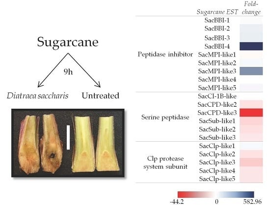
2.1. Macroarray Hybridization

To obtain information regarding the specific roles that peptidase inhibitors and peptidases may play in the sugarcane defense response against herbivores and wounding, a custom-made cDNA macroarray was constructed by spotting 248 selected ESTs on filter membranes (Table S1). The filter membranes were probed with 33P cDNA populations derived from RNA extracted from the leaves of undisturbed sugarcane plants (0 and 9 h time points) and from the leaves of plants attacked by D. saccharalis (9 h time point). Probed membranes were effective for the identification of differentially expressed ESTs as exemplified by SacMPI-like1 (EST spots highlighted with a square in Supplementary Material Figure S1A–C) and SacBBI1 (EST spots highlighted with a circle in Supplementary Material Figure S1A–C). Both SacMPI-like1 and SacBBI1 exhibited stronger signals when probed with 33P cDNA populations derived from the leaves of sugarcane plants attacked by D. saccharalis than when they were probed with populations derived from the leaves of undisturbed plants.
Of the 248 genes represented in the macroarrays, 22 presented consistent and reproducible expression in at least two hybridizations out of six possible comparisons (refer to Materials and Methods for more information regarding membrane randomization). Thirteen upregulated and nine downregulated genes were validated by quantitative real-time PCR; they are listed in Table 1 and Table 2.

2.2. Validation of D. saccharalis-Inducible Genes

To validate the expression of those genes upregulated after D. saccharalis feeding, we performed another set of biological experiments and included one more treatment: mechanical wounding. It was been shown that wounding, either by insects or mechanically, induces a general wounding response in plants [22,52,53,54]. In addition to that, insect wounding provokes a tailored response, specifically induced by some plant components released by insect feeding [3] or present in the insect saliva [53,55]. The objective was to compare the level of transcripts of 10 selected genes, after mechanical wounding and wounding by D. saccharalis.

2.2.1. Sugarcane Bowman-Birk Inhibitor (SacBBI) Genes

Four D. saccharalis-inducible sugarcane genes are homologous to rice and maize Bowman-Birk peptidase inhibitors and are designated here as SacBBI1-4 (Table 1). Real-time quantitative PCR analysis of the SacBBI genes showed that all four SacBBIs are induced by D. saccharalis and mechanical wounding (Figure 1). SacBBI1 is more responsive to mechanical wounding (17 times more highly expressed than the control) than caterpillar feeding (12 times more highly expressed than the control). SacBBI4 was induced by both caterpillar feeding and mechanical wounding; however, in contrast to SacBBI1, SacBBI4 mRNA levels in plants subjected to D. saccharalis feeding were approximately 600 times higher than the levels observed in control undisturbed plants and more than 50 times higher when plants were mechanically wounded (Figure 1). The other two genes, SacBBI2 and SacBBI3, were also induced by wounding and insect feeding, but to a lesser extent. Both the SacBBI2 and SacBBI3 genes were approximately 50 times more highly expressed in plants attacked by the insect and approximately 12 times more highly expressed in plants subjected to wounding.
We further investigated the behavior of all sugarcane BBIs present on the array, independent of their macroarray expression pattern. Gene expression quantification of 14 sugarcane BBI homologs confirmed that the four BBIs identified by our macroarray (SacBBI1 to 4) were the only BBIs induced by mechanical wounding or D. saccharalis feeding. The expression of the remaining 10 genes was unaltered, regardless of the treatment (data not shown).

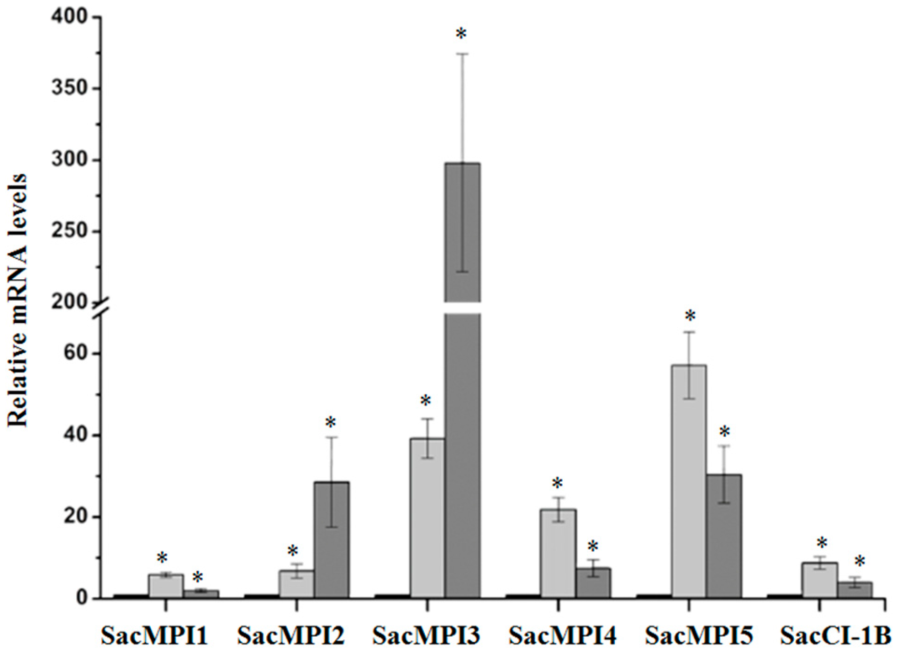
2.2.2. Sugarcane Maize-peptidase-inhibitor-like (SacMPI-like) and Chymotrypsin Inhibitor 1B-like (SacCI1B-like) Genes

Changes in the mRNA levels of five sugarcane maize-peptidase-inhibitor-like genes (SacMPI-like1-5) and one chymotrypsin inhibitor 1B-like gene (SacCI1B-like) in response to D. saccharalis attack and mechanical wounding were evaluated. All SacMPI-like and SacCI1B-like genes were responsive to insect attack and wounding (Figure 2). Insect attack induced higher levels of SacMPI-like2 gene expression (28 times more highly expressed than in the control plants) than did wounding (seven times more highly expressed than in control plants). SacMPI-like 3 exhibited the highest gene induction by D. saccharalis feeding, with mRNA levels averaging 300 times greater than control levels when plants were damaged by D. saccharalis and approximately 60 times greater when plants were mechanically damaged. SacMPI-like1, 4, 5, and SacC1B-like were more responsive to mechanical wounding than insect attack.
All SacMPI-like genes are homologs of the I13 peptidase inhibitor family. To test the possible relationship between SacMPI-like gene evolution and its expression profile, we further investigated the distribution of all sugarcane SacMPI-like sequences within the I13 family via phylogenetic analysis (Supplementary Materials, Figure S2). I13 peptidase inhibitor homologs from sugarcane did not group based on their expression profiles (Supplementary Materials, Figure S2).

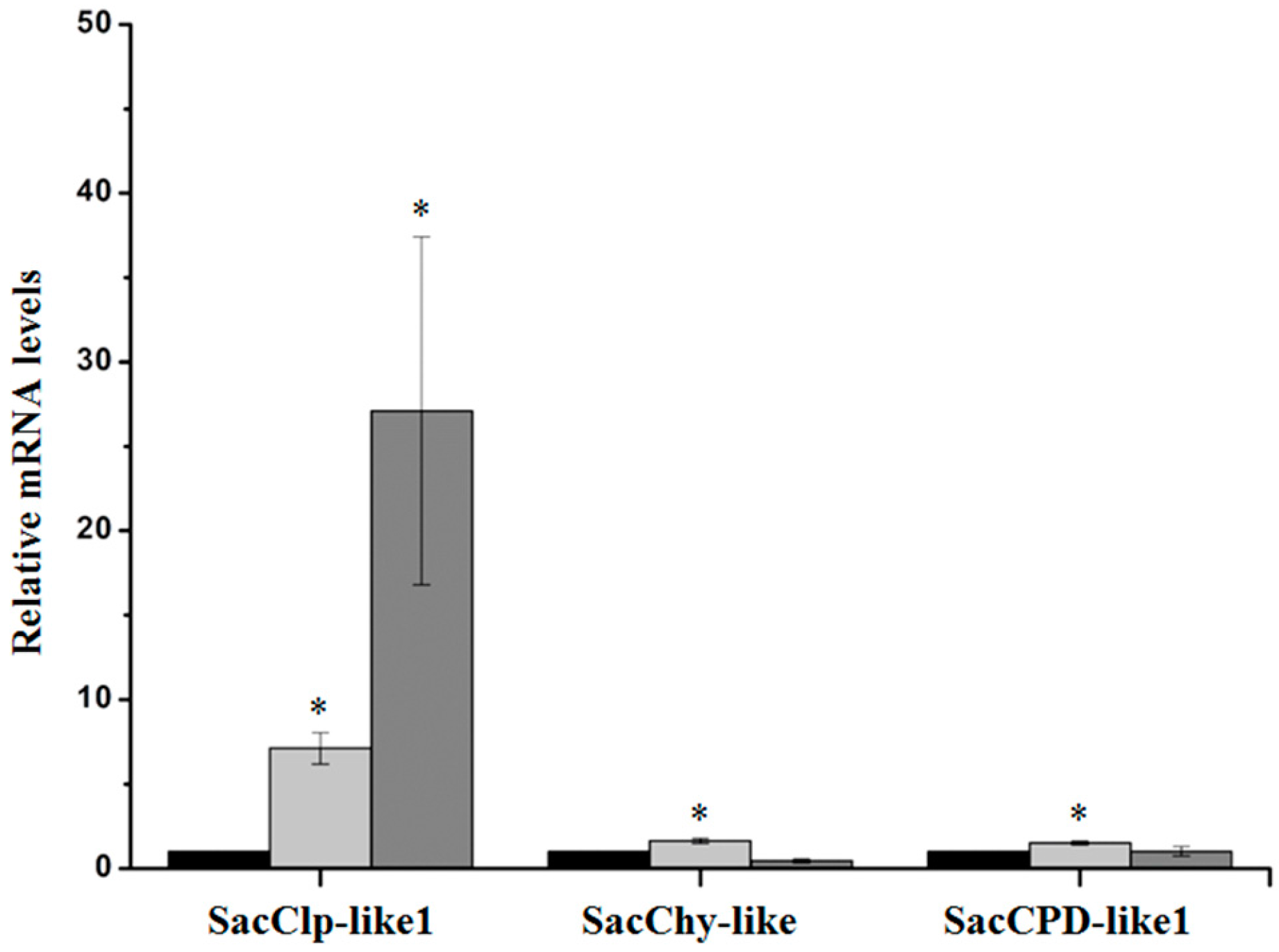
2.2.3. Serine Peptidases and Clp Protease System Subunits

Only two sugarcane serine peptidases and one Clp subunit were identified as induced by D. saccharalis in our macroarray, and their induction by herbivory was validated by performing quantitative real-time PCR analysis (Figure 3).
Among the 16 sugarcane Clp-like subunits analyzed in the macroarray, only the SacClp-like1 subunit (homolog of the Arabidopsis ClpD AAA+ chaperone subunit) was significantly induced by both wounding and insect attack (p < 0.05) (Table 3). Both SacChy-like and SacCPD-like1 were marginally induced by mechanical wounding. The insect wounding was not statistically different from control plants (at level of p < 0.05). SacClp-like1 gene was about 30 times more expressed in D. saccharalis-wounded leaves than in leaves of undisturbed control plants (Figure 3). Mechanical damage caused an increase of the SacClp-like1 mRNA levels up to seven times the levels found in control plants.

2.3. Validation of D. saccharalis-Repressed Genes

Nine sugarcane genes were repressed by D. saccharalis feeding, with the highest reduction observed for the homolog of the Zea mays serine carboxypeptidase K10B2.2 [57] (SacCPD-like3) (Table 4). The sugarcane homolog of the Arabidopsis ClpC2 chaperone subunit of the Clp protease system (SacClp-like3) exhibited a 6.4-fold reduction in mRNA levels in D. saccharalis-treated plants. A sugarcane homolog of a putative subtilase family protein of Zea mays [58] (SacSub-like1) demonstrated a 4.4-fold reduction in insect-attacked plants. The remaining six genes exhibited repression levels below 3-fold.

3. Discussion

In this work, a customized macroarray containing 248 selected sugarcane genes putatively encoding serine peptidase inhibitors, serine peptidases, and Clp protease system subunits was used to identify sugarcane genes involved in the response to D. saccharalis herbivory. The macroarray technique used in this work has proven to be an elegant, rapid, and low-cost method to obtain the sugarcane transcript profile following D. saccharalis feeding. We identified 13 up- and 9 downregulated sugarcane homologs of serine peptidases and serine peptidase inhibitors (Table 1 and Table 2).
Wounding of plant leaves, either by insects or mechanical injury, induces the rapid accumulation of peptidase inhibitors throughout the plant, in both damaged and adjacent tissues [10,49,59,60]. We hypothesized that when D. saccharalis feeds on sugarcane, it will trigger the accumulation of sugarcane serine peptidase inhibitors because D. saccharalis possesses an alkaline pH in its mesenterium, where serine peptidases are most active [20].
Our macroarray results showed that four sugarcane genes homologous to BBIs (SacBBI1 to 4) were induced in response to sugarcane borer herbivory (Figure 1 and Table 1). qPCR positively validated the expression of these genes and revealed that the increases in their expression ranged from 12 to 582 times the levels detected in undisturbed plants. The SacBBI2 gene was previously reported to be induced by insect and mechanical wounding [49]. The remaining 10 sugarcane BBI genes represented in the macroarray were not induced by D. saccharalis feeding. Studies examining the molecular evolution of this group have shown that the sugarcane BBIs can be divided into six subgroups based on amino acid sequence similarity [19]. Curiously, all four sugarcane BBIs induced by wounding diverge phylogenetically from the other ten BBIs that maintained constant expression. We speculate that our phylogenetic analysis, which grouped the BBI sequences into six groups, is accompanied by functional similarity. Some groups have diverged to fulfill a specific biological role in response to wounding (i.e., the four BBIs identified by our study), whereas other groups might possess other biological roles unrelated to plant defense (i.e., the 10 BBIs present in the macroarray that were not induced by wounding).
In addition to the four BBI genes that have already been discussed, our macroarray identified five genes with similarity to the potato inhibitor type I family, including the Maize Proteinase Inhibitor (MPI) and a gene similar to a subtilisin-chymotrypsin inhibitor (Figure 2 and Table 1). MPIs have been shown to contribute to plant insect defense and are induced by insect and mechanical wounding [52,59,60,61,62,63]. The gene SacMPI-like2 CI-1 exhibits strong similarity to the subtilisin-chymotrypsin inhibitor CI-1B. In barley, the related chymotrypsin inhibitor family (CI-2) is associated with pathogen defense [64,65]. The inhibitors identified here may be candidates for incorporation into plant biotechnology programs. For example, when feeding on transgenic sugarcane overexpressing BBI and Kunitz-type PIs, D. saccharalis suffers diminished growth and metabolism [66]. In addition, D. saccharalis larvae raised on an artificial diet supplemented with peptidase inhibitors exhibit diminished growth and development and low fecundity rates [67,68].
Although serine peptidases and Clp subunits represented 81% of the genes present in our array, only two serine peptidases and only one Clp subunit were induced by D. saccharalis feeding (Figure 3 and Table 1).
Our results show that one putative sugarcane Clp protease system subunit (SacClp-like) was highly induced by herbivory. This subunit is homologous to the Arabidopsis Clp AAA+ chaperone subunit ClpD. Interestingly, three other sugarcane homologs of Arabidopsis Clp subunits were downregulated. The Clp protease system plays an important role in chloroplast protein homeostasis and metabolism [69], and Arabidopsis Clp subunits were previously reported to be involved in responses to light and cold acclimation [70,71].
The genes identified in this work can be further characterized and potentially used as molecular markers in sugarcane breeding programs [46] or as candidate genes for transgenic approaches for sugarcane improvement [40], which has been carried out for apples, tomatoes, and previously in sugarcane [66,72,73].

4. Materials and Methods

4.1. Plant Material and Insects

4.1.1. Phase 1—Sugarcane and D. saccharalis Experiments Used for Macroarray Hibridization

Sugarcane plants (Saccharum hybrid cultivar SP80-3280) were kindly provided by Centro de Tecnologia Canavieira (CTC), Piracicaba, SP, Brazil. Sugarcane plants were obtained from vegetative stalk cuttings called setts (nodal buds). One-eyed setts were planted in 200-mL plastic cups containing a commercial planting mix (Plantmax, Eucatex) and cultivated in a greenhouse at temperatures ranging from 18 (night) to 34 °C (day). Diatraea saccharalis was kindly provided by Centro de Tecnologia Canavieira, Piracicaba, SP, Brazil. The caterpillars were maintained on an artificial diet at 25 °C, 60% ± 10% relative humidity, and a 14-h photophase.
Biological experiments were conducted in the greenhouse facility of the Department of Genetics of the ESALQ (Escola Superior de Agricultura “Luiz de Queiroz”). To monitor sugarcane SP80-3280 transcript profiles after D. saccharalis attack, three treatments were applied: (A) non-attacked plants at the 0 h (time point); (B) non-attacked plants at the 9 h time point; and (C) D. saccharalis-attacked plants at the 9 h time point. Fourth-instar caterpillars were removed from diet and kept without food for 24 h before the beginning of the experiment. At this time, each caterpillar was carefully transferred with the aid of forceps, to the base of the stalk of each 20-day-old sugarcane plant. The feeding behavior of the caterpillar was checked for 20 minutes; if it failed to start feeding, the caterpillar was discarded and replaced. At the end of the experiment, the insects were removed. Each treatment had three biological replications, each consisting of a pool of four plants. After each time point, the entire aerial upper portion of the four plants of the replicate was collected, bulked, immediately frozen in liquid nitrogen, and RNA was extracted from this pool of plants. The entire biological experiment was repeated twice to confirm the first result.

4.1.2. Phase 2—qPCR Monitoring of Selected ESTs Identified through Macroarray

Expression of sugarcane ESTs identified through macroarray was validated through real-time qPCR. To perform this, another set of independent biological experiments was conducted.
The experiment was performed as described before (in phase 1), with few modifications. A mechanically-injured treatment was added. Thirty-day-old sugarcane plants were used, and the treatments were: (A) non-attacked plants at the 0 time point; (B) insect attacked plants at the 9 h time point; and (C) mechanically-injured plants at the 9 h time point. In the mechanical injury group, the plants were wounded repeatedly every hour with fine forceps, for 9 h.

4.2. Macroarray Construction

Our macroarray was built by the Brazilian Clone Collection Center using the sugarcane genes of 248 peptidase inhibitors, serine peptidases, and Clp protease system subunits selected from the SUCEST (Sugarcane EST Sequencing Project) database. The serine peptidases and Clp subunits comprised 81% of the total genes, whereas the peptidase inhibitors comprised 13% of the genes. The other 6% were represented by reference genes such as β-actin, GAPDH, eukaryotic initiation factor (eIF) and ubiquitin. The complete list of genes spotted onto the array is given in Supplemental Table S1.
The array was spotted onto six nitrocellulose membranes. The spot pattern consisted of a 3 × 3 array, and each sub-array contained two genes and an empty central spot. Each gene was spotted in quadruplicate. The macroarray technique used was established for filter-based methods [74].

4.3. Macroarray Normalization

To normalize the amount of DNA spotted on the filter membrane and to monitor the amount of DNA that was washed away after probe stripping as well as before and after cDNA probe hybridization, the macroarrays were hybridized with a probe designed to hybridize with a common region of the plasmid vector, specifically the Ampr gene sequence [75] of the pSPORT1 vector, that is used to build the SUCEST libraries [41].
Probes were synthesized with the primers 5′-GTGGTCCTGCAACTTTATCCGC-3′ and 5′-TAGACTGGATGGAGGCGGATAA-3′ in the presence of [α-33P] dCTP for 1 h at room temperature. After purification using ProbeQuant G-50 microcolumns according to the manufacturer’s instructions (Amersham Biosciences, St. Catherine, ON, Canada), the probe was denatured for 3 min at 94 °C and added to the hybridization solution. All filters were placed in the same container, and pre-hybridization was performed for 4 h at 58 °C, followed by 18 h of hybridization (200 mL of 1% BSA, 0.5 mM EDTA (pH 8), 7% SDS, 1 M sodium phosphate (pH 7.2)). The filters were washed with decreasing concentrations of SSC. After washing, filter membranes were sealed with plastic film and were immediately exposed to imaging plates (Fuji Photo Film Co., Tóquio, Japan) for 72 h. Intensity signals were captured using a Storm 860 PhosphorImager (Bio-Rad, Berkeley, CA, USA). Next, the oligo vector probes were removed from the filters as described in [76]. The efficiency of probe removal was monitored by phosphorimager scanning after membrane filter exposition to imaging plates (Fuji Photo Film Co., Tóquio, Japan) for 72 h.
The median values of the signal intensities for all spots were determined. The variation coefficient of those values was estimated to assess the amount of DNA fluctuation among replicates [77]. Only those replicates with variation coefficients lower than 10% were used for subsequent analysis.

4.4. Probe Preparation

The filter membranes were probed with 33P cDNA populations derived from RNA extracted from the leaves of undisturbed sugarcane plants (0 and 9 h time points) and plants attacked by D. saccharalis (9 h time point). To reduce variation among replicate filters, each of the three probes was hybridized, one at a time, with each of the three filter membranes over three rounds of hybridization. To do so, we produced the three probes once, divided the preparations into three aliquots and kept them frozen until hybridization was carried out. After hybridization and the measurement of hybridization signals, only those genes that presented the same expression pattern (up- or downregulation) in at least two hybridizations among the six possible comparisons were selected.
To prepare the probes, total RNA from above-ground sugarcane tissue was used. RNA was extracted using TRIZOL® reagent (Invitrogen, Waltham, MA, USA), followed by deoxyribonucleic acid removal with two units of RNAse-free DNase I (Fermentas, Waltham, MA. USA) at 37 °C for 20 min. The RNA was re-extracted and then quantified by spectrophotometer. The RNA quality was checked by gel electrophoresis. Probes were produced by the reverse transcription of 30 μg of total RNA using SuperScript III (Invitrogen, Waltham, MA, USA) as well as 50 μCi of alphaP33-dCTP and unlabeled dATP, dGTP, and dTTP, following the protocol of Schummer et al. [76]. The cDNA probes were purified by using ProbeQuant G-50 microcolumns according to the manufacturer’s instructions (Amersham Biosciences, St. Catherine, ON, Canada). The probes were synthesized, purified, divided into three fractions, and frozen at −20 °C. For each cycle of hybridization, a new fraction was defrosted for use. Pre-hybridization, hybridization, and washing were performed as described in the Southern protocol [78]. After washing, filter membranes were sealed with plastic film and were immediately exposed to imaging plates (Fuji Photo Film Co., Tóquio, Japan) for 72 h. The intensity signals were captured using a Storm 860 PhosphorImager (Bio-Rad, Berkeley, CA, USA). Then, stripping was performed as described by Schummer et al. [76].

4.5. Macroarray Analysis

All signals were quantified using ArrayVision 8.0 rev 5.0 software (Imaging Research, London, ON, Canada). The grids were predefined and adjusted to obtain optimal spot recognition. For each spot, the AR volume (the spot density, minus artifacts and multiplied by its area), background (background pixel median density), and nARVOL (normalized AR volume) were measured. ArrayVision files were exported and opened using PMmA software [77]. Array normalization was performed with the Arrayflags script using the median of the overgo probe data. Only those genes for which the intensity signal average did not vary significantly among the four replicates were further used. Data analysis was performed using the algorithm ISER [79], which calculates and normalizes the signal intensity geometric average and ratio between the treatment and control. Genes were considered upregulated if the ratio was above the upper limit of the signal intensity threshold. Genes were considered downregulated if the ratio between the treatment and control signals was below the lower limit of the threshold.

4.6. Expression Analysis by Real-Time Quantitative PCR

Total RNA of each replicate was extracted with TRIZOL as described earlier in the macroarray section.
First strand synthesis was performed using Improm-II™ Reverse Transcriptase (Promega, Madison, WI, USA), according to the manufacturer recommendations, from 2 µg of total RNA, in a total volume of 20 µL. Real-time quantitative PCR was performed in a StepOne™ equipment (Applied Biosystems, Waltham, MA, USA) with Platinum SYBR Green qPCR SuperMix-UDG (Invitrogen, Waltham, MA, USA) reagent, following the recommendations of the manufacturer. The primer annealing temperature used was 62 °C and the fluorescence signal was captured at the end of each cycle. The melting curve analysis was performed from 72 to 99 °C, holding for 45 s during the first step and holding for 5 s during subsequent steps.
Primers for qPCR were designed based on the up- and downregulated genes. The Supplemental Table S2 presents the primer sequences used in this analysis. Data analysis was performed using the Pfaffl method [80]. The threshold was manually defined as 0.1 of the normalized fluorescence. Statistical analyses were performed using the Pair Wise Fixed Reallocation Randomization Test © [80] using 2000 randomizations and adopting p < 0.05 as the significance value.

Supplementary Materials

Supplementary materials can be found at www.mdpi.com/1422-0067/17/9/1444/s1.

Acknowledgments

We are grateful to Rafael Colombi for technical assistance with sugarcane plantlets. We also thank Neide G. Zério and José R. P. Parra from the University of São Paulo for providing D. saccharalis larvae, Luís L. Coutinho from the University of São Paulo for the use of the Storm 860 PhosphorImager (Bio-Rad) and S. M. Chabregas and C. Gonçalves of the Centro de Tecnologia Canavieira for providing sugarcane plantlets. This work was supported by a Fundação de Amparo à Pesquisa do Estado de São Paulo (FAPESP) grant (2014/50275-9) and by a Conselho Nacional de Desenvolvimento Científico e Tecnológico (CNPq) grant (482737/2012-3) to Marcio C. Silva-Filho. Ane H. Medeiros, Fabiana B. Mingossi, Marcia O. Mello, and Flávia P. Franco were recipients of FAPESP fellowships (2004/09713-0, 2009/15920-2, 1998/15534-9, 2013/12577-0, respectively). Renata O. Dias was the recipient of a CNPq/PDJ fellowship (168124/2014-9). Marcio C. Silva-Filho, Renato Vicentini, and Daniel S. Moura are also research fellows of CNPq.

Author Contributions

Ane H. Medeiros, Fabiana B. Mingossi, Marcia O. Mello, Renata O. Dias, Daniel S. Moura, and Marcio C. Silva-Filho designed the study and wrote the manuscript. Ane H. Medeiros, Fabiana B. Mingossi, Renata O. Dias, Renato Vicentini, Marcia O. Mello, Daniel S. Moura, and Marcio C. Silva-Filho analyzed the data and contributed to the discussion regarding the statistical aspects of the results. Ane H. Medeiros, Fabiana B. Mingossi, Marcia O. Mello, Renata O. Dias, Flávia P. Franco, Daniel S. Moura, and Marcio C. Silva-Filho contributed to the discussion of the results from a biological point of view. All authors read and approved the final manuscript.

Conflicts of Interest

The authors declare no conflicts of interest.

References

  1. Vendramim, J.; da Silva, F.; César, M.; de Camargo, A. Comparação entre dois métodos para avaliação da infestação pelo complexo broca-podridões em cultivares de cana-de-açúcar. Anais da Escola Superior de Agricultura Luiz de Queiroz 1988, 45, 397–421. [Google Scholar] [CrossRef]
  2. Ogunwolu, E.O.; Reagan, T.E.; Flynn, J.L.; Hensley, S.D. Effects of Diatraea saccharalis (F.) (Lepidoptera: Pyralidae) damage and stalk rot fungi on sugarcane yield in louisiana. Crop Prot. 1991, 10, 57–61. [Google Scholar] [CrossRef]
  3. Schmelz, E.A.; Carroll, M.J.; LeClere, S.; Phipps, S.M.; Meredith, J.; Chourey, P.S.; Alborn, H.T.; Teal, P.E.A. Fragments of ATP synthase mediate plant perception of insect attack. Proc. Natl. Acad. Sci. USA 2006, 103, 8894–8899. [Google Scholar] [CrossRef] [PubMed]
  4. Mattiacci, L.; Dicke, M.; Posthumus, M.A. Beta-glucosidase: An elicitor of herbivore-induced plant odor that attracts host-searching parasitic wasps. Proc. Natl. Acad. Sci. USA 1995, 92, 2036–2040. [Google Scholar] [CrossRef] [PubMed]
  5. Turlings, T.C.J.; Tumlinson, J.H.; Lewis, W.J. Exploitation of herbivore-induced plant odors by host-seeking parasitic wasps. Science 1990, 250, 1251–1253. [Google Scholar] [CrossRef] [PubMed]
  6. Dangl, J.L.; Jones, J.D.G. Plant pathogens and integrated defence responses to infection. Nature 2001, 411, 826–833. [Google Scholar] [CrossRef] [PubMed]
  7. Smith, C.M.; Boyko, E.V. The molecular bases of plant resistance and defense responses to aphid feeding: Current status. Entomol. Exp. Appl. 2007, 122, 1–16. [Google Scholar] [CrossRef]
  8. Mithofer, A.; Boland, W. Recognition of herbivory-associated molecular patterns. Plant Physiol. 2008, 146, 825–831. [Google Scholar] [CrossRef] [PubMed]
  9. Felton, G.W.; Tumlinson, J.H. Plant-insect dialogs: Complex interactions at the plant-insect interface. Curr. Opin. Plant Biol. 2008, 11, 457–463. [Google Scholar] [CrossRef] [PubMed]
  10. Green, T.R.; Ryan, C.A. Wound-induced proteinase inhibitor in plant leaves: A possible defense mechanism against insects. Science 1972, 175, 776–777. [Google Scholar] [CrossRef] [PubMed]
  11. Hartl, M.; Giri, A.P.; Kaur, H.; Baldwin, I.T. The multiple functions of plant serine protease inhibitors: Defense against herbivores and beyond. Plant Signal. Behav. 2011, 6, 1009–1011. [Google Scholar] [CrossRef] [PubMed]
  12. Jongsma, M.A.; Beekwilder, J. Co-evolution of insect proteases and plant protease inhibitors. Curr. Protein Pept. Sci. 2011, 12, 437–447. [Google Scholar] [CrossRef] [PubMed]
  13. Ryan, C.A. Protease inhibitors in plants: Genes for improving defenses against insects and pathogens. Annu. Rev. Phytopathol. 1990, 28, 425–449. [Google Scholar] [CrossRef]
  14. Bowman, D.E. Differentiation of soy bean antitryptic factors. Proc. Soc. Exp. Biol. Med. 1946, 63, 547–550. [Google Scholar] [CrossRef] [PubMed]
  15. Birk, Y.; Gertler, A.; Khalef, S. A pure trypsin inhibitor from soya beans. Biochem. J. 1963, 87, 281–284. [Google Scholar] [CrossRef] [PubMed]
  16. Norioka, S.; Ikenaka, T. Amino acid sequence of a trypsin-chymotrypsin inhibitor, B-III, of peanut (Arachis Hypogaea). J. Biochem. 1983, 93, 479–485. [Google Scholar] [PubMed]
  17. Tanaka, A.S.; Sampaio, M.U.; Marangoni, S.; deOliveira, B.; Novello, J.C.; Oliva, M.L.V.; Fink, E.; Sampaio, C.A.M. Purification and primary structure determination of a bowman-birk trypsin inhibitor from Torresea Cearensis seeds. Biol. Chem. 1997, 378, 273–281. [Google Scholar] [CrossRef] [PubMed]
  18. Odani, S.; Koide, T.; Teruo, O. Wheat germ trypsin inhibitors. Isolation and structural characterization of single-headed and double-headed inhibitors of the bowman-birk type. J. Biochem. 1986, 100, 975–983. [Google Scholar] [PubMed]
  19. Mello, M.O.; Tanaka, A.S.; Silva-Filho, M.C. Molecular evolution of bowman-birk type proteinase inhibitors in flowering plants. Mol. Phylogenet. Evol. 2003, 27, 103–112. [Google Scholar] [CrossRef] [PubMed]
  20. Terra, W.R.; Ferreira, C. Insect digestive enzymes—Properties, compartmentalization and function. Comp. Biochem. Physiol. Part B 1994, 109, 1–62. [Google Scholar] [CrossRef]
  21. Botella, M.A.; Xu, Y.; Prabha, T.N.; Zhao, Y.; Narasimhan, M.L.; Wilson, K.A.; Nielsen, S.S.; Bressan, R.A.; Hasegawa, P.M. Differential expression of soybean cysteine proteinase inhibitor genes during development and in response to wounding and methyl jasmonate. Plant Physiol. 1996, 112, 1201–1210. [Google Scholar] [CrossRef] [PubMed]
  22. Reymond, P.; Weber, H.; Damond, M.; Farmer, E.E. Differential gene expression in response to mechanical wounding and insect feeding in arabidopsis. Plant Cell 2000, 12, 707–719. [Google Scholar] [CrossRef] [PubMed]
  23. Little, D.; Gouhier-Darimont, C.; Bruessow, F.; Reymond, P. Oviposition by pierid butterflies triggers defense responses in arabidopsis. Plant Physiol. 2007, 143, 784–800. [Google Scholar] [CrossRef] [PubMed]
  24. Van der Hoorn, R.A.L.; Jones, J.D. The plant proteolytic machinery and its role in defence. Curr. Opin. Plant Biol. 2004, 7, 400–407. [Google Scholar] [CrossRef] [PubMed]
  25. Moura, D.S.; Bergey, D.R.; Ryan, C.A. Characterization and localization of a wound-inducible type i serine-carboxypeptidase from leaves of tomato plants (Lycopersicon esculentum Mill.). Planta 2001, 212, 222–230. [Google Scholar] [CrossRef] [PubMed]
  26. Tornero, P.; Conejero, V.; Vera, P. Primary structure and expression of a pathogen-induced protease (PR-P69) in tomato plants: Similarity of functional domains to subtilisin-like endoproteases. Proc. Natl. Acad. Sci. USA 1996, 93, 6332–6337. [Google Scholar] [CrossRef] [PubMed]
  27. Horn, M.; Patankar, A.G.; Zavala, J.A.; Wu, J.Q.; Doleckova-Maresova, L.; Vujtechova, M.; Mares, M.; Baldwin, I.T. Differential elicitation of two processing proteases controls the processing pattern of the trypsin proteinase inhibitor precursor in nicotiana attenuata. Plant Physiol. 2005, 139, 375–388. [Google Scholar] [CrossRef] [PubMed]
  28. Schaller, A.; Ryan, C.A. Identification of a 50-kDa systemin-binding protein in tomato plasma-membranes having Kex2p-like properties. Proc. Natl. Acad. Sci. USA 1994, 91, 11802–11806. [Google Scholar] [CrossRef] [PubMed]
  29. Pearce, G.; Strydom, D.; Johnson, S.; Ryan, C.A. A polypeptide from tomato leaves induces wound-inducible proteinase-inhibitor proteins. Science 1991, 253, 895–898. [Google Scholar] [CrossRef] [PubMed]
  30. Ryan, C.A.; Moura, D.S. Systemic wound signaling in plants: A new perception. Proc. Natl. Acad. Sci. USA 2002, 99, 6519–6520. [Google Scholar] [CrossRef] [PubMed]
  31. Pechan, T.; Cohen, A.; Williams, W.P.; Luthe, D.S. Insect feeding mobilizes a unique plant defense protease that disrupts the peritrophic matrix of caterpillars. Proc. Natl. Acad. Sci. USA 2002, 99, 13319–13323. [Google Scholar] [CrossRef] [PubMed]
  32. Pechan, T.; Ye, L.; Chang, Y.-m.; Mitra, A.; Lin, L.; Davis, F.M.; Williams, W.P.; Luthe, D.S. A unique 33-kD cysteine proteinase accumulates in response to larval feeding in maize genotypes resistant to fall armyworm and other lepidoptera. Plant Cell 2000, 12, 1031–1040. [Google Scholar] [CrossRef] [PubMed]
  33. Mohan, S.; Ma, P.W.K.; Pechan, T.; Bassford, E.R.; Williams, W.P.; Luthe, D.S. Degradation of the S. frugiperda peritrophic matrix by an inducible maize cysteine protease. J. Insect. Physiol. 2006, 52, 21–28. [Google Scholar] [CrossRef] [PubMed]
  34. Pautot, V.; Holzer, F.M.; Chaufaux, J.; Walling, L.L. The induction of tomato leucine aminopeptidase genes (LapA) after Pseudomonas syringae pv. tomato infection is primarily a wound response triggered by coronatine. Mol. Plant Microbe Interact. 2001, 14, 214–224. [Google Scholar] [PubMed]
  35. Chao, W.S.; Gu, Y.Q.; Pautot, V.; Bray, E.A.; Walling, L.L. Leucine aminopeptidase rnas, proteins, and activities increase in response to water deficit, salinity, and the wound signals systemin, methyl jasmonate, and abscisic acid. Plant Physiol. 1999, 120, 979–992. [Google Scholar] [CrossRef] [PubMed]
  36. Fowler, J.H.; Narváez-Vásquez, J.; Aromdee, D.N.; Pautot, V.; Holzer, F.M.; Walling, L.L. Leucine aminopeptidase regulates defense and wound signaling in tomato downstream of jasmonic acid. Plant Cell 2009, 21, 1239–1251. [Google Scholar] [CrossRef] [PubMed]
  37. Chen, H.; Wilkerson, C.G.; Kuchar, J.A.; Phinney, B.S.; Howe, G.A. Jasmonate-inducible plant enzymes degrade essential amino acids in the herbivore midgut. Proc. Natl. Acad. Sci. USA 2005, 102, 19237–19242. [Google Scholar] [CrossRef] [PubMed]
  38. Gonzales-Vigil, E.; Bianchetti, C.M.; Phillips, G.N.; Howe, G.A. Adaptive evolution of threonine deaminase in plant defense against insect herbivores. Proc. Natl. Acad. Sci. USA 2011, 108, 5897–5902. [Google Scholar] [CrossRef] [PubMed]
  39. Kessler, A.; Baldwin, I.T. Plant responses to insect herbivory: The emerging molecular analysis. Annu. Rev. Plant Biol. 2002, 53, 299–328. [Google Scholar] [CrossRef] [PubMed]
  40. Chandra, A.; Jain, R.; Solomon, S.; Shrivastava, S.; Roy, A.K. Exploiting EST databases for the development and characterisation of 3425 gene-tagged CISP markers in biofuel crop sugarcane and their transferability in cereals and orphan tropical grasses. BMC Res. Notes 2013, 6. [Google Scholar] [CrossRef] [PubMed]
  41. Vettore, A.L.; da Silva, F.R.; Kemper, E.L.; Arruda, P. The libraries that made sucest. Genet. Mol. Biol. 2001, 24, 1–7. [Google Scholar] [CrossRef]
  42. Vettore, A.L.; da Silva, F.R.; Kemper, E.L.; Souza, G.M.; da Silva, A.M.; Ferro, M.I.T.; Henrique-Silva, F.; Giglioti, E.A.; Lemos, M.V.F.; Coutinho, L.L.; et al. Analysis and functional annotation of an expressed sequence tag collection for tropical crop sugarcane. Genome Res. 2003, 13, 2725–2735. [Google Scholar] [CrossRef] [PubMed]
  43. Casu, R.; Dimmock, C.; Thomas, M.; Bower, N.; Knight, D.; Grof, C.; McIntyre, L.; Jackson, P.; Jordan, D.; Whan, V.; et al. Genetic and expression profiling in sugarcane. Proc. Int. Soc. Sugar Cane Technol. 2001, 24, 542–546. [Google Scholar]
  44. Carson, D.L.; Huckett, B.I.; Botha, F.C. Sugarcane ests differentially expressed in immature and maturing internodal tissue. Plant Sci. 2002, 162, 289–300. [Google Scholar] [CrossRef]
  45. Ma, H.M.; Schulze, S.; Lee, S.; Yang, M.; Mirkov, E.; Irvine, J.; Moore, P.; Paterson, A. An est survey of the sugarcane transcriptome. Theor. Appl. Genet. 2004, 108, 851–863. [Google Scholar] [CrossRef] [PubMed]
  46. Menossi, M.; Silva-Filho, M.C.; Vincentz, M.; Van-Sluys, M.A.; Souza, G.M. Sugarcane functional genomics: Gene discovery for agronomic trait development. Int. J. Plant Genom. 2008, 2008, 458732. [Google Scholar] [CrossRef] [PubMed]
  47. Souza, G.M.; Simoes, A.C.Q.; Oliveira, K.C.; Garay, H.M.; Fiorini, L.C.; Gomes, F.D.; Nishiyama-Junior, M.Y.; da Silva, A.M. The sugarcane signal transduction (SUCAST) catalogue: Prospecting signal transduction in sugarcane. Genet. Mol. Biol. 2001, 24, 25–34. [Google Scholar] [CrossRef]
  48. Falco, M.C.; Marbach, P.A.S.; Pompermayer, P.; Lopes, F.C.C.; Silva-Filho, M.C. Mechanisms of sugarcane response to herbivory. Genet. Mol. Biol. 2001, 24, 113–122. [Google Scholar] [CrossRef]
  49. Medeiros, A.H.; Franco, F.P.; Matos, J.L.; de Castro, P.A.; Santos-Silva, L.K.; Henrique-Silva, F.; Goldman, G.H.; Moura, D.S.; Silva-Filho, M.C. Sugarwin: A sugarcane insect-induced gene with antipathogenic activity. Mol. Plant Microbe Interact. 2012, 25, 613–624. [Google Scholar] [CrossRef] [PubMed]
  50. Rocha, F.R.; Papini-Terzi, F.S.; Nishiyama, M.Y., Jr.; Vencio, R.Z.N.; Vicentini, R.; Duarte, R.D.C.; de Rosa, V.E., Jr.; Vinagre, F.; Barsalobres, C.; Medeiros, A.H.; et al. Signal transduction-related responses to phytohormones and environmental challenges in sugarcane. BMC Genom. 2007, 8, 71. [Google Scholar] [CrossRef] [PubMed] [Green Version]
  51. Altschul, S.F.; Gish, W.; Miller, W.; Myers, E.W.; Lipman, D.J. Basic local alignment search tool. J. Mol. Biol. 1990, 215, 403–410. [Google Scholar] [CrossRef] [PubMed]
  52. Lee, J.S.; Brown, W.E.; Graham, J.S.; Pearce, G.; Fox, E.A.; Dreher, T.W.; Ahern, K.G.; Pearson, G.D.; Ryan, C. Molecular characterization and phylogenetic studies of a wound-inducible proteinase inhibitor i gene in lycopersicon species. Proc. Natl. Acad. Sci. USA 1986, 83, 7277–7281. [Google Scholar] [CrossRef] [PubMed]
  53. Bricchi, I.; Leitner, M.; Foti, M.; Mithofer, A.; Boland, W.; Maffei, M.E. Robotic mechanical wounding (MecWorm) versus herbivore-induced responses: Early signaling and volatile emission in lima bean (Phaseolus lunatus L.). Planta 2010, 232, 719–729. [Google Scholar] [CrossRef] [PubMed]
  54. Howe, G.A.; Lightner, J.; Browse, J.; Ryan, C.A. An octadecanoid pathway mutant (jl5) of tomato is compromised in signaling for defense against insect attack. Plant Cell 1996, 8, 2067–2077. [Google Scholar] [CrossRef] [PubMed]
  55. Alborn, H.T.; Turlings, T.C.J.; Jones, T.H.; Stenhagen, G.; Loughrin, J.H.; Tumlinson, J.H. An elicitor of plant volatiles from beet armyworm oral secretion. Science 1997, 276, 945–949. [Google Scholar] [CrossRef]
  56. Pfaffl, M.W.; Horgan, G.W.; Dempfle, L. Relative expression software tool (REST©) for group-wise comparison and statistical analysis of relative expression results in real-time PCR. Nucleic Acid Res. 2002, 30, e36. [Google Scholar] [CrossRef] [PubMed]
  57. Alexandrov, N.N.; Brover, V.V.; Freidin, S.; Troukhan, M.E.; Tatarinova, T.V.; Zhang, H.; Swaller, T.J.; Lu, Y.-P.; Bouck, J.; Flavell, R.B. Insights into corn genes derived from large-scale cdna sequencing. Plant Mol. Biol. 2009, 69, 179–194. [Google Scholar] [CrossRef] [PubMed]
  58. Schnable, P.S.; Ware, D.; Fulton, R.S.; Stein, J.C.; Wei, F.; Pasternak, S.; Liang, C.; Zhang, J.; Fulton, L.; Graves, T.A. The b73 maize genome: Complexity, diversity, and dynamics. Science 2009, 326, 1112–1115. [Google Scholar] [CrossRef] [PubMed]
  59. Cordero, M.J.; Raventos, D.; San Segundo, B. Expression of a maize proteinase inhibitor gene is induced in response to wounding and fungal infection: Systemic wound-response of a monocot gene. Plant J. 1994, 6, 141–150. [Google Scholar] [CrossRef] [PubMed]
  60. Tamayo, M.C.; Rufat, M.; Bravo, J.M.; San Segundo, B. Accumulation of a maize proteinase inhibitor in response to wounding and insect feeding, and characterization of its activity toward digestive proteinases of spodoptera littoralis larvae. Planta 2000, 211, 62–71. [Google Scholar] [CrossRef] [PubMed]
  61. Plunkett, G.; Senear, D.F.; Zuroske, G.; Ryan, C.A. Proteinase inhibitors I and II from leaves of wounded tomato plants: Purification and properties. Arch. Biochem. Biophys. 1982, 213, 463–472. [Google Scholar] [CrossRef] [PubMed]
  62. Heath, R.L.; McDonald, G.; Christeller, J.T.; Lee, M.; Bateman, K.; West, J.; Van Heeswijck, R.; Anderson, M.A. Proteinase inhibitors from Nicotiana alata enhance plant resistance to insect pests. J. Insect Physiol. 1997, 43, 833–842. [Google Scholar] [CrossRef]
  63. Farag, M.A.; Fokar, M.; Abd, H.; Zhang, H.; Allen, R.D.; Pare, P.W. (Z)-3-hexenol induces defense genes and downstream metabolites in maize. Planta 2005, 220, 900–909. [Google Scholar] [CrossRef] [PubMed]
  64. Wei, F.; Wing, R.A.; Wise, R.P. Genome dynamics and evolution of the Mla (powdery mildew) resistance locus in barley. Plant Cell 2002, 14, 1903–1917. [Google Scholar] [CrossRef] [PubMed]
  65. Beßer, K.; Jarosch, B.; Langen, G.; Kogel, K.H. Expression analysis of genes induced in barley after chemical activation reveals distinct disease resistance pathways. Mol. Plant Pathol. 2000, 1, 277–286. [Google Scholar] [CrossRef] [PubMed]
  66. Falco, M.C.; Silva-Filho, M.C. Expression of soybean proteinase inhibitors in transgenic sugarcane plants: Effects on natural defense against diatraea saccharalis. Plant Physiol. Biochem. 2003, 41, 761–766. [Google Scholar] [CrossRef]
  67. Pompermayer, P.; Lopes, A.R.; Terra, W.R.; Parra, J.R.P.; Falco, M.C.; Silva-Filho, M.C. Effects of soybean proteinase inhibitor on development, survival and reproductive potential of the sugarcane borer, diatraea saccharalis. Entomol. Exp. Appl. 2001, 99, 79–85. [Google Scholar] [CrossRef]
  68. Pompermayer, P.; Falco, M.C.; Parra, J.R.P.; Silva-Filho, M.C. Coupling diet quality and bowman-birk and kunitz-type soybean proteinase inhibitor effectiveness to Diatraea saccharalis development and mortality. Entomol. Exp. Appl. 2003, 109, 217–224. [Google Scholar] [CrossRef]
  69. Zybailov, B.; Friso, G.; Kim, J.; Rudella, A.; Rodriguez, V.R.; Asakura, Y.; Sun, Q.; van Wijk, K.J. Large scale comparative proteomics of a chloroplast clp protease mutant reveals folding stress, altered protein homeostasis, and feedback regulation of metabolism. Mol. Cell Proteom. 2009, 8, 1789–1810. [Google Scholar] [CrossRef] [PubMed]
  70. Zheng, B.; Halperin, T.; Hruskova-Heidingsfeldova, O.; Adam, Z.; Clarke, A.K. Characterization of chloroplast clp proteins in arabidopsis: Localization, tissue specificity and stress responses. Physiol. Plant 2002, 114, 92–101. [Google Scholar] [CrossRef] [PubMed]
  71. Porankiewicz, J.; Schelin, J.; Clarke, A.K. The ATP-dependent Clp protease is essential for acclimation to UV-B and low temperature in the cyanobacterium Synechococcus. Mol. Microbiol. 1998, 29, 275–283. [Google Scholar] [CrossRef] [PubMed]
  72. Abdeen, A.; Virgos, A.; Olivella, E.; Villanueva, J.; Aviles, X.; Gabarra, R.; Prat, S. Multiple insect resistance in transgenic tomato plants over-expressing two families of plant proteinase inhibitors. Plant Mol. Biol. 2005, 57, 189–202. [Google Scholar] [CrossRef] [PubMed]
  73. Maheswaran, G.; Pridmore, L.; Franz, P.; Anderson, M.A. A proteinase inhibitor from Nicotiana alata inhibits the normal development of light-brown apple moth, Epiphyas postvittana in transgenic apple plants. Plant Cell Rep. 2007, 26, 773–782. [Google Scholar] [CrossRef] [PubMed]
  74. Rose, D. Microfluidic technologies and instrumentation for printing DNA microarrays. In Microarray Biochip Technology; Schena, M., Ed.; Eaton: Natic, MA, USA, 2000; pp. 19–37. [Google Scholar]
  75. Ross, M.T.; LaBrie, S.; McPherson, J.; Stanton, V.P. Screening large-insert libraries by hybridization. Curr. Prot. Hum. Genet. 2001. [Google Scholar] [CrossRef]
  76. Schummer, M.; Ng, V.L.V.; Baumgarner, R.E.; Nelson, P.S.; Schummer, B.; Bednarski, D.W.; Hassell, L.; Baldwin, R.L.; Karlan, B.Y.; Hood, L. Comparative hybridization of an array of 21,500 ovarian cDNAs for the discovery of genes overexpressed in ovarian carcinomas. Gene 1999, 238, 375–385. [Google Scholar] [CrossRef] [PubMed]
  77. Vicentini, R.; Menossi, M. Pipeline for macro- and microarray analyses. Braz. J. Med. Biol. Res. 2007, 40, 615–619. [Google Scholar] [CrossRef] [PubMed]
  78. Sambrook, J.; Russel, D.W. Molecular Cloning: A Laboratory Manual; Cold Spring Harbor Laboratory Press: New York, NY, USA, 2001. [Google Scholar]
  79. Drummond, R.; Pinheiro, A.; Rocha, C.; Menossi, M. ISER: Selection of differentially expressed genes from DNA array data by non-linear data transformations and local fitting. Bioinformatics 2005, 21, 4427–4429. [Google Scholar] [CrossRef] [PubMed]
  80. Pfaffl, M.W. A new mathematical model for relative quantification in real-time RT-PCR. Nucleic Acid Res. 2001, 29, e45. [Google Scholar] [CrossRef] [PubMed]
Figure 1. The relative expression levels of sugarcane Bowman-Birk peptidase inhibitors after 9 h of D. saccharalis feeding (dark gray bars), mechanical damage (light-gray bars), and control treatment (black bars). The expression levels were quantified by real-time quantitative PCR. The x-axis indicates the four sugarcane BBIs. The y-axis indicates the fold change in gene expression. The values are the means (±standard errors) of the transcripts from three replications, normalized to the transcript abundance of GAPDH. The regulation of expression was calculated using REST 2008 software [56]. The asterisks above the bars represent significant differences compared with the control at 0 h at a significance level of α < 0.05.
Ijms 17 01444 g001
Figure 2. The relative expression levels of sugarcane maize-peptidase-inhibitor-like genes and the chymotrypsin inhibitor 1B-like gene after 9 h of D. saccharalis feeding (dark gray bars), mechanical damage (light-gray bars), and control treatment (black bars). The expression levels were quantified by real-time quantitative PCR. The x-axis indicates the six sugarcane serine peptidase inhibitors. The y-axis indicates the fold change in gene expression. The values are the means (±standard errors) of the transcripts from three replications, normalized to the transcript abundance of GAPDH. The regulation of expression was calculated using REST 2008 software [56]. The asterisks above the bars represent significant differences compared with the control at 0 (zero) h at a significance level of α < 0.05.
Ijms 17 01444 g002
Figure 3. The relative expression levels of one sugarcane Clp subunit and two serine peptidases after 9 h of D. saccharalis feeding (dark gray bars), mechanical damage (light-gray bars), and control treatment (black bars). The expression levels were quantified by real-time quantitative PCR. The x-axis indicates the three sugarcane genes. The y-axis indicates the fold change in gene expression. The values are the means (±standard errors) of the transcripts from three replications, normalized to the transcript abundance of GAPDH. The regulation of expression was calculated using REST 2008 software [56]. The asterisks above the bars represent significant differences compared with the control at 0 (zero) h at α < 0.05 significance level.
Ijms 17 01444 g003
Table 1. Genes upregulated after 9 h of D. saccharalis feeding.
Sugarcane Clone Identification a E-ValueIdentity (%)BLAST Hit bDescription c
SacBBI1
GI: 34966865
GB: CA113558.1
2 × 10−2473/87 (84%)GI:195610004Bowman–Birk type wound-induced proteinase inhibitor WIP1 precursor [Zea mays]
SacBBI2
GI: 35951517
GB: CA261007.1
1 × 10−4085/98 (87%)GI:115434342Bowman–Birk type proteinase inhibitor Oryza sativa Japonica Group
SacBBI3
GI: 35965021
GB: CA266304.1
5 × 10−4374/88 (84%)GI:195610814Bowman–Birk type bran trypsin inhibitor precursor [Zea mays]
SacBBI4
GI: 35984624
GB: CA272687.1
2 × 10−3985/98 (87%)GI:115434342Bowman–Birk type proteinase inhibitor Oryza sativa Japonica Group
SacMPI-like1
GI: 34922345
GB: CA070500.1
4 × 10−2651/60 (85%)GI:214015177Maize proteinase inhibitor [Zea mays subsp. parviglumis]
SacMPI-like2
GI: 35258606
GB: CA212876.1
6 × 10−3359/66 (89%)GI:75994161Maize protease inhibitor [Zea mays subsp. parviglumis]
SacMPIlike3
GI: 36014330
GB:CA282462.1
1 × 10−3158/70 (83%)GI:214015219Maize proteinase inhibitor [Zea mays subsp. parviglumis]
SacMPI-like4
GI: 36037506
GB: CA288211.1
9 × 10−2955/65 (85%)GI:214015093Maize proteinase inhibitor [Zea mays subsp. parviglumis]
SacMPI-like5
GI: 36065043
GB:CA297188.1
2 × 10−2856/65 (85%)GI:214015177Maize proteinase inhibitor [Zea mays subsp. parviglumis]
SacCI-1B-like
GI: 35010896
GB: CA129230.1
0.06717/26 (65%)GI:226507138Subtilisin-chymotrypsin inhibitor CI-1B [Zea mays]
SacClp-like1
CA136349.1
2 × 10−7794/94 (100%)GI:242061800Clp amino terminal domain; Sorghum bicolor
SacChy-like
GI: 35946811
GB: CA258670.1
1 × 10−153215/221 (97%)GI:242077536PDZ domain of trypsin-like serine proteases
SacCPD-like1
GI: 36002999
GB: CA278685.1
No significant similarity found
a GI: Gene Identification number and GB: GenBank Accession number; b BLAST hit was obtained using the BLASTX algorithm [51]; c Description indicates the putative functions of gene products expected from similar sequences.
Table 2. Genes downregulated after 9 h of D. saccharalis feeding.
Sugarcane Clone Identification aE-ValueIdentity (%)BLAST Hit bDescription c
SacClp-like2
GI: 34940929
GB: CA087622.1
1 × 10−137192/198 (97%)GI:195612324ATP-dependent Clp protease proteolytic subunit 2 [Zea mays]
SacClp-like3
GI: 34966311
GB: CA113004.1
4 × 10−143208/224 (93%)GI:413935895Putative chaperone clp family protein [Zea mays]
SacClp-like4
GI: 35005555
GB: CA126553.1
5 × 10−62131/150 (87%)GI:347602486ATP-dependent Clp protease ATP-binding subunit ClpC homolog 1, Oryza sativa Japonica Group
SacClp-like5
GI: 35081269
GB: CA164148.1
7 × 10−108180/189 (95%)GI:475585607ATP-dependent Clp protease ATP-binding subunit clpA-like CD4A protein, chloroplastic [Aegilops tauschii]
SacCPD-like2
GI: 34966324
GB: CA113017.1
4 × 10−127177/198 (89%)GI:195637388Serine carboxypeptidase K10B2.2 precursor [Zea mays]
SacCPD-like3
GI: 35050806
GB: CA149102.1
7 × 10−5894/105 (90%)GI:226507958Serine carboxypeptidase K10B2.2 precursor [Zea mays]
SacSub-like1
GI: 34948297
GB: CA094990.1
2 × 10−136199/221 (90%)GI:414880317TPA: putative subtilase family protein [Zea mays]
SacSub-like2
GI: 34967468
GB: CA114161.1
2 × 10−4280/100 (80%)GI:42407651Putative subtilisin-like proteinase [Oryza sativa Japonica Group]
SacSub-like3
GI: 34967945
GB: CA114638.1
6 × 10−76124/158 (78%)GI:475577050Subtilisin-like protease [Aegilops tauschii]
a GI: Gene Identification number and GB: GenBank accession number; b BLAST hit was obtained using the BLASTX algorithm [51]; c Description indicated the putative functions of gene products expected from similar sequences.
Table 3. Expression profiles of sugarcane Clp protease system subunit homologs after herbivore attack.
RoleArabidopsis SubunitAccessionSugarcane EST HomologueExpression Profile *
Clp AAA+ chaperonesClpC1AT5G50920CA119085.1-
CA124181.1-
CA126553.1 (SacClp-like4)−1.9 ± 0.07
CA132637.1-
CA164148.1 (SacClp-like5)−1.9 ± 0.06
ClpC2AT3G48870CA113004.1 (SacClp-like3)−6.4 ± 0.03
ClpDAT5G51070CA119462.1-
CA136349.1 (SacClp-like1)27,11 ± 10,31
CA145821.1-
CA194919.1-
CA212375.1-
serine-type ClpPClpP1ATCG00670CA119497.1-
ClpP3AT1G66670CA119729.1-
ClpP4AT5G45390CA107353.1-
ClpP5AT1G02560CA074329.1-
CA183086.1-
ClpP6AT1G11750CA282400.1-
ClpP7AT5G23140--
ClpP-related ClpRClpR1AT1G49970CA108609.1-
CA108695.1-
ClpR2AT1G12410CA087622.1 (SacClp-like2)−2.1 ± 0.14
ClpR3AT1G09130Not found
ClpR4AT4G17040CA113615.1-
* p-value < 0.005.
Table 4. Relative levels of gene repression after 9 h of D. saccharalis feeding.
Sugarcane Clone NameGenBank Accession NumberRelative mRNA Levelp Value
SacClp-like2CA087622.1‒2.1 ± 0.14p < 0.005
SacClp-like3CA113004.1‒6.4 ± 0.03p < 0.005
SacClp-like4CA126553.1‒1.9 ± 0.07p < 0.005
SacClp-like5CA164148.1‒1.9 ± 0.06p < 0.005
SacCPD-like2CA113017.1‒2.1 ± 0.07p < 0.005
SacCPD-like3CA149102.1‒44.2 ± 0.008p < 0.005
SacSub-like1CA094990.1‒4.4 ± 0.04p < 0.005
SacSub-like2CA114161.1‒2.7 ± 0.08p < 0.005
SacSub-like3CA114638.1‒1.6 ± 0.06p < 0.005

