Article

Exploring the Caste-Specific Multi-Layer Defense Mechanism of Formosan Subterranean Termites, Coptotermes formosanus Shiraki

1
Department of Entomology, College of Natural Resources and Environment, South China Agricultural University, Guangzhou 510640, China
2
Laboratory of Bio-control and Molecular Biology, Department of Arid Land Agriculture, College of Agricultural and Food Sciences, King Faisal University, 31982 Hofuf, Al-Ahsa, Saudi Arabia
*
Author to whom correspondence should be addressed.
Int. J. Mol. Sci. 2017, 18(12), 2694; https://0-doi-org.brum.beds.ac.uk/10.3390/ijms18122694
Submission received: 7 November 2017 / Revised: 6 December 2017 / Accepted: 7 December 2017 / Published: 12 December 2017
(This article belongs to the Special Issue Host-Microbe Interaction 2018)

Abstract

:
The survival and foraging of Coptotermes formosanus Shiraki in a microbe-rich environment reflect the adaptation of an extraordinary, sophisticated defense mechanism by the nest-mates. We aimed to explore the host pathogen interaction by studying caste-specific volatile chemistry and genes encoding the antioxidant defense of winged imagoes, nymphs, soldiers and workers of Formosan subterranean termites. Qualitative analyses of C. formosanus Shiraki performed by HS-SPME/GC-MS showed considerable variations in the chemical composition of volatile organic compounds (VOCs) and their proportions among all the castes. Winged imagoes produced the most important compounds such as naphthalene and n-hexanoic acid. The antifungal activity of these compounds along with nonanal, n-pentadecane, n-tetradecane, n-heptadecane and methyl octanoate against the conidial suspensions of Metarhizium anisopliae and Beauveria bassiana isolates enable us to suggest that the failure of natural fungal infection in the nest is due to the antiseptic environment of the nest, which is mainly controlled by the VOCs of nest-mates. In addition, conidial germination of M. anisopliae and B. bassiana isolates evaluated on the cuticle of each caste showed significant variations among isolates and different castes. Our results showed that the conidia of M. anisopliae 02049 exhibited the highest germination on the cuticle of all the inoculated castes. Moreover, we recorded the lowest germination of the conidia of B. bassiana 200436. Caste-specific germination variations enabled us to report for the first time that the cuticle of winged imagoes was found to be the most resistant cuticle. The analysis of the transcriptome of C. formosanus Shiraki revealed the identification of 17 genes directly involved in antioxidant defense. Expression patterns of the identified antioxidant genes by quantitative real-time PCR (qPCR) revealed the significantly highest upregulation of CAT, GST, PRXSL, Cu/Zn-SOD2, TXN1, TXN2, TXNL1, TXNL2, TXNL4A and TPx genes among winged imagoes upon infection with the most virulent isolate, M. anisopliae 02049. Furthermore, soldiers showed the least expression of genes encoding antioxidant defense. Our findings indicated that the volatile chemistry of nest-mates and genes encoding antioxidant defense greatly contribute to the survival and foraging of Formosan subterranean termites in a microbe-rich habitat.

1. Introduction

Formosan subterranean termites, Coptotermes formosanus Shiraki (Isoptera: Rhinotermitidae), a possible threat to the economies of the world, live in complex networks of galleries or tunnels below the soil surface or encased in wood in close social groups consisting of millions of individuals jammed together within the nest. Wood and other plant tissues are the main sources of cellulose, which is the principal component of the termite’s diet. Therefore, termites are considered ecologically important because they facilitate the cellulose degradation that ultimately recycles the nutrients back into the soil [1]. However, their beneficial role as a decomposer changes to “pest” when they encounter commodities important for human consumption including forest and agronomic vegetation used for construction and human consumption, respectively. The huge economic losses caused by the termites all around the world have over-shadowed their beneficial role as decomposers. Previously, a great variety of termiticides were tried due to their cryptic mode of existence [2,3]. However, concerns over human health and environmental pollution by liquid termiticides and the limitations of both the non-repellant and repellent termiticides have provided the impetus to look for alternative methods to control termites.
Naturally-occurring bio-control agents, especially entomopathogenic fungi, are important alternatives in reversing termite management’s dependence on hazardous liquid termiticides. Termite metabolic activity and social behavioral interactions of the nest mates create a conducive nest environment for the development and auto-transmission of soil-borne entomopathogenic fungi dwelling in their surroundings [4,5]. In spite of numerous successful laboratory studies of virulent isolates of entomopathogenic fungi against termites, the colonies of termites are rarely reported to be destroyed by fungal infections. Their failure prompted scientists to investigate the disease resistance mechanism. Recent developments made in this field suggested that C. formosanus Shiraki have adapted a number of defense mechanisms such as behavioral adaptations [4,5,6], immune responses [7,8] and chemical defenses [9,10] to limit the spread of fungal inoculums among the nest-mates. In this regard, C. formosanus Shiraki antioxidant defense genes that respond to reactive oxygen species (ROS) generated after fungal infections were neglected. ROS are produced naturally in the form of superoxide anions and hydroxyl radicals as a consequence of oxidative metabolism. Nature has created an intrinsic balance between ROS and antioxidant processes. However, stressful situations significantly enhance the levels of oxidative damage in the target host that ultimately lead to oxidative stress [11,12]. It is of particular interest to dissect the molecular basis of C. formosanus Shiraki antioxidant defense as termites thrive in an environment thought to offer conditions highly favorable for sustaining fungal infection.
Formosan subterranean termites have in part adapted to the disease risk posed by microbes by employing chemicals capable of suppressing fungal pathogens and competitors [13,14]. These substances permanently flow through the close environments of the nest, which maintain not only the integration of the colony, but also provide defense [9,15]. In addition, it has also been reported that C. formosanus nests contain naphthalene, which shows fungistatic activity [9]. The volatility of naphthalene qualifies it to permeate into the complex network of galleries of termite nests as a chemical defense against natural pathogens. However, their findings did not determine the source of this substance. Later on, they suggested that naphthalene might be produced as a byproduct of cellulose digestion by the termites’ microflora [16].
In the past, several investigations have been carried out in order to identify the hydrocarbons of termite cuticle, nest and feces through surface hexane extraction procedures [9,17,18]. However, there are few reports on the analysis of volatile organic compounds (VOCs) released by termites by SPME/GC-MS, which is a solvent-free form of analysis that provides additional beneficial information from live termites in a more natural state [19]. The range of VOCs produced by C. formosanus Shiraki from different castes has not been fully investigated. Our study aimed to (1) qualitatively analyze the VOCs of nymphs, winged imagoes, soldiers and workers of termites by HS-SPME using GC-MS, (2) test the antifungal response of major identified fractions of Formosan subterranean termites against isolates of M. anisopliae and B. bassiana known to colonize termites, (3) determine the percent viability of M. anisopliae and B. bassiana conidia on the cuticle of each tested caste (nymphs, winged imagoes, soldiers and workers), (4) analyze the expressed sequence tags (ESTs) to compile the antioxidant defense-related genes of C. formosanus Shiraki and (5) validate and quantify the expression pattern of each identified antioxidant gene in each tested caste against entomopathogenic fungal infections to unfold for the first time chemical and antioxidant disease resistance mechanism of Formosan subterranean termites.

2. Results

2.1. Chemical Composition of Volatile Blends of Formosan Subterranean Termites

HS-SPME analyses of the volatiles have revealed an impressive diversity of chemical components in C. formosanus Shiraki (winged imagoes, nymphs, soldiers and workers): fatty acids, aldehydes, aromatics, heterocyclic aromatics, flavanoids, sesquiterpenes, straight-chained and branched alkanes, ketones, esters, alcohols and nitrogen- or sulfur-containing compounds (Table 1). Fourteen compounds ranging from C6–C18 hydrocarbons were identified from winged imagoes with naphthalene as the major component. Hence, volatiles from nymphs were comprised of C10–C16 hydrocarbons, with 1,2,3-trimethyl-4[E]-propenyl-naphthalene being the major component. The blend of soldier volatiles was comprised of C9–C18 hydrocarbons. Among the seventeen compounds, oleic acid was the major component. The volatile blend of workers was comprised of fifteen compounds ranging from C9–C18 hydrocarbons with n-amyl isovalerate being the major component (Table 1).

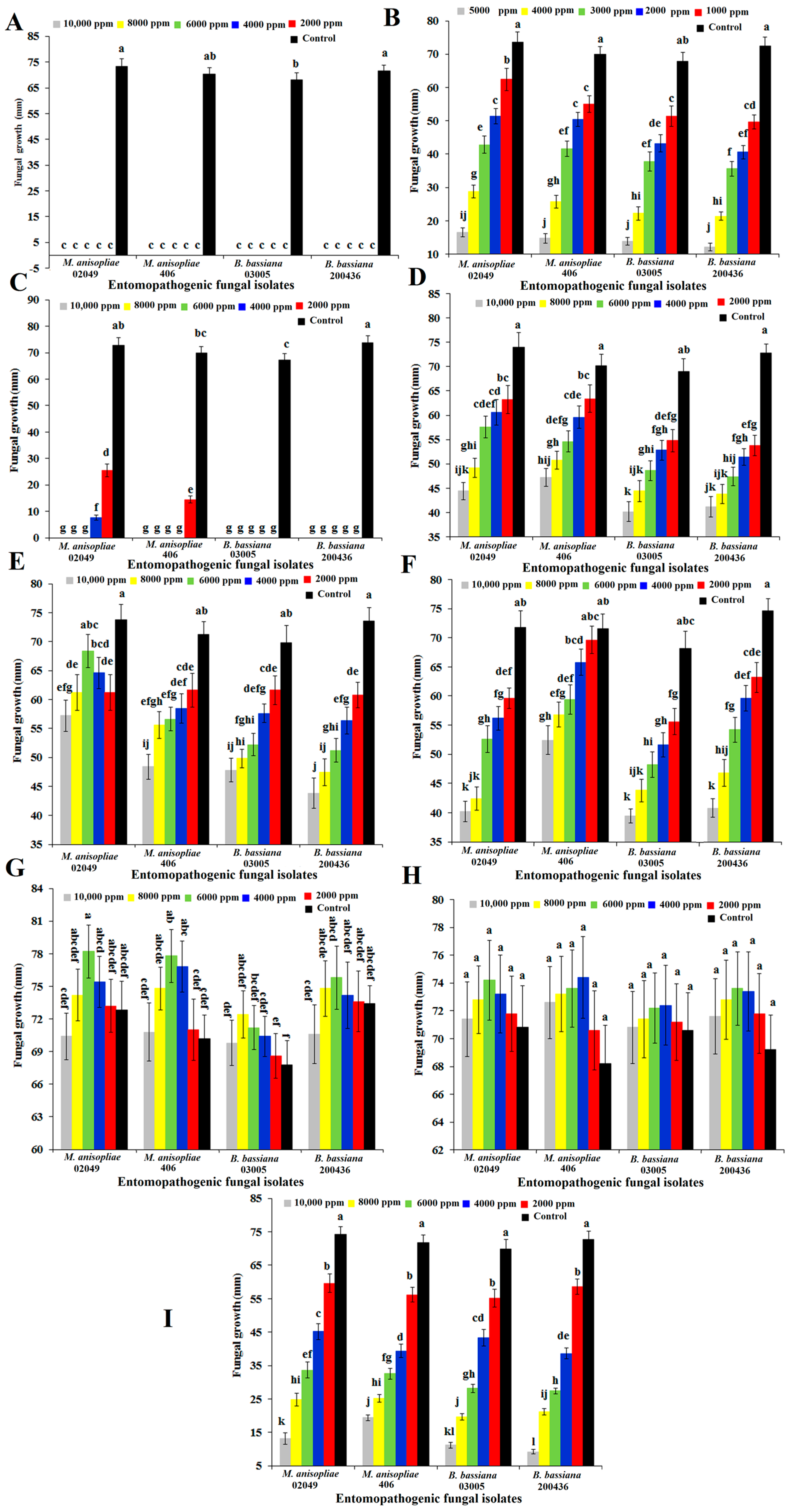
2.2. Antifungal Activity of Selected Compounds against Entomopathogenic Fungi

Isolates of entomopathogenic fungi, viz. M. anisopliae and B. bassiana, were highly sensitive to nonanal and n-hexanoic acid, showing completely or nearly completely suppressed radial fungal growth at all tested doses (Figure 1A,C). Naphthalene at the highest dose (5000 ppm) showed less than 20-mm radial growth of tested isolates. However, naphthalene at all tested doses (F = 355.10; degree of freedom = 5, 96; p < 0.0001) against all tested isolates (F = 14.28; df = 3, 96; p < 0.0001) showed significant differences in their radial growth (Figure 1B). The mycelial growth of straight chain alkanes such as n-pentadecane (Figure 1D) and n-heptadecane (Figure 1F) comparatively showed a lower response even at the highest dose (10,000 ppm). Overall, the inversely proportional relationship of radial growth in response to different doses of n-pentadecane and n-heptadecane was observed. However, there were significant differences in radial growth at different doses of n-pentadecane (F = 78.32; df = 5, 96; p < 0.001), n-heptadecane (F = 87.62; df = 5, 96; p < 0.001) and n-tetradecane (F = 40.53; df = 5, 96; p < 0.001). Similarly, n-pentadecane (F = 16.34; df = 3, 96; p < 0.001), n-heptadecane (F = 30.92; df = 3, 96; p < 0.001) and n-tetradecane (F = 16.36; df = 3, 96; p < 0.001) also showed significant differences in the radial growth of all the tested isolates of entomopathogenic fungi (Figure 1D–F). The incorporation of n-hexadecane (Figure 1G) and oleic acid (Figure 1H) into the growth media (PDA) imparted enhanced radial growth impacts on all the tested isolates. However, n-hexadecane between 4000 and 6000 ppm and oleic acid between 6000 and 8000 ppm showed a growth stimulatory response against all tested isolates. However, the interaction effect of oleic acid (F = 0.09; df = 15, 96; p > 0.05) and n-hexadecane (F = 0.35; df = 15, 96; p > 0.05) at different doses against different isolates showed a non-significant interaction. Methyl octanoate greatly inhibited the radial growth of each tested isolate in a dose-dependent manner (Figure 1I). Radial growth in the presence of methyl octanoate differed significantly among all tested isolates (F = 9.71; df = 3, 96; p < 0.0001), doses (F = 557.46; df = 5, 96; p < 0.0001) and their interaction (F = 3.04; df = 15, 96; p < 0.0001).

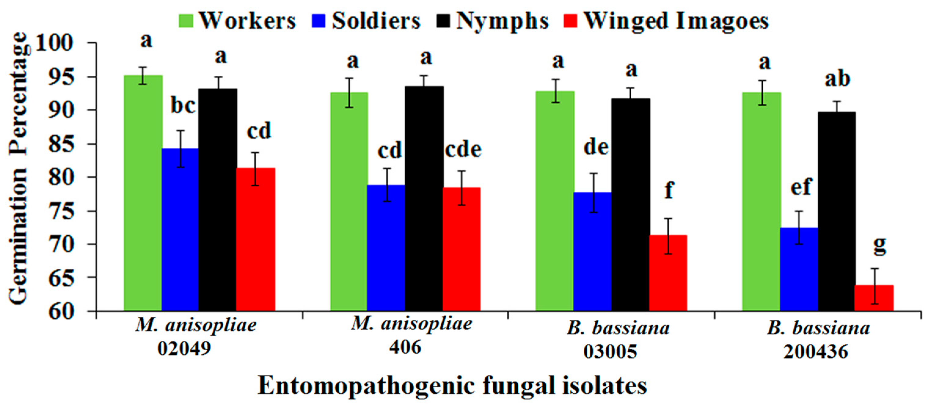
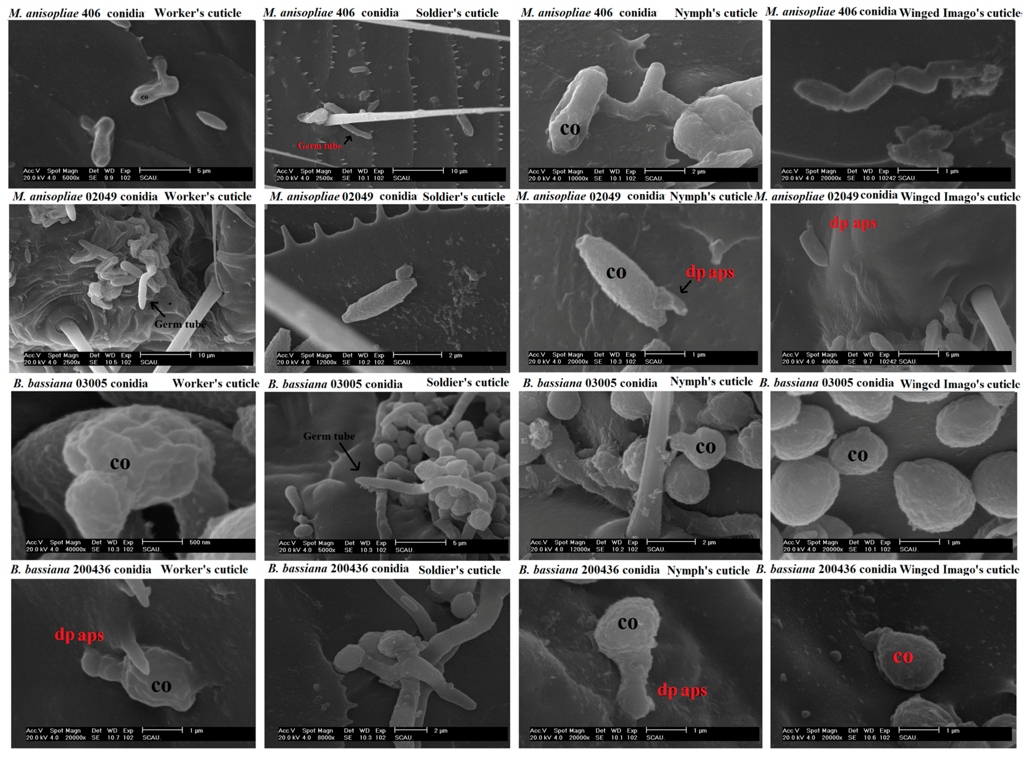
2.3. Conidial Percent Germination of Entomopathogenic Fungi on the Cuticle of Different Castes of FSTs

Conidial germination (%) significantly varied among the tested isolates of M. anisopliae and B. bassiana (F = 11.42; df = 3, 64; p < 0.0001), the cuticle of different castes (F = 77.69; df = 3, 64; p < 0.0001) and their interaction (F = 2.17; df = 9, 64; p < 0.05). Isolate of M. anisopliae 02049 exhibited the highest percent germination on the cuticle of all the inoculated castes compared with other isolates. However, B. bassiana 200436 exhibited significantly the lowest percent germination on the cuticle of each tested caste (Figure 2). The developmental events of each tested isolate on the cuticles of workers, soldiers, nymphs and winged imagoes are shown in Figure 3.
Overall, the cuticle of workers was found to be the most susceptible resulting in the highest percent germination of each tested isolate. However, percent germination of all isolates remained statistically on par with nymphs’ cuticle except isolate B. bassiana 200436. Moreover, the cuticle of winged imagoes was found to be the most resistant cuticle resulting in the lowest conidial percent germination of all tested isolates of entomopathogenic fungi (Figure 2).

2.4. Sequence Annotations for Antioxidant Genes of FSTs

In order to compile an antioxidant defense-related database for Formosan subterranean termites, ESTs were mined resulting in 17 clusters involved in antioxidant defense (Table 2). The clusters of identified antioxidant genes were directly involved in antioxidant defense of C. formosanus Shiraki (Table 2). Overall, antioxidant-related sequences matched with all important antioxidant gene classes such as catalase, glutaredoxin, glutathione S-transferase, peroxiredoxin, superoxide dismutase Cu/Zn, superoxide dismutase Fe (Fe-SOD), thioredoxin family protein, thioredoxin peroxidase and thioredoxin-like [2Fe–2S] ferredoxin (Fd) family protein (Table 2). Identified sequences directly involved in antioxidant defense were deposited to NCBI (GenBank Accession Numbers JX311467, JX876646, JX879124–JX879125, JX915904–JX915909, KC571990, KC632518, KC632520, KC741174–KC741177).

2.5. Quantitative Expression Patterns of FSTs Antioxidant Genes

The expressions of CAT, DUOX1, GRX, GST, PRXS, PRXSL, PRXS1L, Cu/Zn-SOD1, Fe-SOD, Cu/Zn-SOD2, TXN1, TXN2, TXNL1, TXNL2, TXNL4A, TPx and TRx-like-Fd determined in winged imagoes, nymphs, soldiers and workers upon infection with two isolates of B. bassiana and two isolates of M. anisopliae showed caste- and causal agent-specific expression patterns (Table 3). Among all the analyzed genes, CAT showed the highest transcript level. Interestingly, low expression (less than two-fold) of DUOX1, PRXS1L and TXNL1 was observed upon fungal infection among winged imagoes, nymphs, soldiers and workers. CAT, GST, PRXSL, Cu/Zn-SOD2, TXN1, TXN2, TXNL1, TXNL2, TXNL4A and TPx transcript levels were found to be high among winged imagoes infected with M. anisopliae 02049. PRXS, Fe-SOD and TRx-like-Fd showed the highest expression among workers infected with fungal suspensions. Genes encoding dual oxidase 1, glutaredoxin like protein, superoxide dismutase Cu/Zn1 and superoxide dismutase Fe were highly expressed exclusively in nymphs of C. formosanus Shiraki. Overall, soldiers displayed low expression levels of tested antioxidant genes compared with nymphs, workers and winged imagoes. Furthermore, our results suggested that M. anisopliae 02049 greatly enhanced the expression of all antioxidant genes tested, whereas B. bassiana 200436 failed to induce the expression of antioxidant genes.

3. Discussion

The successful foraging and amazing social lives of Formosan subterranean termites in a microbe-rich environment are the manifestation of an extraordinary, sophisticated defense mechanism of the colony members. The identification and origin of important compounds from VOCs of colony members with proven antifungal activity and mining the genes encoding the antioxidant defense of C. formosanus Shiraki and their expression patterns against fungal infections revealed in the current study profoundly widen the knowledge on disease resistance in termites in addition to previously explored social behavioral disease resistance interactions [4,5,20,21] and the immune mechanisms of disease resistance [7].
Host cuticle is the first line of defense against invading fungal pathogens [22]. Conidial germination patterns recorded on the cuticle of winged imagoes, nymphs, soldiers and workers in the current study demonstrated that the percent of germination significantly changed with fungal agent and colony members (Figure 2). The highest estimates of percent germination of all tested isolates on the cuticle of workers indicate their suitability for infection. The least virulent isolate (B. bassiana 200436) reported before against C. formosanus Shiraki workers [4] showed highly significant variations in the current study among the cuticles of tested castes. A high percent of germination on the cuticle of workers and nymphs, while a significantly lesser percent of germination on the cuticle of soldiers and winged imagoes suggest exploring the chemistry of each caste profile in order to discover the chemical defense mechanism of C. formosanus Shiraki.
Volatile organic compound profiles of winged imagoes, nymphs, soldiers and workers analyzed by HS-SPME unveiled an impressive diversity of compounds. Furthermore, our results reflected remarkable differences in the production of each caste’s volatiles suggesting that the proportion and volatile profile of C. formosanus Shiraki vary among the castes (winged imagoes, nymphs, soldiers and workers). This interpretation is consistent with previous work demonstrating that variability exists among the damp-wood termites [23,24]. In contrast, the study of Haverty et al. [25] on inter-colony GC-MS analysis of C. formosanus cuticular hydrocarbon composition revealed little variations among colony members. The difference might be because of the methodology for the extraction of hydrocarbons. Trail-forming n-hexanoic acid and naphthalene for the first time were identified in association with winged imagoes. Previously, naphthalene was found to be associated with nest material [16] and was assumed to be produced as a result of the natural decomposition of wood or fumigation by nest-mates; while n-hexanoic acid was previously detected from the sternal gland of Z. angusticollis [26]. Furthermore, our results for the antifungal investigations of naphthalene against M. anisopliae and B. bassiana are comparable to those obtained in the earlier investigations of Wright et al. [10], who exposed M. anisopliae to several concentrations of naphthalene and fenchone. They observed significant growth inhibition at higher concentrations as observed in our study, enabling us to suggest that M. anisopliae and B. bassiana cultures are highly sensitive to naphthalene at higher doses. Our results confirm and extend those of Wiltz et al. [27], who used naphthalene, butylated hydroxytoluene, dioctyl phthalate and adipic dioctyl ester against saprophytic Mucor sp. and suggested that a high concentration of naphthalene inhibited the growth of Mucor sp. Our results suggested that a high proportion of naphthalene production by winged imagoes (20.02%) favored the evolution of biochemical protection against pathogens. Another important component, n-hexanoic acid (1.35% of total nymphs VOCs), is reported to have antifungal activity against entomopathogenic fungi [26]. Significant reduction in growth in the current research supports the idea that n-hexanoic acid had the original function of controlling microbes within the nest, and their prominent role in communication may have evolved secondarily. On the basis of the above findings, it may be speculated that the versatility of primary reproductive (PR) volatile compounds contributed to the success of incipient colonies.
To date, the inhibitory effect of the volatiles such as nonanal, methyl octanoate, n-tetradecane, n-pentadecane and n-heptadecane against entomopathogenic fungi have not yet been investigated. Complete inhibition of entomopathogenic fungi was observed for nonanal suggesting that the presence of nonanal in the VOC profile of soldiers imparts strong fungistatic activity to the entomopathogenic fungi. In addition, our results suggest that n-hexadecane and oleic acid stimulated the growth of fungal isolates. Stimulation of mycelial growth by n-hexadecane and oleic acid is a novel finding. In addition, previous studies on the cue synergism in termites have reported repellent properties of oleic acid [28]. In contrast, oleic acid was found to be antifungal by significantly reducing the mycelial growth of the plant pathogenic fungus, Pythium ultimum [29]. The differences in biological activity of VOC from different castes suggest the possibility that different castes are capable of emitting specific biologically-active compounds pertinent to their roles in the life of the colony, which is intriguing and important. Normally, it is assumed that the task of defense is performed only by the soldier caste, which is morphologically specialized for this purpose [30], and the workers are involved in nest construction, feeding (trophallaxis) and cleaning (allogrooming). However, the discovery of hazardous compounds (naphthalene and n-hexanoic acid) from the winged imagoes, which may act as a fumigant, reflected the possible function of the production of VOCs by the PRs in a controlled microenvironment having no direct air exchange, which made fumigation a possible defense, not only against pathogens, but also predators.
Fungal infection imparted oxidative stress in the target host by generating reactive oxygen species (ROS). In order to return the cells to homeostasis, efficient elimination of ROS to avoid lipids’, proteins’ and nucleic acids’ damage, insects have evolved an important, complex antioxidant system [12]. The transcriptomic analysis of Formosan subterranean termites revealed the identification of numerous genes involved in antioxidant defense. The identified antioxidant genes (CAT, DUOX1, GRXL, GST, PRXS, PRXSL, PRXS1L, Cu/Zn-SOD1, Fe-SOD, Cu/Zn-SOD2, TXN1, TXN2, TXNL1, TXNL2, TXNL4A, TPx and TRx-like-Fd) showed homology with similar genes from other species. All the identified antioxidant genes have direct antioxidant effects. However, transferrin [31], ferritin [32] and vitellogenin [33], identified from the Formosan subterranean termites, were not included as they mediate indirect antioxidant effects. The molecularly-driven identification of well-developed antioxidant defense for the first time in C. formosanus Shiraki is a corroboration of their adaptation to a microbe-rich environment.
Infection of workers, soldiers, nymphs and winged imagoes of C. formosanus Shiraki with the conidial suspension of B. bassiana and M. anisopliae induced the generation of reactive metabolites such as O2●− (superoxide anion) and H2O2 (hydrogen peroxide). In the current study, we identified three different first-line antioxidant genes such as Cu/Zn-SOD1, Cu/Zn-SOD2 and Fe-SOD that catalyze superoxide radicals (O2●−) into H2O2. Among them, Cu/Zn-SOD2 showed the highest fold expression in winged imagoes against the most pathogenic isolate M. anisopliae 02049. The high upregulation is in agreement with other studies on the first-line insect antioxidant defense genes against fungal infection [34].
To protect biomolecules from oxidative damage by scavenging excessive ROS (Kodrík et al. 2015), multiple components of the thioredoxin system such as TXN1, TXN2, TXNL1, TXNL2, TXNL4A, TPx and TRx-like-Fd identified here are in line with a previous study suggesting their importance in redox-regulatory processes [35]. The expression of components of the thioredoxin system appears to show caste- and isolate-specific patterns: TXNL4A is highly expressed in winged imagoes; TRx-like-Fd exhibits higher expression in workers and winged imagoes against all fungal infections; TXN1 and TPx mainly are expressed against isolate 02049 among workers and winged imagoes; TXN2 is expressed at higher levels among nymphs and winged imagoes against the infection of isolate 02049 (Table 3), implying that thioredoxin components play a crucial role in antioxidant defense against virulent isolates in the mentioned castes. In the current study, another two components of the thioredoxin superfamily such as glutathione-S-transferases (GSTs) and Glutaredoxin like protein (GRXL) were also identified. The expression of GST in winged imagoes and GRXL in nymphs was found to be significantly stimulated by M. anisopliae 02049 infection, which is already reported as a virulent isolate due to low LT50 values [4,5,6].
Catalase (CAT) is an important ROS-scavenging antioxidant mainly involved in the removal of H2O2. CAT and components of peroxiredoxin (PRXS, PRXSL and PRXS1L) prevent oxidative damage by elimination of excessive H2O2 in many organisms [36,37]. The highest induction of CAT and PRXS among winged imagoes and workers against the most virulent isolate 02049 corroborated their role in maintaining redox homeostasis by protecting the host from toxic accumulation of ROS. PRXS1L contrastingly failed to be expressed against all the fungal infections. However, variations among different castes in response to different fungal infections contribute to the infection lethality of the invading pathogen.

4. Materials and Methods

4.1. Termite Collection and Maintenance

Nymphs, workers and soldiers of Formosan subterranean termites, Coptotermes formosanus Shiraki, were collected from Huolu Shan Forest Park, Guangzhou, China. Termites were maintained at 24–27 °C in complete darkness, in glass Petri dishes (115 mm × 20 mm) containing moist filter paper as food. Winged imagoes (alates) for bioassays were collected with an aspirator below street lights during swarms from Huolu Shan Forest Park, Guangzhou, China.

4.2. Extraction and Analysis of VOCs Released by the Termites

Volatile organic compounds were extracted by SPME, with a 1-cm coated fiber (75 µm df) of carboxen/polydimethylsiloxane (CAR/PDMS) from Supelco. Winged imagoes, nymphs, soldiers and workers (100 each, alive) of C. formosanus Shiraki were placed in separate glass vials (20 mL). Termites were equilibrated for 30 min at room temperature prior to inserting in SPME fiber. After this time, SPME fiber was exposed to the head space of the sample (winged imagoes, nymphs, soldiers and workers) for 30 min. The fiber was then retracted and inserted immediately into the inlet of the GC-MS for thermal desorption. The fiber was previously conditioned according to the manufacturer’s instructions and systematically reconditioned before each analysis. Blank injections were obtained by exposing the SPME fiber to clean empty vials. Substances found in the blank were subtracted from the VOC profile of each caste.
The qualitative analysis of the SPME extract of each caste was performed by GC-MS (Finnigan TRACE, Thermo Electron Corporation, Austin, TX, USA). The GC-MS was equipped with the HP1 capillary column having a 1-μm film thickness and 30 m × 0.25 mm internal diameter. Helium gas with 99.99% purity at a constant flow rate of 1.0 mL/min was used as the carrier gas. After injecting each sample, the initial oven temperature (45 °C) was maintained for 1 min. Subsequently, the temperature was raised to 100 °C at a rate of 5 °C/min and held for 2 min, followed by a gradient to 180 °C at a rate of 10 °C/min and held for 10 min. Mass spectra from 35–335 atomic mass units (amu) were repetitively scanned. Electron ionization (EI) was induced at 70 eV. Preliminary identification of VOCs of each C. formosanus Shiraki caste was made through comparison with the spectra in the Wiley275.1 and NIST98 libraries. Peaks of important compounds were confirmed by comparing the retention time and mass spectra with pure standards purchased from different suppliers.

4.3. Origin and Maintenance of Fungal Cultures

Four isolates of entomopathogenic fungi were selected to carry out fungal viability on host cuticle, fungal radial growth inhibition on PDA and host antioxidant defense experimentation. Two isolates, each from Metarhizium anisopliae (EBCL 02049) and Beauveria bassiana (EBCL 03005) isolated from C. formosanus, and two isolates, each from Metarhizium anisopliae (406) and Beauveria bassiana (200436) isolated from soil, were selected for whole experimentation. Each culture was maintained on potato dextrose agar (PDA) at 26 ± 1 °C, in complete darkness as mentioned in previous studies [5,6].

4.4. Preparation of Fungal Conidial Suspensions

Conidial suspension of each tested isolate of entomopathogenic fungi was prepared in sterile distilled water with 0.05% Tween 80 (Sigma-Aldrich, St. Louis, MO, USA) by harvesting 24-day-old sporulating cultures. The conidial suspension (1.3 × 10⁷ conidia/mL) and viability (93–99%) were calculated as described previously [38,39].

4.5. Antifungal Assays

The antifungal response of n-hexanoic acid (International Laboratory IL, San Francisco, CA, USA), naphthalene (Sinopharm Chemical Reagent Company, Shanghai, China), nonanal (International Laboratory IL), n-tetradecane (Sigma-Aldrich), n-pentadecane (Sigma-Aldrich), n-hexadecane (Sigma-Aldrich), n-heptadecane (Sigma-Aldrich), oleic acid (Sigma-Aldrich) and methyl octanoate (Sigma-Aldrich) was determined by the poison food technique. In brief, a specific range of different doses including 2000, 4000, 6000, 8000 and 10,000 ppm by adding 20, 40, 60, 80 and 100 µL of each tested compound (nonanal, n-hexanoic acid, naphthalene, n-tetradecane, n-pentadecane, n-hexadecane, n-heptadecane, oleic acid and methyl octanoate) was mixed with 10 mL PDA after sterilization at 50 °C. In the case of naphthalene, 10, 20, 30, 40 and 50 mg were mixed with sterilized PDA at 50 °C. Ten milliliters of PDA were poured into each Petri dish, separately. A five-microliter conidial suspension (1.3 × 10⁷ conidia/mL) of each tested isolate such as M. anisopliae 02049, M. anisopliae 406, B. bassiana 03005 and B. bassiana 200436 was pipetted to the center of each Petri dish (90 millimeter × 15 mm), separately. The inoculated agar plate was closed and sealed with laboratory film (Parafilm® Pechiney Plastic Packaging; Menasha. WI, USA) and incubated at 26 ± 1 °C, in complete darkness. The radial growth (average of five perpendicular radial lengths) was recorded 12 days post-inoculation. Each treatment was applied to five adjacent Petri dishes for each replicate. The experiment was replicated five times by using separate cultures of each tested isolate. Similarly, a control experiment was also run simultaneously using 0.05% Tween 80. Two factor factorial analysis consisting of four isolates of entomopathogenic fungi and six doses were conducted for radial growth. Significant differences among means were indicated by Fisher’s least significant difference test (α = 0.05) [40].

4.6. Conidial Germination on the Cuticle of Formosan Subterranean Termites

Each caste (nymphs, winged imagoes, soldiers and workers) was infected by immersing them into the conidial suspension (1.3 × 10⁷ conidia/mL) of M. anisopliae 02049, M. anisopliae 406, B. bassiana 03005 and B. bassiana 200436 separately for 5 s into micro-centrifuge tubes by gentle swirling [7]. Five replicates, each from separate insects, were prepared likewise. Germination of each isolate on each caste cuticle was calculated by counting 100 conidia after 18 and 36 h post-inoculation by compound microscope and scanning electron microscope. Percent of germination data on termite’s cuticles were analyzed by two-factor factorial analysis comprised of four castes and four isolates of entomopathogenic fungi. Significant differences among means were indicated by Fisher’s least significant difference test (α = 0.05) [40].

4.7. Scanning Electron Microscope

Infected nymphs, winged imagoes, soldiers and workers were fixed by glutaraldehyde (4%) prepared in phosphate buffered-saline (pH 7) with a strength of 0.1 M overnight at 4 °C. After washing thrice in 0.1 M PBS, termite specimens were subsequently fixed for 60 min in 1% Osmium Tetroxide. The fixed specimens were then washed thrice in 0.1 M PBS for subsequent dehydration in different gradients of ethanol (50%, 70%, 80%, 90% and 100%). Drying of all specimens was performed under CO2 using a critical-point drying apparatus. After mounting on stubs and coating with gold palladium, specimens were observed at 10 kV under a scanning electron microscope (XL30 ESEM, Philips, Amsterdam, The Netherlands).

4.8. Compilation and Analysis of Antioxidant Sequences of Formosan Subterranean Termites

The immunized C. formosanus Shiraki cDNA library was constructed by extracting total RNA by TRIzol reagent (Invitrogen, Waltham, MA, USA). The mRNA was isolated from total RNA using the Oligotex mRNA Mini Kit (QIAGEN, Hilden, Germany) by following the supplier’s protocol. After mRNA purification, first-strand cDNA was synthesized by denaturing mRNA and reverse transcription (In-FusionTM SMARTerTM cDNA library construction kit by Takara Biomedical Technology, Beijing Co., Ltd., Beijing, China). Second-strand cDNA was synthesized by long-distance PCR using a specific primer (5′ PCR Primer II A). The amplified product was purified to normalize the double-strand (ds) cDNAs by the Trimmer-direct cDNA Normalization kit. After purification, the pSMART2IF linearized vector was used to ligate the normalized ds cDNA for subsequent electroporation by competent cells Escherishia coli DH5α (Douglas Hanahan 5α). The individual transformants were grown for overnight on LB broth provided with isopropyl-b-d-thiogalactopyranoside (IPTG) and 5-bromo-4-chloro-3-indolyl-b-d-galac-Topyranoside (X-Gal) for sequencing. Expressed sequence tags (ESTs) were annotated using Blast2GO. Antioxidant defense-related ESTs were separately compiled and annotated by BLASTx using NCBI database.

4.9. Antioxidant Genes Validation and Quantification by qRT-PCR

First-strand cDNAs reverse transcribed from total RNA extracts of nymphs, winged imagoes, soldiers and workers infected with M. anisopliae 02049, M. anisopliae 406, B. bassiana 03005 and B. bassiana 200436 at a concentration of 1.3 × 10⁷ conidia/mL were subjected to qRT-PCR in order to determine the expression patterns of the identified antioxidant genes listed in Table 4. A housekeeping gene, β-Actin (forward primer 5′-AGCGGGAAATCGTGCGTGAC-3′ and reverse primer 5′-CAATAGTGATGACCTGGCCGT-3′), was used as an internal control. The specific primers listed in Table 4 were used for quantification using the CFX96 Real-Time System under the conditions mentioned in the our previous study [7]. Three replicates were prepared using three separate termites of each caste. Numerical values obtained from each experimental unit were compared with those of the control by relative fold expression obtained by transforming the obtained results into absolute values using 2−ΔΔCt [41]. In case of control, relative expression of each gene was set to 1. β-Actin due to its low expression variations was chosen as housekeeping gene. The relative fold expression pattern of each gene was analyzed by two-factor factorial analysis comprised of four castes and four isolates of entomopathogenic fungi. Significant differences among means were indicated by Fisher’s least significant difference test using SAS version 8 (α = 0.05) [40].

5. Conclusions

On the basis of the above findings, we may conclude that the entomopathogenic fungi are only successful in the laboratory because the laboratory evaluation mainly involved the infection of workers, which are not the source of the production of fumigants (naphthalene, n-hexanoic acid, nonanal and some other toxic fumigants) in C. formosanus Shiraki. Furthermore, the identification of a variety of genes encoding antioxidant defense from C. formosanus Shiraki and their upregulation mainly in winged imagoes and workers against the most virulent isolate greatly enhanced the knowledge of the disease resistance mechanism of C. formosanus Shiraki. The identified antioxidant transcriptome might lay the initial ground for the recently-developed precise genome editing CRISPR/Cas9 technique that will target all the castes of Formosan subterranean termites.

Acknowledgments

The authors express their gratitude to Lei Gao, Lei Yanyuan, Liu Yang and Cen Yuan for their assistance. We are very much grateful for the guidance that has been provided by John M Bland (USDA-ARS) in the identification of volatiles. The research project was jointly financed by the Chinese Scholarship Council and the Ministry of Education, Pakistan. In addition, the China Post Doctorate Foundation (20100480764) provided the funds to carry out this experimentation.

Author Contributions

Abid Hussain and Shuo-Yang Wen conceived and designed the experiments. Abid Hussain performed the experiments. Ming-Yi Tian and Shuo-Yang Wen analyzed the data. Ming-Yi Tian and Shuo-Yang Wen contributed reagents/materials/analysis tools. Abid Hussain, Ming-Yi Tian and Shuo-Yang Wen wrote the paper and approved the final manuscript.

Conflicts of Interest

The authors declare no conflict of interest. The founding sponsors had no role in the design of the study; in the collection, analyses or interpretation of data; in the writing of the manuscript; nor in the decision to publish the results.

Abbreviations

HS-SPMEHead space-solid phase micro-extraction
VOCsVolatile organic compounds
dp apsDirectly-penetrating appressorium-like structures
coUnipolar-germinated conidium
CATCatalase
DUOX1Dual oxidase 1
GRXLGlutaredoxin-like protein
GSTGlutathione S-transferase
PRXSPeroxiredoxin
PRXSLPeroxiredoxin-like protein
PRXS1LPeroxiredoxin 1-like protein
Cu/Zn-SODSuperoxide dismutase Cu/Zn
Fe-SODSuperoxide dismutase Fe
TXNThioredoxin family protein
TXNLThioredoxin-like protein
TPxThioredoxin peroxidase
TRx-like-FdThioredoxin-like [2Fe–2S] ferredoxin (Fd) family protein
SEMScanning electron microscope
ESTsExpressed sequence tags

References

  1. Ohkuma, M. Termite symbiotic systems: Efficient bio-recycling of lignocellulose. Appl. Microbiol. Biotechnol. 2003, 61, 1–9. [Google Scholar] [CrossRef] [PubMed]
  2. Su, N.-Y.; Scheffrahn, R.H. A review of subterranean termite control practices and prospects for Integrated Pest Panagement programmes. Integr. Pest Manag. Rev. 1998, 3, 1–13. [Google Scholar] [CrossRef]
  3. Kuswanto, E.; Ahmad, I.; Dungani, R. Threat of subterranean termites attack in the Asian countries and their control: A Review. Asian J. Appl. Sci. 2015, 8, 227–239. [Google Scholar] [CrossRef]
  4. Hussain, A.; Tian, M.Y. Germination pattern and inoculum transfer of entomopathogenic fungi and their role in disease resistance among Coptotermes formosanus (Isoptera: Rhinotermitidae). Int. J. Agric. Biol. 2013, 15, 319–324. [Google Scholar]
  5. Hussain, A.; Tian, M.Y.; He, Y.R.; Bland, J.M.; Gu, W.X. Behavioral and electrophysiological responses of Coptotermes formosanus Shiraki towards entomopathogenic fungal volatiles. Biol. Control 2010, 55, 166–173. [Google Scholar] [CrossRef]
  6. Hussain, A.; Tian, M.Y.; He, Y.R.; Lei, Y.Y. Differential fluctuation in virulence and VOC profiles among different cultures of entomopathogenic fungi. J. Invertebr. Pathol. 2010, 104, 166–171. [Google Scholar] [CrossRef] [PubMed]
  7. Hussain, A.; Li, Y.F.; Cheng, Y.; Liu, Y.; Chen, C.C.; Wen, S.Y. Immune-related transcriptome of Coptotermes formosanus Shiraki workers: The defense mechanism. PLoS ONE 2013, 8, e69543. [Google Scholar] [CrossRef] [PubMed]
  8. Hussain, A.; Wen, S. Induction of immune response among formosan subterranean termites, Coptotermes formosanus Shiraki (Rhinotermitidae: Isoptera). Afr. J. Microbiol. Res. 2012, 6, 995–1000. [Google Scholar] [CrossRef]
  9. Chen, J.; Henderson, G.; Grimm, C.; Lloyd, S.; Laine, R. Termites fumigate their nests with naphthalene. Nature 1998, 392, 558–559. [Google Scholar] [CrossRef]
  10. Wright, M.S.; Lax, A.R.; Henderson, G.; Chen, J. Growth response of Metarhizium anisopliae to two Formosan Subterranean termite nest volatiles, naphthalene and fenchone. Mycologia 2000, 92, 42–45. [Google Scholar] [CrossRef]
  11. Lopez-Martinez, G.; Elnitsky, M.A.; Benoit, J.B.; Lee, R.E.; Denlinger, D.L. High resistance to oxidative damage in the Antarctic midge Belgica antarctica, and developmentally linked expression of genes encoding superoxide dismutase, catalase and heat shock proteins. Insect Biochem. Mol. Biol. 2008, 38, 796–804. [Google Scholar] [CrossRef] [PubMed]
  12. Kodrík, D.; Bednářová, A.; Zemanová, M.; Krishnan, N. Hormonal Regulation of Response to Oxidative Stress in Insects—An Update. Int. J. Mol. Sci. 2015, 16, 25788–25817. [Google Scholar] [CrossRef] [PubMed]
  13. Rosengaus, R.B.; Guldin, M.R.; Traniello, J.F. Inhibitory effect of termite fecal pellets on fungal spore germination. J. Chem. Ecol. 1998, 24, 1697–1706. [Google Scholar] [CrossRef]
  14. Rosengaus, R.B.; Lefebvre, M.L.; Traniello, J.F. Inhibition of fungal spore germination by Nasutitermes: Evidence for a possible antiseptic role of soldier defensive secretions. J. Chem. Ecol. 2000, 26, 21–39. [Google Scholar] [CrossRef]
  15. Howse, P. Sociochemicals of Termites. In Chemical Ecology of Insects; Bell, W.J., Cardé, R.T., Eds.; Chapman & Hall: London, UK, 1984; pp. 475–519. [Google Scholar]
  16. Chen, J.; Henderson, G.; Grimm, C.C.; Lloyd, S.W.; Laine, R.A. Naphthalene in Formosan Subterranean Termite Carton Nests. J. Agric. Food Chem. 1998, 46, 2337–2339. [Google Scholar] [CrossRef]
  17. Kaib, M.; Jmhasly, P.; Wilfert, L.; Durka, W.; Franke, S.; Francke, W.; Leuthold, R.H.; Brandl, R. Cuticular hydrocarbons and aggression in the termite Macrotermes Subhyalinus. J. Chem. Ecol. 2004, 30, 365–385. [Google Scholar] [CrossRef] [PubMed]
  18. Haverty, M.I.; Woodrow, R.J.; Nelson, L.J.; Grace, J.K. Identification of termite species by the Hydrocarbons in their feces. J. Chem. Ecol. 2005, 31, 2119–2151. [Google Scholar] [CrossRef] [PubMed]
  19. Bland, J.M.; Osbrink, W.L.; Cornelius, M.L.; Lax, A.R.; Vigo, C.B. Solid-phase microextraction for the detection of termite cuticular hydrocarbons. J. Chromatogr. A 2001, 932, 119–127. [Google Scholar] [CrossRef]
  20. Zhukovskaya, M.; Yanagawa, A.; Forschler, B. Grooming behavior as a mechanism of insect disease defense. Insects 2013, 4, 609–630. [Google Scholar] [CrossRef] [PubMed]
  21. Yanagawa, A.; Imai, T.; Akino, T.; Toh, Y.; Yoshimura, T. Olfactory cues from pathogenic fungus affect the direction of motion of termites, Coptotermes formosanus. J. Chem. Ecol. 2015, 41, 1118–1126. [Google Scholar] [CrossRef] [PubMed]
  22. Hussain, A.; Rizwan-ul-Haq, M.; Al-Ayedh, H.; AlJabr, A. Susceptibility and immune defence mechanisms of Rhynchophorus ferrugineus (Olivier) (Coleoptera: Curculionidae) against entomopathogenic fungal infections. Int. J. Mol. Sci. 2016, 17, 1518. [Google Scholar] [CrossRef] [PubMed]
  23. Haverty, M.I.; Page, M.; Nelson, L.J.; Blomquist, G.J. Cuticular hydrocarbons of dampwood termites, Zootermopsis: Intra- and intercolony variation and potential as taxonomic characters. J. Chem. Ecol. 1988, 14, 1035–1058. [Google Scholar] [CrossRef] [PubMed]
  24. Sevala, V.L.; Bagnères, A.-G.; Kuenzli, M.; Blomquist, G.J.; Schal, C. Cuticular hydrocarbons of the Dampwood termite, Zootermopsis nevadensis: Caste differences and role of Lipophorin in transport of hydrocarbons and hydrocarbon metabolites. J. Chem. Ecol. 2000, 26, 765–789. [Google Scholar] [CrossRef]
  25. Haverty, M.I.; Grace, J.K.; Nelson, L.J.; Yamamoto, R.T. Intercaste, intercolony, and temporal variation in cuticular hydrocarbons of Copotermes formosanus shiraki (Isoptera: Rhinotermitidae). J. Chem. Ecol. 1996, 22, 1813–1834. [Google Scholar] [CrossRef] [PubMed]
  26. Rosengaus, R.; Traniello, J.; Lefebvre, M.; Maxmen, A. Fungistatic activity of the sternal gland secretion of the dampwood termite Zootermopsis angusticollis. Insectes Soc. 2004, 51, 259–264. [Google Scholar] [CrossRef]
  27. Wiltz, B.A.; Henderson, G.; Chen, J. Effect of naphthalene, butylated hydroxytoluene, dioctyl phthalate, and adipic dioctyl ester, chemicals found in the nests of the Formosan Subterranean Termite (Isoptera: Rhinotermitidae) on a Saprophytic Mucor sp. (Zygomycetes: Mucorales). Environ. Entomol. 1998, 27, 936–940. [Google Scholar] [CrossRef]
  28. Ulyshen, M.D.; Shelton, T.G. Evidence of cue synergism in termite corpse response behavior. Naturwissenschaften 2012, 99, 89–93. [Google Scholar] [CrossRef] [PubMed]
  29. Raynor, L.; Mitchell, A.; Walker, R.; Walker, R. Antifungal activities of four fatty acids against plant pathogenic fungi. Mycopathologia 2004, 157, 87–90. [Google Scholar] [CrossRef]
  30. Noirot, C.; Johanna, P.D. Termite Nests: Architecture, regulation and defence. In Termites: Evolution, Sociality, Symbioses, Ecology; Abe, T., Bignell, D.E., Higashi, M., Eds.; Springer: Dordrecht, The Netherlands, 2000; pp. 121–139. [Google Scholar]
  31. Kucharski, R.; Maleszka, R. Transcriptional profiling reveals multifunctional roles for transferrin in the honeybee, Apis mellifera. J. Insect Sci. 2003, 3, 1–8. [Google Scholar] [CrossRef]
  32. Geiser, D.L.; Chavez, C.A.; Flores-Munguia, R.; Winzerling, J.J.; Pham, D.Q.-D. Aedes aegypti ferritin. A cytotoxic protector against iron and oxidative challenge? Eur. J. Biochem. 2003, 270, 3667–3674. [Google Scholar] [CrossRef] [PubMed]
  33. Seehuus, S.-C.; Norberg, K.; Gimsa, U.; Krekling, T.; Amdam, G.V. Reproductive protein protects functionally sterile honey bee workers from oxidative stress. Proc. Natl. Acad. Sci. USA 2006, 103, 962–967. [Google Scholar] [CrossRef] [PubMed]
  34. Gretscher, R.R.; Streicher, P.E.; Strauß, A.S.; Wielsch, N.; Stock, M.; Wang, D.; Boland, W.; Burse, A. A common theme in extracellular fluids of beetles: Extracellular superoxide dismutases crucial for balancing ROS in response to microbial challenge. Sci. Rep. 2016, 6, 24082. [Google Scholar] [CrossRef] [PubMed]
  35. Zhang, S.; Li, Z.; Nian, X.; Wu, F.; Shen, Z.; Zhang, B.; Zhang, Q.; Liu, X. Sequence analysis, expression profiles and function of thioredoxin 2 and thioredoxin reductase 1 in resistance to nucleopolyhedrovirus in Helicoverpa armigera. Sci. Rep. 2015, 5, 15531. [Google Scholar] [CrossRef] [PubMed]
  36. Robinson, M.W.; Hutchinson, A.T.; Dalton, J.P.; Donnelly, S. Peroxiredoxin: A central player in immune modulation. Parasite Immunol. 2010, 32, 305–313. [Google Scholar] [CrossRef] [PubMed]
  37. Hussain, A.; Rizwan-ul-Haq, M.; Al-Ayedh, H.; Ahmed, S.; Al-Jabr, A.M. Effect of Beauveria bassiana infection on the feeding performance and antioxidant defence of red palm weevil, Rhynchophorus ferrugineus. BioControl 2015, 60, 849–859. [Google Scholar] [CrossRef]
  38. Hussain, A.; Tian, M.Y.; He, Y.R.; Ahmed, S. Entomopathogenic fungi disturbed the larval growth and feeding performance of Ocinara varians (Lepidoptera: Bombycidae) larvae. Insect Sci. 2009, 16, 511–517. [Google Scholar] [CrossRef]
  39. Hussain, A.; Tian, M.Y.; He, Y.R.; Ruan, L.; Ahmed, S. In vitro and in vivo culturing impacts on the virulence characteristics of serially passed entomopathogenic fungi. J. Food Agric. Environ. 2010, 8, 481–487. [Google Scholar]
  40. SAS Institute. SAS User’s Guide: Statistics; SAS Institute: Cary, NC, USA, 2000. [Google Scholar]
  41. Livak, K.J.; Schmittgen, T.D. Analysis of relative gene expression data using real-time quantitative PCR and the 2−ΔΔCt Method. Methods 2001, 25, 402–408. [Google Scholar] [CrossRef] [PubMed]
Figure 1. The effect of different concentrations of n-hexanoic acid (A), naphthalene (B), nonanal (C), n-pentadecane (D), n-tetradecane (E), n-heptadecane (F), n-hexadecane (G), oleic acid (H) and methyl octanoate (I) on the radial growth (12-d colony diameter) of entomopathogenic fungal isolates at 26 ± 1 °C, in complete darkness. Mean ± SE values followed by different letter(s) are significantly different (Fisher’s LSD test, α = 0.05). Values are the means of five replicates, and each replicate is the average of five Petri dishes.
Ijms 18 02694 g001
Figure 2. Percent germination of the conidia of entomopathogenic fungal isolates on the cuticle of workers, soldiers, nymphs and winged imagoes. Values are the means of five replicates. Mean ± SE values followed by different letter(s) are significantly different (Fisher’s LSD test, α = 0.05).
Ijms 18 02694 g002
Figure 3. SEM used for the detection of growth patterns of tested isolates of entomopathogenic fungi on the cuticle of workers, soldiers, nymphs and winged imagoes of C. formosanus Shiraki. dp aps stands for directly-penetrating appressorium-like structure; co stands for unipolar-germinated conidium.
Ijms 18 02694 g003
Table 1. Volatile organic compound profiles of Coptotermes formosanus Shiraki.
Rt (min)Chemical FormulaeCompoundsRA
Winged Imagoes (%)Nymphs (%)Workers (%)Soldiers (%)
8.22C6H12O2n-Hexanoic acid *1.35
11.41C9H18ONonanal *0.54
12.18C9H18O2Methyl octanoate *28.172.70
13.66C10H8Naphthalene *20.02
15.95C10H20O2n-Amyl isovalerate37.533.39
16.42C10H12O31-Methoxyethyl benzoate16.95
16.69C10H12O1-Methoxy-4-(1-propenyl)benzene2.36
16.91C10H14O2-t-Butylphenol2.73
17.05C10H23NS22-Diisopropylaminoethyl ethyl disulfide17.98
17.30C11H22On-Undecanal0.690.10
17.61C11H292,4,6-Trimethyloctane1.03
17.62C12H26n-Dodecane0.15
17.81C12H18O81,2,3,4-Butanetetrol, tetraacetate, [R *,S *]3.27
18.24C12H24O32,2-dimethyl-1-(2-hydroxy-1-isopropyl) propyl isobutanoate1.030.32
19.22C14H28OTetradecanal3.170.30
19.43C14H30n-Tetradecane *4.105.130.50
19.55C15H261,3-Dimethyl-5-n-propyl-adamantane2.92
19.62C15H24β-Caryophyllene1.891.170.30
19.95C15H24Decahydro-1,1,7[1a,a]-trimethyl-4-methylene-1H-cycloprop[e]azulene1.100.15
20.42C15H322,6,10-Trimethyldodecane4.663.99
20.79C15H24OButylated hydroxytoluene2.250.48
20.92C15H32n-Pentadecane *11.299.892.31
21.02C15H22Cadina-1,2,3-triene2.58
21.80C15H323-Methyltetradecane1.28
22.08C15H26OCedrol6.78
22.25C16H34n-Hexadecane *15.953.952.690.49
23.02C16H345-Propyltridecane12.54
23.20C16H343-Methylpentadecane1.66
23.39C16H181,2,3-Trimethyl-4[E]-propenyl-naphthalene3.0034.557.231.77
23.79C17H36n-Heptadecane *12.54
23.96C18H382,6,10-Trimethylpentadecane6.381.21
26.10C18H38n-Octadecane2.97
26.13C18H32O2[Z,Z]-9,12-Octadecadienoic acid12.22
26.95C18H34O2Oleic acid *65.84
29.13C18H36O2Octadecanoic acid8.42
Rt = retention time; RA = relative area (peak area relative to total peak area); * compounds identified by authentic standard.
Table 2. Identified antioxidant defense-related genes of C. formosanus Shiraki based on sequence similarity (E ≤ 10−5).
No.Length (bp)Accession No.AnnotationExpect Value
1821JX876646Catalase (CAT)5 × 10−111
2957KC571990Dual oxidase 1 (DUOX1)7 × 10−121
3847KC741176Glutaredoxin like protein (GRXL)1 × 10−82
4920JX915905Glutathione S-transferase (GST)2 × 10−139
51121JX915906Peroxiredoxin (PRXS)3 × 10−161
6924JX915907Peroxiredoxin-like protein (PRXSL)3 × 10−105
71237JX915909Peroxiredoxin 1-like protein (PRXS1L)5 × 10−137
8852KC741174Superoxide Dismutase Cu/Zn (Cu/Zn–SOD1)5 × 10−103
9640JX311467Superoxide Dismutase Fe (Fe–SOD)3 × 10−85
10909JX915904Superoxide Dismutase Cu/Zn (Cu/Zn–SOD2)4 × 10−75
11892KC632518Thioredoxin family protein (TXN1)1 × 10−58
12919KC741175Thioredoxin family protein (TXN2)1 × 10−178
13910JX879125Thioredoxin-like protein (TXNL1)2 × 10−51
14917KC741177Thioredoxin-like protein (TXNL2)5 × 10−161
15849JX915908Thioredoxin-like protein 4A (TXNL4A)4 × 10−60
16714JX879124Thioredoxin peroxidase (TPx)7 × 10−88
17889KC632520Thioredoxin-like [2Fe–2S] ferredoxin (Fd)
family protein (TRx-like-Fd)
5 × 10−132
Table 3. Expression patterns of C. formosanus Shiraki antioxidant genes in the whole body homogenates of winged imagoes, nymphs, soldiers and workers using quantitative real-time PCR (qRT-PCR).
GenesCastesEntomopathogenic FungiCastes Statistics
M. anisopliae 02049M. anisopliae 406B. bassiana 03005B. bassiana 200436
CATWorkers11.15 ± 0.32 b7.02 ± 0.49 d5.29 ± 0.26 e2.98 ± 0.20 gF = 296.74
Soldiers2.96 ± 0.28 g2.12 ± 0.15 h1.68 ± 0.19 hi1.13 ± 0.13 idf = 3, 32
Nymphs5.05 ± 0.43 e3.94 ± 0.19 f4.92 ± 0.20 e2.09 ± 0.16 hp < 0.0001
Winged Imagoes15.04 ± 0.48 a3.70 ± 0.35 fg9.53 ± 0.34 c1.86 ± 0.16 hi
Infection StatisticsF = 344.94; df = 3, 32; p < 0.0001F = 78.83
df = 9, 32
p < 0.0001
DUOX1Workers1.07 ± 0.14 cde0.81 ± 0.07 efgh1.21 ± 0.11 cd0.59 ± 0.06 hijF = 24.58
Soldiers0.70 ± 0.08 ghi0.49 ± 0.11 ij0.98 ± 0.09 def0.38 ± 0.10 jdf = 3, 32
Nymphs1.81 ± 0.12 a0.89 ± 0.09 efg1.34 ± 0.11 bc0.75 ± 0.05 fghip < 0.0001
Winged Imagoes1.59 ± 0.014 ab0.83 ± 0.08 efgh1.23 ± 0.08 cd0.68 ± 0.10 ghi
Infection StatisticsF = 46.44; df = 3, 32; p < 0.0001F = 3.36
df = 9, 32
p = 0.005
GRXLWorkers2.59 ± 0.16 d1.72 ± 0.12 e1.82 ± 0.12 e1.26 ± 0.13 fgF = 238.99
Soldiers1.57 ± 0.10 ef1.06 ± 0.08 g1.23 ± 0.10 fg0.91 ± 0.07 gdf = 3, 32
Nymphs4.91 ± 0.23 a1.69 ± 0.10 e3.77 ± 0.15 b1.91 ± 0.09 ep < 0.0001
Winged Imagoes4.63 ± 0.16 a3.11 ± 0.10 c3.93 ± 0.08 b1.09 ± 0.09 g
Infection StatisticsF = 219.10; df = 3, 32; p < 0.0001F = 34.80
df = 9, 32
p < 0.0001
GSTWorkers1.95 ± 0.12 ef0.91 ± 0.11 h1.84 ± 0.09 ef0.67 ± 0.08 hiF = 492.93
Soldiers0.88 ± 0.10 hi0.71 ± 0.08 hi0.70 ± 0.08 hi0.61 ± 0.09 idf = 3, 32
Nymphs3.08 ± 0.11 c2.74 ± 0.12 d2.11 ± 0.09 e1.35 ± 0.10 gp < 0.0001
Winged Imagoes5.21 ± 0.13 a1.69 ± 0.10 f3.94 ± 0.10 b2.10 ± 0.08 e
Infection StatisticsF = 202.59; df = 3, 32; p < 0.0001F = 57.27
df = 9, 32
p < 0.0001
PRXSWorkers3.88 ± 0.17 a1.76 ± 0.10 e2.77 ± 0.18 c0.83 ± 0.06 ghF = 114.55
Soldiers1.84 ± 0.09 de1.20 ± 0.10 f1.23 ± 0.11 f0.76 ± 0.09 hdf = 3, 32
Nymphs2.54 ± 0.10 c1.14 ± 0.09 fg2.12 ± 0.10 d0.86 ± 0.09 ghp < 0.0001
Winged Imagoes3.73 ± 0.11 a1.85 ± 0.09 de3.41 ± 0.11 b1.10 ± 0.07 fg
Infection StatisticsF = 294.23; df = 3, 32; p < 0.0001F = 17.60
df = 9, 32
p < 0.0001
PRXSLWorkers2.47 ± 0.12 b2.05 ± 0.08 cd1.90 ± 0.10 d0.96 ± 0.10 efF = 65.96
Soldiers1.85 ± 0.09 d1.23 ± 0.10 e1.07 ± 0.08 ef0.77 ± 0.06 fdf = 3, 32
Nymphs2.06 ± 0.10 cd1.84 ± 0.11 d1.17 ± 0.10 e0.80 ± 0.08 fp < 0.0001
Winged Imagoes3.49 ± 0.16 a1.77 ± 0.11 d2.28 ± 0.13 bc1.18 ± 0.08 e
Infection StatisticsF = 149.75; df = 3, 32; p < 0.0001F = 10.27
df = 9, 32
p < 0.0001
PRXS1LWorkers1.01 ± 0.09 cdef0.79 ± 0.08 fg1.07 ± 0.09 bcde0.48 ± 0.09 hF = 13.70
Soldiers1.86 ± 0.10 a1.03 ± 0.09 cdef0.96 ± 0.08 def0.65 ± 0.08 ghdf = 3, 32
Nymphs0.96 ± 0.10 def0.86 ± 0.08 efg1.11 ± 0.08 bcd1.22 ± 0.09 bcp < 0.0001
Winged Imagoes1.29 ± 0.10 b1.19 ± 0.07 bcd1.14 ± 0.07 bcd1.17 ± 0.08 bcd
Infection StatisticsF = 16.54; df = 3, 32; p < 0.0001F = 11.66
df = 9, 32
p < 0.0001
Cu/Zn–SOD1Workers1.10 ± 0.08 fgh1.24 ± 0.08 ef1.05 ± 0.07 fgh0.84 ± 0.07 hF = 325.13
Soldiers1.09 ± 0.08 fgh0.88 ± 0.09 gh1.21 ± 0.09 ef1.10 ± 0.08 fghdf = 3, 32
Nymphs3.74 ± 0.12 a1.44 ± 0.09 e2.89 ± 0.12 c1.89 ± 0.09 dp < 0.0001
Winged Imagoes3.17 ± 0.10 b2.73 ± 0.10 c2.91 ± 0.12 bc1.10 ± 0.06 fg
Infection StatisticsF = 103.32; df = 3, 32; p < 0.0001F = 45.14
df = 9, 32
p < 0.0001
Fe–SODWorkers3.88 ± 0.15 b3.16 ± 0.13 d3.67 ± 0.11 bc2.66 ± 0.10 eF = 344.02
Soldiers1.23 ± 0.08 g1.11 ± 0.08 g0.92 ± 0.08 gh0.62 ± 0.11 hdf = 3, 32
Nymphs4.21 ± 0.12 a3.69 ± 0.12 bc2.14 ± 0.10 f1.11 ± 0.07 gp < 0.0001
Winged Imagoes3.51 ± 0.13 c2.24 ± 0.12 f2.94 ± 0.13 de2.12 ± 0.10 f
Infection StatisticsF = 137.33; df = 3, 32; p < 0.0001F = 30.98
df = 9, 32
p < 0.0001
Cu/Zn–SOD2Workers4.35 ± 0.19 b2.96 ± 0.10 c1.05 ± 0.09 efg0.77 ± 0.09 ghF = 248.02
Soldiers1.02 ± 0.09 efg1.21 ± 0.08 e0.84 ± 0.09 fgh0.52 ± 0.08 hdf = 3, 32
Nymphs2.88 ± 0.12 c1.93 ± 0.14 d1.66 ± 0.11 d1.12 ± 0.07 efp < 0.0001
Winged Imagoes5.99 ± 0.16 a3.13 ± 0.12 c1.12 ± 0.08 ef1.92 ± 0.12 d
Infection StatisticsF = 422.72; df = 3, 32; p < 0.0001F = 68.00
df = 9, 32
p < 0.0001
TXN1Workers2.74 ± 0.12 ab2.06 ± 0.11 de1.73 ± 0.12 fg1.13 ± 0.08 iF = 32.14
Soldiers1.82 ± 0.17 efg1.55 ± 0.12 gh1.12 ± 0.09 ij0.81 ± 0.07 jdf = 3, 32
Nymphs2.47 ± 0.10 bc1.92 ± 0.10 def2.18 ± 0.12 cd1.27 ± 0.10 hip < 0.0001
Winged Imagoes2.96 ± 0.11 a1.76 ± 0.10 efg1.94 ± 0.12 def1.14 ± 0.07 i
Infection StatisticsF = 111.59; df = 3, 32; p < 0.0001F = 4.34
df = 9, 32
p < 0.0001
TXN2Workers2.13 ± 0.11 c1.89 ± 0.13 cde0.87 ± 0.10 g0.86 ± 0.09 gF = 24.49
Soldiers1.94 ± 0.11 cd1.48 ± 0.11 f1.13 ± 0.06 g0.93 ± 0.11 gdf = 3, 32
Nymphs2.76 ± 0.13 b1.73 ± 0.13 def1.91 ± 0.12 cde1.09 ± 0.08 gp < 0.0001
Winged Imagoes3.75 ± 0.14 a1.17 ± 0.06 g1.60 ± 0.11 ef0.92 ± 0.12 g
Infection StatisticsF = 176.34; df = 3, 32; p < 0.0001F = 19.92
df = 9, 32
p = 0.0009
TXNL1Workers1.76 ± 0.14 ab0.81 ± 0.09 e1.38 ± 0.11 cd0.21 ± 0.05 gF = 0.46
Soldiers1.91 ± 0.11 a0.65 ± 0.09 ef1.17 ± 0.12 d0.47 ± 0.09 fgdf = 3, 32
Nymphs1.50 ± 0.10 bc1.16 ± 0.07 d1.29 ± 0.11 cd0.48 ± 0.08 fgp > 0.05
Winged Imagoes1.93 ± 0.12 a1.15 ± 0.11 d0.84 ± 0.08 e0.26 ± 0.08 g
Infection statisticsF = 142.88; df = 3, 32; p < 0.0001F = 6.06
df = 9, 32
p < 0.0001
TXNL2Workers2.11 ± 0.11 b1.18 ± 0.11 de1.86 ± 0.11 bc0.68 ± 0.09 fF = 15.95
Soldiers1.96 ± 0.14 bc1.13 ± 0.06 de1.72 ± 0.11 c0.79 ± 0.09 fdf = 3, 32
Nymphs2.17 ± 0.13 b1.67 ± 0.12 c0.93 ± 0.07 ef0.65 ± 0.12 fp < 0.0001
Winged Imagoes2.79 ± 0.15 a1.16 ± 0.07 de2.17 ± 0.11 b1.26 ± 0.14 d
Infection StatisticsF = 115.31; df = 3, 32; p < 0.0001F = 9.56
df = 9, 32
p < 0.0001
TXNL4AWorkers4.73 ± 0.15 b2.82 ± 0.14 ef3.46 ± 0.16 d1.13 ± 0.08 jF = 159.16
Soldiers2.09 ± 0.14 gh1.53 ± 0.12 i1.91 ± 0.14 hi1.07 ± 0.07 jdf = 3, 32
Nymphs3.86 ± 0.14 c2.30 ± 0.12 g3.20 ± 0.13 de1.94 ± 0.13 ghp < 0.0001
Winged Imagoes5.31 ± 0.18 a2.72 ± 0.13 f4.76 ± 0.16 b2.13 ± 0.12 gh
Infection StatisticsF = 253.32; df = 3, 32; p < 0.0001F = 20.22
df = 9, 32
p < 0.0001
TPxWorkers3.74 ± 0.15 ab2.46 ± 0.10 g3.35 ± 0.15 cd1.73 ± 0.12 hF = 142.07
Soldiers1.88 ± 0.13 h1.12 ± 0.09 i1.21 ± 0.10 i0.68 ± 0.12 jdf = 3, 32
Nymphs3.47 ± 0.16 bc1.94 ± 0.13 h2.69 ± 0.14 gh1.12 ± 0.08 ip < 0.0001
Winged Imagoes3.95 ± 0.14 a3.09 ± 0.12 de2.93 ± 0.14 ef1.08 ± 0.08 i
Infection StatisticsF = 201.29; df = 3, 32; p < 0.0001F = 9.66
df = 9, 32
p < 0.0001
TRx-like-FdWorkers3.53 ± 0.16 a2.82 ± 0.17 b3.05 ± 0.14 b1.65 ± 0.12 dF = 84.27
Soldiers1.82 ± 0.15 cd1.26 ± 0.13 e1.12 ± 0.07 ef0.73 ± 0.09 gdf = 3, 32
Nymphs2.79 ± 0.16 b2.14 ± 0.10 c1.68 ± 0.13 d0.76 ± 0.09 fgp < 0.0001
Winged Imagoes3.07 ± 0.11 b2.18 ± 0.14 c2.19 ± 0.12 c1.25 ± 0.13 e
Infection StatisticsF = 117.62; df = 3, 32; p < 0.0001F = 7.84
df = 9, 32
p < 0.0001
Relative fold expression values are the means of three replicates. Significant differences among the means were analyzed by two-factor factorial ANOVA under completely randomized design (CRD) (Fisher’s LSD test, α = 0.05). Means ± SE values with the same letter(s) are not significantly different.
Table 4. Antioxidant genes used to study expression patterns of the whole body homogenates of winged imagoes, nymphs, soldiers and workers of C. formosanus Shiraki using quantitative real-time PCR (qRT-PCR).
GeneProduct LengthAccession NumberForward Primer (5′-3′)Reverse Primer (5′-3′)
Catalase (CAT)97 bpJX876646TGGCACCAACTACCTTCAAATCCTTGGTGATGCATTGTTT
Dual oxidase 1 (DUOX1)95 bpKC571990AGTTTGACCTCAGGACCATCCAGTGACTGCTTTCAGCCCAG
Glutaredoxin like protein (GRXL)87 bpKC741176GGAATTGCATCCAGTAGGGAGACTGCTCCCTTGCCTCTTCTT
Glutathione S-transferase (GST)99 bpJX915905GTATTGGCAGGTTCGTGTTGTACAGCCTCAGCTTCCCTTT
Peroxiredoxin (PRXS)106 bpJX915906ACCTGTTGGTCGCAGTGTAGTTCTTGTCCTGGCTTCCAGC
Peroxiredoxin-like protein (PRXSL)112 bpJX915907TTCATGCTGTAGGGCGTGTTTCACAGAGCTCCACACTTGG
Peroxiredoxin 1-like protein (PRXS1L)97 bpJX915909GCATATTCTGACCGTGCAGCTGTTGACCCAAGCAAGGTGA
Superoxide Dismutase Cu/Zn (Cu/Zn-SOD1)93 bpKC741174GTAACGGGGGAAGTGACTGGTCCAGCACTTGTACAGCCAT
Superoxide Dismutase Fe (Fe-SOD)127 bpJX311467ACAGTTGATGCTTGGGAACATGAGACCAGCAGCCTTAAAC
Superoxide Dismutase Cu/Zn (Cu/Zn-SOD2)90 bpJX915904AATGGAGAAGTGGTCAAGGGTGACAGACCAGTCACTTCCC
Thioredoxin family protein (TXN1)81 bpKC632518CTTACACCGGAGGACGACATAACCATCGGGTGCTCGATTC
Thioredoxin family protein (TXN2)75 bpKC741175TCTGAATGTTGCCCGTGTGAGGCACTTCCCAGACTCCAAA
Thioredoxin-like protein (TXNL1)97 bpJX879125ACAAAGCTTACGGAGGCAGGGCTCCTCAATTCTGGGTGCT
Thioredoxin-like protein (TXNL2)81 bpKC741177GGCCACACCACAACTAAAGCATGCAGGTGTTTTGGTCGGA
Thioredoxin-like protein 4A (TXNL4A)80 bpJX915908TGGCCATTGGAAGACAAGCAACTAGACCTCGACCTTTGCG
Thioredoxin peroxidase (TPx)80 bpJX879124TGCCGCAAAACATGGAGAAGTGCTCTCATTCGGCTTAGGA
Thioredoxin-like [2Fe–2S] ferredoxin (Fd) family protein (TRx-like-Fd)106 bpKC632520CTTGTGTCAACGCCCCAATGGGCACTCGGCCATTTTTCAA

