Article

Melatonin and Its Metabolites Ameliorate UVR-Induced Mitochondrial Oxidative Stress in Human MNT-1 Melanoma Cells

by
Konrad Kleszczyński
1,*,†,
Bernadetta Bilska
2,†,
Agatha Stegemann
1,
Damian Jozef Flis
3,
Wieslaw Ziolkowski
3,
Elżbieta Pyza
2,
Thomas A. Luger
1,
Russel J. Reiter
4,
Markus Böhm
1 and
Andrzej T. Slominski
5,6,*
1
Department of Dermatology, University of Münster, Von-Esmarch-Str. 58, 48149 Münster, Germany
2
Department of Cell Biology and Imaging, Institute of Zoology and Biomedical Research, Jagiellonian University, Gronostajowa 9, 30-387 Kraków, Poland
3
Department of Bioenergetics and Nutrition, Gdańsk University of Physical Education and Sport, Górski Str. 1, 80-336 Gdańsk, Poland
4
Department of Cellular and Structural Biology, UT Health Science Center, San Antonio, TX 78229, USA
5
Department of Dermatology, Comprehensive Cancer Center, University of Alabama at Birmingham, Birmingham, AL 35294, USA
6
Pathology and Laboratory Medicine Service, VA Medical Center, Birmingham, AL 35249, USA
*
Authors to whom correspondence should be addressed.
These authors contributed equally to this work.
Int. J. Mol. Sci. 2018, 19(12), 3786; https://0-doi-org.brum.beds.ac.uk/10.3390/ijms19123786
Submission received: 10 October 2018 / Revised: 9 November 2018 / Accepted: 21 November 2018 / Published: 28 November 2018

Abstract

:
Melatonin (Mel) is the major biologically active molecule secreted by the pineal gland. Mel and its metabolites, 6-hydroxymelatonin (6(OH)Mel) and 5-methoxytryptamine (5-MT), possess a variety of functions, including the scavenging of free radicals and the induction of protective or reparative mechanisms in the cell. Their amphiphilic character allows them to cross cellular membranes and reach subcellular organelles, including the mitochondria. Herein, the action of Mel, 6(OH)Mel, and 5-MT in human MNT-1 melanoma cells against ultraviolet B (UVB) radiation was investigated. The dose of 50 mJ/cm2 caused a significant reduction of cell viability up to 48%, while investigated compounds counteracted this deleterious effect. UVB exposure increased catalase activity and led to a simultaneous Ca++ influx (16%), while tested compounds prevented these disturbances. Additional analysis focused on mitochondrial respiration performed in isolated mitochondria from the liver of BALB/cJ mice where Mel, 6(OH)Mel, and 5-MT significantly enhanced the oxidative phosphorylation at the dose of 10−6 M with lower effects seen at 10−9 or 10−4 M. In conclusion, Mel, 6(OH)Mel and 5-MT protect MNT-1 cells, which express melatonin receptors (MT1 and MT2) against UVB-induced oxidative stress and mitochondrial dysfunction, including the uncoupling of oxidative phosphorylation.

Graphical Abstract

1. Introduction

Melatonin (N-acetyl-5-methoxytryptamine) is a ubiquitous physiological mediator that is found throughout the evolutionary scale of animals and is also in plants and unicellular organisms [1,2,3,4,5]. In mammals, it is characterized as a natural neurohormone synthesized in the pineal gland, which is involved in the regulation of circadian rhythms [4]. In addition, many other tissues and cells, including bone marrow [6], lymphocytes [7], retina [8], astrocytes [9], thymus [10], skin [11,12,13], and female reproductive organs (granulosa cells, cumulus cells, and oocytes) [14], can synthesize melatonin. There is also evidence that melatonin is present in follicular fluid [15], and that it can be synthesized by oocytes [16,17]. Chemically, melatonin can function as an endogenous free radical scavenger and a broad spectrum antioxidant, and it can easily reach all cellular compartments because of its well-described amphiphilic nature [18,19,20]. Indeed, melatonin is ubiquitously localized in the cytosolic, membranous, mitochondrial, and nuclear compartments of the cell [21,22]. Interestingly, the highest melatonin concentrations are found in mitochondria [23], raising the possibility of the functional significance for this targeting with involvement in mitochondrial activities. For instance, most apoptotic signals originate in the mitochondria, and melatonin has well-known anti-apoptotic [24,25,26], anti-inflammatory [27] and prodifferentiation effects [28,29,30]. Mitochondria are also the organelles with the highest production rate of reactive oxygen/nitrogen species (ROS/RNS), which are attenuated by melatonin [31] through stimulation of the activity of anti-oxidative enzymes such as catalase (CAT), glutathione peroxidase (GPx), superoxide dismutase (SOD) [32], heme oxygenase-1 (HO-1), γ-glutamylcysteine synthetase (γ-GCS), and NADPH:quinone dehydrogenase-1 (NQO-1), which were induced via activation of nuclear erythroid 2-related factor (Nrf2) [26,33]. UVB itself affects Nrf2 and suppresses Nrf2-dependent gene expression in human skin [34]. It is recognized that mitochondria are the main target of the environmental stress factors, one of them represented by ultraviolet radiation (UVR). Exposure of the skin to UVB induces direct DNA damage [35] such as cyclobutane pyrimidine dimers (CPD) and pyrimidine photoproducts (6-4-PPs) [36,37,38,39] with the additional production of reactive oxygen species (ROS) induced by UVA [40,41]. Collectively, these changes have detrimental effects that include carcinogenesis, cell senescence, and other skin pathologies [42]. Many melatonin actions are mediated through its interaction with membrane bound type 1 and 2 (MT1 and MT2) receptors or through receptor-independent mechanisms [30,43]. However, the metabolites of melatonin can also function as antioxidants [26,33,44], and according to some authors as pro-oxidants [45].
Since melatonin interacts with lipid bilayers, herein on one hand, we investigated the action of melatonin and its selected metabolites under UVR-induced stress conditions. On the other hand, we tested the effect of compounds themselves on the bioenergetics of native isolated mitochondria evaluating their respiratory capacity to understand a “real face” of melatonin’s capability to stabilize mitochondrial homeostasis.

2. Results

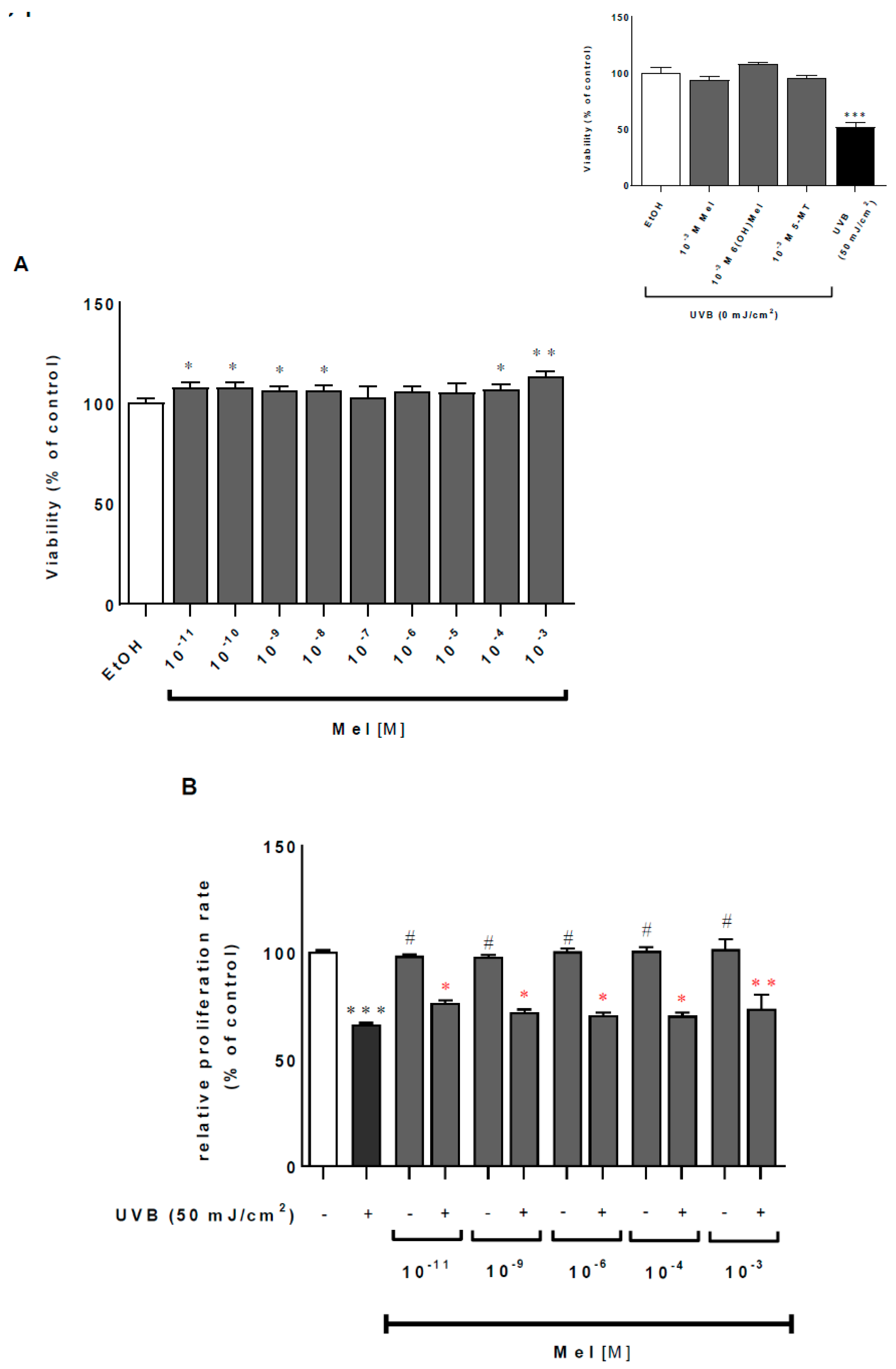
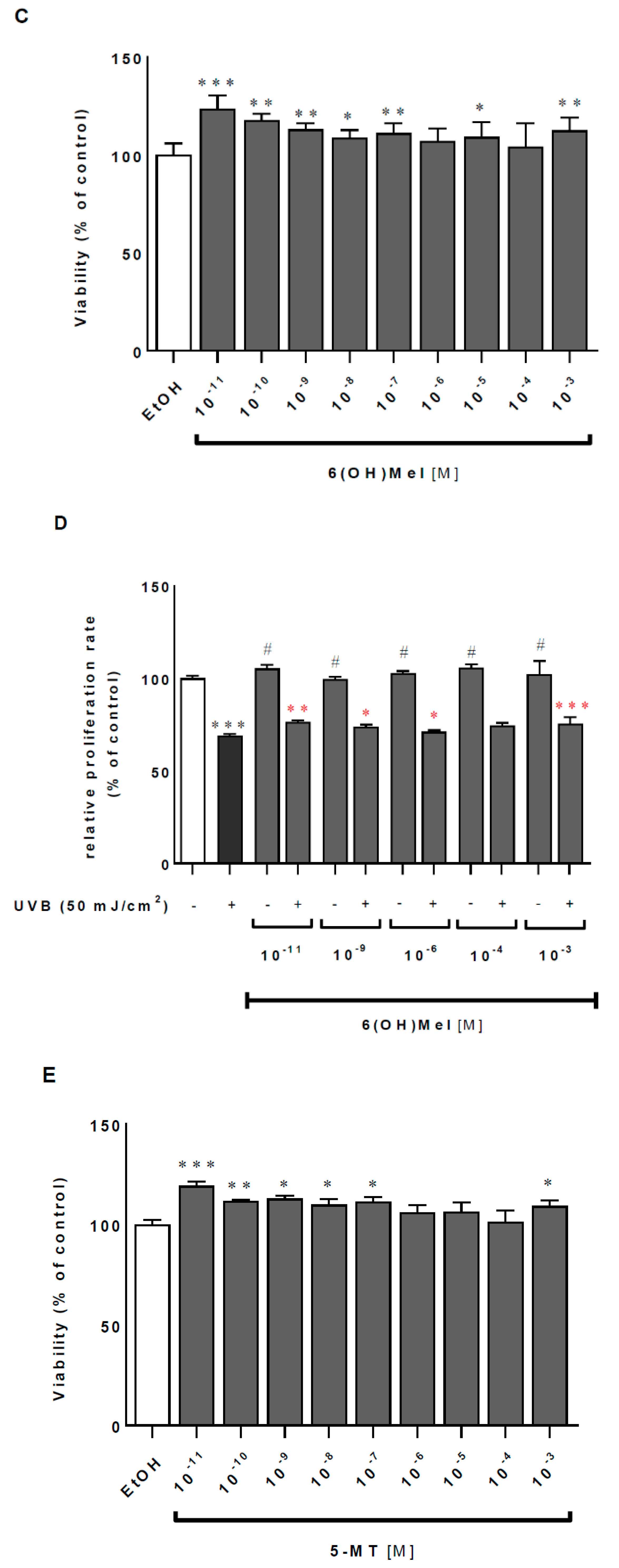
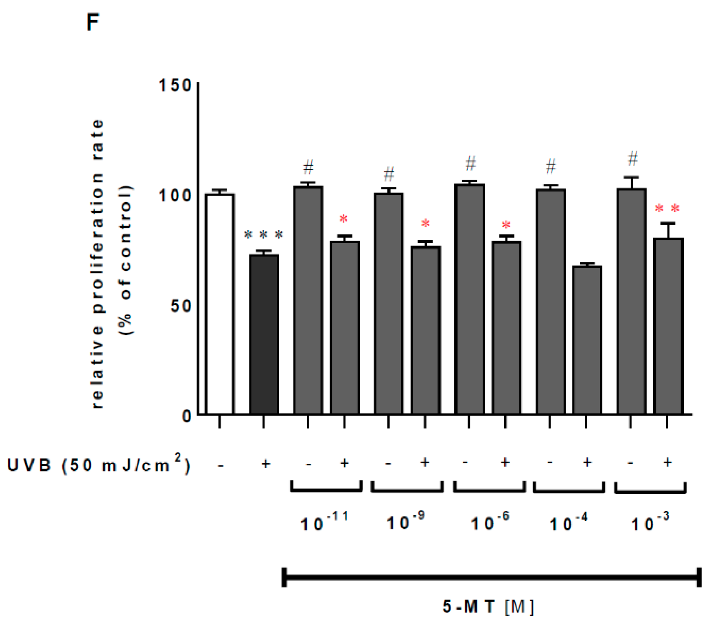
2.1. Melatonin and Its Metabolites Mitigate UVB-Induced Cell Death

UVB induces serious intracellular disturbances such as DNA damage and the production of ROS, leading to cell mortality or carcinogenesis. Firstly, we assessed the effect of UVB on the viability of MNT-1 cells, and noticed a decrease in cell viability of approximately 52% (p < 0.001) compared to the sham-irradiated controls, while melatonin as well as its metabolites themselves did not affect the survival rate of MNT-1 cells, even at the highest tested concentration (10−3 M) (Figure 1, insert). Subsequently, the dose-dependent (10−11–10−3 M) analysis was performed for melatonin (Mel) (Figure 1A), 6-hydroxymelatonin (6(OH)Mel) (Figure 1CE) under ultraviolet B (UVB) exposure. Pre-incubation with all three agents protected UVB-irradiated cells at the physiologic range of day and night plasma levels, i.e. a concentration of 10−11 M by 8% (p < 0.05; Mel), 24% (p < 0.001; 6(OH)Mel), and 19% (p < 0.001; 5-MT) or by 6% (p < 0.05; Mel), 13% (p < 0.01; 6(OH)Mel), and 13% (p < 0.05; 5-MT) for 10−9 M. Middle-range doses (10−8–10−6 M) still revealed the protective action of the tested compounds; however, significant differences were moderate or none (10−6 M) in all of the cases. Finally, the pharmacological doses (10−4 or 10−3 M) ameliorated a decrease in cell viability compared to the UVR-treated cells by 13% (p < 0.01; Mel), 12% (p < 0.01; 6(OH)Mel), and 9% (p < 0.05; 5-MT) for 10−3 M. These data were also supported by crystal violet assessment, where UVB caused a dramatic drop of MNT-1 proliferation ratio by 34% (p < 0.001) compared to the control cells, and pre-incubation with Mel (Figure 1B), 6(OH)Mel (Figure 1D), or 5-MT (Figure 1F) significantly counteracted this effect.

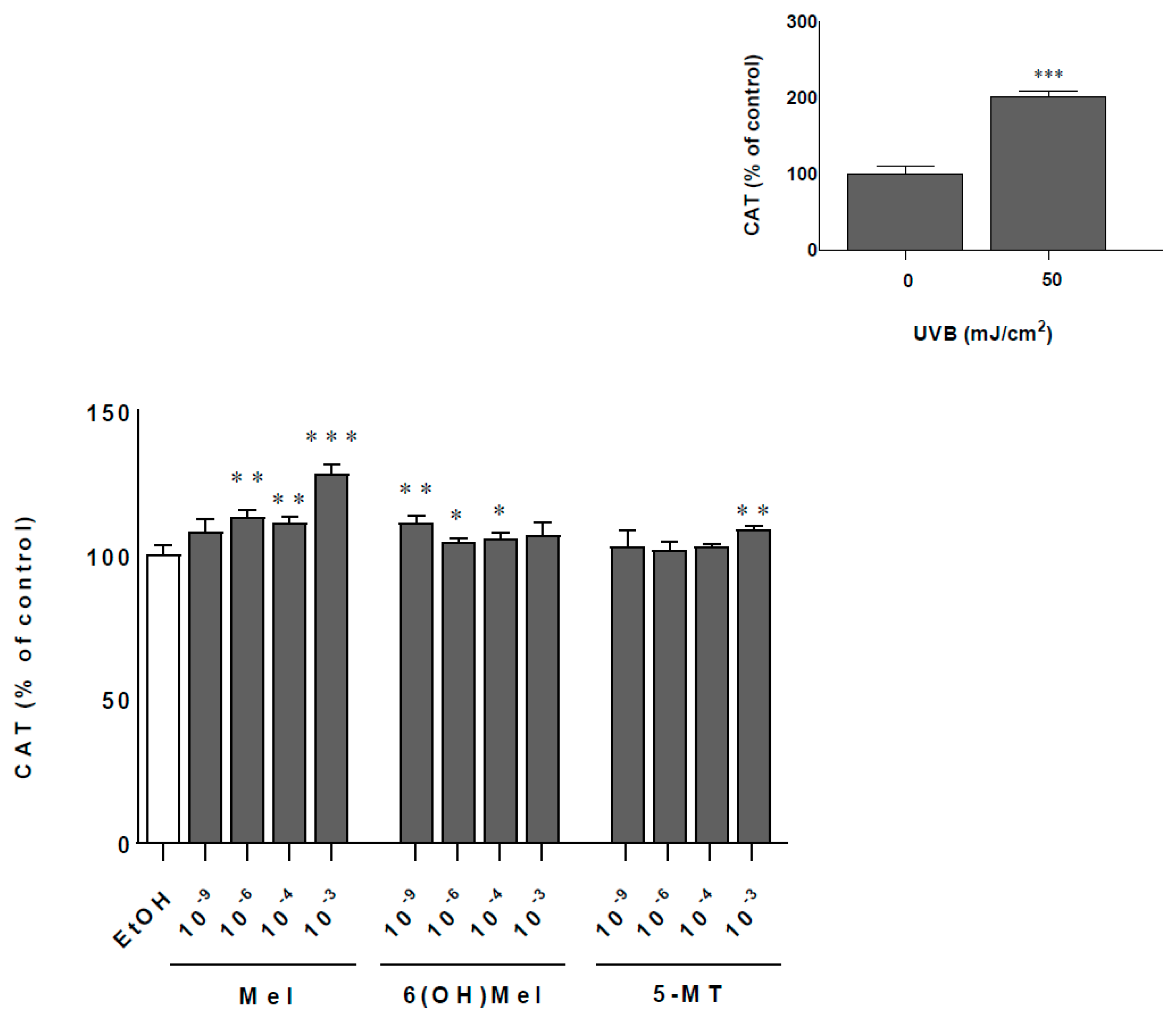
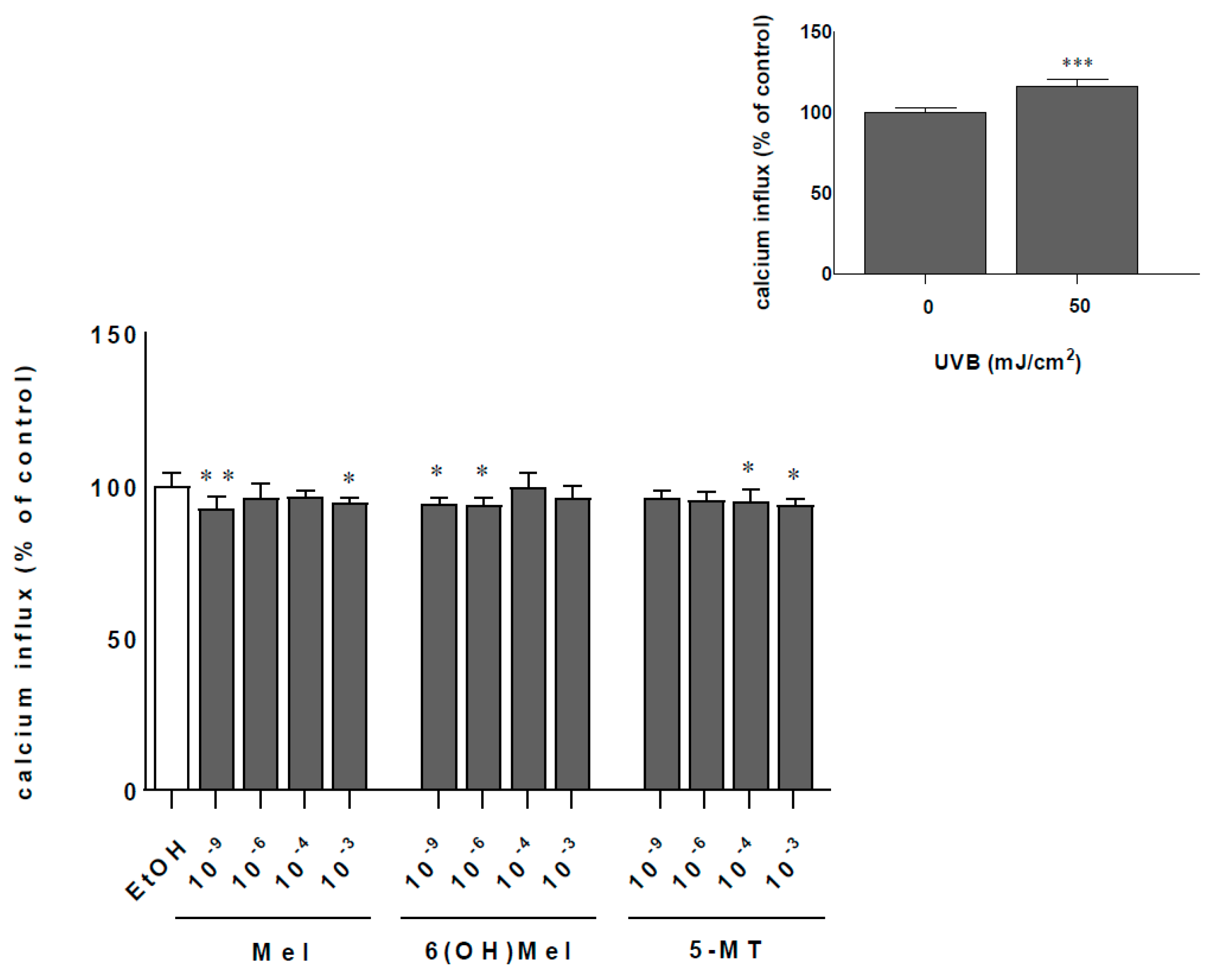
2.2. Melatonin and Its Metabolites Protect MNT-1 Cells from UVB-Induced Oxidative Stress and Disturbances in Calcium Homeostasis

Cells exposed to 50 mJ/cm2 UVB showed a twofold increase (p < 0.001) of catalase activity (CAT) activity compared to sham-irradiated samples (Figure 2; insert). Additionally, comparative analysis of melatonin actions revealed the strongest enhancing effect at a concentration of 10−3 M Mel by 28% (p < 0.001) compared to the control. At lower concentrations, this impact was less pronounced, e.g., 11% (10−4 M), 13% (10−6 M) (p < 0.01), and 9% (10−9 M; not significant). The presence of metabolites of melatonin enhanced significantly CAT activity by 11% (10−9 M; p < 0.01) or by 9% (10−3 M; p < 0.01) for 6(OH)Mel and 5-MT, respectively (Figure 2). UVB irradiation induced a massive calcium influx with 16% (p < 0.001) more calcium inside the cell compared to the non-irradiated cells (Figure 3; insert). Pre-incubation for 1 h with melatonin or its metabolites counteracted these effects modestly by 8% (10−9 M Mel; p < 0.01); 6% (10−9 M 6(OH)Mel; p < 0.05), and 4% (10−9 M 5-MT; not significant). The highest concentration (10−3 M) of the compound showed a similar pattern of regulation arresting calcium influx by 6%, 5%, and by 8%, respectively, for melatonin (p < 0.05), 6(OH)Mel, and 5-MT (p < 0.05) (Figure 3).

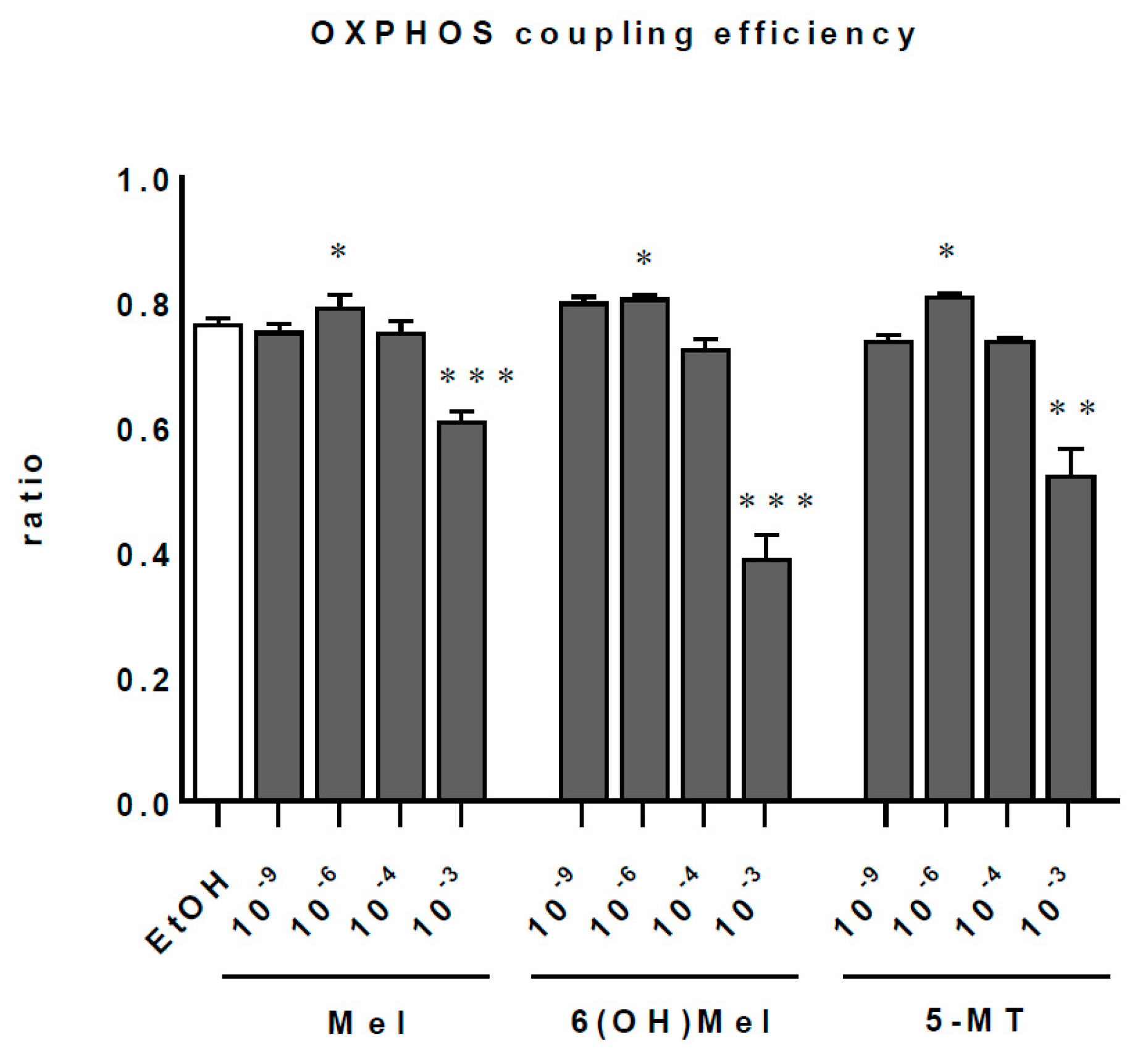
2.3. Melatonin and Its Metabolites Maintain Mitochondrial Function
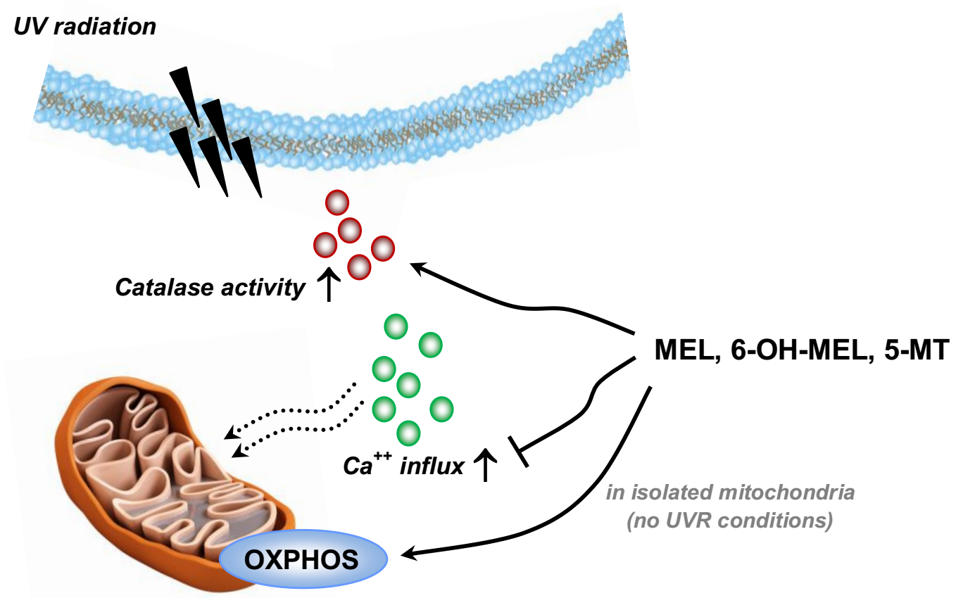
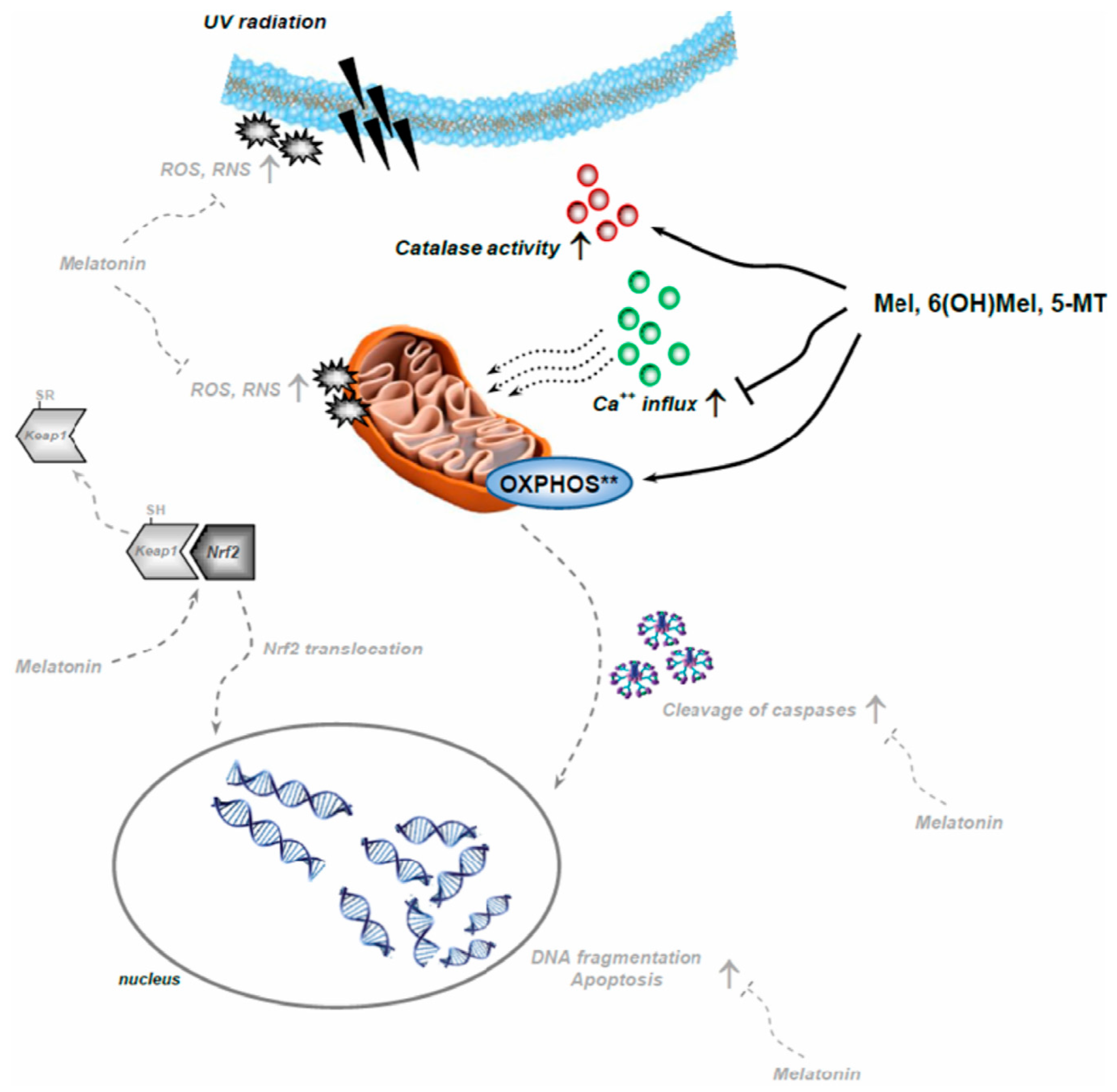
Since melatonin penetrates very easily through cellular membranes reaching numerous intracellular organelles, we assessed the influence of melatonin and its selected metabolites on mitochondrial function (Figure 4; Table 1). These analysis identified Mel, 6(OH)Mel, and 5-MT as the compounds enhancing markedly (p < 0.01) oxidative phosphorylation at the dose of 10−6 M with subtle (not significant) differences at 10−9 or 10−4 M. Surprisingly, the highest dose of 10−3 M for all tested substances decreased prominently the OXPHOS coupling efficiency (p < 0.001 or p < 0.01). Evaluation of the mitochondrial metabolic states (Table 1) showed that in “native” conditions (without stress), lower concentrations (10−9 or 10−6 M) of Mel, 6(OH)Mel, and 5-MT are more optimal than the pharmacological ones for maintenance of the mitochondrial homeostasis. MNT-1 cells express enzymes that play key roles in the synthesis of melatonin, including tryptophan-5-hydroxylase (TPH), and hydroxyindole-O-methyl transferase (HIOMT), and show the presence of MT1 and MT2 (Figure 5). This is consistent with previous reports on the capability of human melanoma cells to produce and metabolize melatonin [46] and the expression of TPH1 in human melanoma skin biopsies [47] and rodent melanomas [48,49].

3. Discussion

Cutaneous cells are under the constant influence of multiple environmental stressors [50], including UVR, which is the main source of generation of reactive oxygen/nitrogen species (ROS/RNS), leading to deleterious oxidative skin damage [40,42,51] that includes cell senescence, photoaging, carcinogenesis, or inflammation. Thus, searching for the sun damage-preventing natural compounds that can counteract UVB-induced increase in levels of ROS/RNS is strongly desired. One of such compounds is melatonin, which possesses enhancing capacities regarding skin protection against UVR [29,32,52]. It is commonly known as a free radical scavenger [53,54]; it detoxifies NO [55], H2O2, O2 [56,57], and OH● [58]. To date, studies have shown the potential beneficial effects of melatonin in the treatment of many chronic diseases, while it exhibits low or absent toxicity [59]. Additionally, it exhibits Nrf2-mediated protective effects in animal models [23] or cutaneous cells [27,33]. Apart from its significant anti-oxidative properties, melatonin also preserves mitochondrial function in different in vitro and in vivo models [23,60,61,62]. Previous reports [63,64] claim that melatonin should be considered as a critical modulator of mitochondrial integrity and physiology in skin cells [26], and this function can be also assigned to its metabolites [65]. This assumption has been substantiated in several mitochondrial-related disorders such as murine models of Parkinson’s disease [66,67,68], and the direct action of melatonin and metabolites on mitochondrial homeostasis [69,70]. This experimental evidence identifies mitochondria as a likely site of melatonin synthesis [71], placing this compound as a strategic molecule during the evolution of aerobic eukaryotes [72]. Our data confirmed that melatonin and its selected metabolites can protect cells from UVB-mediated oxidative stress by enhancing the activity of catalase, decreasing Ca++ influx, and maintaining mitochondrial function. It should be still noted that melatonin, 6(OH)Mel, or 5-MT, particularly at pharmacological doses (10−3 M), seem to affect mitochondrial function in the absence of any exogenous stressors (Figure 4). Interestingly, these concentrations were found to be the most protective under UVB treatment. Thus, we assume that in normal conditions at higher concentrations (10−3/10−4 M), the mitochondrial fission could occur, but this hypothesis remains to be clarified. On the other hand, we propose that melatonin may act as a sort of “standby molecule” or “cellular guardian” whose anti-oxidative capacities are activated in case of cell stress. Indeed, it was reported in rat heart mitochondria that melatonin inhibits stress-induced mitochondrial permeability transition pore (mPTP) opening and release of cytochrome c [73,74]. It is also known that treatment with melatonin at a pharmacological dose (10−3 M) affects mitochondrial function [75,76], which is consistent with our results performed on isolated mitochondria. Moreover, Odinokova et al. [77] showed that melatonin drastically decreases the expression of complex I subunit, which is accompanied by the suppression of ROS production and the inhibition of mPTP opening. As we noticed here, the mechanism of action of melatonin is complexed, and G protein-coupled melatonin type 1 and type 2 receptors (MT1 and MT2) as well as key enzymes (TPH1 and HIOMT) for its synthesis are expressed in MNT-1 cells. According to the earlier detections, melatonin receptors have been reported in melanocytes (normal and malignant), keratinocytes (normal), or dermal fibroblasts [30,78,79,80,81]. The expression of both MT1 and MT2 receptors in MNT-1 melanoma cells suggests that they might be involved in a regulation of calcium homeostasis in this cell line under stress conditions. However, it is well described that calmodulin is another melatonin-binding protein, which may be physiologically relevant to affinity after Ca++ binding and interaction with calmodulin-controlled enzymes, including calmodulin kinase II (CaMKII) and calcineurin (Cn), which regulate intracellular calcium homeostasis [11,82]. These observations may partially explain the role of melatonin and its metabolites in the endoplasmic reticulum stress response, as well as the regulation of apoptosis, autophagy, and mitochondrial function [83]. In addition, Martin et al. [62] observed that melatonin arrested ruthenium red (RR)-mediated oxidative stress and mitochondrial uncoupling where RR is a molecule that inhibits the mitochondrial Ca++ uniporter. Also, our previous experiments [28,84] showed that melatonin in the skin is rapidly metabolized and/or degraded through both indolic and kynuric pathways. The indolic pathway produces 6(OH)Mel as the main product and 5-MT as a minor one, and these two metabolites of melatonin are produced in skin cells [28]; similar effects are also observed in the kidney. Thus, the predominant pathway of melatonin metabolism in all of the main skin cell populations (human normal epidermal keratinocytes and melanocytes, human normal dermal fibroblasts) and malignant melanoma cells is similar to that described at the systemic level, in which 6(OH)Mel is the main degradation product [65].
As mentioned above, mitochondria are one of the main target organelles for melatonin and play the key role in disease initiation and/or progression. Although restoring mitochondrial function and redox balance/signaling is not a straightforward process, mitochondria can convert external stimuli into alterations in antioxidant defense mechanisms and energetic demands, making cells resistant to stress. This phenomenon is known as mitohormesis, and is detrimental for restoring mitochondrial function and control/regulation in non-physiological mtROS production. Since melatonin and its metabolites over the years have been described as effective anti-inflammatory, anti-oxidative and anti-apoptotic agents, also under UVR conditions (Figure 6), the development of novel mitochondria-targeted multi-functional antioxidants based on melatonin are of great interest in skin biology and physiology [11,26]. These compounds maintaining mitochondrial homeostasis by direct scavenging mtROS can increase cell resistance to stress and viability; therefore, it should be taken under consideration as possible future drugs to improve mitochondrial health in primary and/or secondary mitochondrial-related diseases.

4. Materials and Methods

4.1. Reagents

Dulbecco’s modified Eagle’s medium (DMEM) with high glucose (4500 mg/L), 1% penicillin-streptomycin solution (10,000 units of penicillin and 10 mg of streptomycin in 1 mL 0.9% NaCl), EGTA, ethanol, HEPES (1 M), isopropanol, KH2PO4, mannitol, MgCl2·6H2O, MTT, non-essential amino acids (NEAA) (100×), l-glutamic acid, l-malic acid, potassium lactobionate, sodium pyruvate, sodium pyruvate solution (100 mM), succinic acid, and taurine were purchased from Sigma (St. Louis, MO, USA). Fetal bovine serum, 0.05% trypsin/0.53 mM EDTA solution, 1× PBS (pH 7.4), l-glutamine (200 mM) were supplied by Thermo Fisher Scientific (Waltham, MA, USA).

4.2. Cell Culture

MNT-1 human melanotic cells were acquired as a gift from Dr. Cédric Delevoye (Institute Curie, Paris, France). Cells were maintained in T-75 or T-175 flasks in DMEM medium supplemented with 20% (v/v) heat-inactivated fetal bovine serum, 2 mM of l-glutamine, 10 mM of HEPES, 1 mM of sodium pyruvate, 1% (v/v) NEAA, and 1% (v/v) streptomycin-penicillin solution at 37 °C in a humidified atmosphere of 5% CO2 in air. Cells in the logarithmic growth phase were used in all of the experiments, while 80–90% monolayers of confluent MNT-1 cells were harvested with a mixture of 0.05% trypsin–EDTA solution.

4.3. Pre-incubation with Melatonin, Its Metabolites, and UV Irradiation

Prior to the start of the experimental treatments, cells were cultured in normal medium for 24 h in order to attach to the bottom of the culture dish. Thereafter, culture medium was replaced with medium containing Mel, 6(OH)Mel, 5-MT, or solvent (control) (Sigma). Compounds were dissolved in absolute ethanol and further diluted with phosphate-buffered saline (1× PBS, pH 7.4) to yield 10−2 M stock solution, then diluted 1:10 with culture medium to yield a final wide concentration range of powers of 10 from 10−3 to 10−11 M for cell culture pre-incubation. Melatonin-free, 6(OH)Mel-free, or 5-MT-free medium served as controls. After 1 h pre-incubation with test compounds, cells were washed twice with 1× PBS to remove remnants of medium, and fresh 1× PBS was added prior to UVR exposure. Irradiation was performed using an irradiation bank consisting of six UV bulbs (TL12, Philips, The Netherlands) that emit most of their energy within the UVB range (280–320 nm) with an emission peak of 313 nm. UVB doses of 0 and 50 mJ/cm2 were used, and UVR-treated cells were examined 48 h post-UVR. Sham-irradiated samples (0 mJ/cm2) were prepared using a culture dish covered with aluminum foil and were considered as the control value. After UVR exposure, cells were cultured and then subjected to the particular measurements.

4.4. MTT Viability Assay

Tested cells were seeded on 96-well plates at the density of 0.15 × 10⁵ cells/well in the culture medium, and thereafter grown to subconfluence (as judged from light microscopy). Following the pre-incubation with tested compounds, cells were UV-irradiated in presence of 100 μL of pre-warmed 1× PBS, and then cultured until the desired time end-point. Cell viability was evaluated by MTT assay based on the protocol described earlier by Carmichael et al. [85]. MTT (5 mg/mL) was dissolved in 1× PBS, sterilized by filtration through a 0.22-μM Millipore® filter and stored at 4 °C. After treatment as described above, cells were washed twice with 1×PBS; then, 100 μL of MTT solution in culture medium (the final dilution, 1:10) was added to each well. The cells were subsequently incubated for 3 h at 37 °C to allow MTT metabolism. The formazan produced was dissolved in 100 μL of isopropanol/0.04 N HCl, and absorbance was measured at 595 nm using a BioTek ELx808™ microplate reader (BioTek Instruments, Inc., Winooski, VT, USA).

4.5. Crystal Violet Assessment

The proliferation of MNT-1 cells was assessed using ready-to-use crystal violet (CV) solution (Cell Biolabs, Inc., San Diego, CA, USA). Cells were seeded on six-well plates (0.4–0.5 × 10⁶ cells/cm2) and experiments were performed when cultures reached 70–80% confluence. At the desired times, cells were washed twice with 1× PBS, stained with 0.1% CV solution in 10% ethanol for 1 min, and remnants were washed again with 1× PBS. Plates were photographed and the quantification of cells was performed using ImageJ 1.51f software (National Institute of Health, Bethesda, MD, USA).

4.6. Catalase Activity Assay

Evaluation of catalase (CAT) activity was conducted by colorimetric assays (BioVision Research Products, Inc., Mountain View, CA, USA). Briefly, cells were seeded on six-well plates at the density and cultured until reached confluency as described in point 4.5. At the desired times, cells were lysed with catalase assay buffer and then centrifuged for 15 min at 10,000× g at 4 °C. Resultant supernatant was proceeded for catalase reaction with one mM of H2O2 for 30 min at 25 °C, and arrested subsequently with a stop solution. Catalase activity was determined by incubation at room temperature (RT) for 15 min with a develop mix containing catalase assay buffer, OxiRed™ Probe, and HRP solution. The absorbance of tested samples was measured at 595 nm using a BioTek ELx808™ microplate reader (BioTek Instruments, Inc., Winooski, VT, USA).

4.7. Calcium Assay

Intracellular calcium homeostasis was assessed by colorimetric assay supplied by BioVision Research Products, Inc. After reaching desired confluency, cells were lysed and centrifuged for five min at 16,000× g at 4 °C. The resultant supernatant was mixed with the chromogenic reagent, calcium assay buffer, and then incubated at RT for 15 min protecting from light. The absorbance of tested samples was measured at 595 nm using a BioTek ELx808™ microplate reader (BioTek Instruments, Inc.)

4.8. Animals and Isolation of Liver Mitochondria

All of the experimental procedures were performed in accordance with the principles of the European animal research laws (European Communities Council Directive 2010/63/EU). Wild-type female BALB/cJ mice (n = 8; weight: 20–25 g) were kept in an environmentally controlled room (23 ± 1 °C with a 12-h light-dark cycle) receiving standard mice chow and water ad libitum. The mice were euthanized by cervical dislocation. The liver mitochondria were isolated, as previously described by Broekemeier et al. [86] with slight modification [87]. The liver was rapidly removed, weighed, and placed in ice-cold mitochondrial buffer A (230 mM of mannitol, 70 mM of sucrose, three mM of HEPES, 0.1 mM of EGTA; pH 7.4), and subsequently rinsed three times with buffer A. The liver was minced with scissors and homogenized using a Teflon pestle homogenizer in buffer A (20 mL/1 g of liver). The homogenate was centrifuged at 800× g for 10 min; then, the supernatant was decanted and centrifuged at 4000× g for 10 min. The pellet was resuspended in 30 mL of suspension buffer B (230 mM of mannitol, 70 mM of sucrose, 3 mM of HEPES), and centrifuged again at 7000× g for 10 min. This step was repeated twice. The final mitochondrial pellet was resuspended in 0.5 mL of buffer B. All of the steps were performed at 4 °C. Total protein concentration (20 to 30 mg of mitochondrial protein per mL) was determined with the Bradford reagent (Bio-Rad Laboratories, Inc., Hercules, CA, USA) [88].

4.9. High-Resolution Respirometry

Respiration was measured at 37 °C under constant stirring (750 rpm), which ensured a homogenous oxygen distribution in the medium in a high-resolution respirometer using an Oxygraph-2k (O2k, Oroboros Instruments, Innsbruck, Austria), a modular system for high-resolution respirometry (HRR) according to Volani et al. [89]. Oxygen concentration (μM) and oxygen flux per mass (pmol O2● s−1·mg−1) were recorded in real-time, while obtained data were evaluated using DatLab software (Oroboros Instruments, Innsbruck, Austria). Briefly, mitochondrial respiration was measured in MiR05 (mitochondrial respiration medium containing 0.5 mM of EGTA, 3 mM of MgCl2·6H2O, 60 mM of potassium lactobionate, 20 mM of taurine, 10 mM of KH2PO4, 20 mM of HEPES, 110 mM of sucrose, and 1 g/L of BSA fatty acid-free (pH 7.1) (MiR05; Oroboros Instruments, Innsbruck, Austria). Freshly isolated liver mitochondria (0.1 mg of mitochondrial protein per each 2-mL chamber) used for respiration measurement were first incubated for 5 min in the medium without (control) or in presence of Mel, 6(OH)Mel, or 5-MT.
For the assessment of mitochondrial respiration, the following protocol was used:
(1)
Non-phosphorylating LEAK respiration was assessed by injecting 10 mM of sodium pyruvate, 10 mM of l-glutamic acid (neutralized with KOH) and 2 mM of l-malic acid (neutralized with KOH) as NADH (N)-linked substrates: state NL;
(2)
OXPHOS capacity was induced by adding 1.25 mM of ADP at saturating concentration: state NP;
(3)
10 μM of cytochrome c was added to test the integrity of the outer mitochondrial membrane: state NC;
(4)
NADH and succinate (NS)-linked OXPHOS capacity was measured by adding 10 mM of succinic acid (neutralized with KOH): state NSP.

4.10. Mitochondrial Quality and Control

The OXPHOS coupling efficiency was calculated as measures of mitochondrial quality and control. OXPHOS coupling efficiency, calculated with the formula (1)—(state NL)/(state NP), reflects the coupling of respiration supported by electron transferring flavoprotein (ETF) with pyruvate, glutamate, and malate as substrates before (state NL) and after the addition of ADP (state NP). The OXPHOS coupling efficiency (OCE) can be compared with the respiratory control ratio (RCR). The formula of OCE could be rewritten as:
O C E = ( 1 1 R C R )
The OCE is noted between 0 and 1, while the RCR could be from 0 to infinite. A lower value of OCE means lesser coupling of the oxidation and phosphorylation after the addition of ADP. As the OCE decreases, it is therefore less coupled [90].

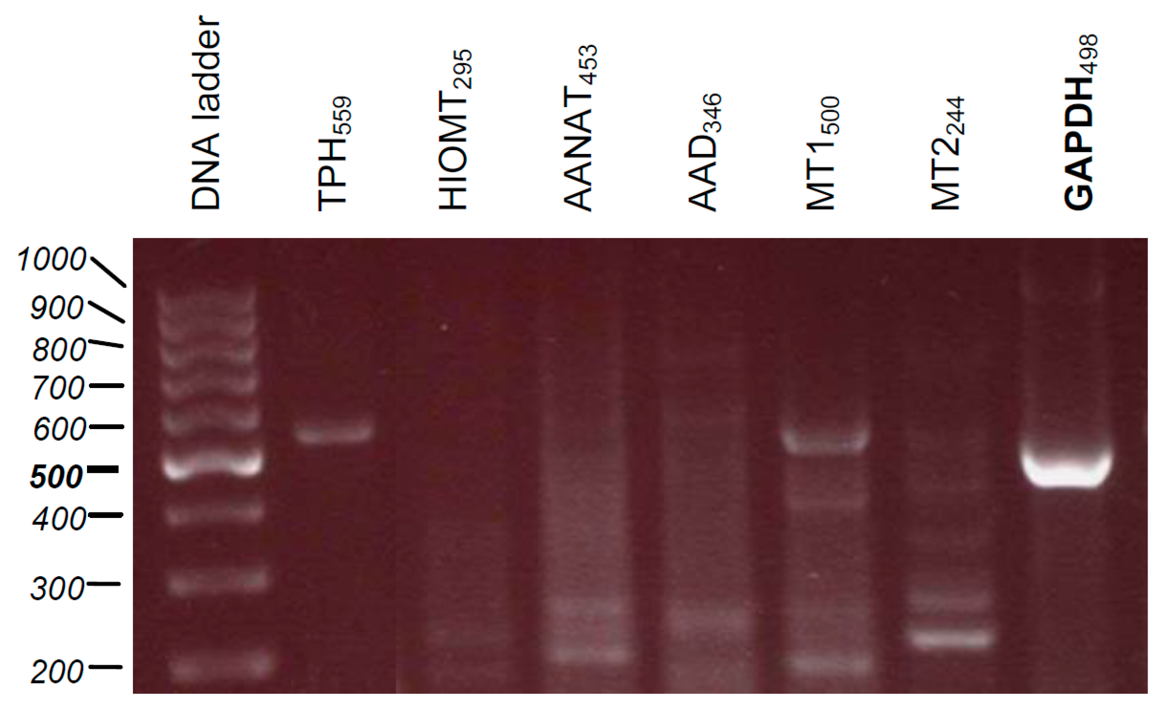
4.11. RNA Isolation, cDNA Synthesis, and PCR

RNA from MNT-1 cell pellet (5 × 10⁶ cells) was extracted accordingly to the manufacturer’s instructions using the innuPREP RNA Mini Kit (Analytik Jena, Berlin, Germany). The amount of RNA was determined using BioPhotometer (Eppendorf, Hamburg, Germany). cDNA synthesis was conducted using RevertAid™ First Strand cDNA Synthesis Kit (Thermo Fisher Scientific, Waltham, MA, USA) in the presence of oligo(dT) primers as follows: 65 °C for 5 min, 42 °C for 60 min, 70 °C for 5 min in Thermomixer (Eppendorf), and the resultant cDNA was stored at −20 °C prior to PCR reaction. Reactions were carried out by GoTaq® PCR Master Mix (Promega GmbH, Mannheim, Germany) in the presence of sequences of primers (Table 2). Amplification was performed using 10 min of initial denaturation at 95 °C followed by three-step 39-cycling of 60 s at 95 °C (denaturation), 60 s at 60 °C (annealing), and 60 s at 72 °C (extension). PCR products were separated on 1.8% agarose gel containing RedSafe™ Nucleic Acid Staining solution (iNtRON Biotechnology, Sangdaewon-Dong, Korea), and afterwards visualized using the Fusion-FX7 UV transilluminator (Vilber GmbH, Eberhardzell, Germany). Samples were standardized by amplification of the housekeeping gene glyceraldehyde phosphate dehydrogenase (GAPDH) [12]. DNA-standard O’GeneRuler™ 100 bp DNA Ladder Mix from Fermentas International (Burlington, ON, Canada) was used.

4.12. Statistical Analysis

Data were expressed as pooled means (at least n = 3) ±standard error of the mean (SEM). The data were analyzed using t-Test (for two groups) or with one-way ANOVA (for multiple groups) with GraphPad Prism 7.05 software (La Jolla, CA, USA). Obtained data for MTT viability assay, crystal violet assessment, CAT activity, or Ca++ homeostasis were normalized and expressed as a percentage of the control value i.e., sham-irradiated sample (0 mJ/cm2, inserts) or UVB-exposed samples. The results of mitochondrial metabolism were presented in arbitrary units. A p-value of less than 0.05 was considered statistically significant.

Author Contributions

K.K. conceived, designed and with support of B.B. performed the experiments, analyzed obtained data, and then K.K. wrote the manuscript. A.S. supported with PCR analysis and described them. D.J.F. together with W.Z. carried out mitochondrial experiments and analyzed data. E.P. and A.T.S. analysed the data and they together with T.A.L., M.B., R.J.R., K.K. revised the final version of the manuscript.

Funding

The present study was mainly supported by the grant of the German Research Foundation (Deutsche Forschungsgemeinschaft (DFG)); grant number: KL2900/2-1 (K.K.) with partial funding from the Jagiellonian University Research Grant, Kraków, Poland (K/ZDS/008070/2018) (E.P.), the Ministry of Science and Higher Education of Poland, Gdańsk, Poland (DSRiK/2018) (W.Z.), in part by grants 1R01AR056666-01A2, and to some degree 1R01AR073004-01A1 and 1RO1AR071189-01A1 from NIH (A.T.S.).

Acknowledgments

The authors would like to express their gratitude to Dr. Cédric Delevoye (Structure and Membrane Compartments, Institute Curie, Paris Sciences & Lettres Research University, Centre National de la Recherche Scientifique, UMR144, Paris, France) for kind gift of MNT-1 cells.

Conflicts of Interest

The authors declare no conflict of interest.

Abbreviations

5-MT5-Methoxytryptamine
6(OH)Mel6-Hydroxymelatonin
CATCatalase
DMEMDulbecco’s modified Eagle’s medium
HIOMTHydroxyindole-O-methyl transferase
HRRHigh-resolution respirometry
MelMelatonin
mPTPMitochondrial permeability transition pore
OCEOXPHOS coupling efficiency
RCRRespiratory control ratio
RNSReactive nitrogen species
ROSReactive oxygen species
TEMTransmission electron microscopy
TPHTryptophan-5-hydroxylase
UVRUltraviolet radiation

References

  1. Hardeland, R.; Fuhrberg, B. Ubiquitous melatonin—Presence and effects in unicells, plants and animals. Trends Comp. Biochem. Physiol. 1996, 2, 25–45. [Google Scholar]
  2. Kolar, J.; Machackova, I. Melatonin in higher plants: Occurrence and possible functions. J. Pineal Res. 2005, 39, 333–341. [Google Scholar] [CrossRef] [PubMed]
  3. Paredes, S.D.; Korkmaz, A.; Manchester, L.C.; Tan, D.X.; Reiter, R.J. Phytomelatonin: A review. J. Exp. Bot. 2009, 60, 57–69. [Google Scholar] [CrossRef] [PubMed]
  4. Reiter, R.J. Pineal melatonin: Cell biology of its synthesis and of its physiological interactions. Endocr. Rev. 1991, 12, 151–180. [Google Scholar] [CrossRef] [PubMed]
  5. Tan, D.X.; Hardeland, R.; Manchester, L.C.; Paredes, S.D.; Korkmaz, A.; Sainz, R.M.; Mayo, J.C.; Fuentes-Broto, L.; Reiter, R.J. The changing biological roles of melatonin during evolution: From an antioxidant to signals of darkness, sexual selection and fitness. Biol. Rev. Camb. Philos. Soc. 2010, 85, 607–623. [Google Scholar] [CrossRef] [PubMed]
  6. Tan, D.X.; Manchester, L.C.; Reiter, R.J.; Qi, W.B.; Zhang, M.; Weintraub, S.T.; Cabrera, J.; Sainz, R.M.; Mayo, J.C. Identification of highly elevated levels of melatonin in bone marrow: Its origin and significance. Biochim. Biophys. Acta 1999, 1472, 206–214. [Google Scholar] [CrossRef]
  7. Carrillo-Vico, A.; Calvo, J.R.; Abreu, P.; Lardone, P.J.; García-Mauriño, S.; Reiter, R.J.; Guerrero, J.M. Evidence of melatonin synthesis by human lymphocytes and its physiological significance: Possible role as intracrine, autocrine, and/or paracrine substance. FASEB J. 2004, 18, 537–539. [Google Scholar] [CrossRef] [PubMed]
  8. Iuvone, P.M.; Tosini, G.; Pozdeyev, N.; Haque, R.; Klein, D.C.; Chaurasia, S.S. Circadian clocks, clock networks, arylalkylamine N-acetyltransferase, and melatonin in the retina. Prog. Retin. Eye Res. 2005, 24, 433–456. [Google Scholar] [CrossRef] [PubMed]
  9. Liu, Y.J.; Zhuang, J.; Zhu, H.Y.; Shen, Y.X.; Tan, Z.L.; Zhou, J.N. Cultured rat cortical astrocytes synthesize melatonin: Absence of a diurnal rhythm. J. Pineal Res. 2007, 43, 232–238. [Google Scholar] [CrossRef] [PubMed]
  10. Naranjo, M.C.; Guerrero, J.M.; Rubio, A.; Lardone, P.J.; Carrillo-Vico, A.; Carrascosa-Salmoral, M.P.; Jiménez-Jorge, S.; Arellano, M.V.; Leal-Noval, S.R.; Leal, M.; et al. Melatonin biosynthesis in the thymus of humans and rats. Cell. Mol. Life Sci. 2007, 64, 781–790. [Google Scholar] [CrossRef] [PubMed]
  11. Slominski, A.T.; Hardeland, R.; Zmijewski, M.A.; Slominski, R.M.; Reiter, R.J.; Paus, R. Melatonin: A cutaneous perspective on its production, metabolism, and functions. J. Investig. Dermatol. 2018, 138, 490–499. [Google Scholar] [CrossRef] [PubMed]
  12. Slominski, A.; Pisarchik, A.; Semak, I.; Sweatman, T.; Wortsman, J.; Szczesniewski, A.; Slugocki, G.; McNulty, J.; Kauser, S.; Tobin, D.J.; et al. Serotoninergic and melatoninergic systems are fully expressed in human skin. FASEB J. 2002, 16, 896–898. [Google Scholar] [CrossRef] [PubMed]
  13. Slominski, A.; Tobin, D.J.; Zmijewski, M.A.; Wortsman, J.; Paus, R. Melatonin in the skin: Synthesis, metabolism and functions. Trends Endocrinol. Metab. 2008, 19, 17–24. [Google Scholar] [CrossRef] [PubMed]
  14. Reiter, R.J.; Tamura, H.; Tan, D.X.; Xu, X.Y. Melatonin and the circadian system: Contributions to successful female reproduction. Fertil. Steril. 2014, 102, 321–328. [Google Scholar] [CrossRef] [PubMed]
  15. Shi, J.M.; Tian, X.Z.; Zhou, G.B.; Wang, L.; Gao, C.; Zhu, S.E.; Zeng, S.M.; Tian, J.H.; Liu, G.S. Melatonin exists in porcine follicular fluid and improves in vitro maturation and parthenogenetic development of porcine oocytes. J. Pineal Res. 2009, 47, 318–323. [Google Scholar] [CrossRef] [PubMed]
  16. El-Raey, M.; Geshi, M.; Somfai, T.; Kaneda, M.; Hirako, M.; Abdel-Ghaffar, A.E.; Sosa, G.A.; El-Roos, M.E.; Nagai, T. Evidence of melatonin synthesis in the cumulus oocyte complexes and its role in enhancing oocyte maturation in vitro in cattle. Mol. Reprod. Dev. 2011, 78, 250–262. [Google Scholar] [CrossRef] [PubMed]
  17. Sakaguchi, K.; Itoh, M.T.; Takahashi, N.; Tarumi, W.; Ishizuka, B. The rat oocyte synthesises melatonin. Reprod. Fertil. Dev. 2013, 25, 674–682. [Google Scholar] [CrossRef] [PubMed]
  18. Allegra, M.; Reiter, R.J.; Tan, D.X.; Gentile, C.; Tesoriere, L.; Livrea, M.A. The chemistry of melatonin’s interaction with reactive species. J. Pineal Res. 2003, 34, 1–10. [Google Scholar] [CrossRef] [PubMed]
  19. Rodriguez, C.; Mayo, J.C.; Sainz, R.M.; Antolin, I.; Herrera, F.; Martín, V.; Reiter, R.J. Regulation of antioxidant enzymes: A significant role for melatonin. J. Pineal Res. 2004, 36, 1–9. [Google Scholar] [CrossRef] [PubMed]
  20. Tan, D.X.; Reiter, R.J.; Manchester, L.C.; Yan, M.T.; El-Sawi, M.; Sainz, R.M.; Mayo, J.C.; Kohen, R.; Allegra, M.; Hardeland, R. Chemical and physical properties and potential mechanisms: Melatonin as a broad spectrum antioxidant and free radical scavenger. Curr. Top. Med. Chem. 2002, 2, 181–197. [Google Scholar] [CrossRef] [PubMed]
  21. Menendez-Pelaez, A.; Reiter, R.J. Distribution of melatonin in mammalian tissues: The relative importance of nuclear versus cytosolic localization. J. Pineal Res. 1993, 15, 59–69. [Google Scholar] [CrossRef] [PubMed] [Green Version]
  22. Costa, E.J.; Shida, C.S.; Biaggi, M.H.; Ito, A.S.; Lamy-Freund, M.T. How melatonin interacts with lipid bilayers: A study by fluorescence and ESR spectroscopies. FEBS Lett. 1997, 416, 103–106. [Google Scholar] [CrossRef]
  23. Martín, M.; Macías, M.; Escames, G.; León, J.; Acuña-Castroviejo, D. Melatonin but not vitamins C and E maintains glutathione homeostasis in t-butyl hydroperoxide-induced mitochondrial oxidative stress. FASEB J. 2000, 14, 1677–1679. [Google Scholar] [CrossRef] [PubMed] [Green Version]
  24. Andrabi, S.A.; Sayeed, I.; Siemen, D.; Wolf, G.; Horn, T.F. Direct inhibition of the mitochondrial permeability transition pore: A possible mechanism responsible for anti-apoptotic effects of melatonin. FASEB J. 2004, 18, 869–871. [Google Scholar] [CrossRef] [PubMed]
  25. Janjetovic, Z.; Nahmias, Z.P.; Hanna, S.; Jarrett, S.G.; Kim, T.K.; Reiter, R.J.; Slominski, A.T. Melatonin and its metabolites ameliorate ultraviolet B-induced damage in human epidermal keratinocytes. J. Pineal Res. 2014, 57, 90–102. [Google Scholar] [CrossRef] [PubMed] [Green Version]
  26. Slominski, A.T.; Zmijewski, M.A.; Semak, I.; Kim, T.K.; Janjetovic, Z.; Slominski, R.M.; Zmijewski, J.W. Melatonin, mitochondria, and the skin. Cell. Mol. Life Sci. 2017, 74, 3913–3925. [Google Scholar] [CrossRef] [PubMed]
  27. Kleszczyński, K.; Zillikens, D.; Fischer, T.W. Melatonin enhances mitochondrial ATP synthesis, reduces reactive oxygen species formation, and mediates translocation of the nuclear erythroid 2-related factor 2 resulting in activation of phase-2 antioxidant enzymes (γ-GCS, HO-1, NQO1) in ultraviolet radiation-treated normal human epidermal keratinocytes (NHEK). J. Pineal Res. 2016, 61, 187–197. [Google Scholar] [PubMed]
  28. Kim, T.K.; Kleszczyński, K.; Janjetovic, Z.; Sweatman, T.; Lin, Z.; Li, W.; Reiter, R.J.; Fischer, T.W.; Slominski, A.T. Metabolism of melatonin and biological activity of intermediates of melatoninergic pathway in human skin cells. FASEB J. 2013, 27, 2742–2755. [Google Scholar] [CrossRef] [PubMed]
  29. Slominski, A.T.; Kleszczyński, K.; Semak, I.; Janjetovic, Z.; Zmijewski, M.A.; Kim, T.K.; Slominski, R.M.; Reiter, R.J.; Fischer, T.W. Local melatoninergic system as the protector of skin integrity. Int. J. Mol. Sci. 2014, 15, 17705–17732. [Google Scholar] [CrossRef] [PubMed]
  30. Slominski, R.M.; Reiter, R.J.; Schlabritz-Loutsevitch, N.; Ostrom, R.S.; Slominski, A.T. Melatonin membrane receptors in peripheral tissues: Distribution and functions. Mol. Cell. Endocrinol. 2012, 351, 152–166. [Google Scholar] [CrossRef] [PubMed] [Green Version]
  31. Reiter, R.J.; Tan, D.X.; Cabrera, J.; D’Arpa, D. Melatonin and tryptophan derivatives as free radical scavengers and antioxidants. Adv. Exp. Med. Biol. 1999, 467, 379–387. [Google Scholar] [PubMed]
  32. Fischer, T.W.; Kleszczyński, K.; Hardkop, L.H.; Kruse, N.; Zillikens, D. Melatonin enhances antioxidative enzyme gene expression (CAT, GPx, SOD), prevents their UVR-induced depletion, and protects against the formation of DNA damage (8-hydroxy-2′-deoxyguanosine) in ex vivo human skin. J. Pineal Res. 2013, 54, 303–312. [Google Scholar] [CrossRef] [PubMed]
  33. Janjetovic, Z.; Jarrett, S.G.; Lee, E.F.; Duprey, C.; Reiter, R.J.; Slominski, A.T. Melatonin and its metabolites protect human melanocytes against UVB-induced damage: Involvement of NRF2-mediated pathways. Sci. Rep. 2017, 7, 1274. [Google Scholar] [CrossRef] [PubMed] [Green Version]
  34. Kokot, A.; Metze, D.; Mouchet, N.; Galibert, M.D.; Schiller, M.; Luger, T.A.; Böhm, M. α-Melanocyte-stimulating hormone counteracts the suppressive effect of UVB on Nrf2 and Nrf-dependent gene expression in human skin. Endocrinology 2009, 150, 3197–3206. [Google Scholar] [CrossRef] [PubMed]
  35. Wondrak, G.T.; Roberts, M.J.; Cervantes-Laurean, D.; Jacobson, M.K.; Jacobson, E.L. Proteins of the extracellular matrix are sensitizers of photo-oxidative stress in human skin cells. J. Investig. Dermatol. 2003, 121, 578–586. [Google Scholar] [CrossRef] [PubMed]
  36. Ahmed, N.U.; Ueda, M.; Nikaido, O.; Osawa, T.; Ichihashi, M. High levels of 8-hydroxy-2′-deoxyguanosine appear in normal human epidermis after a single dose of ultraviolet radiation. Br. J. Dermatol. 1999, 140, 226–231. [Google Scholar] [CrossRef] [PubMed]
  37. Gilchrest, B.A.; Bohr, V.A. Aging processes, DNA damage, and repair. FASEB J. 1997, 11, 322–330. [Google Scholar] [CrossRef] [PubMed]
  38. Mouret, S.; Baudouin, C.; Charveron, M.; Favier, A.; Cadet, J.; Douki, T. Cyclobutane pyrimidine dimers are predominant DNA lesions in whole human skin exposed to UVA radiation. Proc. Natl. Acad. Sci. USA 2006, 103, 13765–13770. [Google Scholar] [CrossRef] [PubMed] [Green Version]
  39. Pfeifer, G.P.; Besaratinia, A. UV wavelength-dependent DNA damage and human non-melanoma and melanoma skin cancer. Photochem. Photobiol. Sci. 2012, 11, 90–97. [Google Scholar] [CrossRef] [PubMed] [Green Version]
  40. Heck, D.E.; Vetrano, A.M.; Mariano, T.M.; Laskin, J.D. UVB light stimulates production of reactive oxygen species: Unexpected role for catalase. J. Biol. Chem. 2003, 278, 22432–22436. [Google Scholar] [CrossRef] [PubMed]
  41. Swalwell, H.; Latimer, J.; Haywood, R.M.; Birch-Machin, M.A. Investigating the role of melanin in UVA/UVB- and hydrogen peroxide-induced cellular and mitochondrial ROS production and mitochondrial DNA damage in human melanoma cells. Free Radic. Biol. Med. 2012, 52, 626–634. [Google Scholar] [CrossRef] [PubMed]
  42. Slominski, A.T.; Zmijewski, M.A.; Plonka, P.M.; Szaflarski, J.P.; Paus, R. How UV light touches the brain and endocrine system through skin, and why. Endocrinology 2018, 159, 1992–2007. [Google Scholar] [CrossRef] [PubMed]
  43. Lee, S.; Jin, J.X.; Taweechaipaisankul, A.; Kim, G.A.; Lee, B.C. Stimulatory effects of melatonin on porcine in vitro maturation are mediated by MT2 receptor. Int. J. Mol. Sci. 2018, 19, 1581. [Google Scholar] [CrossRef] [PubMed]
  44. Skobowiat, C.; Brożyna, A.A.; Janjetovic, Z.; Jeayeng, S.; Oak, A.S.W.; Kim, T.K.; Panich, U.; Reiter, R.J.; Slominski, A.T. Melatonin and its derivatives counteract the ultraviolet B radiation-induced damage in human and porcine skin ex vivo. J. Pineal Res. 2018, 65, e12501. [Google Scholar] [CrossRef] [PubMed]
  45. Gurer-Orhan, H.; Suzen, S. Melatonin, its metabolites and its synthetic analogs as multi-faceted compounds: Antioxidant, prooxidant and inhibitor of bioactivation reactions. Curr. Med. Chem. 2015, 22, 490–499. [Google Scholar] [CrossRef] [PubMed]
  46. Slominski, A.; Semak, I.; Pisarchik, A.; Sweatman, T.; Szczesniewski, A.; Wortsman, J. Conversion of l-tryptophan to serotonin and melatonin in human melanoma cells. FEBS Lett. 2002, 511, 102–106. [Google Scholar] [CrossRef]
  47. Slominski, A.; Pisarchik, A.; Johansson, O.; Jing, C.; Semak, I.; Slugocki, G.; Wortsman, J. Tryptophan hydroxylase expression in human skin cells. Biochim. Biophys. Acta. 2003, 1639, 80–86. [Google Scholar] [CrossRef]
  48. Slominski, A.; Pisarchik, A.; Semak, I.; Sweatman, T.; Szczesniewski, A.; Wortsman, J. Serotoninergic system in hamster skin. J. Investig. Dermatol. 2002, 119, 934–942. [Google Scholar] [CrossRef] [PubMed]
  49. Slominski, A.; Pisarchik, A.; Semak, I.; Sweatman, T.; Wortsman, J. Characterization of the serotoninergic system in the C57BL/6 mouse skin. Eur. J. Biochem. 2003, 270, 3335–3344. [Google Scholar] [CrossRef] [PubMed] [Green Version]
  50. Slominski, A.T.; Zmijewski, M.A.; Skobowiat, C.; Zbytek, B.; Slominski, R.M.; Steketee, J.D. Sensing the environment: Regulation of local and global homeostasis by the skin’s neuroendocrine system. Adv. Anat. Embryol. Cell Biol. 2012, 212, 1–115. [Google Scholar]
  51. Bickers, D.R.; Athar, M. Oxidative stress in the pathogenesis of skin disease. J. Investig. Dermatol. 2006, 126, 2565–2575. [Google Scholar] [CrossRef] [PubMed]
  52. Slominski, A.; Wortsman, J.; Tobin, D.J. The cutaneous serotoninergic/melatoninergic system: Securing a place under the sun. FASEB J. 2005, 19, 176–194. [Google Scholar] [CrossRef] [PubMed]
  53. Galano, A.; Tan, D.X.; Reiter, R.J. On the free radical scavenging activities of melatonin’s metabolites, AFMK and AMK. J. Pineal Res. 2013, 54, 245–257. [Google Scholar] [CrossRef] [PubMed]
  54. Reiter, R.J.; Melchiorri, D.; Sewerynek, E.; Poeggeler, B.; Barlow-Walden, L.; Chuang, J.; Ortiz, G.G.; Acuña-Castroviejo, D. A review of the evidence supporting melatonin’s role as an antioxidant. J. Pineal Res. 1995, 18, 1–11. [Google Scholar] [CrossRef] [PubMed]
  55. Blanchard, B.; Pompon, D.; Ducrocq, C. Nitrosation of melatonin by nitric oxide and peroxynitrite. J. Pineal Res. 2000, 29, 184–192. [Google Scholar] [CrossRef] [PubMed]
  56. Tan, D.X.; Manchester, L.C.; Reiter, R.J.; Plummer, B.F.; Limson, J.; Weintraub, S.T.; Qi, W. Melatonin directly scavenges hydrogen peroxide: A potentially new metabolic pathway of melatonin biotransformation. Free Radic. Biol. Med. 2000, 29, 1177–1185. [Google Scholar] [CrossRef]
  57. Tan, D.X.; Manchester, L.C.; Terron, M.P.; Flores, L.J.; Reiter, R.J. One molecule, many derivatives: A never-ending interaction of melatonin with reactive oxygen and nitrogen species? J. Pineal Res. 2007, 42, 28–42. [Google Scholar] [CrossRef] [PubMed] [Green Version]
  58. Tan, D.X.; Chen, L.D.; Poeggeler, B.; Manchester, L.C.; Reiter, R.J. Melatonin: A potent, endogenous hydroxyl radical scavenger. Endocr. J. 1993, 1, 57–60. [Google Scholar]
  59. Rondanelli, M.; Faliva, M.A.; Perna, S.; Antoniello, N. Update on the role of melatonin in the prevention of cancer tumorigenesis and in the management of cancer correlates, such as sleep-wake and mood disturbances: Review and remarks. Aging Clin. Exp. Res. 2013, 25, 499–510. [Google Scholar] [CrossRef] [PubMed]
  60. Jou, M.J.; Peng, T.I.; Yu, P.Z.; Jou, S.B.; Reiter, R.J.; Chen, J.Y.; Wu, H.Y.; Chen, C.C.; Hsu, L.F. Melatonin protects against common deletion of mitochondrial DNA-augmented mitochondrial oxidative stress and apoptosis. J. Pineal Res. 2007, 43, 389–403. [Google Scholar] [CrossRef] [PubMed]
  61. León, J.; Acuña-Castroviejo, D.; Escames, G.; Tan, D.X.; Reiter, R.J. Melatonin mitigates mitochondrial malfunction. J. Pineal Res. 2005, 38, 1–9. [Google Scholar] [CrossRef] [PubMed] [Green Version]
  62. Martín, M.; Macías, M.; Escames, G.; Reiter, R.J.; Agapito, M.T.; Ortiz, G.G.; Acuña-Castroviejo, D. Melatonin-induced increased activity of the respiratory chain complexes I and IV can prevent mitochondrial damage induced by ruthenium red in vivo. J. Pineal Res. 2000, 28, 242–248. [Google Scholar] [CrossRef] [PubMed]
  63. Hardeland, R.; Coto-Montes, A.; Poeggeler, B. Circadian rhythms, oxidative stress, and antioxidative defense mechanisms. Chronobiol. Int. 2003, 20, 921–962. [Google Scholar] [CrossRef] [PubMed]
  64. León, J.; Acuña-Castroviejo, D.; Sainz, R.M.; Mayo, J.C.; Tan, D.X.; Reiter, R.J. Melatonin and mitochondrial function. Life Sci. 2004, 75, 765–790. [Google Scholar] [CrossRef] [PubMed]
  65. Slominski, A.T.; Semak, I.; Fischer, T.W.; Kim, T.K.; Kleszczyński, K.4; Hardeland, R.; Reiter, R.J. Metabolism of melatonin in the skin: Why is it important? Exp. Dermatol. 2017, 26, 563–568. [Google Scholar] [CrossRef] [PubMed] [Green Version]
  66. Antolín, I.; Mayo, J.C.; Sainz, R.M.; del Brío, M.A.; Herrera, F.; Martín, V.; Rodríguez, C. Protective effect of melatonin in a chronic experimental model of Parkinson’s disease. Brain Res. 2002, 943, 163–173. [Google Scholar] [CrossRef]
  67. Mayo, J.C.; Sainz, R.M.; Tan, D.X.; Antolín, I.; Rodríguez, C.; Reiter, R.J. Melatonin and Parkinson’s disease. Endocrine 2005, 27, 169–178. [Google Scholar] [CrossRef]
  68. Tapias, V.; Escames, G.; López, L.C.; López, A.; Camacho, E.; Carrión, M.D.; Entrena, A.; Gallo, M.A.; Espinosa, A.; Acuña-Castroviejo, D. Melatonin and its brain metabolite N1-acetyl-5-methoxykynuramine prevent mitochondrial nitric oxide synthase induction in Parkinsonian mice. J. Neurosci. Res. 2009, 87, 3002–3010. [Google Scholar] [CrossRef] [PubMed]
  69. Semak, I.; Naumova, M.; Korik, E.; Terekhovich, V.; Wortsman, J.; Slominski, A. A novel metabolic pathway of melatonin: Oxidation by cytochrome c. Biochemistry 2005, 44, 9300–9307. [Google Scholar] [CrossRef] [PubMed]
  70. Semak, I.; Korik, E.; Antonova, M.; Wortsman, J.; Slominski, A. Metabolism of melatonin by cytochrome P450s in rat liver mitochondria and microsomes. J. Pineal Res. 2008, 45, 515–523. [Google Scholar] [CrossRef] [PubMed] [Green Version]
  71. Ahluwalia, A.; Brzozowska, I.M.; Hoa, N.; Jones, M.K.; Tarnawski, A.S. Melatonin signaling in mitochondria extends beyond neurons and neuroprotection: Implications for angiogenesis and cardio/gastroprotection. Proc. Natl. Acad. Sci. USA 2018, 115, E1942–E1943. [Google Scholar] [CrossRef] [PubMed]
  72. Tan, D.X.; Manchester, L.C.; Liu, X.; Rosales-Corral, S.A.; Acuña-Castroviejo, D.; Reiter, R.J. Mitochondria and chloroplasts as the original sites of melatonin synthesis: A hypothesis related to melatonin’s primary function and evolution in eukaryotes. J. Pineal. Res. 2013, 54, 127–138. [Google Scholar] [CrossRef] [PubMed]
  73. Petrosillo, G.; Colantuono, G.; Moro, N.; Ruggiero, F.M.; Tiravanti, E.; di Venosa, N.; Fiore, T.; Paradies, G. Melatonin protects against heart ischemia-reperfusion injury by inhibiting mitochondrial permeability transition pore opening. Am. J. Physiol. Heart Circ. Physiol. 2009, 297, 1487–1493. [Google Scholar] [CrossRef] [PubMed]
  74. Suofu, Y.; Li, W.; Jean-Alphonse, F.G.; Jia, J.; Khattar, N.K.; Li, J.; Baranov, S.V.; Leronni, D.; Mihalik, A.C.; He, Y.; et al. Dual role of mitochondria in producing melatonin and driving GPCR signaling to block cytochrome c release. Proc. Natl. Acad. Sci. USA 2017, 114, E7997–E8006. [Google Scholar] [CrossRef] [PubMed]
  75. Cheshchevik, V.T.; Dremza, I.K.; Lapshina, E.A.; Zabrodskaya, S.V.; Kujawa, J.; Zavodnik, I.B. Corrections by melatonin of liver mitochondrial disorders under diabetes and acute intoxication in rats. Cell Biochem. Funct. 2011, 29, 481–488. [Google Scholar] [CrossRef] [PubMed]
  76. Petrosillo, G.; di Venosa, N.; Pistolese, M.; Casanova, G.; Tiravanti, E.; Colantuono, G.; Federici, A.; Paradies, G.; Ruggiero, F.M. Protective effect of melatonin against mitochondrial dysfunction associated with cardiac ischemia-reperfusion: Role of cardiolipin. FASEB J. 2006, 20, 269–276. [Google Scholar] [CrossRef] [PubMed]
  77. Odinokova, I.; Baburina, Y.; Kruglov, A.; Fadeeva, I.; Zvyagina, A.; Sotnikova, L.; Akatov, V.; Krestinina, O. Effect of melatonin on rat heart mitochondria in acute heart failure in aged rats. Int. J. Mol. Sci. 2018, 19, 1555. [Google Scholar] [CrossRef] [PubMed]
  78. Slominski, A.; Pisarchik, A.; Zbytek, B.; Tobin, D.J.; Kauser, S.; Wortsman, J. Functional activity of serotoninergic and melatoninergic systems expressed in the skin. J. Cell. Physiol. 2003, 196, 144–153. [Google Scholar] [CrossRef] [PubMed] [Green Version]
  79. Cabrera, J.; Negrín, G.; Estévez, F.; Loro, J.; Reiter, R.J.; Quintana, J. Melatonin decreases cell proliferation and induces melanogenesis in human melanoma SK-MEL-1 cells. J. Pineal Res. 2010, 49, 45–54. [Google Scholar] [CrossRef] [PubMed]
  80. Slominski, A.; Chassalevris, N.; Mazurkiewicz, J.; Maurer, M.; Paus, R. Murine skin as a target for melatonin bioregulation. Exp. Dermatol. 1994, 3, 45–50. [Google Scholar] [CrossRef] [PubMed]
  81. Slominski, A.; Pisarchik, A.; Wortsman, J. Expression of genes coding melatonin and serotonin receptors in rodent skin. Biochim. Biophys. Acta. 2004, 1680, 67–70. [Google Scholar] [CrossRef] [PubMed]
  82. León, J.; Macías, M.; Escames, G.; Camacho, E.; Khaldy, H.; Martín, M.; Espinosa, A.; Gallo, M.A.; Acuña-Castroviejo, D. Structure-related inhibition of calmodulin-dependent neuronal nitric-oxide synthase activity by melatonin and synthetic kynurenines. Mol. Pharmacol. 2000, 58, 967–975. [Google Scholar] [CrossRef] [PubMed]
  83. Fernández, A.; Ordóñez, R.; Reiter, R.J.; González-Gallego, J.; Mauriz, J.L. Melatonin and endoplasmic reticulum stress: Relation to autophagy and apoptosis. J. Pineal Res. 2015, 59, 292–307. [Google Scholar] [CrossRef] [PubMed]
  84. Slominski, A.; Baker, J.; Rosano, T.G.; Guisti, L.W.; Ermak, G.; Grande, M.; Gaudet, S.J. Metabolism of serotonin to N-acetylserotonin, melatonin, and 5-methoxytryptamine in hamster skin culture. J. Biol. Chem. 1996, 271, 12281–12286. [Google Scholar] [CrossRef] [PubMed]
  85. Carmichael, J.; DeGraff, W.G.; Gazdar, A.F.; Minna, J.D.; Mitchell, J.B. Evaluation of a tetrazolium-based semiautomated colorimetric assay; assessment of chemosensitivity testing. Cancer Res. 1987, 47, 936–942. [Google Scholar] [PubMed]
  86. Broekemeier, K.M.; Schmid, P.C.; Schmid, H.H.; Pfeiffer, D.R. Effects of phospholipase A2 inhibitors on ruthenium red-induced Ca2+ release from mitochondria. J. Biol. Chem. 1985, 260, 105–113. [Google Scholar] [PubMed]
  87. Flis, D.J.; Olek, R.A.; Kaczor, J.J.; Rodziewicz, E.; Halon, M.; Antosiewicz, J.; Wozniak, M.; Gabbianelli, R.; Ziolkowski, W. Exercise-induced changes in caveolin-1, depletion of mitochondrial cholesterol, and the inhibition of mitochondrial swelling in rat skeletal muscle but not in the liver. Oxid. Med. Cell. Longev. 2016, 2016, 3620929. [Google Scholar] [CrossRef] [PubMed]
  88. Bradford, M.M. A rapid and sensitive method for the quantitation of microgram quantities of protein utilizing the principle of protein-dye binding. Anal. Biochem. 1976, 72, 248–254. [Google Scholar] [CrossRef]
  89. Volani, C.; Doerrier, C.; Demetz, E.; Haschka, D.; Paglia, G.; Lavdas, A.A.; Gnaiger, E.; Weiss, G. Dietary iron loading negatively affects liver mitochondrial function. Metallomics. 2017, 9, 1634–1644. [Google Scholar] [CrossRef] [PubMed]
  90. Van Schaardenburgh, M.; Wohlwend, M.; Rognmo, Ø.; Mattsson, E.J.R. Exercise in claudicants increase or decrease walking ability and the response relates to mitochondrial function. J. Transl. Med. 2017, 15, 130. [Google Scholar] [CrossRef] [PubMed] [Green Version]
Figure 1. Ultraviolet radiation (UVR)-mediated reduction of viability is attenuated by melatonin, 6-hydroxymelatonin (6(OH)Mel), and 5-methoxytryptamine (5-MT) in MNT-1 melanoma cells. Ultraviolet B (UVB)-irradiated (50 mJ/cm2) and non-irradiated cells (presented as inserts) were treated with graded concentrations of melatonin and its selected metabolites for 1 h prior to UVR. Viability was determined 48 h post-UVR by MTT assay (A,C,E) and crystal violet assessment (B,D,F), as described in the Section 4. Values were expressed as a percentage of the UVR-irradiated (50 mJ/cm2) or sham-irradiated sample (insert). Statistically significant differences versus control were indicated as * p < 0.05, ** p < 0.01, *** p < 0.001; with (# p < 0.001) indicating a significant difference between sham-irradiated cell and UVR-exposed samples at particular concentrations. Red labeling indicates the effect of presence of tested compounds compared to UV-treated cells.
Ijms 19 03786 g001aIjms 19 03786 g001bIjms 19 03786 g001c
Figure 2. Melatonin, 6(OH)Mel, and 5-MT affect catalase activity (CAT) under UVR-induced stress condition in MNT-1 cells. Ultraviolet B (UVB)-irradiated (50 mJ/cm2) cells were pre-treated with graded concentrations of melatonin and its selected metabolites for 1 h prior to UVR. CAT activity was determined 48 h post-UVR by the colorimetric assay as described in the Section 4. Values were expressed as percentage of the UVR-irradiated (50 mJ/cm2) or sham-irradiated sample (insert). Statistically significant differences were indicated as * p < 0.05, ** p < 0.01, *** p < 0.001.
Ijms 19 03786 g002
Figure 3. UVR-mediated disturbances in Ca++ homeostasis are attenuated by melatonin, 6(OH)Mel, or 5-MT in MNT-1 cells. UVB-irradiated (50 mJ/cm2) were pre-treated with graded concentrations of melatonin and its selected metabolites for 1 h prior to UVR. Calcium influx was determined 48 h post-UVR by the colorimetric assay, as it was described in the Section 4. Values were normalized and expressed as a percentage of the UVR-irradiated (50 mJ/cm2) or sham-irradiated sample (insert). Statistically significant differences were indicated as * p < 0.05, ** p < 0.01, *** p < 0.001.
Ijms 19 03786 g003
Figure 4. Effect of melatonin and its selected metabolites on bioenergetics. Parallel analysis of mitochondrial coupling efficiency (OXPHOS coupling efficiency) measured in liver mitochondria incubated without (control) or in presence of Mel, 6(OH)Mel, 5-MT. Results are presented as the means + SEM. Statistically significant differences compared to mitochondria without tested compounds (control) are as follows: * p < 0.05, ** p < 0.01, *** p < 0.001.
Ijms 19 03786 g004
Figure 5. Evaluation of expression of enzymes regulating cutaneous melatonin biosynthetic pathway (tryptophan-5-hydroxylase (TPH1) and hydroxyindole-O-methyl transferase (HIOMT)) as well as melatonin membrane receptors (MT1 and MT2) in parallel to housekeeping gene glyceraldehyde phosphate dehydrogenase (GAPDH) in intact MNT-1 cells using specific primers (Table 2).
Ijms 19 03786 g005
Figure 6. Schematic diagram of the complex mechanisms of inductive (arrows) and inhibiting (T line) action of melatonin counteracting UVR-induced intracellular alterations based on earlier reports [25,27,28,32,33,44] (labeled in grey and dashed lines) with modification based on the presented data (labeled in black). ** - experiments conducted in isolated mitochondria (without UV exposure).UVR, reported as one of the main deleterious environmental skin stressors, is prominently counteracted and/or modulated by melatonin in the context of a complex intracutaneous melatoninergic anti-oxidative system with UVR-enhanced or UVR-independent melatonin metabolites [29,52]. Therefore, endogenous intracutaneous melatonin production, together with topically applied exogenous melatonin or metabolites would be expected to represent anti-oxidative defense systems against the UVB-induced skin damages [13,52]. In summary, a key question is whether melatonin maintaining mitochondrial homeostasis can be exploited therapeutically as a protective agent, as a general “skin survival factor” or as a “defender” of the genome and cellular integrity with clinical applications in UVR-induced pathology, including carcinogenesis and skin aging.
Ijms 19 03786 g006
Table 1. Effect of melatonin and its metabolites on the bioenergetics of mitochondria. Non-phosphorylating LEAK respiration (state NL), OXPHOS capacity (state NP), integrity of the outer mitochondrial membrane: (state NC), and NADH and succinate (NS)-linked OXPHOS capacity (state NSP) were measured in liver mitochondria incubated without or in presence of melatonin (Mel), 6(OH)Mel, or 5-MT. Results are presented as the mean ± SEM. Statistically significant differences compared to mitochondria without tested compounds (control) were presented as follows: * p < 0.05, ** p < 0.01, *** p < 0.001.
EtOH (control)10−9 M10−6 M10−4 M10−3 M
State NL
Melatonin14.95 ± 0.5012.87 ± 0.84 *12.76±1.25 *14.60±0.9418.13±1.03
6-OH-melatonin15.31 ± 0.7014.40 ± 0.5416.45 ± 0.6518.97 ± 0.97
5-MT12.80 ± 0.39 **11.97 ± 0.32 **16.91 ± 0.4824.25 ± 0.88 **
State NP
Melatonin79.04 ± 2.3464.90 ± 2.34 *74.19 ± 2.8573.84 ± 3.5064.56 ± 3.16 *
6-OH-melatonin91.75 ± 1.02 *88.82 ± 2.75 *76.72 ± 2.5550.27 ± 1.79 **
5-MT62.16 ± 2.52 **74.67 ± 0.8181.61 ± 2.1475.62 ± 1.72
State NC (% of corresponding state NP)
Melatonin121.10 ± 2.01123.80 ± 1.15116.30 ± 0.73119.10 ± 1.83112.80 ± 1.27
6-OH-melatonin118.30 ± 0.74125.20 ± 2.44150.60 ± 1.61 ***203.60 ± 15.11 **
5-MT124.80 ± 1.68115.20 ± 4.27119.60 ± 0.93116.00 ± 0.93
State NSP
Melatonin300.00 ± 9.64248.90 ± 8.65 *262.00 ± 8.30267.30 ± 12.61266.10 ± 9.90
6-OH-melatonin327.10 ± 3.51 *328.00 ± 3.95 **368.90 ± 5.22 ***311.60 ± 9.66
5-MT260.00 ± 9.53280.60 ± 3.97313.30 ± 1.30295.90 ± 3.41
Table 2. The primers used for PCR, oriented 5’→3’.
GenePrimer SequencesProduct Size (bp)
HIOMTF: TTCCAGGAAGGGGATTTCT
R: GAAGCCAGCAGAAGAGAGGA
295
MT1F: GCGTCCTCATCTTCACCATC
R: GACGAGGAAGTGGAAAACCA
500
MT2F: TATCACTGCCATCGCCATTA
R: GAGGAGGAAGTGGATGACCA
244
TPHF: GACAACGTCCCCCATACTCT
R: CATAGCCAAGTCCGCAAAAT
559
GAPDHF: AAGGTCATCCCTGAGCTGAA
R: CCCCTCTTCAAGGGGTCTAC
498

