Prodrugs for Skin Delivery of Menahydroquinone-4, an Active Form of Vitamin K2(20), Could Overcome the Photoinstability and Phototoxicity of Vitamin K2(20)

Abstract
:1. Introduction
2. Results
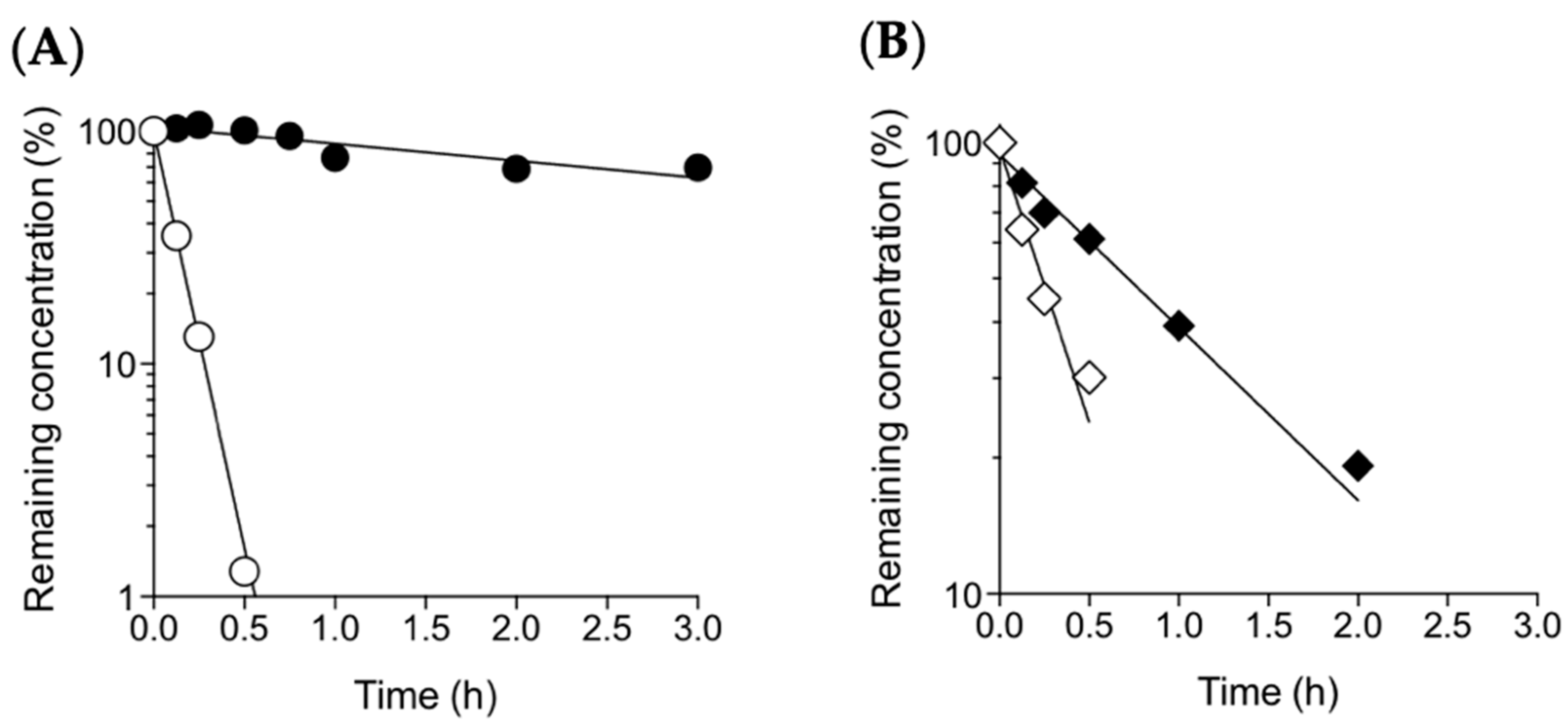
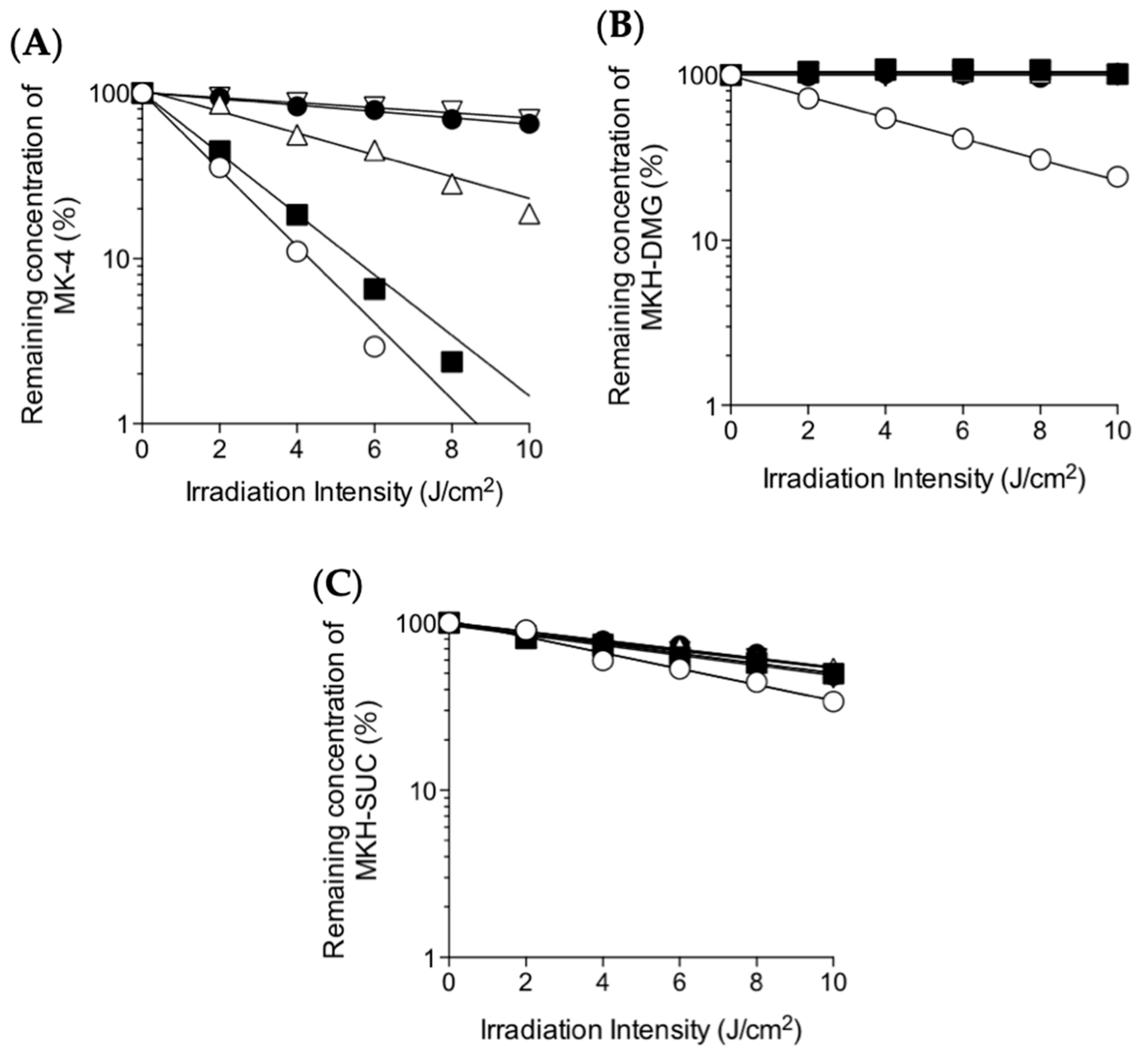
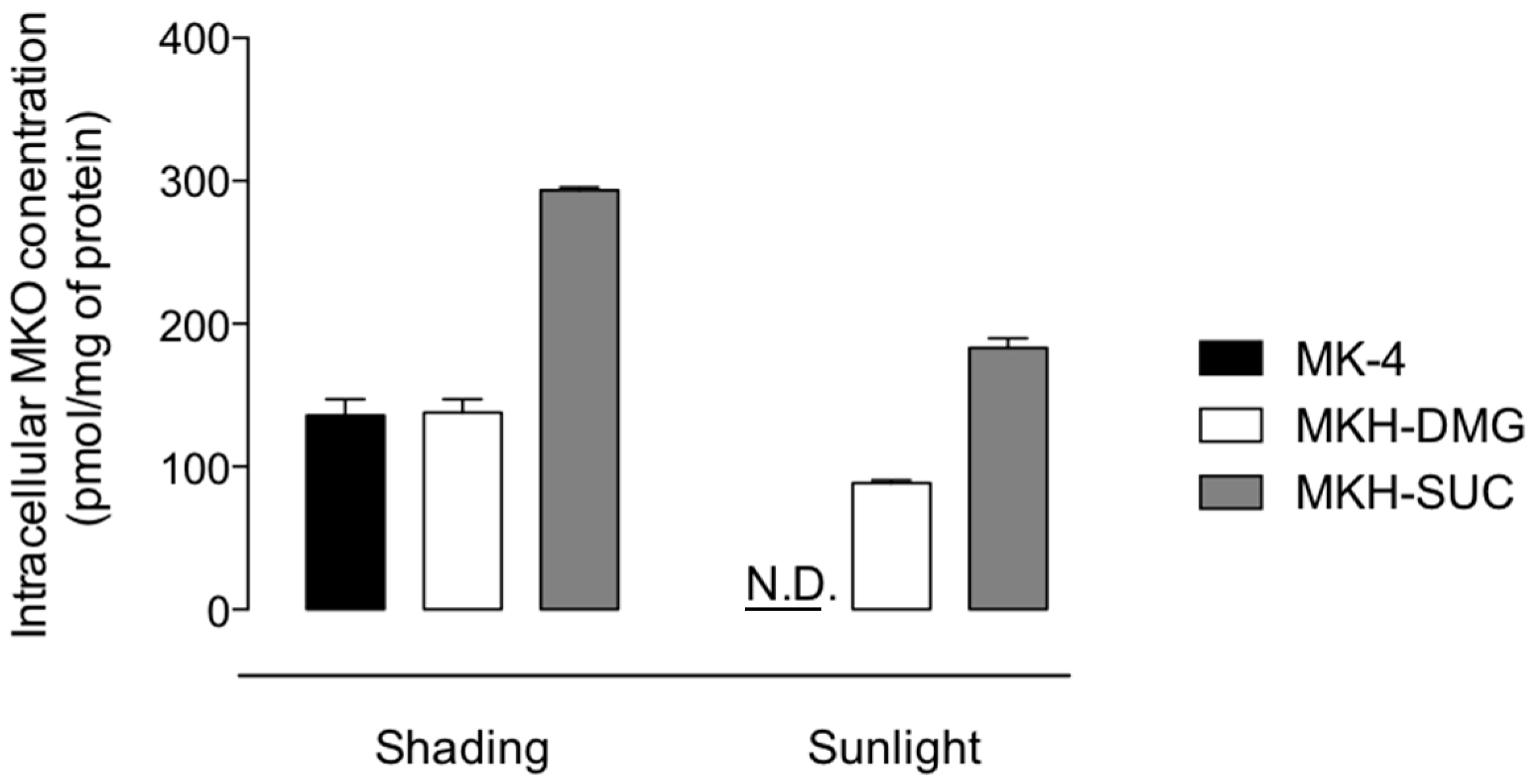
2.1. Photostability of MKH-Ester Derivatives in Artificial Sunlight
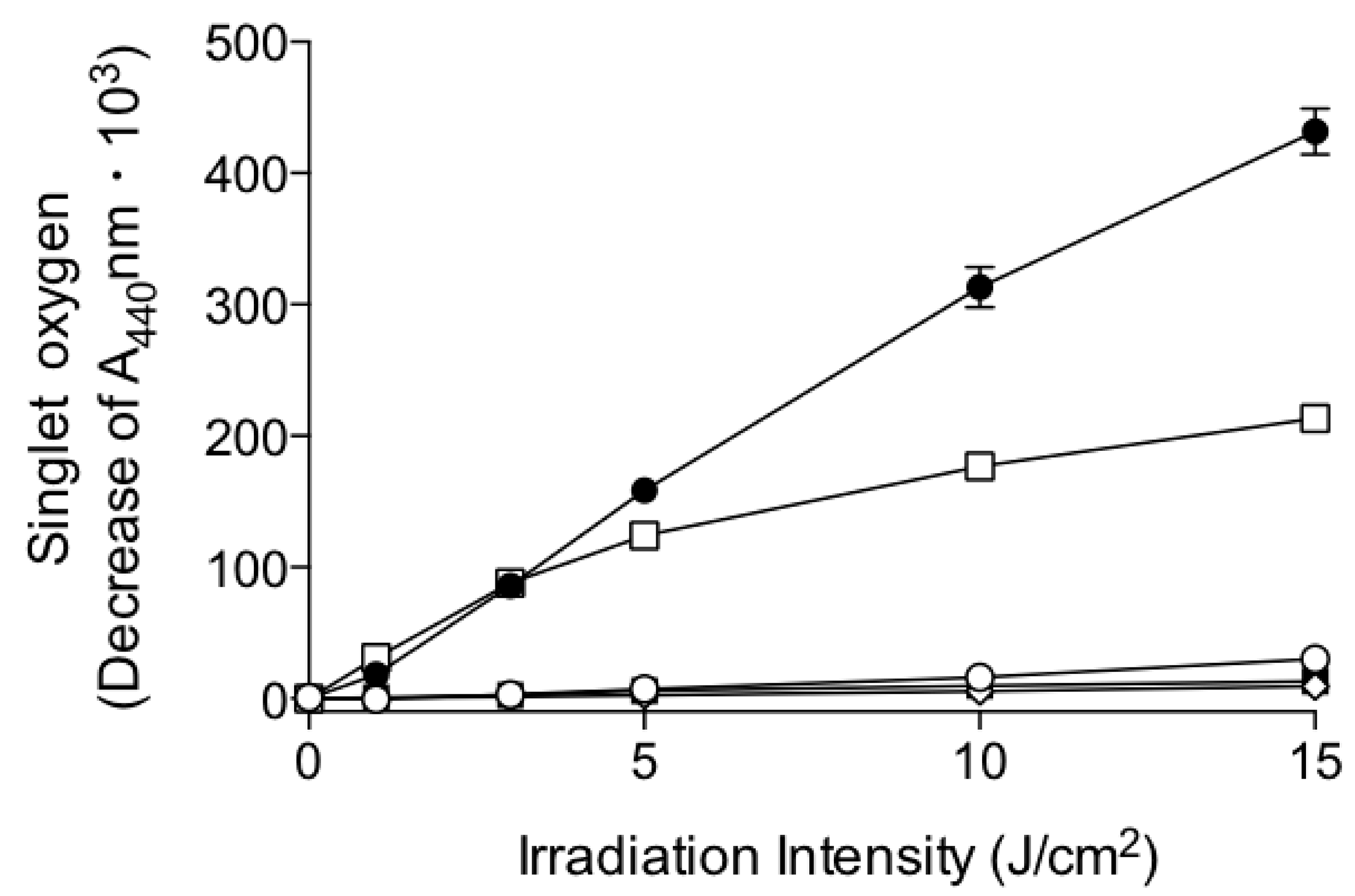
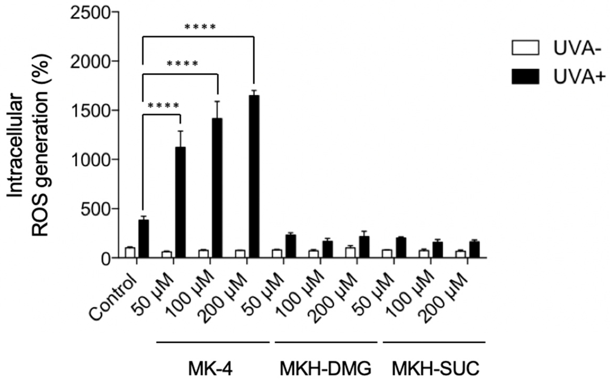
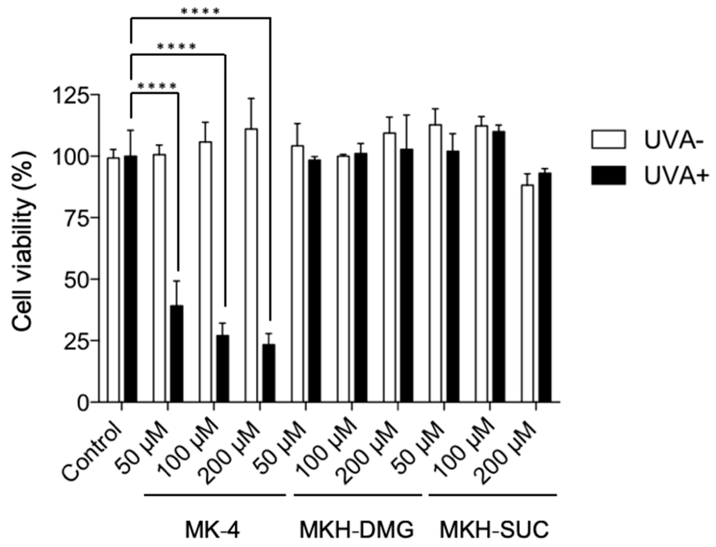
2.2. Phototoxicity of MK-4 and MKH-Ester Derivatives
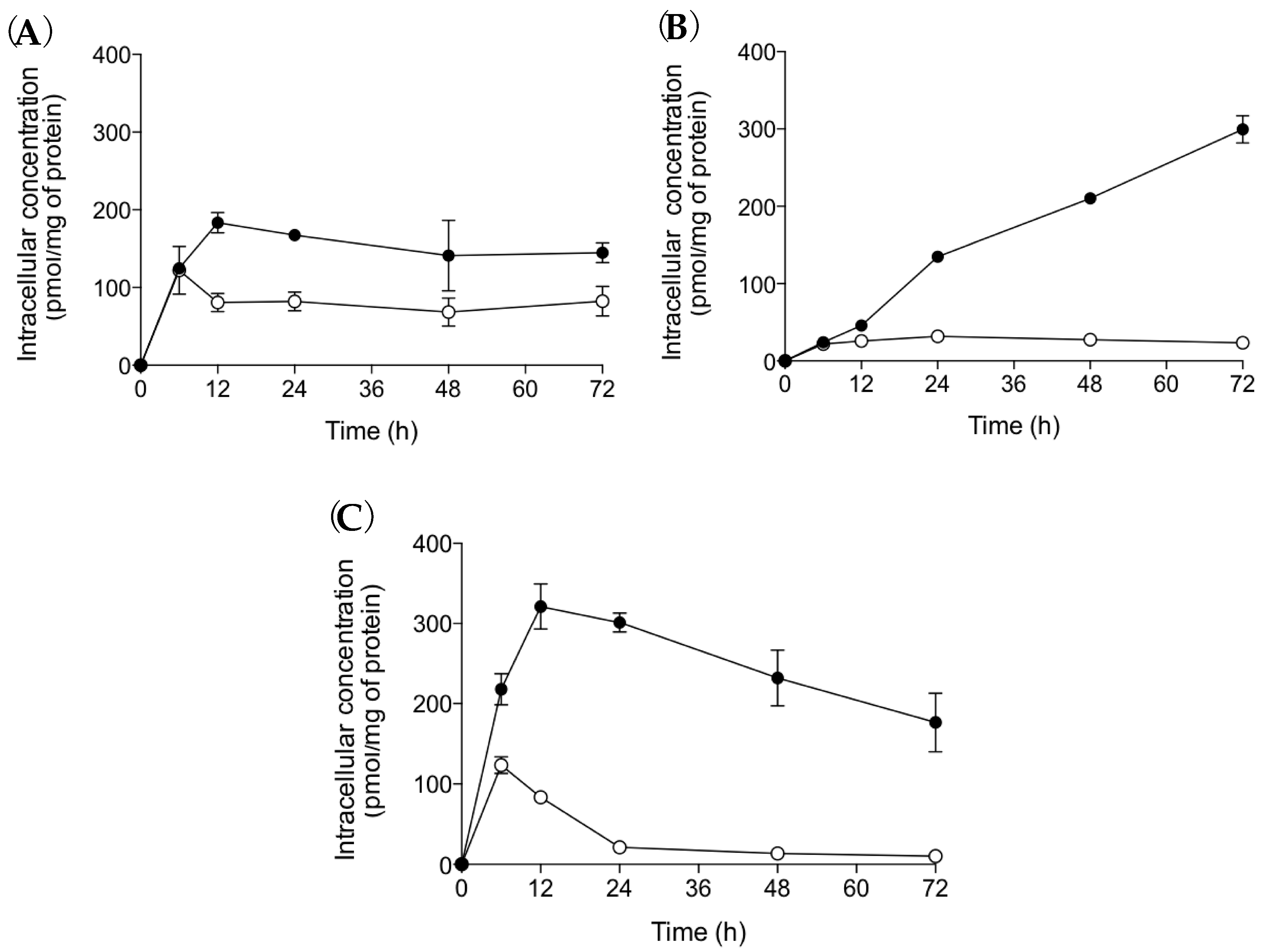
2.3. MKH Delivery into HaCaT Cells Using MK-4 and MKH-Ester Derivatives
3. Discussion
4. Materials and Methods
4.1. Chemicals
4.2. Cell Culture
4.3. Photostability
4.4. Singlet Oxygen Generation Assay
4.5. Intracellular ROS Generation Assay
4.6. Cell Viability Assay
4.7. Determination of Intracellular MKO Levels after Treatment With MK-4 and MKH Derivatives
4.8. Determination of Intracellular MKO Levels After Treatment With Sunlight-Irradiated MK-4 and MKH Derivatives
4.9. LC-MS/MS
4.10. Statistical Analysis
Supplementary Materials
Author Contributions
Acknowledgments
Conflicts of Interest
Abbreviations
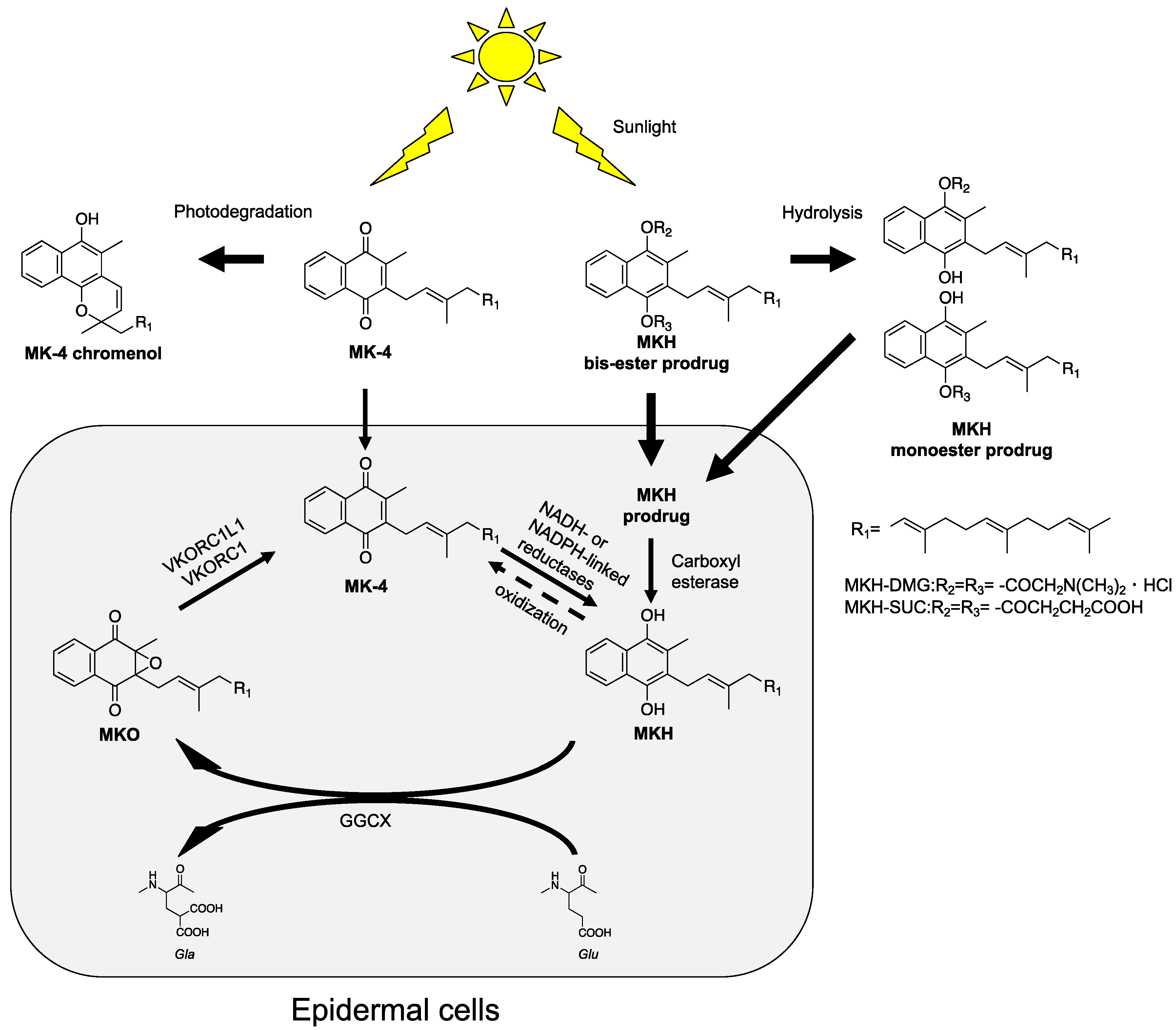
| MKH | menahydroquinone-4 |
| MK-4 | menaquinone-4 |
| MKC | menaquinone-4 chromenol |
| MKH-DMG | menahydroquinone-4 1,4-bis-N,N-dimethylglycinate hydrochloride |
| MKH-mono-DMG | menahydroquinone-4 1-mono N,N-dimethylglycinate hydrochloride menahydroquinone-4 4-mono N,N-dimethylglycinate hydrochloride |
| MKH-SUC | menahydroquinone-4 1,4-bis-hemisuccinate |
| MKH-mono-SUC | menahydroquinone-4 1-mono hemisuccinate menahydroquinone-4 4-mono hemisuccinate |
| MKO | menaquinone-4 epoxide |
| GGCX | γ-glutamyl carboxylase |
| VKORC1 | vitamin K epoxide reductase complex subunit 1 |
| VKORC1L1 | vitamin K epoxide reductase complex subunit 1 like 1 |
| ROS | reactive oxygen species |
| DCFH-DA | 2′,7′-dichlorofluorescein diacetate |
| AUC | area under the concentration versus time curve |
| MRM | MS/MS-multiple reaction monitoring mode |
References
- Nakagawa, K.; Hirota, Y.; Sawada, N.; Yuge, N.; Watanabe, M.; Uchino, Y.; Okuda, N.; Shimomura, Y.; Suhara, Y.; Okano, T. Identification of UBIAD1 as a novel human menaquinone-4 biosynthetic enzyme. Nature 2010, 468, 117. [Google Scholar] [CrossRef] [PubMed]
- Lou, W.W.; Quintana, A.T.; Geronemus, R.G.; Grossman, M.C. Effects of topical vitamin K and retinol on laser-induced purpura on nonlesional skin. Dermatol. Surg. 1999, 25, 942–944. [Google Scholar] [CrossRef]
- Lopes, L.B.; Speretta, F.F.F.; Vitoria, M.; Bentley, L.B. Enhancement of skin penetration of vitamin K using monoolein-based liquid crystalline systems. Eur. J. Pharm. Sci. 2007, 32, 209–215. [Google Scholar] [CrossRef] [PubMed]
- Shah, N.S.; Lazarus, M.C.; Bugdodel, R.; Hsia, S.L.; He, J.; Duncan, R.; Baumann, L. The effects of topical vitamin K on bruising after laser treatment. J. Am. Acad. Dermatol. 2002, 47, 241–244. [Google Scholar] [CrossRef] [PubMed]
- Ocvirk, J. Management of cetuximab-induced skin toxicity with the prophylactic use of topical vitamin K1 cream. Radiol. Oncol. 2010, 44, 265–266. [Google Scholar] [CrossRef] [PubMed] [Green Version]
- Pinta, F.; Ponzetti, A.; Spadi, R.; Fanchini, L.; Zanini, M.; Mecca, C.; Sonetto, C.; Ciuffreda, L.; Racca, P. Pilot Clinical Trial on the Efficacy of Prophylactic Use of Vitamin K-1-Based Cream (Vigorskin) to Prevent Cetuximab-Induced Skin Rash in Patients With Metastatic Colorectal Cancer. Clin. Colorectal Cancer 2014, 13, 62–67. [Google Scholar] [CrossRef] [PubMed]
- Tan, E.H.; Chan, A. Evidence-Based Treatment Options for the Management of Skin Toxicities Associated with Epidermal Growth Factor Receptor Inhibitors. Ann. Pharm. 2009, 43, 1658–1666. [Google Scholar] [CrossRef] [PubMed]
- Li, T.H.; Perez-Soler, R. Skin toxicities associated with epidermal growth factor receptor inhibitors. Targeted Oncol. 2009, 4, 107–119. [Google Scholar] [CrossRef] [PubMed]
- Hemmati, A.A.; Houshmand, G.; Ghorbanzadeh, B.; Nemati, M.; Behmanesh, M.A. Topical vitamin K-1 promotes repair of full thickness wound in rat. Indian J. Pharmacol. 2014, 46, 409–412. [Google Scholar] [CrossRef] [PubMed]
- Teraoka, R.; Matsuda, Y. Stabilization-oriented preformulation study of photolabile menatetrenone (vitamin K2). Int. J. Pharm. 1993, 93, 85–90. [Google Scholar] [CrossRef]
- Fujisawa, S.; Kawabata, S.; Yamamoto, R. Photo-degradation and stabilization of vitamin K1. I. Degradation product in ethanol and in non-ionic surfactant solution. Yakugaku Zasshi 1967, 87, 1451–1456. [Google Scholar] [CrossRef] [PubMed]
- Hangarter, M.A.; Hormann, A.; Kamdzhilov, Y.; Wirz, J. Primary photoreactions of phylloquinone (vitamin K-1) and plastoquinone-1 in solution. Photochem. Photobiol. Sci. 2003, 2, 524–535. [Google Scholar] [CrossRef] [PubMed]
- Scientific Committees on Consumer Safety of the European Comission. Opinion on Vitamin K1 (Phytonadione). 23 March 2010. Available online: https://ec.europa.eu/health/scientific_committees/consumer_safety/docs/sccs_o_014.pdf (accessed on 23 May 2019). [CrossRef]
- Takata, J.; Karube, Y.; Hanada, M.; Matsunaga, K.; Matsushima, Y.; Sendo, T.; Aoyama, T. Vitamin K prodrugs: 1. Synthesis of amino acid esters of menahydroquinone-4 and enzymatic reconversion to an active form. Pharm. Res. 1995, 12, 18–23. [Google Scholar] [CrossRef] [PubMed]
- Takata, J.; Karube, Y.; Hanada, M.; Matsunaga, K.; Matsushima, Y.; Sendo, T.; Oishi, R. Vitamin K prodrugs: 2. Water-soluble prodrugs of menahydroquinone-4 for systemic site-specific delivery. Pharm. Res. 1995, 12, 1973–1979. [Google Scholar] [CrossRef] [PubMed]
- Takata, J.; Karube, Y.; Hanada, M.; Matsunaga, K.; Iwasaki, H. Prodrug for bioreductive activation-independent delivery of menahydroquinone-4: Human liver enzymatic activation and its action in warfarin-poisoned human liver. Biol. Pharm. Bull. 1999, 22, 172–178. [Google Scholar] [CrossRef] [PubMed]
- Setoguchi, S.; Watase, D.; Matsunaga, K.; Yamakawa, H.; Goto, S.; Terada, K.; Ohe, K.; Enjoji, M.; Karube, Y.; Takata, J. Antitumor Effects and Delivery Profiles of Menahydroquinone-4 Prodrugs with Ionic or Nonionic Promoiety to Hepatocellular Carcinoma Cells. Molecules 2018, 23, 1738. [Google Scholar] [CrossRef] [PubMed]
- Ryu, A.; Arakane, K.; Koide, C.; Arai, H.; Nagano, T. Squalene as a Target Molecule in Skin Hyperpigmentation Caused by Singlet Oxygen. Biol. Pharm. Bull. 2009, 32, 1504–1509. [Google Scholar] [CrossRef] [PubMed] [Green Version]
- Prusakiewicz, J.J.; Ackermann, C.; Voorman, R. Comparison of skin esterase activities from different species. Pharm. Res. 2006, 23, 1517–1524. [Google Scholar] [CrossRef] [PubMed]
- Batz, F.M.; Klipper, W.; Korting, H.C.; Henkler, F.; Landsiedel, R.; Luch, A.; von Fritschen, U.; Weindl, G.; Schafer-Korting, M. Esterase activity in excised and reconstructed human skin—Biotransformation of prednicarbate and the model dye fluorescein diacetate. Eur. J. Pharm. Biopharm. 2013, 84, 374–385. [Google Scholar] [CrossRef] [PubMed]
- Fu, J.; Sadgrove, M.; Marson, L.; Jay, M. Biotransformation Capacity of Carboxylesterase in Skin and Keratinocytes for the Penta-Ethyl Ester Prodrug of DTPA. Drug Metab. Dispos. 2016, 44, 1313–1318. [Google Scholar] [CrossRef] [PubMed] [Green Version]
- ROS Assay Validation Management Team of Japanese Center for the Validation of Alternative Methods (JaCVAM). Reactive Oxygen Species (Ros) Assay to Examine Photoreactivity of Chemicals. Version 3.2. 28 November 2014. Available online: http://www.jacvam.jp/files/doc/02_03/02_03_E3.pdf (accessed on 23 May 2019).


| Compound a | Irradiation Conditions | k (h−1) | t1/2 (h) |
|---|---|---|---|
| MK-4 | Sunlight | 8.239 | 0.084 |
| MKH-DMG | Sunlight | 0.167 | 4.150 |
| MKH-SUC | Sunlight | 2.796 | 0.248 |
| Shading b | 0.883 | 0.785 |
| Compound a | Wavelength (nm) | k (J−1·cm2) | E1/2 (J−1·cm2) |
|---|---|---|---|
| MK-4 | 279 | 0.533 | 1.301 |
| 341 | 0.422 | 1.643 | |
| 373 | 0.151 | 4.583 | |
| 404 | 0.049 | 15.800 | |
| 435 | 0.035 | 19.738 | |
| MKH-DMG | 279 | 0.146 | 4.750 |
| 341 | - b | - b | |
| 373 | - b | - b | |
| 404 | - b | - b | |
| 435 | - b | - b | |
| MKH-SUC | 279 | 0.110 | 6.323 |
| 341 | 0.069 | 10.036 | |
| 373 | 0.059 | 11.792 | |
| 404 | 0.061 | 11.296 | |
| 435 | 0.068 | 10.253 |
| Compound a | AUCMKO (0–72 h) (nmol·h/mg of Protein) | AUCMK-4 (0–72 h) (nmol·h/mg of Protein) | AUCMKO/AUCMK-4 |
|---|---|---|---|
| MK-4 | 10.543 ± 0.795 | 5.628 ± 0.698 | 1.873 ± 0.272 |
| MKH-DMG | 10.786 ± 1.696 | 1.878 ± 0.088 | 5.743 ± 0.942 |
| MKH-SUC | 17.304 ± 1.068 | 2.316 ± 0.095 | 7.471 ± 0.554 |
© 2019 by the authors. Licensee MDPI, Basel, Switzerland. This article is an open access article distributed under the terms and conditions of the Creative Commons Attribution (CC BY) license (http://creativecommons.org/licenses/by/4.0/).
Share and Cite
Goto, S.; Setoguchi, S.; Yamakawa, H.; Watase, D.; Terada, K.; Matsunaga, K.; Karube, Y.; Takata, J. Prodrugs for Skin Delivery of Menahydroquinone-4, an Active Form of Vitamin K2(20), Could Overcome the Photoinstability and Phototoxicity of Vitamin K2(20). Int. J. Mol. Sci. 2019, 20, 2548. https://0-doi-org.brum.beds.ac.uk/10.3390/ijms20102548
Goto S, Setoguchi S, Yamakawa H, Watase D, Terada K, Matsunaga K, Karube Y, Takata J. Prodrugs for Skin Delivery of Menahydroquinone-4, an Active Form of Vitamin K2(20), Could Overcome the Photoinstability and Phototoxicity of Vitamin K2(20). International Journal of Molecular Sciences. 2019; 20(10):2548. https://0-doi-org.brum.beds.ac.uk/10.3390/ijms20102548
Chicago/Turabian StyleGoto, Shotaro, Shuichi Setoguchi, Hirofumi Yamakawa, Daisuke Watase, Kazuki Terada, Kazuhisa Matsunaga, Yoshiharu Karube, and Jiro Takata. 2019. "Prodrugs for Skin Delivery of Menahydroquinone-4, an Active Form of Vitamin K2(20), Could Overcome the Photoinstability and Phototoxicity of Vitamin K2(20)" International Journal of Molecular Sciences 20, no. 10: 2548. https://0-doi-org.brum.beds.ac.uk/10.3390/ijms20102548