Article

Silencing of the Chitin Synthase Gene Is Lethal to the Asian Citrus Psyllid, Diaphorina citri

1
College of Life Sciences, Gannan Normal University, Ganzhou 341000, China
2
National Navel Orange Engineering Research Center, Ganzhou 341000, China
3
Saskatoon Research Center, Agriculture and Agri-Food Canada, Saskatoon, SK S7N 0X2, Canada
*
Author to whom correspondence should be addressed.
These authors contributed equally to this work.
Int. J. Mol. Sci. 2019, 20(15), 3734; https://0-doi-org.brum.beds.ac.uk/10.3390/ijms20153734
Submission received: 29 June 2019 / Revised: 27 July 2019 / Accepted: 29 July 2019 / Published: 31 July 2019
(This article belongs to the Special Issue Molecular Ecology, Physiology and Biochemistry of Insects)

Abstract

:
Chitin synthase is a critical enzyme that catalyzes N-acetylglucosamine to form chitin, which plays an important role in the growth and development of insects. In this study, we identified a chitin synthase gene (CHS) with a complete open reading frame (ORF) of 3180 bp from the genome database of Diaphorina citri, encoding a protein of 1059 amino acid residues with the appropriate signature motifs (EDR and QRRRW). Reverse transcription-quantitative PCR (RT-qPCR) analysis suggested that D. citri CHS (DcCHS) was expressed throughout all developmental stages and all tissues. DcCHS had the highest expression level in the integument and fifth-instar nymph stage. Furthermore, the effects of diflubenzuron (DFB) on D. citri mortality and DcCHS expression level were investigated using fifth-instar nymph through leaf dip bioassay, and the results revealed that the nymph exposed to DFB had the highest mortality compared with control group (Triton-100). Silencing of DcCHS by RNA interference resulted in malformed phenotypes and increased mortality with decreased molting rate. In addition, transmission electron microscopy (TEM) and fluorescence in situ hybridization (FISH) also revealed corresponding ultrastructural defects. Our results suggest that DcCHS might play an important role in the development of D. citri and can be used as a potential target for psyllid control.

Graphical Abstract

1. Introduction

The Asian citrus psyllid (ACP), Diaphorina citri Kuwayama (Hemiptera: Liviidae), is a notorious pest across Asia, USA, and Brazil, causing severe economic losses in the citrus industry [1]. However, the greatest threat of ACP arises from the transmission of “Candidatus Liberibacter asiaticus (CLas)”, which causes citrus greening disease, also called huanglongbing (HLB) [2]. Up to this date, psyllid control plays a leading role to prevent HLB from spreading. At present, the application of insecticides is the most widely followed option for reducing ACP populations. However, the improper use of insecticide will lead to pest resistance, human poisoning, and environmental pollution [3,4]. Therefore, there is a great need to find environmentally friendly methods to control the citrus psyllid.
During the growth and development of insects, periodic molting, the process of shedding and replacing the rigid insect exoskeleton is required. During this process, the degraded old cuticle is shed and replaced with a newly synthesized cuticle. Insects are covered by this cuticle, which is a composite structure consisting of chitin filaments embedded in a protein matrix. The insect cuticle undertakes various functional roles, including protection, support, movement, and as a shield against environmental stress [5]. Chitin, a polymer of N-acetylglucosamine, is an essential component of the cuticle, which plays a critical role in maintaining insect shape and protecting it from external forces [6]. Chitin is mainly found in arthropod exoskeletons, fungal cell walls, and nematode eggshells, but is not found in vertebrates [7]. During insect molting, it is key for the insect to keep a dynamic balance of chitin content, which is regulated by chitin synthase and chitin degradation related enzymes, including chitinase and chitin deacetylase [8]. Based on these crucial roles during insect molting, chitin serves as a selective target for pest control [9].
Chitin formation arises from trehalose and is catalyzed by chitin synthase, a conserved enzyme found in every chitin-synthesizing organism. Chitin synthase belongs to the large family of glycosyltransferases, a ubiquitous group of enzymes that catalyze the transfer of sugar moieties from activated sugar donors to specific acceptors, thereby forming a glycosidic bond [10]. Two chitin synthase (CHS) genes have been identified in insects, including CHS1 and CHS2. CHS1 encodes the isoform of the enzyme for the synthesis of chitin in the cuticle and tracheae, while CHS2 is produced by midgut epithelial cells for chitin synthesis in the peritrophic membrane (PM) [11]. In recent years, the CHS genes have been identified in various insects, including Lepidopteras, Dipterans, Coleopterans, Orthopterans, and Hemipterans [12]. Zhuo et al. identified two chitin synthases genes (CHS1 and CHS2) from Bombyx mori, the results suggested that BmCHS1 is an epidermis-specific gene that is expressed during the molting stage, while BmCHS2 is midgut specific and highly expressed during the feeding process in the larva [13,14]. In Tribolium castaneum, silencing of CHS1 by RNA interference (RNAi) will disrupt molting and greatly reduce whole-body chitin content, while CHS2 specific RNAi has no effect on metamorphosis or on total body chitin content [15]. Interestingly, many studies have revealed that CHS2 is not present in phloem-sucking hemipterans due to the absence of PM [16]. Still, research into the function of CHS in D. citri has not been reported.
As insects grow in increments, and each stage of growth ends with molting, the inhibition of chitin synthesis is considered as an attractive target site for insect control [10]. Insecticides based on chitin synthesis inhibition are safe to humans as chitin synthesis pathway is absent in vertebrates. Diflubenzuron (DFB) was the first commercial insecticide that acts by inhibiting chitin synthesis in insects [17]. In Aphis glycines, when fed with soybean leaves previously dipped in 50 ppm DFB solution, the nymphs suffered significantly higher mortality compared to controls [18]. In Toxoptera citricida, fourth-instar nymphs were exposed to 5 and 500 mg/L concentrations of DFB for 48 h and had the highest mortality at the 500 mg/L concentration [19]. Tiwari et al. have revealed that DFB could effectively suppress D. citri adult emergence, and fifth-instar nymphs are more susceptible than first-third instar nymphs [4]. However, the exact mode of action of DFB in D. citri still remains unclear.
In this study, we identified a cDNA encoding the whole ORF of a chitin synthase (D. citri CHS (DcCHS)) from D. citri and analyzed the expression patterns of DcCHS in different tissues and at different developmental stages. Fluorescence in situ hybridization (FISH) analysis also suggested that the DcCHS was evenly distributed in whole integument. The mortality of fifth-instar nymphs significantly increased after exposed to DFB. Furthermore, we demonstrated that dsRNA-mediated gene-specific silencing resulted in a strong reduction in relative expression of DcCHS and molting rate of nymphs, as well as an increase in mortality. These results will provide a new target for citrus psyllid control.

2. Results

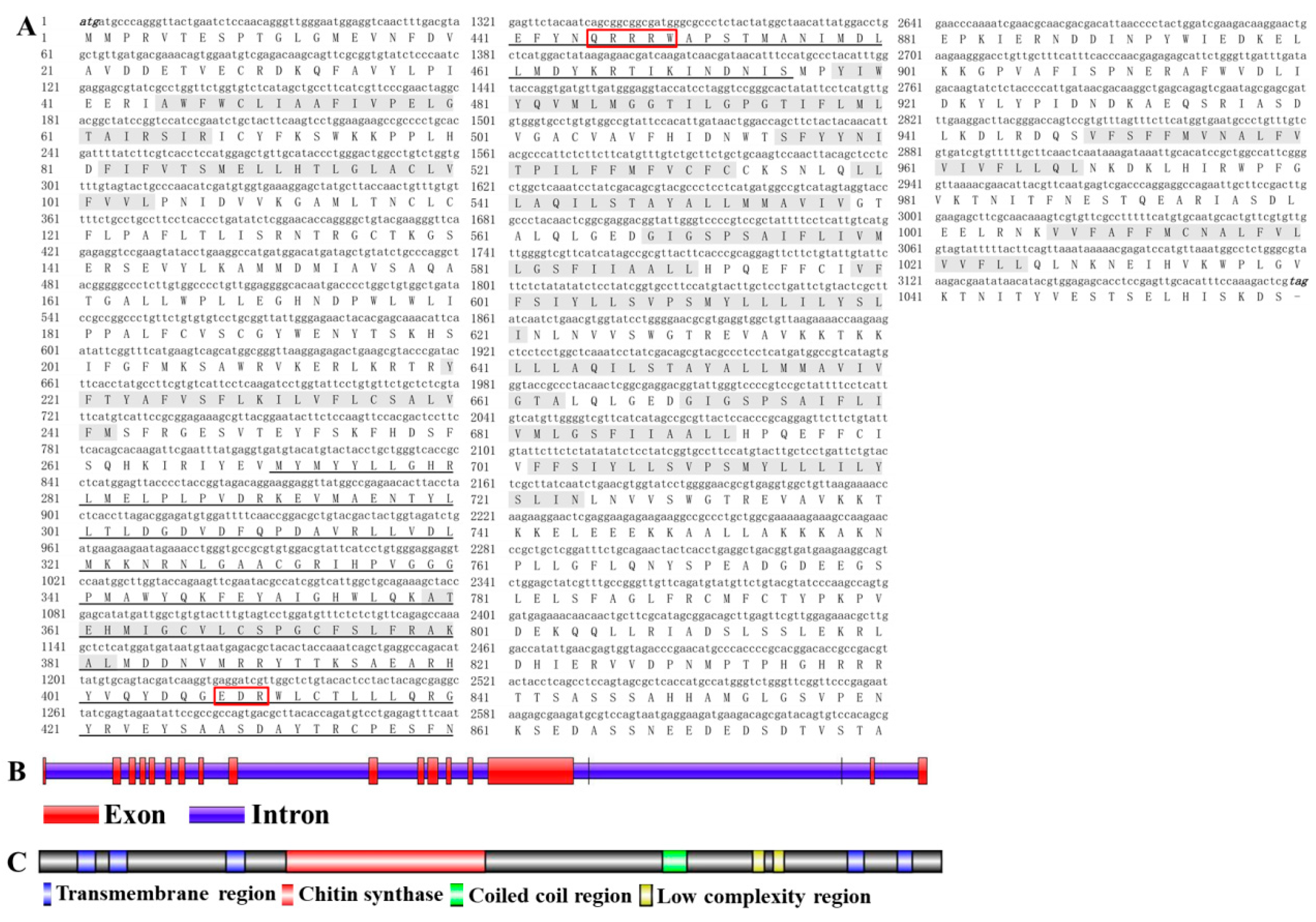
2.1. Analysis of the cDNA and Protein Sequences of DcCHS

The cDNA sequence of DcCHS (XP_017303059) contains an ORF of 3180 bp encoding a protein of 1059 amino acid residues with a predicted MW of 129.9 kDa and pI of 5.02 (Figure 1A). Gene structure analysis showed that DcCHS contains 19 exons and 18 introns (Figure 1B). In terms of protein structure, a total of 14 transmembrane helices, two low-complexity regions, and a coiled-coil region were identified (Figure 1C). A BLASTP search of the NCBI databases indicated that the amino acid sequence of DcCHS shared 42.75%, 42.67%, and 42.30% with Acyrthosiphon pisum, Toxoptera citricida, and Aphis glycines, respectively. Based on the amino acid sequences of CHS from different insect species, a phylogenetic tree was generated using MEGA 5.0 to investigate the evolutionary relationship of DcCHS among the selected insect species. The results showed that DcCHS has a close relationship with the CHSs of sap-sucking hemipterans, including A. pisum, T. citricida, and A. glycines (Figure 2).

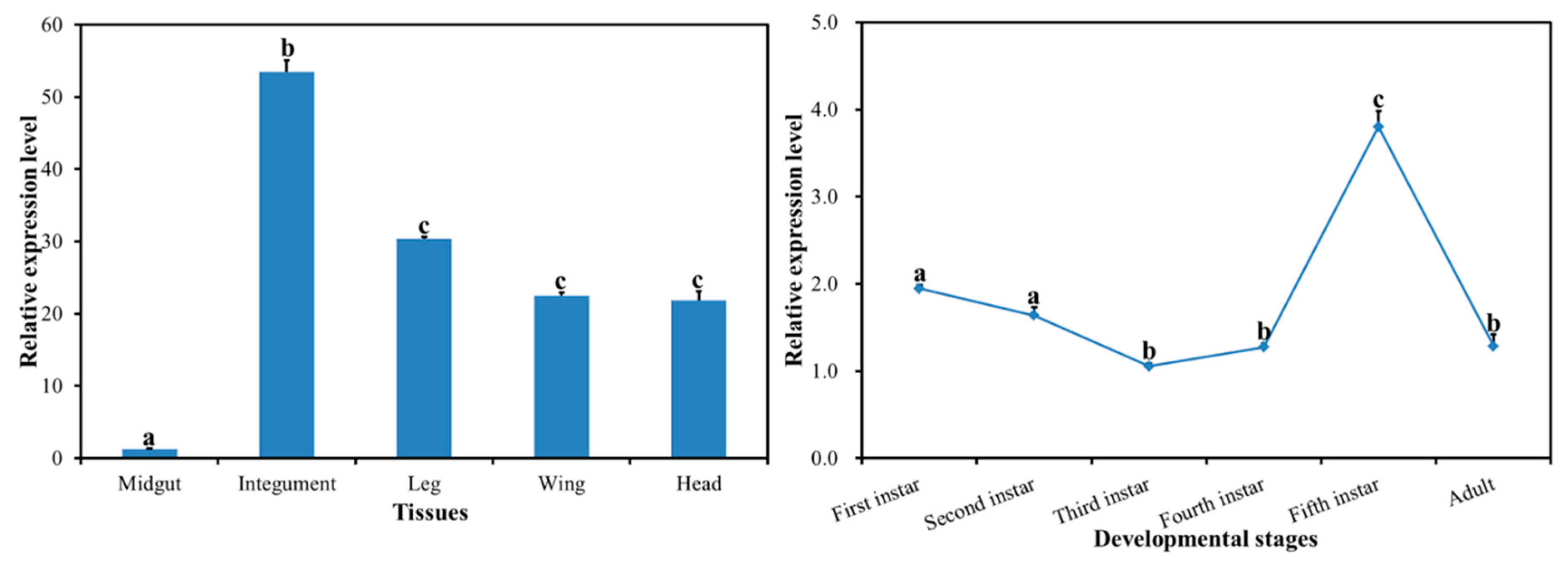
2.2. Spatiotemporal Expression Profiles of DcCHS

The expression profiles of DcCHS in different developmental stages and different tissues were investigated by RT-qPCR. The results showed that the DcCHS gene was expressed in all tissues, including midgut, integument, leg, wing, and head. It is notable that DcCHS had high expression in the integument, while it had low expression in the midgut. The expression level of DcCHS in the integument was 44.3 times of that in the midgut, and its expression in the leg was 25.1 times of that in the midgut (Figure 3). The expression of DcCHS decreased from first-instar nymph to third-instar nymph, while it increased from third-instar nymph to fifth-instar nymph, and decreased sharply from fifth-instar nymph stage to adult stage (Figure 3). The expression level of DcCHS in the fifth-instar nymph was 3.6 times that of the third-instar nymph stage.

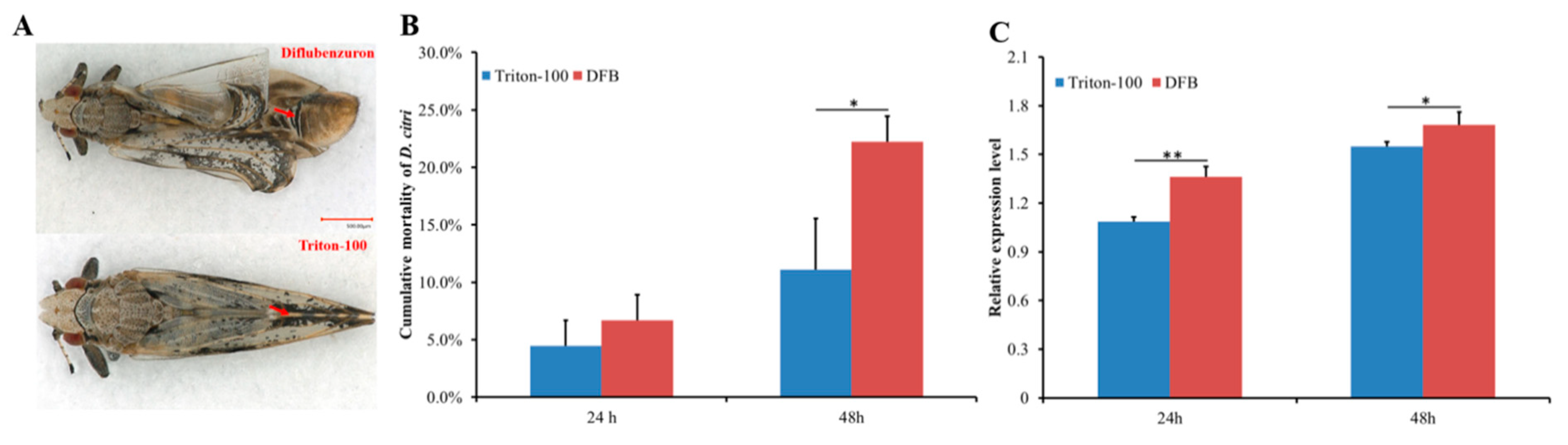
2.3. Effect of DFB on D. citri Survival and DcCHS Expression Level

A leaf-dip bioassay was used to assay the effect of DFB on D. citri survival and DcCHS expression level. After being exposed for 24 h, the cumulative mortality of D. citri had no significant change between DFB treatment group and control (Triton-100 treatment group). However, the cumulative mortality of DFB sharply increased after exposure to DFB for 48 h (Figure 4B). In addition, D. citri in the DFB treatment group showed an abnormal phenotype after molting and the wing of adult exhibited crimp (Figure 4A). The relative expression level of DcCHS was significantly upregulated after DFB exposure at 24 h and 48 h (Figure 4C).

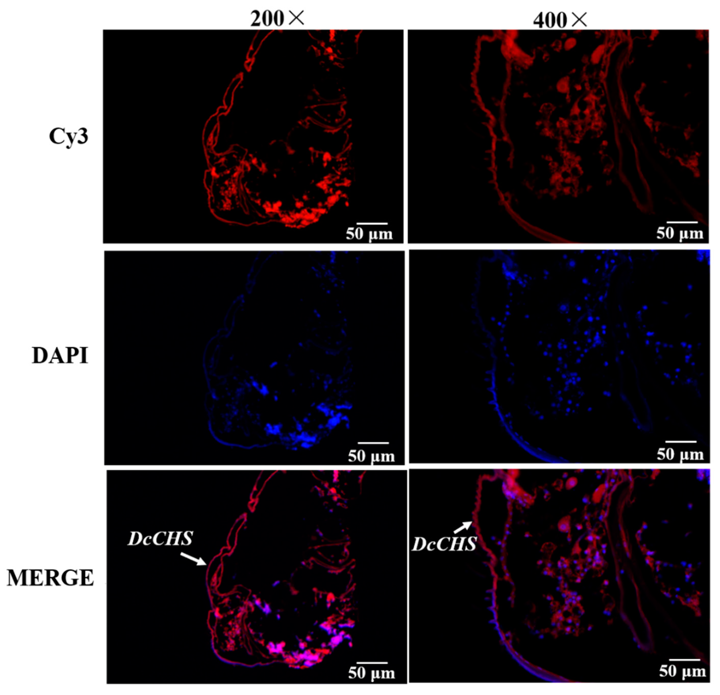
2.4. Localization of DcCHS Transcript by FISH

In order to confirm localization of DcCHS transcript in D. citri, FISH was performed using DcCHS RNA probes. The results showed that DcCHS transcript was evenly distributed in the epidermal cells (Figure 5). The fluorescence signal is the strongest in exocuticle epidermal cells of D. citri, indicating DcCHS is mainly involved in synthesis of chitin in new cuticle.

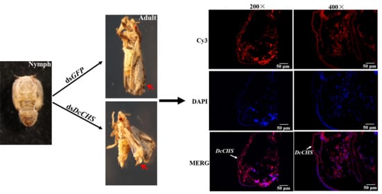
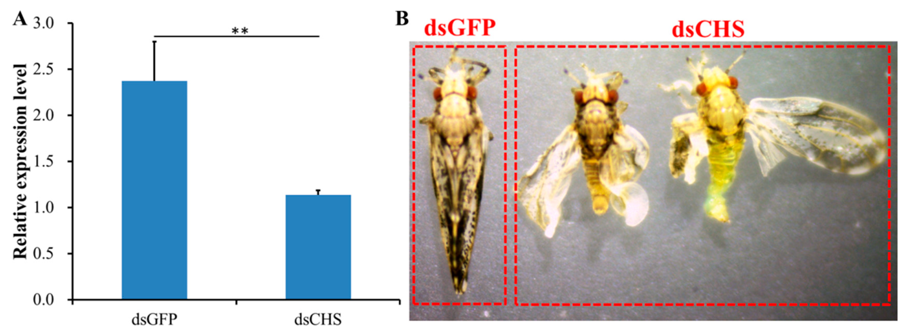
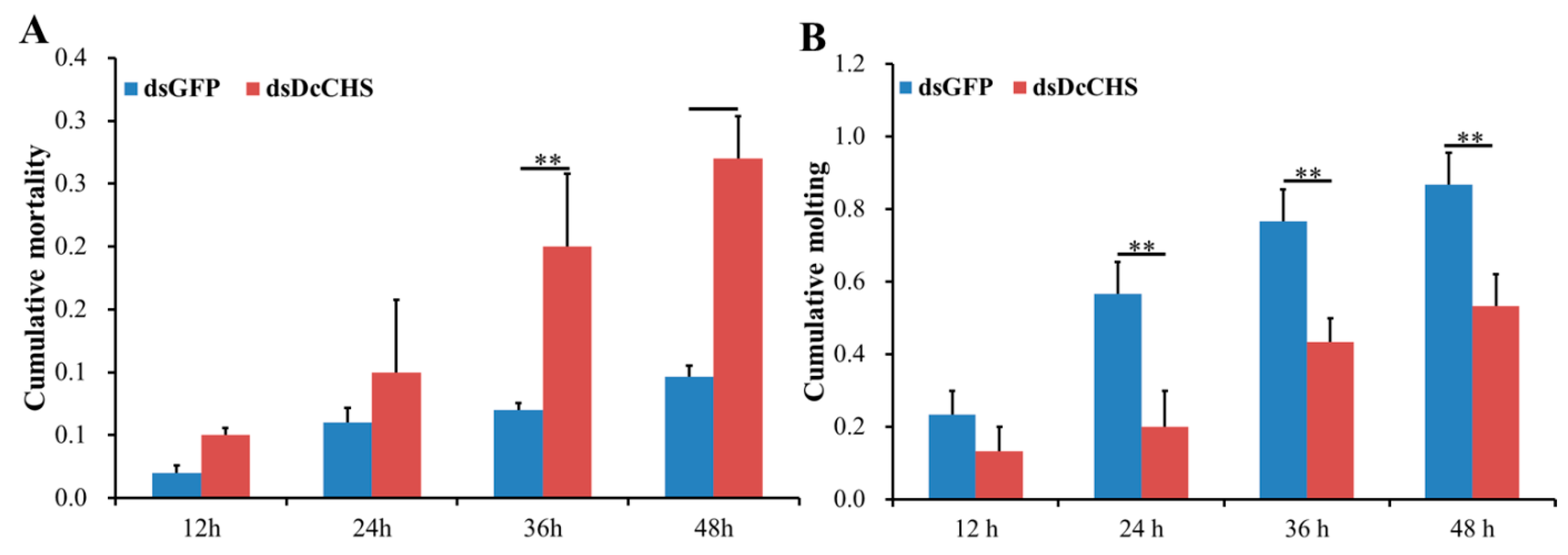
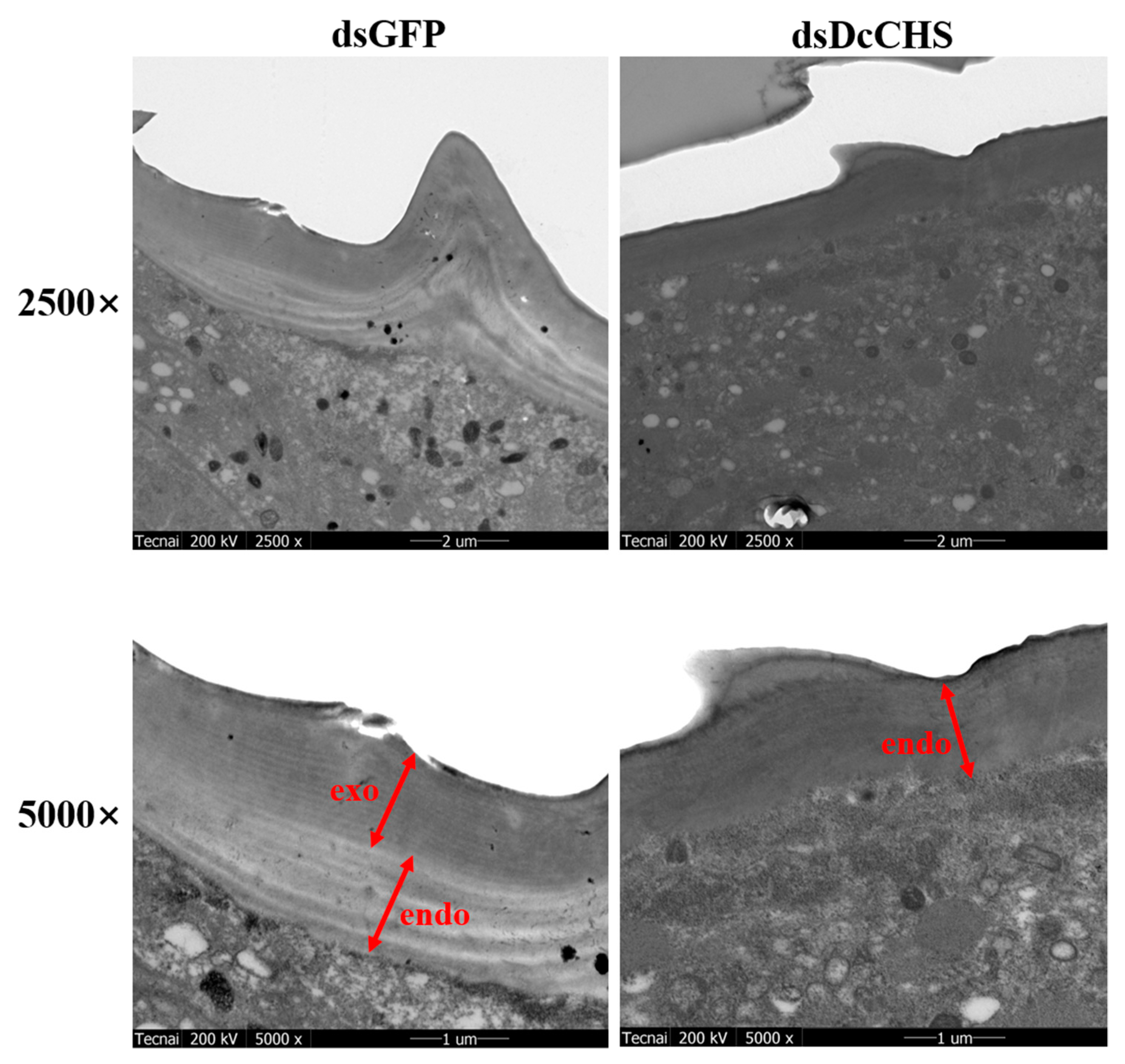
2.5. RNAi-Based Silencing of DcCHS and Epidermal Structure Analysis

To determine the effect of DcCHS on D. citri molting, RNAi was performed by ingestion of dsRNA. At 24 h after feeding D. citri with double-stranded DcCHS (dsDcCHS), the expression level of DcCHS was significantly downregulated compared with the controls (double-stranded green fluorescent protein (dsGFP) treatment group) (Figure 6A). In the dsDcCHS treatment group, many adults did not complete wing development and presented an abnormal wing phenotype after molting (Figure 6B). Importantly, the cumulative mortality in the dsDcCHS treatment group was 27% compared to 10% mortality in the group at 48 hpt. The mortality of D. citri increased from 24 hpt to 48 hpt in the treatment group, whereas control group had no significant change (Figure 7A). In addition, the cumulative molting of D. citri was 53.3% in the treatment group at 48 hpt. However, in the control group, the molting could reach 86.7% (Figure 7B). These results indicated that RNAi effectively inhibited the relative expression level of DcCHS and resulted in the abnormal D. citri phenotype.
To further analyze the effect of epidermal structure after silencing of DcCHS, TEM observation was conducted at 24 h after RNAi treatment. The results showed that the formation of exocuticle was inhibited compared with the control group, while the structure of endocuticle has no significant change (Figure 8). Insect cuticle is primarily composed of epicuticle and procuticle, and procuticle is comprised of the exocuticle and endocuticle. Chitin is found mainly in exocuticle and endocuticle. These results suggested that knockdown of DcCHS will inhibit the synthesis of chitin and further disrupt the structure of D. citri cuticle.

3. Discussion

Chitin is a major component of the exoskeleton and the peritrophic matrix of insects. The synthesis of chitin is catalyzed by many enzymes. Among them, chitin synthases (CHSs) play important roles during insect development and metamorphosis [8]. To date, CHS genes have been identified from many insect species, including those in the Lepidoptera, Hemiptera, Hymenoptera, Diptera, and Coleoptera. The number of genes encoding CHS in different fungal species can range from 1 to 20, while in insect genomes characterized so far individual species contain only two CHS genes (CHS-1 and CHS-2) [20]. CHS-A is responsible for chitin synthesis in the cuticle and cuticular lining of the foregut, hindgut, and trachea, whereas CHS-B is dedicated to chitin synthesis in the PM [21]. In this study, only one CHS gene was identified from the genome of D. citri, which was named DcCHS (Figure 1). In a previous report, many hemipterous insects only contain a single CHS gene, including A. glycines, Sogatella furcifera, and A. pisum [18]. We considered that Hemiptera insects lack the PM structure. Terry et al. revealed that the insect PM was lost, leading to the compartmentalization of the digestive process and ultimately increased digestion of polymers during the course of evolution [22]. The insect CHS proteins usually contain 15 transmembrane helices that flank the catalytic domain located on the cytoplasmic side of the plasma membrane [10]. We found that DcCHS contains 14 transmembrane helices (Figure 1). Depending on the number of predicted transmembrane helices, the N-terminus faces either the extracellular space or the cytoplasm. However, the C-terminus region is predicted to face the extracellular space and may be involved in protein–protein interaction or oligomerization [5,23]. In addition, DcCHS has two conserved domains with chitin synthase signature motifs, i.e., EDR and QRRRW in the catalytic domain, which is essential for the catalytic mechanism [24]. The phylogenetic tree analysis indicated that CHS from D. citri has a close relationship with A. pisum and belongs to the CHS1 group. Interestingly, many studies have proved that the CHS1 gene contains alternative splicing [25,26]. In the present study, the genome analysis of D. citri revealed no splice variants exist. Bansal et al. also found that splice variants of CHS1 do not exist in the A. glycines genome [18]. However, the specific reasons for this apparent loss need to be researched in depth.
The relative expression level of DcCHS was determined in different tissues and different developmental stages. Results suggested that DcCHS had a relatively higher expression in the integument, followed by the leg, but it had low expression in the midgut (Figure 3). Our results are in agreement with the roles of CHS in chitin production in the insect exoskeleton [27]. In insects, chitin functions as a scaffold material, supporting the cuticles of the epidermis [5]. In Sogatella furcifera, CHS1 was also predominantly expressed in the integument [28]. Therefore, we speculated that DcCHS may play a critical role in the process of cuticle formation. Additionally, the phenomenon of low expression of CHS in the midgut was also observed in Plutella xylostella and Nilaparvata lugens [6,29]. At different developmental stages, DcCHS had a higher expression in the fifth-instar nymph stage (Figure 3). The fifth-instar nymph stage is a critical period that involves progressing from nymph stage into adult stage [30]. During molting period, the synthesis of chitin is required to maintain rigid structure of new cuticle in D. citri.
Chitin is synthesized by insects and fungi but not by vertebrates, so chitin synthase presents a novel target for pest control. In recent years, the exploration of inhibitors of chitin synthesis has received extensive attention [17]. DFB is an insect growth regulator acting on chitin synthesis, which was discovered in the 1970s [31]. It was used as a potential insecticide for pest control in forestry and agriculture [32]. In a previous research, Tiwari et al. revealed that DFB could effectively suppress D. citri adult emergence [4]. However, the specific molecular mechanisms are unclear. In this study, we found that DFB can increase the expression of DcCHS and lead to higher mortality of D. citri (Figure 4). In many insect species, DFB has been found to elevate CHS expression level. Zhang et al. revealed a significant increase of CHS1 mRNA level in Anopheles quadrimaculatus larvae exposed to DFB at 100 and 500 μg/L [33]. In T. citricida, the mRNA expression levels of TCiCHS were significantly increased upon the exposure of nymphs to both low and high DFB concentrations [19]. At 48 h post DFB treatment (hpt), the cumulative mortality in the treatment group was significantly greater than the control group, whereas it showed no obvious change at 24 hpt. Therefore, we speculated that DFB can reduce the chitin content by inhibition of chitin synthase activity. The increasing DcCHS expression levels may also indicate the existence of a feedback regulatory mechanism that compensates for the low enzyme content.
RNA interference (RNAi) has already proven its usefulness in functional genomics research on insects, but it also has considerable potential for the control of pest insects [34]. When monitoring RNAi responses using different delivery methods, variable efficiency is very common in different insect species, e.g., the physiological pH of hemolymph significantly affected the efficiency of RNAi in Locusta migratoria [35]. In this study, we performed RNAi experiments through the feeding of dsRNA. The results showed that DcCHS was silenced effectively (Figure 5). In recent years, RNAi combined with RT-qPCR has been widely used to research the functions of target genes. Yu et al. revealed that silencing of transformer-2 gene influenced female reproduction and offspring sex in D. citri [36]. Kishk et al. used RNAi to silence genes implicated in pesticide resistance in order to increase susceptibility, and the results suggested that the treatments with dsRNA caused concentration-dependent nymph mortality [37]. In addition, we found that D. citri showed increased cumulative mortality and abnormal phenotypes after silencing of DcCHS (Figure 6). In this regard, many reports on RNAi mediated knockdown of insect CHS genes resulting in lethal phenotypes are encouraging [15,38,39]. Chitin is the main component of exocuticle and endocuticle [40]. We found that the formation of exocuticle was inhibited after knockdown of DcCHS, while the structure of endocuticle had no significant change, indicating the decrease of chitin content will influence the structure of cuticle. These results further indicated that DcCHS could be used as a new target for control of D. citri.

4. Materials and Methods

4.1. Diaphorina citri Rearing and Collection

The ACP was reared at the National Navel Orange Engineering Research Center (NORC), Gannan Normal University, Ganzhou, China. D. citri was reared in mesh cages (50 × 50 × 50 cm) on Murraya exotica with 27 ± 1 °C, 70 ± 5% relative humidity, and a 14:10 (light: dark) photocycle.
D. citri nymphs were classified to different stages according to their morphological features and collected using an aspirator. D. citri adults were divided into three groups, and then each group was dissected to obtain the midgut, head, leg, wing, and integument. The collected tissues were cleaned with DEPC water and stored at −80 °C until use. Each experiment was conducted in three biological replicates.

4.2. RNA Isolation and cDNA Synthesis

To obtain the cDNA template for spatiotemporal expression analysis of DcCHS, total RNA was isolated from different tissues of adult insects (head, midgut, leg, wing, and integument) and at different developmental stages of the nymph (first-instar, second-instar, third-instar, fourth-instar, and fifth-instar nymphs) using a RNA extraction kit (TaKaRa Biotechnology Co. Ltd., Dalian, China). The integrity of total RNA was confirmed using standard agarose gel electrophoresis with ethidium bromide staining. RNA quantification was performed using a NanoDrop 2000 spectrophotometer (Thermo Fisher Scientific, New York, NY, USA). The purity of all RNA samples was assessed at an absorbance ratio of A260/230 and A260/280. Total RNA was reverse-transcribed in a 20 μL reaction system using a cDNA synthesis master mix kit according to the manufacturer’s instructions (Simgen, Hangzhou, Zhejiang, China). In brief, 2.0 μL of 5 × gRNA buffer and 1 μg of total RNA were mixed, then RNase-free water was added to reach 10 μL, which was then incubated at 42 °C for 3 min. Afterward, 4 μL of 5 × RT buffer, 2 μL of RT enzyme mix, and 4 μL RT primer mix were added to the tube. The mixture was incubated at 42 °C for 15 min and then incubated at 95 °C for 3 min. The cDNA was stored at −20 °C for later use.

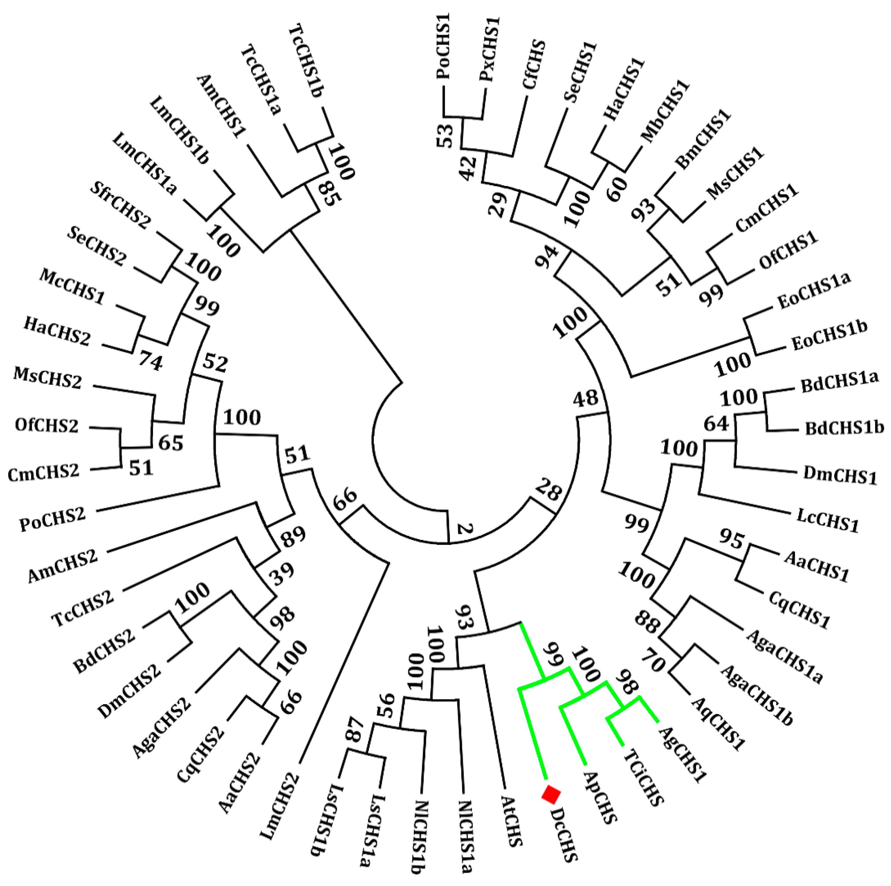
4.3. Identification of DcCHS and Bioinformatics Analysis

To identify chitin synthase genes in D. citri, sequences of Nilaparvata lugens CHS (NlCHS: AEL88648.1) and A. pisum CHS (ApCHS: XP_003247517.1) were used as query in a TBLASTN search of genome database of D. citri. The cDNA and deduced amino acid sequence of DcCHS were analyzed by using DNASTAR. The open reading frame (ORF) was identified according to the ORF finder tool (https://0-www-ncbi-nlm-nih-gov.brum.beds.ac.uk/orffinder/) at the National Center for Biotechnology Information (NCBI). The molecular weight (MW) and isogenic point (pI) of DcCHS were calculated using ExPASy (http://web.expasy.org/compute_pi). The signal peptide of DcCHS was predicted using SignalP 4.1 Server (http://www.cbs.dtu.dk/services/SignalP). The functional domain was predicted by using SMART software (http://smart.embl-heidelberg.de/). The membrane-spanning domain was predicted by TMHMM Server v. 2.0. The phylogenetic tree was constructed with MEGA 7.0 software using the neighbor-joining method with 1000-fold bootstrap resampling. Protein sequences from different species were downloaded from GenBank (http://0-www-ncbi-nlm-nih-gov.brum.beds.ac.uk/) and used in phylogenetic analysis (Table S1).

4.4. RT-qPCR Analysis of DcCHS Expression Levels

RT-qPCR was conducted to confirm the relative expression levels of DcCHS in various tissues and developmental phases. The primers were designed using Primer Premier 5.0 software (Premier Biosoft, www.premierbiosoft.com) (Table 1). The 20 μL of reaction mixture for the RT-qPCR contained 10 μL of SYBR II, 8 μL of ddH2O, 0.5 μL of forward primer, 0.5 μL of reverse primer, and 1.0 μL of cDNA template. The thermal cycling profile consisted of an initial denaturation at 95 °C for 60 s, and 40 cycles of 95 °C for 10 s, 60 °C for 10 s, and 72 °C for 10 s. The reactions were performed with a LightCycle®96 PCR Detection System (Roche, Basel, Switzerland). Relative expression levels were calculated by using the 2−ΔΔCt method. There were three biological replicates and three technique replicates for each sample. The reference gene chosen for analysis of DcCHS in different tissues and different developmental phases was glyceraldehyde-3-phosphate dehydrogenase (GAPDH). This gene was cited as a reference in previous studies involving expression analyses in D. citri [41]. All ANOVAs were followed by Fisher’s protected least significant difference (LSD) tests.

4.5. Leaf-Dip Bioassay

To determine whether DFB can affect the expression of DcCHS and increase the mortality of D. citri, we used a leaf-dip bioassay according to a previous protocol with some modifications [32]. In brief, about 0.05 g of DFB (LKT Laboratories Inc., Saint Paul, MN, USA) was added into 10 mL acetone and dissolved completely as a stock solution (5000 mg/L). The stock solution was diluted into a low concentration (300 mg/L) using ddH2O, and 50 μL of Triton-100 (0.1%) was added. Detached tender leaves of Murraya koenigii were dipped into the DFB working solution for 5 min and air-dried for 1 h, after which 40 fifth-instar D. citri nymphs were transferred to each leaf. Leaves treated with ddH2O containing 0.1% Triton-100 were used as a control. The number of deaths and molts were counted at 24 h and 48 h after DFB exposure. In addition, the surviving D. citri were kept at −80 °C for RNA isolation. The relative expression levels of DcCHS were analyzed by RT-qPCR as described above. All experiments were performed using three biological replicates.

4.6. dsRNA Synthesis and DcCHS RNAi Analysis

The primers for DcCHS were designed to synthesize dsRNA using the T7 RioMAX Express RNAi System (Promega, San Luis Obispo, CA, USA) based on the manufacturer’s instructions. GFP dsRNA was used as a control. The dsRNA delivery was performed by using an artificial diet according to a previous method [42]. In brief, 30 newly emerged fifth-instar D. citri nymphs were used in dsRNA treatment. All nymphs were allowed to feed on an aliquot of the artificial diet (150 μL) placed between two layers of stretched Parafilm. The artificial diet consisting of 20% (w:v) sucrose was mixed with dsDcCHS at a final concentration of 150 ng/μL. After 24 h, all live insects were collected to isolate total RNA and synthesize cDNA. The effect of dsDcCHS on gene expression was evaluated by RT-qPCR. A total of three biological replicates were used for each experiment.

4.7. Fluorescence In Situ Hybridization (FISH) Analysis

FISH was performed to confirm the specific distribution of DcCHS transcript in D. citri. The RNA probes of DcCHS (5′-Cy3-CGUAAGUCCUUCAAAUCGCUCGUAAUUCGACUCUG-3′) were synthesized by Rochen Biotechnology (Rochen, Shanghai, China). FISH was conducted according to previous protocol with some modification [43]. In brief, the dissected integument samples were fixed in 4% paraformaldehyde for about 8 h at 4 °C and then washed using PBST (PBS+Triton X-100) for three times (5 min each), treated with proteinase K (50 μg/mL in PBST). The fixed tissues were prehybridized in hybridization buffer at 37 °C for 1 h and then hybridized in the same hybridization buffer containing 10 μg/mL RNA probes overnight at 37 °C. After hybridization, the samples were washed using 2 × SSC at 37 °C for 10 min, 1 × SSC at 37 °C for 5 min, and 0.5 × SSC at 37 °C for 10 min to remove remaining hybridization buffer, and then moved to a fresh microscope slide containing 30 μL of new hybridization buffer supplemented with DAPI and kept with a liquid blocker. At last, the samples were visualized under an Olympus fluorescence microscope equipped with Cy3 filter set.

4.8. Transmission Election Microscopy (TEM) Analysis

To investigate the transformation of epidermal structure after silencing of DcCHS, TEM was used following a previous report [21]. Abdominal integument of a hatched adult was dissected and fixed in 2.5% glutaraldehyde of PBS at 4 °C overnight and then fixed in 1% osmic acid at 4 °C for 3 h. After fixation, all samples were washed three times using 0.1 M PBS. The fixed integuments were dehydrated through incubation in a graded series of ethanol washes (50%, 70%%, 80%, 85%, 90%, 95%, and 100%, v/v) for 15 min each and then further dehydrated twice using 100% ethanol for 10 min each. The dehydrated samples were consecutively soaked by penetrant 1 (2:1 mixture of acetone and epoxy resin), penetrant 2 (1:1 mixture of acetone and epoxy resin), and penetrant 3 (epoxy resin) at 37 °C for 12 h. Finally, the samples were embedded at 60 °C for 48 h. The ultrathin (100 nm) sections were cut with a Leica microtome (Leica, Wetzlar, Germany). The sections were stained with 3% uranyl acetate and alkaline lead citrate and observed using TEM with a mode (Tecnai G2 20 S-TWIN, EDX: GENESIS 2000) at an accelerating voltage of 200 kV.

5. Conclusions

In conclusion, the cDNA sequence of DcCHS was identified from the genome database of D. citri. Spatiotemporal expression analysis suggested that DcCHS was highly expressed in the integument and fifth-instar nymph stage. In addition, DcCHS was upregulated and the cumulative mortality of D. citri increased after exposure to DFB, an inhibitor of chitin synthesis. Furthermore, RNAi-based gene silencing inhibited the expression of DcCHS and influenced the structure of cuticle, resulting in malformed phenotypes. Taken together, these results indicated DcCHS as a new target for control of D. citri.

Supplementary Materials

Author Contributions

Conceived and designed this study: H.-Z.Y. and Z.-J.L. Collected the samples: Y.-L.H., N.-Y.L., Y.-X.X., X.-D.Z., and Q.Z. Performed experiments: H.-Z.Y. and Y.-L.H. Analyzed the data: H.-Z.Y., Y.-L.H., and Z.-J.L. Wrote the manuscript: H.-Z.Y., H.H. and Z.-J.L. Investigation: A.-J.H. Supervision: L.Y. Software: H.-N.S. All authors contributed to discussions and revisions.

Funding

This work was supported by the Natural Science Foundation of China (31560602), Ministry of Science and Technology of China (2018YFD0201504), the Educational Commission of Jiangxi Province of China (GJJ180747), and Science and Technology Department of Jiangxi Province (20171BCB23074).

Acknowledgments

We would like to thank Pei Zhang and Du An-Na from the core facility and technical support, Wuhan Institute of Virology, for their help with producing TEM micrographs.

Conflicts of Interest

The authors declare no conflict of interest.

Abbreviations

CHSChitin synthase
TEMTransmission electron microscopy
FISHFluorescence in situ hybridization
ACPAsian citrus psyllid
CLasCandidatus Liberibacter asiaticus
HLBHuanglongbing
PMPeritrophic membrane
RNAiRNA interference
DFBDiflubenzuron

References

  1. Chen, X.D.; Ashfaq, M.; Stelinski, L.L. Susceptible of Asian citrus psyllid, Diaphorina citri (Hemiptera: Liviidae), to the insecticide afidopyropen: A new and potent modulator of insect transient receptor potential channels. Appl. Entomol. Zool. 2018, 53, 1–9. [Google Scholar] [CrossRef]
  2. Wang, N.; Pierson, E.A.; Setubal, J.C.; Xu, J.; Levy, J.G.; Zhang, Y.; Li, J.; Rangel, L.T.; Martins, J.J. The Candidatus liberibacter-host interface: Insights into pathogenesis mechanisms and disease control. Annu. Rev. Phytopathol. 2017, 55, 451–482. [Google Scholar] [CrossRef] [PubMed]
  3. Huang, J.K.; Hu, R.F.; Pray, C.; Qiao, F.B.; Rozelle, S. Biotechnology as an alternative to chemical pesticides: A case study of Bt cotton in China. Agr. Econ. 2003, 29, 55–67. [Google Scholar] [CrossRef]
  4. Tiwari, S.; Clayson, P.J.; Kuhns, E.H.; Stelinski, L.L. Effects of buprofezin and diflubenzuron on various developmental stages of Asian citrus psyllid, Diaphorina citri. Pest. Manag. Sci. 2012, 68, 1405–1412. [Google Scholar] [CrossRef] [PubMed]
  5. Merzendorfer, H.; Zimoch, L. Chitin metabolism in insects: Structure, function and regulation of chitin synthases and chitinases. J. Exp. Biol. 2003, 206, 4393–4412. [Google Scholar] [CrossRef] [PubMed]
  6. Wang, Y.; Fan, H.W.; Huang, H.J.; Xue, J.; Wu, W.J.; Bao, Y.Y.; Xu, H.J.; Zhu, Z.R.; Cheng, J.A.; Zhang, C.X. Chitin synthase 1 gene and its two alternative splicing variants from two sap-sucking insects, Nilaparvata lugens and Laodelphax striatellus (Hemiptera: Delphacidae). Insect Biochem. Mol. Biol. 2012, 42, 637–646. [Google Scholar] [CrossRef] [PubMed]
  7. Tobias, P.A.; Christie, N.; Naidoo, S.; Guest, D.I.; Kulheim, C. Identification of the Eucalyptus grandis chitinase gene family and expression characterization under different biotic stress challenges. Tree Physiol. 2017, 37, 565–582. [Google Scholar] [CrossRef] [PubMed]
  8. Zhu, K.Y.; Merzendorfer, H.; Zhang, W.Q.; Zhang, J.Z.; Muthukrishnan, S. Biosynthesis, turnover, and functions of chitin in insects. Annu. Rev. Entomol. 2016, 61, 177–196. [Google Scholar] [CrossRef] [PubMed]
  9. Choi, H.K.; Choi, K.H.; Kramer, K.J.; Muthukrishnan, S. Isolation and characterization of a genomic clone for the gene of an insect molting enzyme, chitinase. Insect Biochem. Mol. Biol. 1997, 27, 37–47. [Google Scholar] [CrossRef]
  10. Merzendorfer, H. Insect chitin synthases: A review. J. Comp. Physiol. B. 2006, 176, 1–15. [Google Scholar] [CrossRef]
  11. Mansur, J.F.; Alvarenga, E.S.; Figueira-Mansur, J.; Franco, T.A.; Ramos, I.B.; Masuda, H.; Melo, A.C.; Moreira, M.F. Effects of chitin synthase double-stranded RNA on molting and oogenesis in the Chagas disease vector Rhodnius prolixus. Insect Biochem. Mol. Biol. 2014, 51, 110–121. [Google Scholar] [CrossRef] [PubMed]
  12. Wang, Y.W.; Zuber, R.; Oehl, K.; Norum, M.; Moussian, B. Report on Drosophila melanogaster larvae without functional tracheae. J. Zool. 2015, 296, 139–145. [Google Scholar] [CrossRef]
  13. Zhuo, W.W.; Fang, Y.; Kong, L.F.; Li, X.; Sima, Y.H.; Xu, S.Q. Chitin synthase A: A novel epidermal development regulation gene in the larvae of Bombyx mori. Mol. Biol. Rep. 2014, 41, 4177–4186. [Google Scholar] [CrossRef] [PubMed]
  14. Zhuo, W.W.; Chu, F.; Kong, L.F.; Tao, H.; Sima, Y.H.; Xu, S.Q. Chitin synthase B: A midgut-specific gene induced by insect hormones and involved in food intake in Bombyx mori larvae. Arch. Insect Biochem. Physiol. 2014, 85, 36–47. [Google Scholar] [CrossRef] [PubMed]
  15. Arakane, Y.; Muthukrishnan, S.; Kramer, K.J.; Specht, C.A.; Tomoyasu, Y.; Lorenzen, M.D.; Kanost, M.; Beeman, R.W. The Tribolium chitin synthase genes TcCHS1 and TcCHS2 are specialized for synthesis of epidermal cuticle and midgut peritrophic matrix. Insect Mol. Biol. 2005, 14, 453–463. [Google Scholar] [CrossRef] [PubMed]
  16. Silva, C.P.; Silva, J.R.; Vasconcelos, F.F.; Petretski, M.D.; Damatta, R.A.; Ribeiro, A.F.; Terra, W.R. Occurrence of midgut perimicrovillar membranes in paraneopteran insect orders with comments on their function and evolutionary significance. Arthropod Struct. Dev. 2004, 33, 139–148. [Google Scholar] [CrossRef] [PubMed]
  17. Cohen, E. Chitin synthesis and inhibition: A revisit. Pest. Manag. Sci. 2001, 57, 946–950. [Google Scholar] [CrossRef] [PubMed]
  18. Bansal, R.; Mian, M.A.; Mittapalli, O.; Michel, A.P. Characterization of a chitin synthase encoding gene and effect of diflubenzuron in soybean aphid, Aphis glycines. Int. J. Biol. Sci. 2012, 8, 1323–1334. [Google Scholar] [CrossRef]
  19. Shang, F.; Xiong, Y.; Xia, W.K.; Wei, D.D.; Wei, D.; Wang, J.J. Identification, characterization and functional analysis of a chitin synthase gene in the brown citrus aphid, Toxoptera citricida (Hemiptera, Aphididae). Insect Mol. Biol. 2016, 25, 422–430. [Google Scholar] [CrossRef]
  20. Merzendorfer, H. The cellular basis of chitin synthesis in fungi and insects: Common principles and differences. Eur. J. Cell Biol. 2011, 90, 759–769. [Google Scholar] [CrossRef]
  21. Zimoch, L.; Merzendorfer, H. Immunolocaliation of chitin synthase in the tobacco hornworm. Cell Tissue Res. 2002, 308, 287–297. [Google Scholar] [CrossRef] [PubMed]
  22. Terra, W.R.; Ferreira, C. Insect digestive enzymes: Properties, compartmentalization and function. Comp. Biochem. Phys. B. 1994, 109, 1–62. [Google Scholar] [CrossRef]
  23. Burkhard, P.; Stetefeld, J.; Strelkov, S.V. Coiled coils: A highly versatile protein folding motif. Trends Cell Biol. 2001, 11, 82–88. [Google Scholar] [CrossRef]
  24. Zhu, Y.C.; Specht, C.A.; Dittmer, N.T.; Muthukrishnan, S.; Kanost, M.R.; Kramer, K.J. Sequence of a cDNA and expression of the gene encoding a putative epidermal chitin synthase of Manduca sexta. Insect Biochem. Mol. Biol. 2002, 32, 1497–1506. [Google Scholar] [CrossRef]
  25. Chen, X.F.; Yang, X.; Kumar, N.S.; Tang, B.; Sun, X.J.; Qiu, X.M.; Hu, J.; Zhang, W.Q. The class A chitin synthase gene of Spodoptera exigua: Molecular cloning and expression patterns. Insect Biochem. Mol. Biol. 2007, 37, 409–417. [Google Scholar] [CrossRef] [PubMed]
  26. Arakane, Y.; Hogenkamp, D.G.; Zhu, Y.C.; Kramer, K.J.; Specht, C.A.; Beeman, R.W.; Kanost, M.R.; Muthukrishnan, S. Characterization of two chitin synthase genes of the red flour beetle, Tribolium castaneum, and alternate exon usage in one of the genes during development. Insect Biochem. Mol. Biol. 2004, 34, 291–304. [Google Scholar] [CrossRef] [PubMed]
  27. Zhang, X.; Zhang, J.Z.; Zhu, K.Y. Chitosan/double-stranded RNA nanoparticle-mediated RNA interference to silencing chitin synthase genes through larval feeding in the African malaria mosquito (Anopheles gambiae). Insect Mol. Biol. 2010, 19, 683–693. [Google Scholar] [CrossRef] [PubMed]
  28. Wang, Z.; Zhou, C.; Long, G.Y.; Yang, H.; Jin, D.C. Sublethal effects of buprofezin on development, reproduction, and chitin synthase 1 gene (SfCHS1) expression in the white-backed planthopper, Sogatella furcifera (Hemiptera: Delphacidae). J. Asia Pac. Entomol. 2018, 21, 585–591. [Google Scholar] [CrossRef]
  29. Ashfaq, M.; Sonoda, S.; Tsumuki, H. Developmental and tissue-specific expression of CHS1 from Plutella xylostella and its response to chlorfluazuron. Pestic Biochem. Phys. 2007, 89, 20–30. [Google Scholar] [CrossRef]
  30. Boina, D.R.; Rogers, M.E.; Wang, N.; Stelinski, L.L. Effect of pyriproxyfen, a juvenile hormone mimic, on egg hatch, nymph development, adult emergence and reproduction of the Asian citrus psyllid, Diaphorina citri. Pest. Manag. Sci. 2010, 66, 349–357. [Google Scholar] [CrossRef]
  31. Mayer, R.T.; Chen, A.C.; Deloach, J.R. Chitin synthesis inhibiting insect growth regulators do not inhibit chitin synthase. Experientia 1981, 37, 337–338. [Google Scholar] [CrossRef]
  32. Xie, W.K.; Ding, T.B.; Niu, J.Z.; Liao, C.Y.; Zhong, R.; Yang, W.J.; Liu, B.; Dou, W.; Wang, J.J. Exposure to diflubenzuron results in an up-regulation of a chitin synthase 1 gene in citrus red mite, Panonychus citri (Acari: Tetranychidae). Int. J. Mol. Sci. 2014, 15, 3711–3728. [Google Scholar] [CrossRef] [PubMed]
  33. Zhang, J.Z.; Zhu, K.Y. Characterization of a chitin synthase cDNA and its increased mRNA level associated with decreased chitin synthesis in Anopheles quadrimaculatus exposed to diflubenzuron. Insect Biochem. Mol. Biol. 2006, 36, 712–725. [Google Scholar] [CrossRef] [PubMed]
  34. Huvenne, H.; Smagghe, G. Mechanisms of dsRNA uptake in insects and potential of RNAi for pest control: A review. J. Insect Physiol. 2010, 56, 227–235. [Google Scholar] [CrossRef] [PubMed]
  35. Song, H.F.; Fan, Y.H.; Zhang, J.Q.; Cooper, A.M.; Silver, K.; Li, D.Q.; Li, T.; Ma, E.B.; Zhu, K.Y.; Zhang, J.Z. Contributions of dsRNases to differential RNAi efficiencies between the injection and oral delivery of dsRNA in Locusta migratoria. Pest. Manag. Sci. 2019, 75, 1707–1717. [Google Scholar] [CrossRef] [PubMed]
  36. Yu, X.D.; Killiny, N. Effect of parental RNA interference of a transformer-2 homologue on female reproduction and offspring sex determination in Asian citrus psyllid. Physiol. Entomol. 2018, 43, 42–50. [Google Scholar] [CrossRef]
  37. Kishk, A.; Anber, H.A.; AbdEI-Raof, T.K.; EI-Sherbeni, A.D.; Hamed, S.; Gowda, S.; Killiny, N. RNA interference of carboxyesterases causes nymph mortality in the Asian citrus psyllid, Diaphorina citri. Arch. Insect Biochem. Physiol. 2017, 94, e21377. [Google Scholar] [CrossRef] [PubMed]
  38. Tian, H.G.; Peng, H.; Yao, Q.; Chen, H.X.; Xie, Q.; Tang, B.; Zhang, W.Q. Developmental control of a Lepidopteran pest Spodoptera exigua by ingestion of bacteria expressing dsRNA of a non-midgut gene. PLoS ONE 2009, 4, e6225. [Google Scholar] [CrossRef] [PubMed]
  39. Qu, M.B.; Yang, Q. A novel alternative splicing site of class A chitin synthase from the insect Ostrinia furnacalis-Gene organization, expression pattern and physiological significance. Insect Biochem. Mol. Biol. 2011, 41, 923–931. [Google Scholar] [CrossRef] [PubMed]
  40. Rudall, K.M. The chitin/protein complexes of insect cuticle. Adv. Insect Physiol. 1963, 1, 257–313. [Google Scholar]
  41. Wu, Z.Z.; Zhang, H.; Bin, S.Y.; Chen, L.; Han, Q.X.; Lin, J.T. Antennal and abdominal transcriptomes reveal chemosensory genes in the Asian Citrus psyllid, Diaphorina citri. PLoS ONE 2016, 11, e0159372. [Google Scholar] [CrossRef] [PubMed]
  42. Lu, Z.J.; Zhou, C.H.; Yu, H.Z.; Huang, Y.L.; Liu, Y.X.; Xie, Y.X.; Wang, J.; Hu, P.; Hu, W.; Huang, A.J.; et al. Potential roles of insect tropomyosin1-X1 isoform in the process of Candidatus Liberibacter asiaticus infection of Diaphorina citri. J. Insect Physiol. 2019, 114, 125–135. [Google Scholar] [CrossRef] [PubMed]
  43. Zhang, X.; Zhang, J.Z.; Park, Y.; Zhu, K.Y. Identification and characterization of two chitin synthase genes in African malaria mosquito, Anopheles gambiae. Insect Biochem. Mol. Biol. 2012, 42, 674–682. [Google Scholar] [CrossRef] [PubMed]
Figure 1. Bioinformatics analysis of Diaphorina citri chitin synthase (DcCHS). (A) Complete nucleotide and deduced amino acid sequence of the DcCHS cDNA. Numbers on the left side represent nucleotide and amino acid positions. The initiation codon (ATG) and termination codon (TAG) are indicated in black italics. The Chitin_synth_2 domain is indicated in a single line. (B) Gene structure analysis of DcCHS by using Splign online software. (C) Domain analysis of DcCHS by Illustrator for Biological Sequences (IBS) software.
Ijms 20 03734 g001
Figure 2. Phylogenetic relationships of DcCHS in different insect species using the neighbor-joining method with a bootstrap value of 1000. Chitin synthases were from Acyrthosiphon pisum (Ap), Aphis glycines (Ag), Laodelphax striatellus (Ls), Nilaparvata lugens (Nl), Bombyx mori (Bm), Choristoneura fumiferana (Cf), Cnaphalocrocis medinalis (Cm), Toxoptera citricida (TCi), Aedes aegypti (Aa), Culex quinquefasciatus (Cq), Drosophila melanogaster (Dm), Ectropis obliqua (Eo), Helicoverpa armigera (Ha), Mamestra brassicae (Mb), Apis mellifera (Am), Phthorimaea operculella (Po), Anopheles gambiae (Aga), Anopheles quadrimaculatus (Aq), Lucilia cuprina (Lc), Locusta migratoria manilensis (Lm), Manduca sexta (Ms), Plutella xylostella (Px), Spodoptera exigua (Se), Tribolium castaneum (Tc), Ostrinia furnacalis (Of), Spodoptera frugiperda (Sf), Anasa tristis (At), and Bactrocera dorsalis (Bd).
Ijms 20 03734 g002
Figure 3. Expression profiles of DcCHS at different developmental stages and in different tissues of the adult of D. citri. Relative mRNA levels of DcCHS as examined using RT-qPCR. Data were normalized using glyceraldehyde-3-phosphate dehydrogenase (GAPDH) and are represented as the means ± standard errors of the means from three independent experiments. Relative expression levels were calculated using the 2−∆∆Ct method. Statistical analysis was performed using SPSS software. The significant differences are indicated by a different letter, for example, a, b, and c (P < 0.05).
Ijms 20 03734 g003
Figure 4. Effect of diflubenzuron (DFB) on Diaphorina citri morphology, mortality rate, and DcCHS expression levels. (A) Phenotypes of D. citri at 24 h of DFB treatment and Triton-100. (B) Cumulative mortality of D. citri following feeding with DFB and Triton-100 (Control). (C) Relative expression of DcCHS in D. citri feeding with DFB and Triton-100 (Control). The mean expression level represented for three biological replicates. Relative expression levels were calculated using the 2−∆∆Ct method. Statistical analysis was performed using SPSS software. The significant differences are indicated by * (P < 0.05) or ** (P < 0.01).
Ijms 20 03734 g004
Figure 5. Fluorescence in situ hybridization (FISH) analysis of DcCHS in psyllid integument using DcCHS probe. The red color indicated the distribution of DcCHS transcript. The blue color indicated the distribution of cell nucleus in D. citri.
Ijms 20 03734 g005
Figure 6. Effects on Diaphorina citri chitin synthase (DcCHS) after RNA interference (RNAi) of DcCHS. (A) Relative expression levels of DcCHS when D. citri was treated with double-stranded CHS (dsDcCHS) and double-stranded green fluorescent protein (dsGFP). The mean expression level represented for three biological replicates. Relative expression levels were calculated using the 2−∆∆Ct method. Statistical analysis was performed using SPSS software. The significant differences are indicated by ** (P < 0.01). (B) Representative phenotypes of D. citri at 24 h post RNAi treatment.
Ijms 20 03734 g006
Figure 7. Effects on survival of D. citri after RNA interference (RNAi) of DcCHS. (A) Cumulative mortality of D. citri in the treatment (dsDcCHS) and control (dsGFP) groups at 12, 24, 36, and 48 h post RNAi treatment. (B) Cumulative molting of D. citri in the treatment (dsDcCHS) and control (dsGFP) groups at 12, 24, 36, and 48 h post RNAi treatment. The significant differences are indicated by ** (P < 0.01).
Ijms 20 03734 g007
Figure 8. TEM observation of the integument of adult D. citri treated with dsDcCHS. D. citri treated with dsGFP as a control. Images were acquired using a Tecnai TEM. Three biological replicates were performed for each condition. (Endo: endocuticle; Exo: exocuticle).
Ijms 20 03734 g008
Table 1. Primers used in this study.
PrimersSequencesPurpose
DcCHS-RT-FTCAGCATGGCGGGTTAAGRT-qPCR
DcCHS-RT-RCTCCGCGGAATGACATGAATA
GAPDH-FCATGGCAAGTTCAACGGTGA
GAPDH-RCGATGCCTTCTCAATGGTGG
ds-DcCHS-FTAATACGACTCACTATAGGGAGACAGGAAGGAGGTTATGdsRNA synthesis
ds-DcCHS-RTAATACGACTCACTATAGGGCATCTGGTGTAAGCGTCA
ds-GFP-FGGATCCTAATACGACTCACTATAGGCAGTGCTTCAGCCGCTACCC
ds-GFP-RGGATCCTAATACGACTCACTATAGGACTCCAGCAGGACCATGTGAT

