Time-Resolved Spectroscopic Study of N,N–Di(4–bromo)nitrenium Ions in Acidic Aqueous Solution


Abstract
:1. Introduction
2. Results
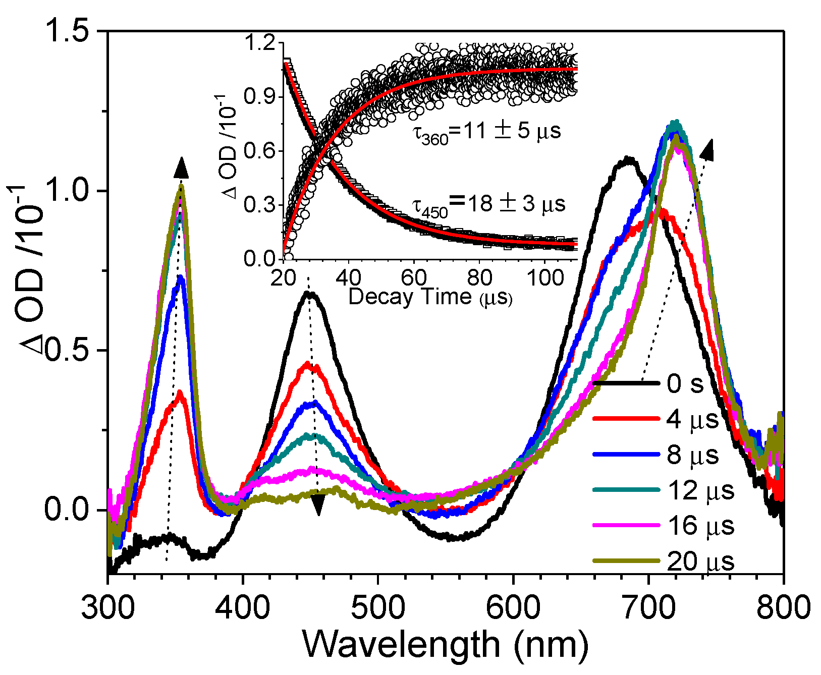
Time-Resolved Spectroscopic Study
3. Discussion
4. Materials and Methods
4.1. Fs-TA and Ns-TA Experiments
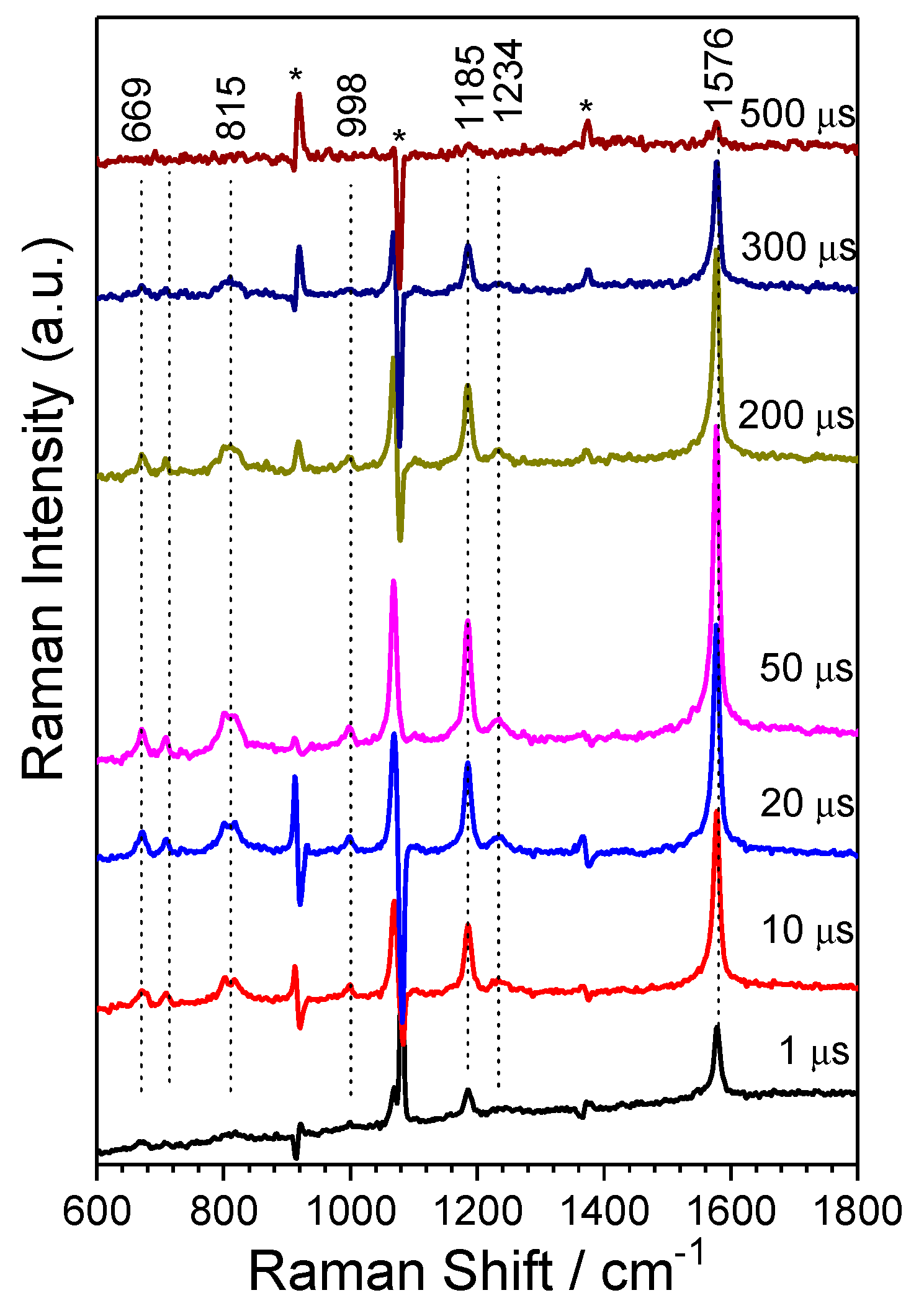
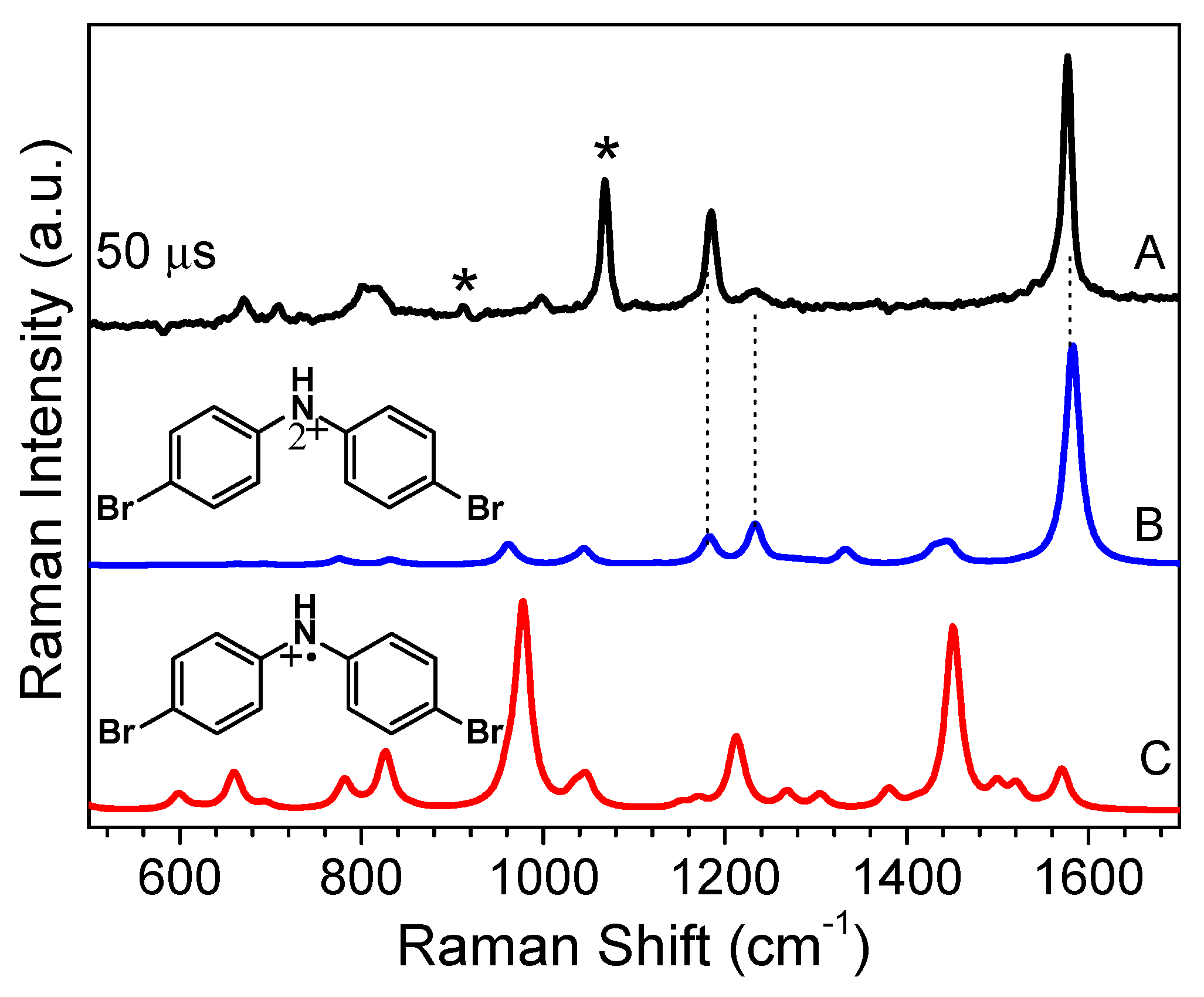
4.2. Ns-TR3 Experiments
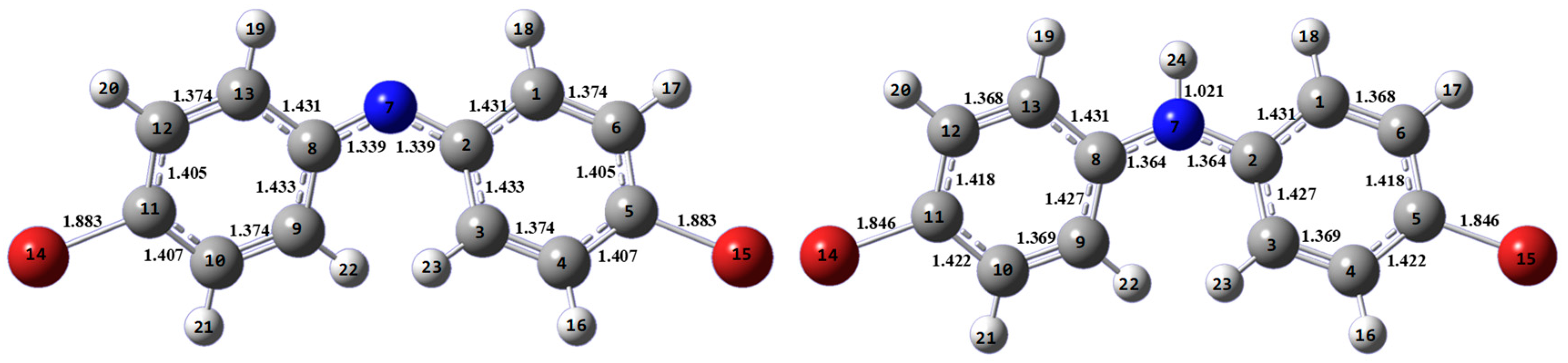
4.3. DFT Calculations
Supplementary Materials
Author Contributions
Funding
Acknowledgments
Conflicts of Interest
References
- Miller, E.C.; Lotlikar, P.D.; Miller, J.A.; Butler, B.W.; Irving, C.C.; Hill, J.T. Reactions in vitro of some tissue nucleophiles with the glucuronide of the carcinogen N-hydroxy-2-acetylaminofluorene. Mol. Pharmacol. 1968, 4, 147–154. [Google Scholar] [PubMed]
- Miller, E.C.; Miller, J.A. Mechanisms of chemical carcinogenesis: Nature of proximate carcinogens and interactions with macromolecules. Pharmacol. Rev. 1966, 18, 805–838. [Google Scholar] [PubMed]
- Poirier, L.A.; Miller, J.A.; Miller, E.C.; Sato, K. N-benzoyloxy-N-methyl-4-aminoazobenzene: Its carcinogenic activity in the rat and its reactions with proteins and nucleic acids and their constituents in vitro. Cancer Res. 1967, 27, 1600–1613. [Google Scholar] [PubMed]
- Novak, M.; Kennedy, S.A. Inhibitory effect of DNA structure on the efficiency of reaction of guanosine moieties with a nitrenium ion. J. Phys. Org. Chem. 1998, 11, 71–76. [Google Scholar] [CrossRef]
- Novak, M.; Kennedy, S.A. Selective trapping of N-acetyl-N-(4-biphenylyl)nitrenium and N-acetyl-N-(2-fluorenyl)nitrenium ions by 2’-deoxyguanosine in aqueous solution. J. Am. Chem. Soc. 1995, 117, 574–575. [Google Scholar] [CrossRef]
- McClelland, R.A.; Ahmad, A.; Dicks, A.P.; Licence, V.E. Spectroscopic characterization of the initial C8 intermediate in the reaction of the 2-fluorenylnitrenium ion with 2‘-deoxyguanosine. J. Am. Chem. Soc. 1999, 121, 3303–3310. [Google Scholar] [CrossRef]
- Humphreys, W.G.; Kadlubar, F.F.; Guengerich, F.P. Mechanism of C8 alkylation of guanine residues by activated arylamines: Evidence for initial adduct formation at the N7 position. Proc. Natl. Acad. Sci. USA 1992, 89, 8278–8282. [Google Scholar] [CrossRef]
- McClelland, R.A.; Gadosy, T.A.; Ren, D. 1997 Alfred Bader Award Lecture Reactivities of arylnitrenium ions with guanine derivatives and other nucleophiles. Can. J. Chem. 1998, 76, 1327–1337. [Google Scholar] [CrossRef]
- Chan, P.Y.; Kwok, W.M.; Lam, S.K.; Chiu, P.; Phillips, D.L. Time-resolved resonance Raman observation of the 2-fluorenylnitrenium ion reaction with guanosine to form a C8 intermediate. J. Am. Chem. Soc. 2005, 127, 8246–8247. [Google Scholar] [CrossRef]
- McIlroy, S.; Moran, R.J.; Falvey, D.E. Photogenerated nitrenium ions: A search for triplet-state reactivity in the chemistry of the diphenylnitrenium ion. J. Phys. Chem. A 2000, 104, 11154–11158. [Google Scholar] [CrossRef]
- Moran, R.J.; Falvey, D.E. Photogenerated diarylnitrenium ions: Laser flash photolysis and product studies on diphenylnitrenium ion generated from photolysis of 1-(N,N-diphenylamino)pyridinium ions. J. Am. Chem. Soc. 1996, 118, 8965–8966. [Google Scholar] [CrossRef]
- Kung, A.C.; McIlroy, S.P.; Falvey, D.E. Diphenylnitrenium ion: Cyclization, electron transfer, and polymerization reactions. J. Org. Chem. 2005, 70, 5283–5290. [Google Scholar] [CrossRef] [PubMed]
- Thomas, S.I.; Falvey, D.E. N,N-di(4-halophenyl)nitrenium ions: Nucleophilic trapping, aromatic substitution, and hydrogen atom transfer. J. Org. Chem. 2007, 72, 4626–4634. [Google Scholar] [CrossRef] [PubMed]
- Srivastava, S.; Ruane, P.H.; Toscano, J.P.; Sullivan, M.B.; Cramer, C.J.; Chiapperino, D.; Reed, E.C.; Falvey, D.E. Structures of reactive nitrenium ions: Time-resolved infrared laser flash photolysis and computational studies of substituted N-methyl-N-arylnitrenium ions. J. Am. Chem. Soc. 2000, 122, 8271–8278. [Google Scholar] [CrossRef]
- Du, L.; Lan, X.; Yan, Z.; Zhu, R.; Phillips, D.L. Time-resolved spectroscopic study of N,N-Di(4-bromo)nitrenium ions in selected solutions. Molecules 2018, 23, 3182. [Google Scholar] [CrossRef] [PubMed]
- Xue, J.D.; Li, Y.F.; Du, L.L.; Du, Y.; Tang, W.J.; Zheng, X.M.; Phillips, D.L. Direct observation of 4-phenoxyphenylnitrenium ion: A transient absorption and transient resonance Raman study. J. Phys. Chem. B 2015, 119, 14720–14727. [Google Scholar] [CrossRef] [PubMed]
- Xue, J.; Du, L.; Zhu, R.; Huang, J.; Phillips, D.L. Direct time-resolved spectroscopic observation of arylnitrenium ion reactions with guanine-containing DNA oligomers. J. Org. Chem. 2014, 79, 3610–3614. [Google Scholar] [CrossRef] [PubMed]
- Guo, Z.; Lin, X.F.; Zhao, C.Y.; PhillipS, D.L. Density functional theory study of water-assisted deprotonation of the C8 intermediate in the reaction of the 2-fluorenylnitrenium ion with guanosine to form a C8 adduct. J. Mol. Struc.-Theochem 2008, 848, 119–127. [Google Scholar] [CrossRef]
- Xue, J.D.; Guo, Z.; Chan, P.Y.; Chu, L.M.; But, T.Y.S.; Phillips, D.L. Time-resolved resonance Raman study of the reaction of the 2-fluorenylnitrenium ion with 2-fluorenylazide. J. Phys. Chem. A 2007, 111, 1441–1451. [Google Scholar] [CrossRef]
- Xue, J.D.; Chan, P.Y.; Du, Y.; Guo, Z.; Chung, C.W.Y.; Toy, P.H.; Phillips, D.L. Time-resolved resonance Raman investigation of the 2-fluorenylnitrenium ion reactions with C8 guanosine derivatives. J. Phys. Chem. B 2007, 111, 12676–12684. [Google Scholar] [CrossRef]
- McClelland, R.A.; Kahley, M.J.; Davidse, P.A.; Hadzialic, G. Acid-base properties of arylnitrenium ions. J. Am. Chem. Soc. 1996, 118, 4794–4803. [Google Scholar] [CrossRef]
- Srivastava, S.; Toscano, J.P.; Moran, R.J.; Falvey, D.E. Experimental confirmation of the iminocyclohexadienyl cation-like structure of arylnitrenium ions: Time-resolved IR studies of diphenylnitrenium ion. J. Am. Chem. Soc. 1997, 119, 11552–11553. [Google Scholar] [CrossRef]
- Du, L.; Li, M.D.; Zhang, Y.; Xue, J.; Zhang, X.; Zhu, R.; Cheng, S.C.; Li, X.; Phillips, D.L. Photoconversion of β-Lapachone to α-Lapachone via a protonation-assisted singlet excited state pathway in aqueous solution: A time-resolved spectroscopic study. J. Org. Chem. 2015, 80, 7340–7350. [Google Scholar] [CrossRef]
- Du, L.L.; Zhu, R.X.; Xue, J.D.; Du, Y.; Phillips, D.L. Time-resolved spectroscopic and density functional theory investigation of the photochemistry of suprofen. J. Raman Spectrosc. 2015, 46, 117–125. [Google Scholar] [CrossRef]
- Du, L.L.; Zhang, X.T.; Xue, J.D.; Tang, W.J.; Li, M.D.; Lan, X.; Zhu, J.R.; Zhu, R.X.; Weng, Y.X.; Li, Y.L.; et al. Influence of water in the photogeneration and properties of a bifunctional quinone methide. J. Phys. Chem. B 2016, 120, 11132–11141. [Google Scholar] [CrossRef]
- Du, L.L.; Qiu, Y.F.; Lan, X.; Zhu, R.X.; Phillips, D.L.; Li, M.D.; Dutton, A.S.; Winter, A.H. Direct detection of the open-shell singlet phenyloxenium ion: An atom-centered diradical reacts as an electrophile. J. Am. Chem. Soc. 2017, 139, 15054–15059. [Google Scholar] [CrossRef]
- Xiong, W.J.; Du, L.L.; Lo, K.C.; Shi, H.T.; Takaya, T.; Iwata, K.; Chan, W.K.; Phillips, D.L. Control of electron flow direction in photoexcited cycloplatinated complex containing conjugated polymer-single-walled carbon nanotube hybrids. J. Phys. Chem. Lett. 2018, 9, 3819–3824. [Google Scholar] [CrossRef]
- Chan, K.T.; Lam, T.L.; Yu, D.; Du, L.; Phillips, D.; Kwong, C.-L.; Tong, G.; Cheng, G.; Che, C.M. Strongly luminescent tungsten emitters with emission quantum yields up to 84%: TADF and high-efficiency molecular tungsten OLEDs. Angew. Chem. Int. Ed. 2019, 58, 14896–14900. [Google Scholar] [CrossRef]
- Yan, Z.P.; Du, L.L.; Lan, X.; Li, Y.C.; Wang, W.C.; Phillips, D.L. Time-resolved spectroscopic and density functional theory investigation of the photogeneration of a bifunctional quinone methide in neutral and basic aqueous solutions. Molecules 2018, 23, 3102. [Google Scholar] [CrossRef]
- Frisch, M.J.; Trucks, G.W.; Schlegel, H.B.; Scuseria, G.E.; Robb, M.A.; Cheeseman, J.R.; Scalmani, G.; Barone, V.; Mennucci, B.; Petersson, G.A.; et al. Gaussian 09; Gaussian, Inc.: Wallingford, CT, USA, 2009. [Google Scholar]


© 2019 by the authors. Licensee MDPI, Basel, Switzerland. This article is an open access article distributed under the terms and conditions of the Creative Commons Attribution (CC BY) license (http://creativecommons.org/licenses/by/4.0/).
Share and Cite
Du, L.; Yan, Z.; Bai, X.; Liang, R.; Phillips, D.L. Time-Resolved Spectroscopic Study of N,N–Di(4–bromo)nitrenium Ions in Acidic Aqueous Solution. Int. J. Mol. Sci. 2019, 20, 5512. https://0-doi-org.brum.beds.ac.uk/10.3390/ijms20215512
Du L, Yan Z, Bai X, Liang R, Phillips DL. Time-Resolved Spectroscopic Study of N,N–Di(4–bromo)nitrenium Ions in Acidic Aqueous Solution. International Journal of Molecular Sciences. 2019; 20(21):5512. https://0-doi-org.brum.beds.ac.uk/10.3390/ijms20215512
Chicago/Turabian StyleDu, Lili, Zhiping Yan, Xueqin Bai, Runhui Liang, and David Lee Phillips. 2019. "Time-Resolved Spectroscopic Study of N,N–Di(4–bromo)nitrenium Ions in Acidic Aqueous Solution" International Journal of Molecular Sciences 20, no. 21: 5512. https://0-doi-org.brum.beds.ac.uk/10.3390/ijms20215512