Article

In Vitro Activity of the Bacteriophage Endolysin HY-133 against Staphylococcus aureus Small-Colony Variants and Their Corresponding Wild Types

1
Institute of Medical Microbiology, University Hospital Münster (UKM), 48149 Münster, Germany
2
HYpharm GmbH, 82347 Bernried, Germany
*
Author to whom correspondence should be addressed.
Present address: Ursula Kaspar, Landeszentrum Gesundheit Nordrhein-Westfalen, 44801 Bochum, Germany.
Present address: Dennis Knaack, St. Franziskus-Hospital GmbH, 48145 Münster, Germany.
§
Present address: Christof von Eiff, Pfizer Pharma GmbH, 10785 Berlin, Germany.
Int. J. Mol. Sci. 2019, 20(3), 716; https://0-doi-org.brum.beds.ac.uk/10.3390/ijms20030716
Submission received: 20 December 2018 / Revised: 1 February 2019 / Accepted: 2 February 2019 / Published: 7 February 2019
(This article belongs to the Special Issue Drug Resistance: Mechanisms and New Strategies)

Abstract

:
Nasal carriage of methicillin-susceptible (MSSA) and methicillin-resistant Staphylococcus aureus (MRSA) represents both a source and a risk factor for subsequent infections. However, existing MRSA decolonization strategies and antibiotic treatment options are hampered by the duration of administration and particularly by the emergence of resistance. Moreover, beyond classical resistance mechanisms, functional resistance as the formation of the small-colony variant (SCV) phenotype may also impair the course and treatment of S. aureus infections. For the recombinant bacteriophage endolysin HY-133, rapid bactericidal and highly selective in vitro activities against MSSA and MRSA has been shown. In order to assess the in vitro efficacy of HY-133 against the SCV phenotype, minimal inhibitory (MIC) and minimal bactericidal concentrations (MBC) were evaluated on clinical SCVs, their isogenic wild types, as well as on genetically derived and gentamicin-selected SCVs. For all strains and growth phases, HY-133 MIC and MBC ranged between 0.12 and 1 mg/L. Time-kill studies revealed a fast-acting bactericidal activity of HY-133 resulting in a ≥3 − log10 decrease in CFU/mL within 1 h compared to oxacillin, which required 4–24 h. Since the mode of action of HY-133 was independent of growth phase, resistance pattern, and phenotype, it is a promising candidate for future S. aureus decolonization strategies comprising rapid activity against phenotypic variants exhibiting functional resistance.

1. Introduction

Staphylococcus aureus represents a widely disseminated and invasive pathogen causing clinically important and life-threatening infectious diseases [1,2]. Since S. aureus colonizes 30% or more of all humans, primarily the vestibules of the nasal cavity, the risk for subsequent infections originating from the patients’ own flora is significantly increased [2,3,4,5,6,7]. The possession of healthcare- (HA-), community- (CA-), and livestock-associated (LA-) methicillin-resistant S. aureus (MRSA) drastically aggravates the treatment by limiting the therapeutic options and necessitates expansive preventive measures, therefore resulting in additional morbidity, mortality, and costs [1,8,9]. Resistances also emerged against remaining or newly developed antibiotic classes with anti-MRSA activity such as glycopeptides, oxazolidinones, cyclic lipopeptides, and anti-MRSA cephalosporins, often shortly after their introduction [10,11,12,13]. Current strategies to prevent nosocomial S. aureus infections, particularly those due to MRSA, are frequently based on the topical application of the antibiotic agent mupirocin [14,15,16], but a considerable number of patients show recolonization [17,18,19]. Possible reasons include its bacteriostatic mode of action and the rising emergence of resistance against this substance [20,21,22,23,24].
Beyond these classical resistance mechanisms, resistance based on microbial biofilm lifestyle or the formation of the small-colony variant (SCV) phenotype may render microbial isolates functionally resistant even if they are susceptible to antibiotics. Moreover, the administration of antibiotics, in particular long-term exposure, may select for the generation of SCVs [25,26]. The naturally occurring SCV phenotype is capable of evading the host´s immune response by switching from an extracellular to an intracellular lifestyle, resulting in chronic, relapsing, and often therapy-refractory infections [27,28,29]. Furthermore, nasal colonization by SCVs and intracellular persistence in nasal epithelium have been shown [30,31].
The ability of SCVs to persist within eukaryotic cells and their changed physiology is also of particular therapeutic significance, as the activity of many antibiotic compounds may be attenuated. In particular, β-lactams, which in general represent the most effective anti-staphylococcal agents, are not bioavailable in the intracellular milieu [32]. Moreover, SCVs may be tolerant against β-lactams (e.g., oxacillin). The tolerance phenomenon was already shown for dormant and slow-growing cells and might also result in treatment failure [33,34,35,36,37]. Reduced susceptibilities of staphylococcal SCVs to antifolate antibiotics, aminoglycosides, and cationic antimicrobial compounds are also common traits of this phenotype compared to isolates displaying the normal phenotype [38,39]. Compounds of other antibiotic classes usually show minimal inhibitory concentrations that are similar to those measured for strains with a normal phenotype, but they are frequently less bactericidal in the case of SCVs [40,41]. Typical SCV colonies differ from the parental wild type (WT) not only in slower growth with less or no pigmentation and hemolytic activity, but also in several biochemical traits, such as altered expression of virulence factors, decreased respiration, as well as different forms of auxotrophisms, e.g., for hemin, menadione, and/or thymidine [42,43,44]. Moreover, SCVs are characterized by major ultrastructural alterations of the cell wall and cell separation structures [45,46,47].
Obviously, the limited treatment options of S. aureus infections due to classical and functional resistance mechanisms necessitate alternative prophylactic and therapeutic options. Bacteriophage-derived endolysins exhibit the ability to disrupt bacterial cell walls in a highly specific manner [48]. Recombinant endolysins are characterized by a modular design comprising an enzymatically active domain (EAD) connected by a short linker region with specific cell wall recognition site (cell wall-binding domain, CBD) [49]. In the pre-antibiotic era, phages were successfully used for the treatment of various bacterial infections; however, the introduction of antibiotics stopped their use in Western medicine [50]. Only in recent years has research on this subject been revived, resulting in numerous recombinant endolysins that are active in vitro and/or in vivo against several gram-negative and gram-positive bacteria including Pseudomonas aeruginosa [51], Streptococcus spp. [52,53,54,55], and Staphylococcus spp. [56,57,58,59,60,61,62]. Moreover, phage endolysins were shown to be active against difficult-to-treat bacterial infections caused by phenotypically resistant bacterial populations, e.g., by being embedded in biofilms [63,64,65].
The recombinant endolysin HY-133 (HYpharm GmbH, Bernried, Germany) used in this study represents a stability-optimized derivative of endolysin PRF-119 [59,60]. The modular structure of HY-133 comprises a CBD from lysostaphin, responsible for the specific attachment to the peptidoglycan of S. aureus [59,62]. Its EAD consists of the cysteine, histidine-dependent amidohydrolase/peptidase (CHAP) originating from the recombinant endolysin LysK of phage K [62,66]. Within the peptidoglycan of S. aureus, this CHAP domain cleaves between d-alanine at the termini of the tetrapeptide and glycine of the pentaglycine crossbridge, and is thereby responsible for the lytic mode of action [67]. HY-133 was previously shown to be highly active against methicillin-susceptible Staphylococcus aureus (MSSA) and MRSA isolates [59,60,61,62]. However, the efficacy of Hy-133 against SCVs has yet to be determined. Here, the activity of HY-133 by the determination of the minimal inhibitory (MIC) and minimal bactericidal concentrations (MBC) and time-kill analyses was systematically analyzed against a well-characterized collection of clinically derived MSSA SCVs and mutants displaying the SCV phenotype in comparison with their parental WTs. Oxacillin was used as comparator agent.

2. Results

2.1. Genotyping by Pulsed-Field Gel Electrophoresis (PFGE)

We previously demonstrated the very fast and high bactericidal in vitro activity of the recombinant endolysin HY-133 against several clinical MSSA and (LA-)MRSA isolates [59,60,61,62]. To further investigate the activity of HY-133, it was challenged with 12 clinical S. aureus strain pairs, each comprising WT and stable SCV phenotypes. Since the pulsed-field gel electrophoresis (PFGE) fragment patterns of the WT-SCV pairs were indistinguishable or varied in only one band, all strain pairs were considered clonally identical according to published guidelines [68] (data not shown).

2.2. Antimicrobial Susceptibility Testing

MIC50, MBC50, as well as MIC90 and MBC90 with the corresponding ranges were calculated for the different growth phases and phenotypes for HY-133 and oxacillin and are listed in Table 1 for the 12 clinical strain pairs (n = 24). Table 2 lists the MIC and MBC values evaluated for the isogenic 6850 triplet consisting of the WT isolate 6850, its gentamicin-selected SCV JB1, and its hemB mutant SCV IIb13. MIC50/90 and MBC50/90 for HY-133 tested against the 12 clinical strain pairs were similar for the different phenotypes or growth phases. The same applied for oxacillin which, however, exhibited slightly higher MIC50/90 and MBC50/90 values for clinical strains than HY-133 (Table 1).
Within the 6850 triplet, the MIC and MBC of HY-133 were similar for the different phenotypes (0.12–0.25 mg/L) when measured from cultures in stationary growth phase. Measurements from logarithmic growth cultures revealed the same MIC and MBC values against 6850 (WT) and IIb13 (SCV), being 0.25 mg/L for both strains and reflecting values detected during the stationary phase. However, HY-133 MIC and MBC values measured against the gentamicin-selected SCV JB1 were increased (1 mg/L) in this growth phase. For oxacillin, MIC and MBC were substantially lower against the 6850-derived SCVs JB1 and IIb13 than against the corresponding WT and the clinical strains, irrespective of the growth phase (Table 2).
Furthermore, for HY-133, the median MBCs were higher than the median MICs in 4.2% (n = 2/48) of cases with an MBC:MIC ratio of 2. In either case, the SCV phenotype tested from stationary growth cultures was affected. However, when strains were treated with oxacillin, 20.8% (n = 10/48) showed a higher median MBC than MIC with 40% (n = 4/10) being WTs and 60% (n = 6/10) being SCVs, respectively, and an MBC:MIC ratio of 2 in each case.
The quality control (QC) strain ATCC 29213 showed oxacillin values in the range as required by the Clinical & Laboratory Standards Institute (CLSI) guidelines [69]. There is no official QC range available for HY-133, but the MICs detected for the QC strain ATCC 29213 were always within the in-house established limits.

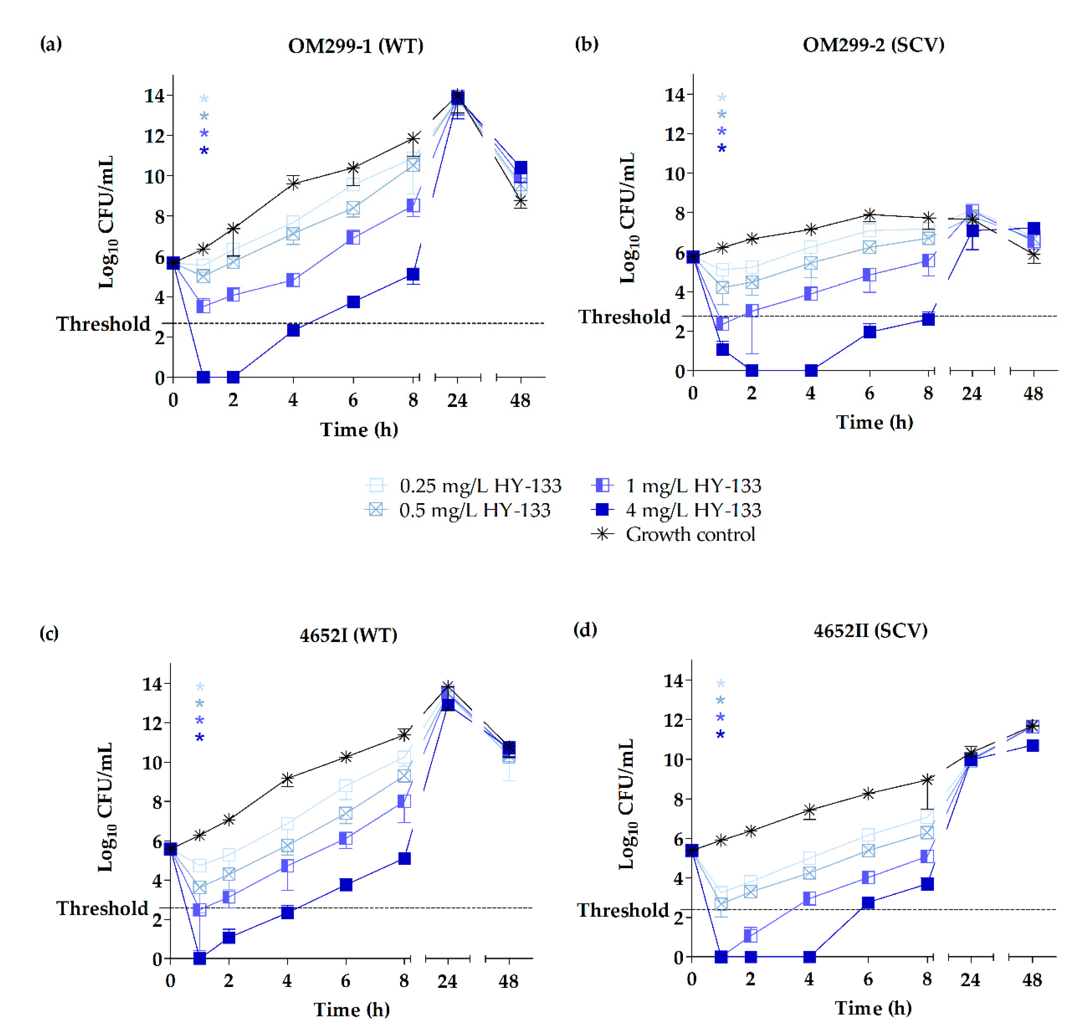
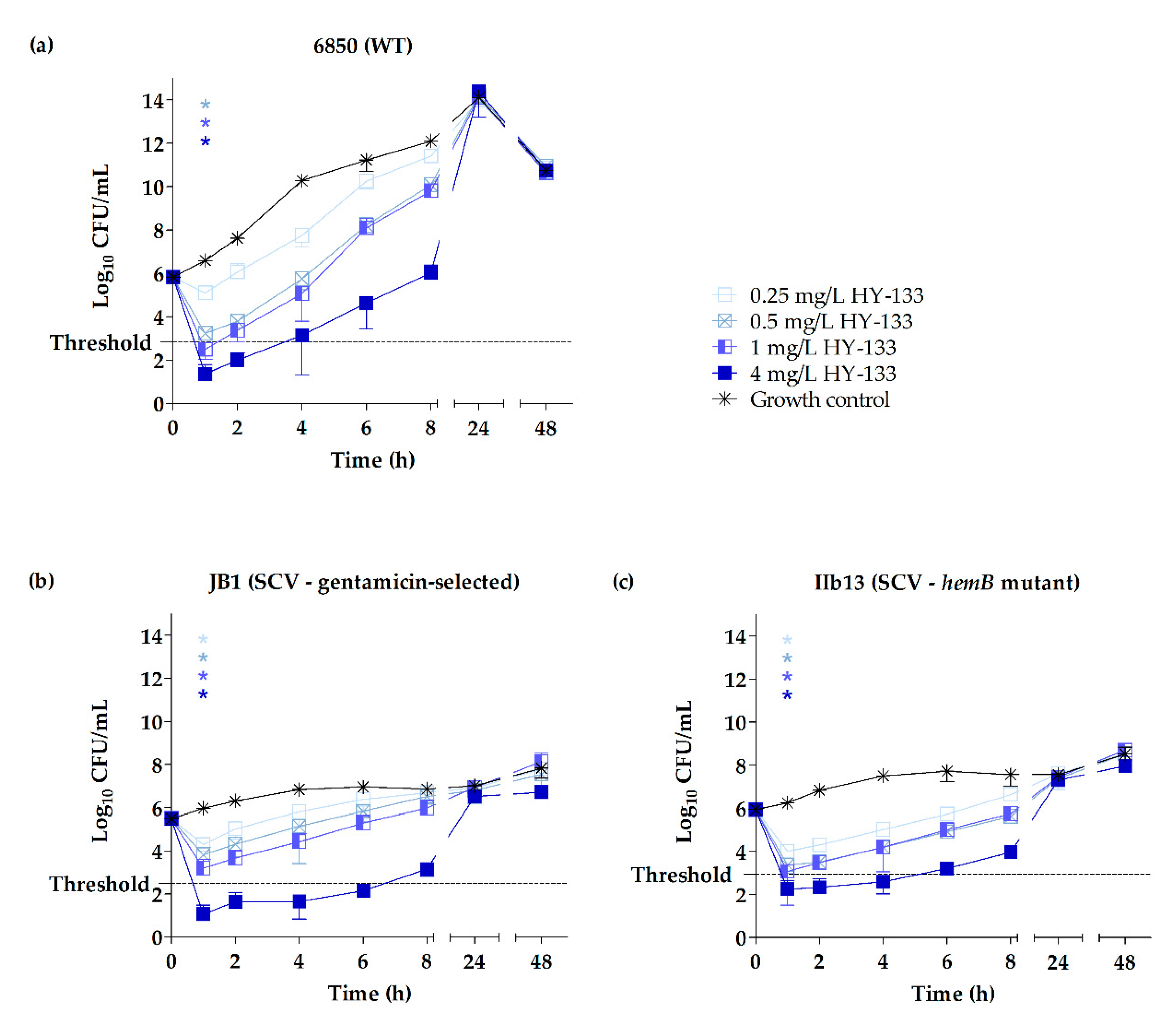
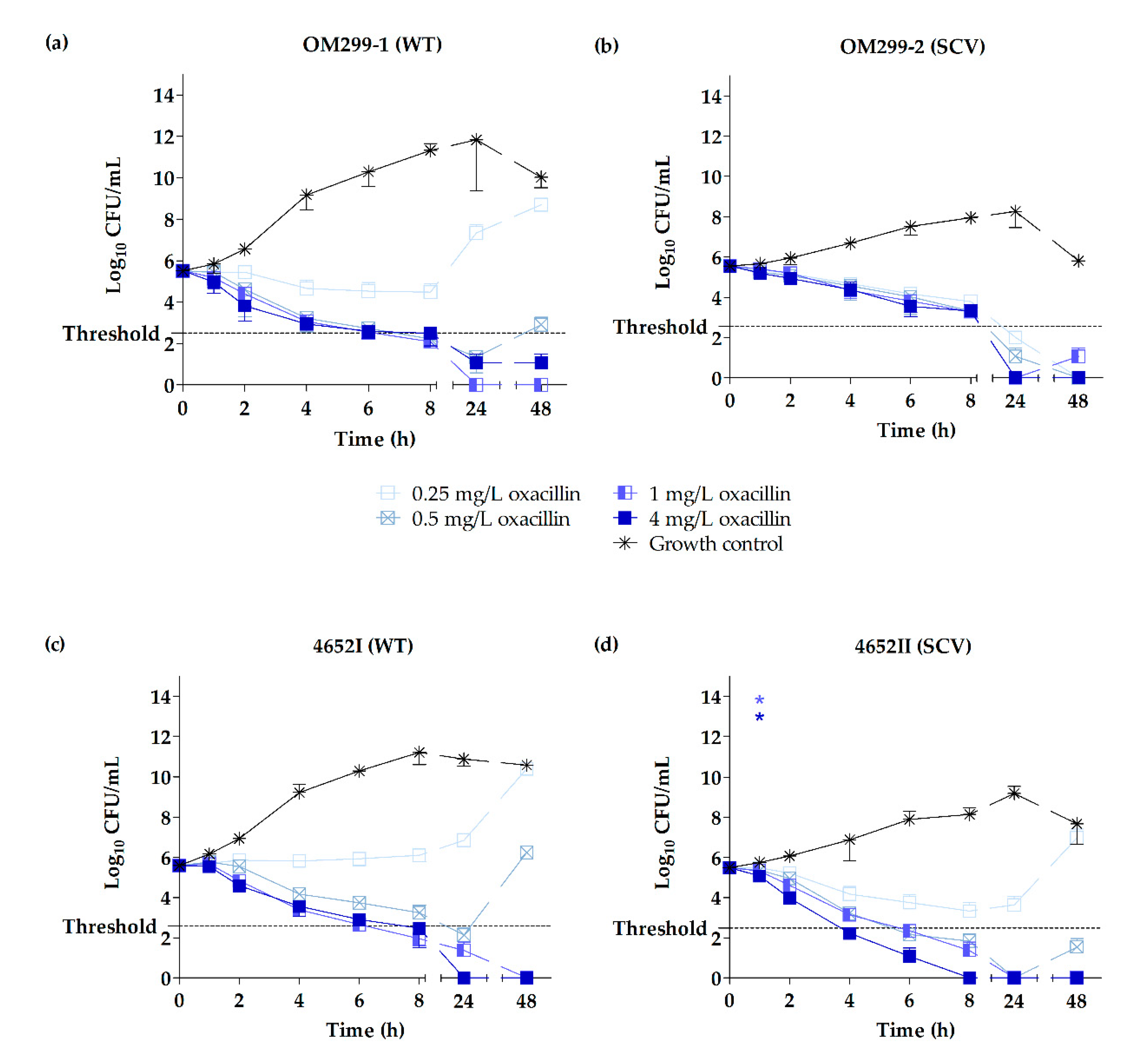
2.3. Time-Kill Studies

Time-kill curves were performed for the two representative clinical strain pairs OM299 and 4652 as well as for the strains of triplet 6850. Time-kill curves revealed distinct growth characteristics of the two phenotypes with clinical and 6850-derived SCVs exhibiting a clear growth retardation compared to the WTs, as described in previous studies [42,43,70]. For the clinical strain pairs OM299 and 4652, this difference in growth was more pronounced for strain pair OM299.
Killing kinetics of HY-133 against the clinical and 6850-derived strains revealed significant differences in the killing rates of HY-133 compared to the untreated growth controls (p ≤ 0.001 for all concentrations, except for 0.25 mg/L HY-133 against strain 6850), already after 1 h of incubation. At this time point, a ≥3 − log10 decrease in CFU/mL (99.9% killing) was shown for all strains irrespective of the phenotype when applying a concentration of 4 mg/L (Figure 1 and Figure 2, Table 3, and Table S1 in the Supplementary Material). Furthermore, this concentration led to a reduction of cell numbers below the detection limit for all clinical isolates tested for at least 1 h (Figure 1). However, regarding the strain 6850-derived triplet, neither the WT nor the in vitro-generated SCVs reached this limit (Figure 2). Corresponding levels of oxacillin (4 mg/L) were bactericidal against the clinical WTs after a time interval of 8 h. By contrast, the time until a bactericidal effect was detected in the clinical SCVs varied between 4 h for SCV 4652II and 24 h for SCV OM299-2. The detection limit for oxacillin (4 mg/L) was reached between 8–24 h for all strains and phenotypes (Figure 3, Table 3 and Table S1). After 1 h of incubation, significant differences in the killing rate of oxacillin versus the untreated growth control were observed only for the SCV strain 4652II applying 1 and 4 mg/L of the substance (p ≤ 0.001). A concentration of 1 mg/L of HY-133 was bactericidal for most of the strains within 1 h except for the clinical WT strain OM299-1 as well as for SCVs JB1 and IIb13 (Table 3). All other HY-133 concentrations applied caused no bactericidal effect. For oxacillin, a bactericidal effect was also reached for concentrations of 1 and 0.5 mg/L. For these concentrations, time intervals until reaching bactericidal effect differed between 6 and 24 h irrespective of the phenotype. Moreover, for SCV OM299-2, a concentration of 0.25 mg/L oxacillin was bactericidal after 24 h (Table 3).
The killing effect for HY-133 was similar for SCVs and their respective WTs (Figure 1 and Figure 2). After prolonged incubation (4–24 h for 4 mg/L HY-133), time-kill curves for HY-133 revealed a regrowth phenomenon for all tested strains and phenotypes, even for concentrations initially resulting in values below the detection limit. For clinical SCVs as well as for SCVs JB1 and IIb13, this regrowth appeared slower than for WTs when applying 4 mg/L HY-133 (Figure 1 and Figure 2). For oxacillin, regrowth above the threshold after prolonged incubation (>24 h for 0.5 mg/L oxacillin) could also be detected for concentrations less than or equal to 0.5 mg/L for both WTs, whereas for the SCVs, regrowth above the threshold could only be observed for the lowest concentration (0.25 mg/L) for strain 4652 (Figure 3).

3. Discussion

In recent years, several in vitro and in vivo studies revealed the high potential of endolysins for the treatment of several bacterial infections [49]. HY-133 represents one of its promissing candidates in anti-S. aureus treatment and prevention regimens. This recombinant phage endolysin and its precursor PRF-119 were previously shown to be highly active against different strains of MSSA, MRSA (including strains possessing mecA, mecB, or mecC genes), borderline oxacillin-resistant isolates, as well as isolates that were found to be resistant to ceftaroline/ceftobiprole [59,60,61,62]. Recently, we demonstrated that HY-133 is as effective against S. aureus isolates as mupirocin or daptomycin, with an even faster mode of killing [61].
In contrast to the vast majority of S. aureus clonal lineages possessing polyribitol phosphate wall teichoic acid (WTA) [71], the unusual polyglycerol phosphate WTA-producing S. aureus PS 187, which belongs to a distant S. aureus lineage (phage type 187, biotype E) often isolated from dogs [72], was less susceptible to PRF-119. Thus, changes of the cell wall composition or structure may influence the endolysin activity [59]. Also SCVs, functionally resistant to several antibiotic classes, are notorious for gross ultrastructural changes of the cell wall. As shown by transmission and scanning electron microscopy, they are characterized by the formation of pleomorphic cocci up to eight times larger than those of WTs and impaired cell separation with multiple cross walls [45,46,47,73]. Furthermore, SCVs exhibit slower growth characteristics in contrast to their parental WT isolates. Lastly, SCVs are characterized by altered metabolism [42,43,74]. As the mechanism in which HY-133 binds to the cell wall is very specific and its mode of action does not depend on the target’s metabolic processes, the analysis of its efficacy against the SCV phenotype in comparison to the corresponding WT is a further necessary step in the determination of the anti-staphylococcal activity of this substance.
To consider the divergent growth characteristics of SCVs [27], the determination of MIC and MBC values was not only performed from cultures in stationary growth, but also from the logarithmic growth phase. MIC and MBC determinations revealed a highly bactericidal effect of HY-133 against SCVs, independent of growth phase and to the same extent as that observed for the corresponding WTs. In contrast, the endolysin encoded by LM12, a Kayvirus bacteriophage with broad S. aureus host range, was shown to exhibit growth phase-dependent activity [75]. The killing kinetics of HY-133 revealed a very fast mode of action until 99.9% of the bacterial population was killed. This rapid mode of action within 1 h was also observed for the gentamicin-selected and the genetically defined SCVs of the highly cytotoxic strain 6850. This finding reflects the observation of another study, where the exceptionally fast onset of action of HY-133 within the first minutes of application for MSSA and MRSA WT phenotypes was demonstrated [61]. The observed prolongation of the bactericidal effect for the SCVs treated with HY-133 (4 mg/L) is most likely due to the slower growth of the SCVs.
Whereas MIC and MBC values for oxacillin did not show significant differences to MIC and MBC values of HY-133, time-kill studies revealed that the onset of bactericidal effect of oxacillin (4 mg/L) was not as fast as that detected for HY-133 within 1 h and was reached only after 4 to 24 h. For strain pair OM299, including an SCV exhibiting a considerable growth retardation compared to its WT, the detected killing effect of the SCV by oxacillin was clearly delayed compared to its WT. For β-‍lactams, tolerance is a known phenomenon in slow-growing or dormant bacteria [33,34,35], which appears due to the slower assembly of the bacterial cell wall and necessitates a longer minimum duration of antimicrobial treatment to reach the same bactericidal effect [37]. However, the tolerance phenomenon can result in treatment failures [36,37]. Since the CLSI standard definition of antimicrobial tolerance is defined as an MBC:MIC ratio of ≥32 (M26-A) [76], a tolerance phenomenon could not be confirmed by the MBC:MIC ratio calculation in this case. However, according to the terminology used by Brauner et al. [37], SCV OM299-2 exhibited a longer minimum duration of treatment and therefore might be tolerant. On the contrary, SCV 4652II—exhibiting only a minor growth retardation compared to its WT—showed no signs for tolerance, suggesting that tolerance towards β-lactams might not be a general phenomenon for SCVs. Moreover, for a methicillin-resistant menD SCV mutant, hyper-susceptibility to β-lactams in a macrophage cell model has been shown [77]. For HY-133, no tolerance phenomenon was detected for any of the SCVs including the gentamicin-selected and genetically defined SCVs.
This study highlights one of the most relevant advantages of HY-133, i.e., its particular mode of action. The modular design of HY-133 is responsible for the species-specific cleavage of the peptidoglycan of S. aureus, thus inducing cellular lysis. This form of cell death occurs independently of cellular processes. In contrast, most conventional antibiotics operate by inhibiting essential cellular functions [78], e.g., inhibition of the cell wall biosynthesis by the β-lactam oxacillin. However, in SCVs, these processes can be altered, leading to failures in antibiotic treatment. Long-term treatment with trimethoprim-sulfamethoxazole (TMP-SXT) that inhibits the dihydrofolate reductase within the bacterial folate pathway may result in SCVs auxotrophic for thymidine and resistant to TMP-SXT [38]. Furthermore, SCVs with deficiencies in electron-transport show resistance to aminoglycosides. Due to their lack in membrane potential that is normally generated by electron-transport, the bacterial uptake of aminoglycosides is impaired [25,39,40,79].
For HY-133, the killing kinetics revealed an in vitro regrowth phenomenon for all strains tested appearing after prolonged incubation. This regrowth occurred even after an initial killing of 99.9% of the bacerial population. Its extent depended on both the concentration of HY-133 and the strain used. A similar in vitro regrowth phenomenon was previously shown for clinical MSSA and MRSA isolates [61]. Further investigations are warranted to understand whether the inactivation of HY-133 is an exclusive in vitro phenomenon caused, e.g., by environmental conditions (medium, shearing forces etc.) which may irreversibly degrade the endolysin, complete the consumption of HY-133, or escape by remnant susceptible cells via hiding mechanisms.
The present study demonstrates that HY-133 caused very rapid cell death independent of the rate of bacterial growth, the consistency of the cell wall, and the distinct growth phase. Although mutants resistant to the bacteriocin lysostaphin have been created [80,81], selection pressure for resistances against endolysins seems to be absent or very low [49,55,82,83]. This is most likely due to the fact that the respective regions of the bacterial cell wall are highly conserved [84,85]. Thus, since there were also no adverse side effects reported until now [86], the recombinant phage endolysin HY-133 may serve as a future narrow-spectrum antimicrobial drug used in treatment and infection prevention purposes for the eradication of normal-growing S. aureus cells as well as for bacterial cells with impaired growth. S. aureus SCVs possess the capacity to enter and persist in host cells [73,79,87,88]. Therefore, further cell-culture experiments are necessary to elucidate the cell permeability of HY-133. Recently, the penetration of mammalian cells was demonstrated for the anti-streptococcal endolysin PlyC that was able to readily kill intracellular Streptococcus pyogenes without damaging the host cell [86].
In summary, HY-133 was shown to be highly bactericidal in the logarithmic as well as in the stationary growth phases of all tested clinical S. aureus WT and SCV isogenic strain combinations, irrespective of their exhibited phenotype or genetic background. Due to its highly bactericidal activity and its high specificity, HY-133 is a promising candidate for future anti-staphylococcal therapy, decolonization, and prevention, particularly for difficult-to-treat phenotypes such as SCVs.

4. Materials and Methods

4.1. Bacterial Strains and Maintenance

Twelve clinical S. aureus WT isolates and their corresponding stable SCVs were obtained from patients of the University Hospital Münster (UKM), Münster, Germany (see Table 4) [39,89,90]. Each strain pair was isolated from one patient either consecutively or concurrently. All isolates were categorized as methicillin-susceptible by (i) disk diffusion methodology (oxacillin susceptibility disks, Oxoid, Hampshire, United Kingdom; Mueller Hinton II agar, MHA, Becton Dickinson, Franklin Lakes, NJ, USA) and (ii) absence of methicillin resistance-encoding mecA, mecB, and mecC genes, as described elsewhere [91,92,93]. Isolates were characterized as SCVs according to the following criteria: pinpoint colonies on Columbia blood agar (BBLTM Columbia agar with 5% sheep blood, Becton Dickinson, Franklin Lakes, NJ, USA) after approximately 48 h of incubation, decreased pigmentation, and reduced hemolytic activity [27]. The phenotypic stability of the SCVs was verified by parallel cultivation on Columbia blood agar throughout all experiments. Unstable SCVs showing revertants after several passages on Columbia blood agar were excluded from analysis.
Furthermore, the highly cytotoxic MSSA 6850—originally isolated from a patient with a complicated S. aureus skin abscess [94]—and the corresponding SCVs JB1 and IIb13 were tested (see Table 4).
Strains were cultivated on Columbia blood agar at 37 °C for 24–48 h. Unless otherwise stated, liquid cultures were grown aerobically overnight in 10 mL cation-adjusted Mueller Hinton II broth (CAMHB, Becton Dickinson, Franklin Lakes, NJ, USA) in 100-mL glass baffled flasks at 37 °C and 160 rpm.

4.2. Genotyping by PFGE

Insofar as this has not already been done in previous studies [39,89,90], the clonal relationship between the strain pairs was identified by SmaI macrorestriction analyses of total bacterial DNA and resolving the digests with the use of PFGE, as previously described [96]. Strains were considered clonal according to published guidelines [68].

4.3. Antimicrobial Susceptibility Testing

The MIC and MBC values of HY-133 (HYpharm GmbH, Bernried, Germany) and oxacillin (Sigma-Aldrich, St. Louis, MO, USA) were determined in sterile 96-well microplates (Greiner Bio One International, Kremsmünster, Austria) using standard CLSI broth microdilution methodology for staphylococci [69,97]. CAMHB was used for HY-133 and CAMHB with 2% NaCl for oxacillin. Both antimicrobials were used in a 2-fold dilution series with concentrations ranging from 0.016 to 8 mg/L. Direct colony suspensions from stationary phase were prepared in accordance with CLSI methodology for staphylococci [69,97]. Briefly, colonies from overnight Columbia blood agar cultures were adjusted to McFarland 0.5 in 2 mL of NaCl. Subsequently, cultures were diluted in either CAMHB for HY-133 or CAMHB with 2% NaCl for oxacillin followed by the addition of the appropriate amount of antimicrobial substance to obtain the final starting inoculum of 5 × 10⁵ CFU/mL. Furthermore, to test the activity of HY-133 and oxacillin under logarithmic growth conditions, colonies from overnight Columbia blood agar cultures were used to prepare 10 mL tryptic soy broth (TSB, Becton Dickinson, Franklin Lakes, NJ, USA) cultures with a starting OD578 of 0.05. After 3 h of incubation at 37 °C and 160 rpm, starting inocula of 5 × 10⁵ CFU/mL were prepared. Microplates were incubated at 37 °C for 18–20 h for HY-133 and 24 h for oxacillin.
For the evaluation of the MBC, 10 µL of culture medium from each microwell displaying the MIC and at least one concentration above were inoculated onto tryptic soy agar (TSA, Becton Dickinson, Franklin Lakes, NJ, USA) plates. After incubation at 37 °C overnight, CFUs were counted. The MBC was defined as the concentration of antimicrobial substance generating <500 CFU/mL (killing rate of 99.9%; this corresponds to a ≥3-log10 decrease in CFU/mL) [76]. MICs and MBCs were determined in triplicate and the median MIC and MBC values were calculated for further analyses. S. aureus ATCC 29213 was used as a QC strain.

4.4. Time-Kill Studies

In addition to MIC and MBC determination, the killing kinetics of HY-133 and oxacillin were evaluated by means of time-kill curves for two representative strain pairs (OM299 and 4652). Moreover, the killing kinetics of HY-133 were determined for triplet 6850. Time-kill curves were performed in triplicate with the macrodilution method in accordance with CLSI guideline M26-A [76]. Briefly, overnight cultures were grown on Columbia blood agar plates at 37 °C. For the detection of bactericidal activity, colonies were inoculated in 10 mL TSB and incubated for 3 h at 37 °C and 160 rpm. Subsequently, cultures were adjusted to McFarland 0.5 in 2 mL of NaCl. To obtain final inoculum suspensions with approximately 5 × 10⁵ CFU/mL, the adjusted cultures were diluted in either CAMHB for HY-133 or CAMHB with 2% NaCl for oxacillin and supplemented with the appropriate amount of antimicrobial substance. MICs of the strains used for time-kill studies differed slightly depending on the growth phase tested (see Table S1 in the Supplementary Material). For a direct comparison of the activity of both antimicrobials against the WTs versus the corresponding SCVs, detailed time-kill kinetics were determined for 0.25, 0.5, 1, and 4 mg/L of HY-133 and oxacillin. Viability counts were carried out at 0, 1, 2, 4, 6, 8, 24, and 48 h of incubation at 37 °C and 160 rpm via plating culture aliquot dilutions on TSA in triplicate and incubation over night at 37 °C. A growth control without antimicrobial substance and a sterile control were included in each experiment. Mean colony counts (log10 of the numbers of CFU/mL) versus the time were plotted in graphs for each analyzed strain and antimicrobial (mean ± standard deviation).

4.5. Statistical Analysis

Statistical analysis and graphs were made using GraphPad PRISM software version 5.0 (GraphPad Software, LLC, San Diego, CA, USA). Changes in the number of CFU/mL after 1 h of incubation analyzed by time-kill curves were compared by one-way analysis of variance (ANOVA) with Dunnett’s test. A p value of ≤ 0.001 was considered significant.

Supplementary Materials

The following is available online at https://0-www-mdpi-com.brum.beds.ac.uk/1422-0067/20/3/716/s1, Table S1: MIC and MBC values of HY-133 and oxacillin for clinical S. aureus pairs OM299 and 4652 comprising WT isolates and their clonally identical SCVs as well as of the 6850-derived triplet for the evaluation of time-kill curves.

Author Contributions

Conceptualization, E.A.I. and K.B.; Funding acquisition, K.B.; Investigation, N.S.; Methodology, N.S., D.K., C.v.E., S.M., H.G., and E.A.I.; Project administration, N.S. and E.A.I.; Resources, K.B.; Software, N.S. and D.K.; Supervision, K.B.; Validation, N.S., U.K., and E.A.I.; Visualization, N.S.; Writing—original draft, N.S.; Writing—review and editing, N.S., U.K., D.K., C.v.E., S.M., H.G., E.A.I. and K.B.

Funding

This research was funded in part by the German Federal Ministry of Education and Research (BMBF) and by the German Center for Infection Research (DZIF), TTU 08.807 (8037808809 to HYpharm GmbH and 8037808907 to K.B.).

Acknowledgments

We thank Daniela Kuhn and Melanie Bach for excellent technical assistance and Maik Bartelheimer for statistical advice. Parts of the study were presented as poster presentations at the Annual Meeting of the German Society for Hygiene and Microbiology (DGHM) in 2015, Münster, Germany and in 2017, Würzburg, Germany as well as at the Joint Annual Meeting of the German Society of Infectious Diseases (DGI) and the German Center for Infection Research (DZIF) in 2015, Munich, Germany, and at the DZIF Annual Meeting in 2016, Cologne, Germany.

Conflicts of Interest

S.M. and H.G. are employees of HYpharm GmbH. The other authors declare no conflict of interest.

Abbreviations

CBDCell wall-binding domain
CHAPHistidine-dependent amidohydrolases/peptidases
EADEnzymatically active domain
MBC50/90Minimum bactericidal concentration required to kill 50% and 90%, respectively, of the tested organisms
MIC50/90Minimum inhibitory concentration required to inhibit growth of 50% and 90%, respectively, of the tested organisms
SCVSmall-colony variant
TMP-SXTTrimethoprim-sulfamethoxazole
WTAWall teichoic acid
WTWild type

References

  1. Köck, R.; Becker, K.; Cookson, B.; van Gemert-Pijnen, J.E.; Harbarth, S.; Kluytmans, J.; Mielke, M.; Peters, G.; Skov, R.L.; Struelens, M.J.; et al. Methicillin-resistant Staphylococcus aureus (MRSA): Burden of disease and control challenges in Europe. Euro Surveill. 2010, 15, 19688. [Google Scholar] [CrossRef] [PubMed]
  2. Tong, S.Y.C.; Davis, J.S.; Eichenberger, E.; Holland, T.L.; Fowler, V.G., Jr. Staphylococcus aureus Infections: Epidemiology, Pathophysiology, Clinical Manifestations, and Management. Clin. Microbiol. Rev. 2015, 28, 603–661. [Google Scholar] [CrossRef]
  3. Kaspar, U.; Kriegeskorte, A.; Schubert, T.; Peters, G.; Rudack, C.; Pieper, D.H.; Wos-Oxley, M.; Becker, K. The culturome of the human nose habitats reveals individual bacterial fingerprint patterns. Environ. Microbiol. 2016, 18, 2130–2142. [Google Scholar] [CrossRef] [PubMed]
  4. Von Eiff, C.; Becker, K.; Machka, K.; Stammer, H.; Peters, G. Nasal Carriage as a Source of Staphylococcus aureus Bacteremia. N. Engl. J. Med. 2001, 344, 11–16. [Google Scholar] [CrossRef]
  5. Wertheim, H.F.; Vos, M.C.; Ott, A.; van Belkum, A.; Voss, A.; Kluytmans, J.A.; van Keulen, P.H.; Vandenbroucke-Grauls, C.M.; Meester, M.H.; Verbrugh, H.A. Risk and outcome of nosocomial Staphylococcus aureus bacteraemia in nasal carriers versus non-carriers. Lancet 2004, 364, 703–705. [Google Scholar] [CrossRef]
  6. Becker, K.; Schaumburg, F.; Fegeler, C.; Friedrich, A.W.; Köck, R.; Prevalence of Multiresistant Microorganisms (PMM) Study Group. Staphylococcus aureus from the German general population is highly diverse. Int. J. Med. Microbiol. 2017, 307, 21–27. [Google Scholar] [CrossRef]
  7. Köck, R.; Werner, P.; Friedrich, A.W.; Fegeler, C.; Becker, K.; Bindewald, O.; for the Prevalence of Multiresistant Microorganisms (PMM) Study Group. Persistence of nasal colonization with human pathogenic bacteria and associated antimicrobial resistance in the German general population. New Microbes New Infect. 2016, 9, 24–34. [Google Scholar] [CrossRef] [Green Version]
  8. Humphreys, H.; Becker, K.; Dohmen, P.M.; Petrosillo, N.; Spencer, M.; van Rijen, M.; Wechsler-Fördös, A.; Pujol, M.; Dubouix, A.; Garau, J. Staphylococcus aureus and surgical site infections: Benefits of screening and decolonization before surgery. J. Hosp. Infect. 2016, 94, 295–304. [Google Scholar] [CrossRef]
  9. Köck, R.; Becker, K.; Cookson, B.; van Gemert-Pijnen, J.E.; Harbarth, S.; Kluytmans, J.; Mielke, M.; Peters, G.; Skov, R.L.; Struelens, M.J.; et al. Systematic literature analysis and review of targeted preventive measures to limit healthcare-associated infections by meticillin-resistant Staphylococcus aureus. Euro Surveill. 2014, 19, 20860. [Google Scholar] [CrossRef]
  10. Centers for Disease Control and Prevention. Staphylococcus aureus resistant to vancomycin. MMWR. Morb. Mortal. Wkly. Rep. 2002, 51, 565–567. [Google Scholar]
  11. Tsiodras, S.; Gold, H.S.; Sakoulas, G.; Eliopoulos, G.M.; Wennersten, C.; Venkataraman, L.; Moellering, R.C.; Ferraro, M.J. Linezolid resistance in a clinical isolate of Staphylococcus aureus. Lancet 2001, 358, 207–208. [Google Scholar] [CrossRef]
  12. Mangili, A.; Bica, I.; Snydman, D.R.; Hamer, D.H. Daptomycin-Resistant, Methicillin-Resistant Staphylococcus aureus Bacteremia. Clin. Infect. Dis. 2005, 40, 1058–1060. [Google Scholar] [CrossRef]
  13. Schaumburg, F.; Peters, G.; Alabi, A.; Becker, K.; Idelevich, E.A. Missense mutations of PBP2a are associated with reduced susceptibility to ceftaroline and ceftobiprole in African MRSA. J. Antimicrob. Chemother. 2016, 71, 41–44. [Google Scholar] [CrossRef]
  14. Thomas, C.M.; Hothersall, J.; Willis, C.L.; Simpson, T.J. Resistance to and synthesis of the antibiotic mupirocin. Nat. Rev. Microbiol. 2010, 8, 281–289. [Google Scholar] [CrossRef] [PubMed]
  15. Reagan, D.R.; Doebbeling, B.N.; Pfaller, M.A.; Sheetz, C.T.; Houston, A.K.; Hollis, R.J.; Wenzel, R.P. Elimination of Coincident Staphylococcus aureus Nasal and Hand Carriage with Intranasal Application of Mupirocin Calcium Ointment. Ann. Intern. Med. 1991, 114, 101–106. [Google Scholar] [CrossRef] [PubMed]
  16. White, D.G.; Collins, P.O.; Rowsell, R.B. Topical antibiotics in the treatment of superficial skin infections in general practice—A comparison of mupirocin with sodium fusidate. J. Infect. 1989, 18, 221–229. [Google Scholar] [CrossRef]
  17. Hill, R.L.; Duckworth, G.J.; Casewell, M.W. Elimination of nasal carriage of methicillin-resistant Staphylococcus aureus with mupirocin during a hospital outbreak. J. Antimicrob. Chemother. 1988, 22, 377–384. [Google Scholar] [CrossRef]
  18. Cederna, J.E.; Terpenning, M.S.; Ensberg, M.; Bradley, S.F.; Kauffman, C.A. Staphylococcus aureus Nasal Colonization in a Nursing Home: Eradication With Mupirocin. Infect. Control Hosp. Epidemiol. 1990, 11, 13–16. [Google Scholar] [CrossRef]
  19. Doebbeling, B.N.; Reagan, D.R.; Pfaller, M.A.; Houston, A.K.; Hollis, R.J.; Wenzel, R.P. Long-term Efficacy of Intranasal Mupirocin Ointment—A Prospective Cohort Study of Staphylococcus aureus Carriage. Arch. Intern. Med. 1994, 154, 1505–1508. [Google Scholar] [CrossRef]
  20. Yun, H.-J.; Lee, S.W.; Yoon, G.M.; Kim, S.Y.; Choi, S.; Lee, Y.S.; Choi, E.-C.; Kim, S.Y. Prevalence and mechanisms of low- and high-level mupirocin resistance in staphylococci isolated from a Korean hospital. J. Antimicrob. Chemother. 2003, 51, 619–623. [Google Scholar] [CrossRef] [Green Version]
  21. Patel, J.B.; Gorwitz, R.J.; Jernigan, J.A. Mupirocin Resistance. Clin. Infect. Dis. 2009, 49, 935–941. [Google Scholar] [CrossRef] [Green Version]
  22. Hetem, D.J.; Bonten, M.J.M. Clinical relevance of mupirocin resistance in Staphylococcus aureus. J. Hosp. Infect. 2013, 85, 249–256. [Google Scholar] [CrossRef] [PubMed]
  23. Coates, T.; Bax, R.; Coates, A. Nasal decolonization of Staphylococcus aureus with mupirocin: Strengths, weaknesses and future prospects. J. Antimicrob. Chemother. 2009, 64, 9–15. [Google Scholar] [CrossRef]
  24. Kavi, J.; Andrews, J.M.; Wise, R.; Smith, M.D.; Sanghrajka, M.; Lock, S. Mupirocin-Resistant Staphylococcus aureus. Lancet 1987, 330, 1472–1473. [Google Scholar] [CrossRef]
  25. Miller, M.H.; Wexler, M.A.; Steigbigel, N.H. Single and Combination Antibiotic Therapy of Staphylococcus aureus Experimental Endocarditis: Emergence of Gentamicin-Resistant Mutants. Antimicrob. Agents Chemother. 1978, 14, 336–343. [Google Scholar] [CrossRef] [PubMed]
  26. Kriegeskorte, A.; Lorè, N.I.; Bragonzi, A.; Riva, C.; Kelkenberg, M.; Becker, K.; Proctor, R.A.; Peters, G.; Kahl, B.C. Thymidine-Dependent Staphylococcus aureus Small-Colony Variants Are Induced by Trimethoprim-Sulfamethoxazole (SXT) and Have Increased Fitness during SXT Challenge. Antimicrob. Agents Chemother. 2015, 59, 7265–7272. [Google Scholar] [CrossRef]
  27. Proctor, R.A.; von Eiff, C.; Kahl, B.C.; Becker, K.; McNamara, P.; Herrmann, M.; Peters, G. Small colony variants: A pathogenic form of bacteria that facilitates persistent and recurrent infections. Nat. Rev. Microbiol. 2006, 4, 295–305. [Google Scholar] [CrossRef] [PubMed]
  28. Kahl, B.C.; Becker, K.; Löffler, B. Clinical Significance and Pathogenesis of Staphylococcal Small Colony Variants in Persistent Infections. Clin. Microbiol. Rev. 2016, 29, 401–427. [Google Scholar] [CrossRef] [PubMed] [Green Version]
  29. Proctor, R.A.; van Langevelde, P.; Kristjansson, M.; Maslow, J.N.; Arbeit, R.D. Persistent and Relapsing Infections Associated with Small-Colony Variants of Staphylococcus aureus. Clin. Infect. Dis. 1995, 20, 95–102. [Google Scholar] [CrossRef]
  30. Von Eiff, C.; Lubritz, G.; Heese, C.; Peters, G.; Becker, K. Effect of trimethoprim-sulfamethoxazole prophylaxis in AIDS patients on the formation of the small colony variant phenotype of Staphylococcus aureus. Diagn. Microbiol. Infect. Dis. 2004, 48, 191–194. [Google Scholar] [CrossRef]
  31. Sachse, F.; Becker, K.; von Eiff, C.; Metze, D.; Rudack, C. Staphylococcus aureus invades the epithelium in nasal polyposis and induces IL-6 in nasal epithelial cells in vitro. Allergy 2010, 65, 1430–1437. [Google Scholar] [CrossRef]
  32. Van Bambeke, F.; Barcia-Macay, M.; Lemaire, S.; Tulkens, P.M. Cellular pharmacodynamics and pharmacokinetics of antibiotics: Current views and perspectives. Curr. Opin. Drug Discov. Dev. 2006, 9, 218–230. [Google Scholar]
  33. Hobby, G.L.; Dawson, M.H. Effect of Rate of Growth of Bacteria on Action of Penicillin. Proc. Soc. Exp. Biol. Med. 1944, 56, 181–184. [Google Scholar] [CrossRef]
  34. Kirby, W.M. Bacteriostatic And Lytic Actions Of Penicillin On Sensitive And Resistant Staphylococci. J. Clin. Invest. 1945, 24, 165–169. [Google Scholar] [CrossRef]
  35. Tuomanen, E.; Cozens, R.; Tosch, W.; Zak, O.; Tomasz, A. The Rate of Killing of Escherichia coli by β-Lactam Antibiotics Is Strictly Proportional to the Rate of Bacterial Growth. J. Gen. Microbiol. 1986, 132, 1297–1304. [Google Scholar] [CrossRef] [PubMed]
  36. Sandberg, A.; Lemaire, S.; van Bambeke, F.; Tulkens, P.M.; Hughes, D.; von Eiff, C.; Frimodt-Møller, N. Intra- and Extracellular Activities of Dicloxacillin and Linezolid against a Clinical Staphylococcus aureus Strain with a Small-Colony-Variant Phenotype in an In Vitro Model of THP-1 Macrophages and an In Vivo Mouse Peritonitis Model. Antimicrob. Agents Chemother. 2011, 55, 1443–1452. [Google Scholar] [CrossRef] [PubMed]
  37. Brauner, A.; Fridman, O.; Gefen, O.; Balaban, N.Q. Distinguishing between resistance, tolerance and persistence to antibiotic treatment. Nat. Rev. Microbiol. 2016, 14, 320–330. [Google Scholar] [CrossRef] [PubMed]
  38. Kahl, B.; Herrmann, M.; Everding, A.S.; Koch, H.G.; Becker, K.; Harms, E.; Proctor, R.A.; Peters, G. Persistent Infection with Small Colony Variant Strains of Staphylococcus aureus in Patients with Cystic Fibrosis. J. Infect. Dis. 1998, 177, 1023–1029. [Google Scholar] [CrossRef]
  39. Von Eiff, C.; Bettin, D.; Proctor, R.A.; Rolauffs, B.; Lindner, N.; Winkelmann, W.; Peters, G. Recovery of Small Colony Variants of Staphylococcus aureus Following Gentamicin Bead Placement for Osteomyelitis. Clin. Infect. Dis. 1997, 25, 1250–1251. [Google Scholar] [CrossRef]
  40. Baumert, N.; von Eiff, C.; Schaaff, F.; Peters, G.; Proctor, R.A.; Sahl, H.-G. Physiology and Antibiotic Susceptibility of Staphylococcus aureus Small Colony Variants. Microb. Drug Resist. 2002, 8, 253–260. [Google Scholar] [CrossRef]
  41. Garcia, L.G.; Lemaire, S.; Kahl, B.C.; Becker, K.; Proctor, R.A.; Denis, O.; Tulkens, P.M.; van Bambeke, F. Antibiotic activity against small-colony variants of Staphylococcus aureus: Review of in vitro, animal and clinical data. J. Antimicrob. Chemother. 2013, 68, 1455–1464. [Google Scholar] [CrossRef]
  42. Kriegeskorte, A.; König, S.; Sander, G.; Pirkl, A.; Mahabir, E.; Proctor, R.A.; von Eiff, C.; Peters, G.; Becker, K. Small colony variants of Staphylococcus aureus reveal distinct protein profiles. Proteomics 2011, 11, 2476–2490. [Google Scholar] [CrossRef]
  43. Kriegeskorte, A.; Grubmüller, S.; Huber, C.; Kahl, B.C.; von Eiff, C.; Proctor, R.A.; Peters, G.; Eisenreich, W.; Becker, K. Staphylococcus aureus small colony variants show common metabolic features in central metabolism irrespective of the underlying auxotrophism. Front. Cell. Infect. Microbiol. 2014, 4. [Google Scholar] [CrossRef] [PubMed]
  44. Proctor, R.A.; Kriegeskorte, A.; Kahl, B.C.; Becker, K.; Löffler, B.; Peters, G. Staphylococcus aureus Small Colony Variants (SCVs): A road map for the metabolic pathways involved in persistent infections. Front. Cell. Infect. Microbiol. 2014, 4. [Google Scholar] [CrossRef]
  45. Bulger, R.J.; Bulger, R.E. Ultrastructure of Small Colony Variants of a Methicillin-Resistant Staphylococcus aureus. J. Bacteriol. 1967, 94, 1244–1246. [Google Scholar]
  46. Kahl, B.C.; Belling, G.; Reichelt, R.; Herrmann, M.; Proctor, R.A.; Peters, G. Thymidine-Dependent Small-Colony Variants of Staphylococcus aureus Exhibit Gross Morphological and Ultrastructural Changes Consistent with Impaired Cell Separation. J. Clin. Microbiol. 2003, 41, 410–413. [Google Scholar] [CrossRef]
  47. Forbes, S.; Latimer, J.; Bazaid, A.; McBain, A.J. Altered Competitive Fitness, Antimicrobial Susceptibility, and Cellular Morphology in a Triclosan-Induced Small-Colony Variant of Staphylococcus aureus. Antimicrob. Agents Chemother. 2015, 59, 4809–4816. [Google Scholar] [CrossRef]
  48. Fenton, M.; McAuliffe, O.; O’Mahony, J.; Coffey, A. Recombinant bacteriophage lysins as antibacterials. Bioeng. Bugs 2010, 1, 9–16. [Google Scholar] [CrossRef] [PubMed] [Green Version]
  49. Kashani, H.H.; Schmelcher, M.; Sabzalipoor, H.; Hosseini, E.S.; Moniri, R. Recombinant Endolysins as Potential Therapeutics against Antibiotic-Resistant Staphylococcus aureus: Current Status of Research and Novel Delivery Strategies. Clin. Microbiol. Rev. 2018, 31, e00071-17. [Google Scholar] [CrossRef]
  50. Kakasis, A.; Panitsa, G. Bacteriophage therapy as an alternative treatment for human infections. A comprehensive review. Int. J. Antimicrob. Agents 2018, 53, 16–21. [Google Scholar] [CrossRef] [PubMed]
  51. Briers, Y.; Lavigne, R. Breaking barriers: Expansion of the use of endolysins as novel antibacterials against Gram-negative bacteria. Future Microbiol. 2015, 10, 377–390. [Google Scholar] [CrossRef]
  52. Gilmer, D.B.; Schmitz, J.E.; Thandar, M.; Euler, C.W.; Fischetti, V.A. The Phage Lysin PlySs2 Decolonizes Streptococcus suis from Murine Intranasal Mucosa. PLoS ONE 2017, 12, e0169180. [Google Scholar] [CrossRef] [PubMed]
  53. Lood, R.; Raz, A.; Molina, H.; Euler, C.W.; Fischetti, V.A. A Highly Active and Negatively Charged Streptococcus pyogenes Lysin with a Rare D-Alanyl-L-Alanine Endopeptidase Activity Protects Mice Against Streptococcal Bacteremia. Antimicrob. Agents Chemother. 2014, 58, 3073–3084. [Google Scholar] [CrossRef] [PubMed]
  54. Nelson, D.; Loomis, L.; Fischetti, V.A. Prevention and elimination of upper respiratory colonization of mice by group A streptococci by using a bacteriophage lytic enzyme. Proc. Natl. Acad. Sci. USA 2001, 98, 4107–4112. [Google Scholar] [CrossRef] [Green Version]
  55. Loeffler, J.M.; Nelson, D.; Fischetti, V.A. Rapid killing of Streptococcus pneumoniae with a bacteriophage cell wall hydrolase. Science 2001, 294, 2170–2173. [Google Scholar] [CrossRef]
  56. Yang, H.; Zhang, Y.; Yu, J.; Huang, Y.; Zhang, X.-E.; Wei, H. Novel Chimeric Lysin with High-Level Antimicrobial Activity against Methicillin-Resistant Staphylococcus aureus In Vitro and In Vivo. Antimicrob. Agents Chemother. 2014, 58, 536–542. [Google Scholar] [CrossRef] [PubMed]
  57. Totté, J.E.E.; van Doorn, M.B.; Pasmans, S.G.M.A. Successful Treatment of Chronic Staphylococcus aureus-Related Dermatoses with the Topical Endolysin Staphefekt SA.100: A Report of 3 Cases. Case Rep. Dermatol. 2017, 9, 19–25. [Google Scholar] [CrossRef]
  58. Fan, J.; Zeng, Z.; Mai, K.; Yang, Y.; Feng, J.; Bai, Y.; Sun, B.; Xie, Q.; Tong, Y.; Ma, J. Preliminary treatment of bovine mastitis caused by Staphylococcus aureus, with trx-SA1, recombinant endolysin of S. aureus bacteriophage IME-SA1. Vet. Microbiol. 2016, 191, 65–71. [Google Scholar] [CrossRef]
  59. Idelevich, E.A.; von Eiff, C.; Friedrich, A.W.; Iannelli, D.; Xia, G.; Peters, G.; Peschel, A.; Wanninger, I.; Becker, K. In Vitro Activity against Staphylococcus aureus of a Novel Antimicrobial Agent, PRF-119, a Recombinant Chimeric Bacteriophage Endolysin. Antimicrob. Agents Chemother. 2011, 55, 4416–4419. [Google Scholar] [CrossRef]
  60. Idelevich, E.A.; Schaumburg, F.; Knaack, D.; Scherzinger, A.S.; Mutter, W.; Peters, G.; Peschel, A.; Becker, K. The Recombinant Bacteriophage Endolysin HY-133 Exhibits In Vitro Activity against Different African Clonal Lineages of the Staphylococcus aureus Complex, Including Staphylococcus schweitzeri. Antimicrob. Agents Chemother. 2016, 60, 2551–2553. [Google Scholar] [CrossRef]
  61. Knaack, D.; Idelevich, E.A.; Schleimer, N.; Molinaro, S.; Kriegeskorte, A.; Peters, G.; Becker, K. Bactericidal activity of bacteriophage endolysin HY-133 against Staphylococcus aureus. Diagn. Microbiol. Infect. Dis. 2018. [Google Scholar] [CrossRef] [PubMed]
  62. Kaspar, U.; de Haro Sautto, J.A.; Molinaro, S.; Peters, G.; Idelevich, E.A.; Becker, K. The Novel Phage-Derived Antimicrobial Agent HY-133 Is Active against Livestock-Associated Methicillin-Resistant Staphylococcus aureus. Antimicrob. Agents Chemother. 2018. [Google Scholar] [CrossRef]
  63. Sharma, U.; Vipra, A.; Channabasappa, S. Phage-derived lysins as potential agents for eradicating biofilms and persisters. Drug Discov. Today 2018, 23, 848–856. [Google Scholar] [CrossRef] [PubMed]
  64. Meng, X.; Shi, Y.; Ji, W.; Meng, X.; Zhang, J.; Wang, H.; Lu, C.; Sun, J.; Yan, Y. Application of a Bacteriophage Lysin To Disrupt Biofilms Formed by the Animal Pathogen Streptococcus suis. Appl. Environ. Microbiol. 2011, 77, 8272–8279. [Google Scholar] [CrossRef] [PubMed]
  65. Son, J.-S.; Lee, S.-J.; Jun, S.Y.; Yoon, S.J.; Kang, S.H.; Paik, H.R.; Kang, J.O.; Choi, Y.-J. Antibacterial and biofilm removal activity of a podoviridae Staphylococcus aureus bacteriophage SAP-2 and a derived recombinant cell-wall-degrading enzyme. Appl. Microbiol. Biotechnol. 2010, 86, 1439–1449. [Google Scholar] [CrossRef] [PubMed]
  66. O’Flaherty, S.; Ross, R.P.; Meaney, W.; Fitzgerald, G.F.; Elbreki, M.F.; Coffey, A. Potential of the Polyvalent Anti-Staphylococcus Bacteriophage K for Control of Antibiotic-Resistant Staphylococci from Hospitals. Appl. Environ. Microbiol. 2005, 71, 1836–1842. [Google Scholar] [CrossRef] [PubMed]
  67. Becker, S.C.; Dong, S.; Baker, J.R.; Foster-Frey, J.; Pritchard, D.G.; Donovan, D.M. LysK CHAP endopeptidase domain is required for lysis of live staphylococcal cells. FEMS Microbiol. Lett. 2009, 294, 52–60. [Google Scholar] [CrossRef] [Green Version]
  68. Tenover, F.C.; Arbeit, R.D.; Goering, R.V.; Mickelsen, P.A.; Murray, B.E.; Persing, D.H.; Swaminathan, B. Interpreting Chromosomal DNA Restriction Patterns Produced by Pulsed-Field Gel Electrophoresis: Criteria for Bacterial Strain Typing. J. Clin. Microbiol. 1995, 33, 2233–2239. [Google Scholar]
  69. CLSI Supplement M100. Performance Standards for Antimicrobial Susceptibility Testing, 28th ed.; Clinical and Laboratory Standards Institute: Wayne, PA, USA, 2017; ISBN 1562388045.
  70. Abu-Qatouseh, L.; Chinni, S.; Seggewiß, J.; Proctor, R.A.; Brosius, J.; Rozhdestvensky, T.S.; Peters, G.; von Eiff, C.; Becker, K. Identification of differentially expressed small non-protein-coding RNAs in Staphylococcus aureus displaying both the normal and the small-colony variant phenotype. J. Mol. Med. 2010, 88, 565–575. [Google Scholar] [CrossRef]
  71. Weidenmaier, C.; Kokai-Kun, J.F.; Kristian, S.A.; Chanturiya, T.; Kalbacher, H.; Gross, M.; Nicholson, G.; Neumeister, B.; Mond, J.J.; Peschel, A. Role of teichoic acids in Staphylococcus aureus nasal colonization, a major risk factor in nosocomial infections. Nat. Med. 2004, 10, 243–245. [Google Scholar] [CrossRef]
  72. Piechowicz, L.; Garbacz, K.; Galiński, J. Staphylococcus aureus of phage type 187 isolated from people occurred to be a genes carrier of enterotoxin C and toxic shock syndrome toxin-1 (TSST-1). Int. J. Hyg. Environ. Health 2008, 211, 273–282. [Google Scholar] [CrossRef]
  73. Von Eiff, C.; Becker, K.; Metze, D.; Lubritz, G.; Hockmann, J.; Schwarz, T.; Peters, G. Intracellular Persistence of Staphylococcus aureus Small-Colony Variants within Keratinocytes: A Cause for Antibiotic Treatment Failure in a Patient with Darier’s Disease. Clin. Infect. Dis. 2001, 32, 1643–1647. [Google Scholar] [CrossRef] [PubMed]
  74. Von Eiff, C.; McNamara, P.; Becker, K.; Bates, D.; Lei, X.-H.; Ziman, M.; Bochner, B.R.; Peters, G.; Proctor, R.A. Phenotype Microarray Profiling of Staphylococcus aureus menD and hemB Mutants with the Small-Colony-Variant Phenotype. J. Bacteriol. 2006, 188, 687–693. [Google Scholar] [CrossRef] [PubMed]
  75. Melo, L.D.R.; Brandão, A.; Akturk, E.; Santos, S.B.; Azeredo, J. Characterization of a New Staphylococcus aureus Kayvirus Harboring a Lysin Active against Biofilms. Viruses 2018, 10, 182. [Google Scholar] [CrossRef] [PubMed]
  76. NCCLS M26-A. Methods for Determining Bactericidal Activity of Antimicrobial Agents; Approved Guideline; The National Committee for Clinical Laboratory Standards: Wayne, PA, USA, 1999; Volume 19, ISBN 1562383841.
  77. Garcia, L.G.; Lemaire, S.; Kahl, B.C.; Becker, K.; Proctor, R.A.; Tulkens, P.M.; Van Bambeke, F. Intracellular forms of menadione-dependent small-colony variants of methicillin-resistant Staphylococcus aureus are hypersusceptible to beta-lactams in a THP-1 cell model due to cooperation between vacuolar acidic pH and oxidant species. J. Antimicrob. Chemother. 2012, 67, 2873–2881. [Google Scholar] [CrossRef]
  78. Walsh, C. Antibiotics: Actions, Origins, Resistance; American Society for Microbiology (ASM): Washington, WA, USA, 2003; ISBN 1555812546. [Google Scholar]
  79. Von Eiff, C.; Heilmann, C.; Proctor, R.; Woltz, C.; Peters, G.; Götz, F. A Site-Directed Staphylococcus aureus hemB Mutant Is a Small-Colony Variant Which Persists Intracellularly. J. Bacteriol. 1997, 179, 4706–4712. [Google Scholar] [CrossRef] [PubMed]
  80. DeHart, H.P.; Heath, H.E.; Heath, L.S.; LeBlanc, P.A.; Sloan, G.L. The Lysostaphin Endopeptidase Resistance Gene (epr) Specifies Modification of Peptidoglycan Cross Bridges in Staphylococcus simulans and Staphylococcus aureus. Appl. Environ. Microbiol. 1995, 61, 1475–1479. [Google Scholar]
  81. Gründling, A.; Missiakas, D.M.; Schneewind, O. Staphylococcus aureus Mutants with Increased Lysostaphin Resistance. J. Bacteriol. 2006, 188, 6286–6297. [Google Scholar] [CrossRef]
  82. Schuch, R.; Nelson, D.; Fischetti, V.A. A bacteriolytic agent that detects and kills Bacillus anthracis. Nature 2002, 418, 884–889. [Google Scholar] [CrossRef]
  83. Pastagia, M.; Euler, C.; Chahales, P.; Fuentes-Duculan, J.; Krueger, J.G.; Fischetti, V.A. A Novel Chimeric Lysin Shows Superiority to Mupirocin for Skin Decolonization of Methicillin-Resistant and -Sensitive Staphylococcus aureus Strains. Antimicrob. Agents Chemother. 2011, 55, 738–744. [Google Scholar] [CrossRef]
  84. Schmelcher, M.; Donovan, D.M.; Loessner, M.J. Bacteriophage endolysins as novel antimicrobials. Future Microbiol. 2012, 7, 1147–1171. [Google Scholar] [CrossRef] [Green Version]
  85. Fischetti, V.A. Bacteriophage lytic enzymes: Novel anti-infectives. Trends Microbiol. 2005, 13, 491–496. [Google Scholar] [CrossRef]
  86. Shen, Y.; Barros, M.; Vennemann, T.; Gallagher, D.T.; Yin, Y.; Linden, S.B.; Heselpoth, R.D.; Spencer, D.J.; Donovan, D.M.; Moult, J.; et al. A bacteriophage endolysin that eliminates intracellular streptococci. Elife 2016, 5, e13152. [Google Scholar] [CrossRef] [PubMed]
  87. Tuchscherr, L.; Medina, E.; Hussain, M.; Völker, W.; Heitmann, V.; Niemann, S.; Holzinger, D.; Roth, J.; Proctor, R.A.; Becker, K.; et al. Staphylococcus aureus phenotype switching: An effective bacterial strategy to escape host immune response and establish a chronic infection. EMBO Mol. Med. 2011, 3, 129–141. [Google Scholar] [CrossRef] [PubMed]
  88. Balwit, J.M.; Langevelde, P.v.; Vann, J.M.; Proctor, R.A. Gentamicin-Resistant Menadione and Hemin Auxotrophic Staphylococcus aureus Persist within Cultured Endothelial Cells. J. Infect. Dis. 1994, 170, 1033–1037. [Google Scholar] [CrossRef]
  89. Moenninghoff, C. Untersuchungen zur klinischen Relevanz von Staphylococcus aureus Small Colony Variants bei Patienten mit Osteomyelitis; Westfälische Wilhelms-Universität: Münster, Germany, 2006. [Google Scholar]
  90. Becker, K.; Kahl, B.; von Eiff, C.; Roth, R.; Peters, G. Exotoxin production by small colony variants (SCV) of Staphylococcus aureus. In Proceedings of the 9th European Congress of Clinical Microbiology and Infectious Diseases, Berlin, Germany, 16–19 May 1999; pp. 333–334. [Google Scholar]
  91. Kriegeskorte, A.; Ballhausen, B.; Idelevich, E.A.; Köck, R.; Friedrich, A.W.; Karch, H.; Peters, G.; Becker, K. Human MRSA Isolates with Novel Genetic Homolog, Germany. Emerg. Infect. Dis. 2012, 18, 1016–1018. [Google Scholar] [CrossRef] [PubMed] [Green Version]
  92. Kipp, F.; Becker, K.; Peters, G.; von Eiff, C. Evaluation of Different Methods To Detect Methicillin Resistance in Small-Colony Variants of Staphylococcus aureus. J. Clin. Microbiol. 2004, 42, 1277–1279. [Google Scholar] [CrossRef]
  93. Becker, K.; van Alen, S.; Idelevich, E.A.; Schleimer, N.; Seggewiß, J.; Mellmann, A.; Kaspar, U.; Peters, G. Plasmid-Encoded Transferable mecB-Mediated Methicillin Resistance in Staphylococcus aureus. Emerg. Infect. Dis. 2018, 24, 242–248. [Google Scholar] [CrossRef]
  94. Vann, J.M.; Proctor, R.A. Ingestion of Staphylococcus aureus by Bovine Endothelial Cells Results in Time- and Inoculum-Dependent Damage to Endothelial Cell Monolayers. Infect. Immun. 1987, 55, 2155–2163. [Google Scholar]
  95. Vaudaux, P.; Francois, P.; Bisognano, C.; Kelley, W.L.; Lew, D.P.; Schrenzel, J.; Proctor, R.A.; McNamara, P.J.; Peters, G.; von Eiff, C. Increased Expression of Clumping Factor and Fibronectin-Binding Proteins by hemB Mutants of Staphylococcus aureus Expressing Small Colony Variant Phenotypes. Infect. Immun. 2002, 70, 5428–5437. [Google Scholar] [CrossRef]
  96. Goering, R.V.; Winters, M.A. Rapid Method for Epidemiological Evaluation of Gram-Positive Cocci by Field Inversion Gel Electrophoresis. J. Clin. Microbiol. 1992, 30, 577–580. [Google Scholar] [PubMed]
  97. CLSI M07. Methods for Dilution Antimicrobial Susceptibility Tests for Bacteria That Grow Aerobically, 11th ed.; Clinical and Laboratory Standards Institute: Wayne, PA, USA, 2018; ISBN 1562388363. [Google Scholar]
Figure 1. Time-kill curves of HY-133 shown by plots of mean values for the log10 of the numbers of CFU/mL versus time for two representative clinical Staphylococcus aureus WTs (a,c) and corresponding SCVs (b,d) tested against HY-133. The threshold implicates a ≥3 − log10 decrease in CFU/mL. Time-kill curves for each strain were performed in triplicate (mean ± standard deviation). Asterisks denote statistical difference of the respective concentration of HY-133 used (defined by matching colors) with respect to the untreated growth control at 1 h; p ≤ 0.001 by one-way ANOVA.
Ijms 20 00716 g001
Figure 2. Time-kill curves of HY-133 shown by plots of mean values for the log10 of the numbers of CFU/mL versus time for the highly cytotoxic and clinically virulent S. aureus WT strain 6850 (a) and corresponding SCVs JB1 (b) and IIb13 (c) tested against HY-133. The threshold implicates a ≥3 − log10 decrease in CFU/mL. Time-kill curves for each strain were performed in triplicate (mean ± standard deviation). Asterisks denote statistical difference of the respective concentration of HY-133 used (defined by matching colors) with respect to the untreated growth control at 1 h; p ≤ 0.001 by one-way ANOVA.
Ijms 20 00716 g002
Figure 3. Time-kill curves of oxacillin shown by plots of mean values for the log10 of the numbers of CFU/mL versus time for two representative clinical S. aureus WTs (a,c) and corresponding SCVs (b,d) tested against oxacillin. The threshold implicates a ≥3 − log10 decrease in CFU/mL. Time-kill curves for each strain were performed in triplicate (mean ± standard deviation). Asterisks denote statistical difference of the respective concentration of oxacillin used (defined by matching colors) with respect to the untreated growth control at 1 h; p ≤ 0.001 by one-way ANOVA.
Ijms 20 00716 g003
Table 1. Antimicrobial activities of HY-133 and oxacillin for 12 clinical wild type (WT) isolates compared with their clonally identical small-colony variants (SCVs).
Antimicrobial AgentGrowth PhasePhenotype (No. of Strains)Median MIC (mg/L) 1Median MBC (mg/L) 1
50%90%Range50%90%Range
HY-133Stationary growth 2WT (12)0.120.50.12–0.50.120.50.12–0.5
SCV (12)0.250.50.12–0.50.250.50.12–0.5
Logarithmic growth 3WT (12)0.250.50.25–0.50.250.50.25–0.5
SCV (12)0.120.50.12–0.50.120.50.12–0.5
OxacillinStationary growth 2WT (12)0.510.25–20.510.25–2
SCV (12)0.2510.25–10.2510.25–2
Logarithmic growth 3WT (12)0.510.25–10.510.25–2
SCV (12)0.250.50.12–10.2510.25–1
1 50% and 90%, MIC and MBC for 50% and 90% of strains tested, respectively. 2 Determination of MIC and MBC with direct colony suspension method. 3 Determination of MIC and MBC from log phase after 3 h of incubation. MIC and MBC were performed in triplicate for each strain and growth phase, and the determined medians were used to calculate the given MIC50/90 and MBC50/90 values and ranges.
Table 2. Comparison of antimicrobial activities of HY-133 and oxacillin against WT strain 6850, its gentamicin-selected SCV JB1, and its hemB mutant SCV IIb13.
Antimicrobial AgentGrowth PhasePhenotypeMedian MIC (mg/L)Median MBC (mg/L)
HY-133Stationary growth 16850 (WT)0.120.12
JB1 (selected SCV)0.250.25
IIb13 (mutant SCV)0.120.12
Logarithmic growth 26850 (WT)0.250.25
JB1 (selected SCV)11
IIb13 (mutant SCV)0.250.25
OxacillinStationary growth 16850 (WT)0.50.5
JB1 (selected SCV)0.060.06
IIb13 (mutant SCV)0.060.06
Logarithmic growth 26850 (WT)0.50.5
JB1 (selected SCV)0.030.03
IIb13 (mutant SCV)0.030.06
1 Determination of MIC and MBC with direct colony suspension method. 2 Determination of MIC and MBC from log phase after 3 h of incubation. MIC and MBC of each strain and growth condition were determined in triplicate, and the calculated median MIC and MBC values are given.
Table 3. Times to achieve 50%, 90%, and 99.9% reductions in growth from starting inoculum when HY-133 or oxacillin was used.
Strain (Phenotype)Growth ReductionTime (h) when Respective Growth Reduction Was Reached for the Following Concentrations (mg/L) of Antimicrobial Used
HY-133Oxacillin
0.250.5140.250.514
OM299-1 (WT)90%NRNR118422
99%NRNR11NR444
99.9%NRNRNR1NR888
OM299-2 (SCV)90%NR1116644
99%NRNR1124888
99.9%NRNR1124242424
4652I (WT)90%NR111NR442
99%NRNR11NR844
99.9%NRNR11NR2488
4652II (SCV)90%11114442
99%11118444
99.9%NRNR11NR664
6850 (WT)90%NR111NPNPNPNP
99%NR111NPNPNPNP
99.9%NRNR11NPNPNPNP
JB1 (SCV)90%1111NPNPNPNP
99%NRNR11NPNPNPNP
99.9%NRNRNR1NPNPNPNP
IIb13 (SCV)90%1111NPNPNPNP
99%NR111NPNPNPNP
99.9%NRNRNR1NPNPNPNP
NR, not reached; NP, not performed. Data were extracted from time-kill curve measurements.
Table 4. Characteristics of methicillin-susceptible S. aureus (MSSA) strains analyzed in this study.
Strain No.PhenotypeUnderlying Disease/DescriptionSourceReference
A22616/5WTOsteomyelitisTissue 1[39]
A22616/3SCVTissue 1[39]
OM1aWTSternoclavicular joint arthritis with abscessTissue 1[39]
OM1bSCVTissue 1[39]
OM184/1WTAcute osteomyelitisBone (distal radius)[89]
OM184/2SCVBone (distal radius)[89]
OM299-1 2WTFemur osteomyelitisTissue (femur)[39]
OM299-2 2SCVTissue (femur)[39]
OM420/1WTKnee arthrodesis-associated chronic osteomyelitisTissue (tibia)[89]
OM420/3SCVTissue (tibia)[89]
4652I 2WTAcute osteomyelitis with tibia abscessAbscess (tibia)[89]
4652II 2SCVAbscess (tibia)[89]
K3515IWTSepsisBloodThis study
K3515IISCVBloodThis study
A9380IIWTLumbar spondylitisSwab (lumbar disc)This study
A9379ISCVSwab (lumbar disc)[89]
OM372/1WTChronic osteomyelitisTissue (femur)This study
OM372/2SCVTissue (femur)[89]
14799WTChronic osteomyelitisTissue (femur exostosis)This study
OM40/1SCVTissue (femur exostosis)[89]
OM234WTHip osteoarthritisSwab (joint)This study
OM235/2SCVSwab (bone)This study
A5382IWTHip TEP infectionSwab (joint)This study
A5382IIISCVSwab (joint)This study
6850 2WTSkin abscess-[94]
JB1 2SCVSCV, in vitro selected with gentamicin from 6850-[88]
IIb13 2SCVΔhemB (hemB::ermB) mutant from 6850-[95]
ATCC 29213WTReference strain, S. aureus subsp. aureusWoundATCC
1 Not further classified. 2 Strains used in time-kill studies. ATCC, American Type Culture Collection (LGC Standards GmbH, Wesel, Germany); QC, quality control; TEP, total endoprosthesis.

