Article

Lemon Balm Extract ALS-L1023 Regulates Obesity and Improves Insulin Sensitivity via Activation of Hepatic PPARα in High-Fat Diet-Fed Obese C57BL/6J Mice

1
Department of Biomedical Engineering, Mokwon University, Daejeon 35349, Korea
2
Department of Microbiology and Immunology, Pusan National University School of Medicine, Busan 50612, Korea
3
Genome Research Center, Korea Research Institute of Bioscience and Biotechnology, Daejeon 34141, Korea
4
Department of Formula Sciences, College of Oriental Medicine, Dongeui University, Busan 47340, Korea
*
Authors to whom correspondence should be addressed.
These authors contributed equally to this work.
Int. J. Mol. Sci. 2020, 21(12), 4256; https://0-doi-org.brum.beds.ac.uk/10.3390/ijms21124256
Submission received: 20 April 2020 / Revised: 24 May 2020 / Accepted: 11 June 2020 / Published: 15 June 2020
(This article belongs to the Special Issue Medicines for the Treatment of Obesity)

Abstract

:
Our previous studies demonstrated that peroxisome proliferator-activated receptor α (PPARα) activation reduces weight gain and improves insulin sensitivity in obese mice. Since excess lipid accumulation in non-adipose tissues is suggested to be responsible for the development of insulin resistance, this study was undertaken to examine whether the lemon balm extract ALS-L1023 regulates hepatic lipid accumulation, obesity, and insulin resistance and to determine whether its mechanism of action involves PPARα. Administration of ALS-L1023 to high-fat-diet-induced obese mice caused reductions in body weight gain, visceral fat mass, and visceral adipocyte size without changes of food consumption profiles. ALS-L1023 improved hyperglycemia, hyperinsulinemia, glucose and insulin tolerance, and normalized insulin-positive β-cell area in obese mice. ALS-L1023 decreased hepatic lipid accumulation and concomitantly increased the expression of PPARα target genes responsible for fatty acid β-oxidation in livers. In accordance with the in vivo data, ALS-L1023 reduced lipid accumulation and stimulated PPARα reporter gene expression in HepG2 cells. These effects of ALS-L1023 were comparable to those of the PPARα ligand fenofibrate, while the PPARα antagonist GW6471 inhibited the actions of ALS-L1023 on lipid accumulation and PPARα luciferase activity in HepG2 cells. Higher phosphorylated protein kinase B (pAkt)/Akt ratios and lower expression of gluconeogenesis genes were observed in the livers of ALS-L1023-treated mice. These results indicate that ALS-L1023 may inhibit obesity and improve insulin sensitivity in part through inhibition of hepatic lipid accumulation via hepatic PPARα activation.

Graphical Abstract

1. Introduction

Obesity is the consequence of a caloric imbalance caused by an elevated ratio of caloric input to energy consumption. Obesity and its related metabolic disorders, such as insulin resistance and type 2 diabetes (T2D), have become global health problems. In an insulin-resistant state, pancreatic islet cells temporarily maintain normoglycemia via β-cell compensation that includes greater secretion of insulin [1,2,3]. This compensatory response eventually fails over time. A progressive decline in β-cell function and diminishing tolerance to glucose interact to contribute to the development of T2D [2,3].
Excess lipid deposition in non-adipose tissues, such as liver and skeletal muscle, has been associated with insulin resistance and T2D [4,5,6,7]. It is suggested that there is a strong correlation between nonalcoholic fatty liver disease (NAFLD) and T2D: more than 90% of T2D patients have NAFLD [8]. Patients with NAFLD almost globally exhibit hepatic insulin resistance, which elevates fasting glucose levels and increases the risk of T2D [9,10]. An association between NAFLD and T2D is thought to be due to the fact that excess hepatic lipid impairs insulin signaling, leading to decreased glycogen synthesis and increased gluconeogenesis [11]. Evidence has been increasing that insulin resistance is accompanied by mitochondrial dysfunction in liver and skeletal muscle, and that this could be the cause of damaged fat oxidation and deposition of intracellular lipids [12,13,14,15,16]. Failure of fat oxidation is a predictor of weight gain and T2D [17]. Impaired fatty acid oxidation is a feature of T2D progression, leading to increased lipid accumulation and further insulin resistance [5,18].
Study results suggest that the family of peroxisome proliferator-activated receptors (PPARs) is involved in energy homeostasis regulation. Of the three PPAR isoforms, PPARα is a ligand-activated transcription factor that controls the expression of genes crucial for lipid and lipoprotein metabolism [19,20]. The target genes of PPARα include those related to plasma triglyceride hydrolysis, fatty acid uptake and binding, and fatty acid β-oxidation. Activation of PPARα target genes therefore promotes fatty acid oxidation. Use of animal models of obesity revealed that the PPARα activator fenofibrate also induces weight reduction and inhibits hepatic lipid accumulation [21,22]. This result suggests that PPARα activation may suppress obesity and insulin resistance via eventual reduction of excess hepatic lipids through higher induction of fatty acid β-oxidation enzymes in livers.
Lemon balm (Melissa officinalis L.) is traditionally used as a medicinal herb to cure anxiety, insomnia, and Alzheimer’s disease [23,24]. We recently found that the lemon balm extract ALS-L1023 reduces adipose tissue mass in high-fat diet (HFD)-fed obese mice [25,26,27]. We thus hypothesized that hepatic ALS-L1023 actions would alleviate obesity, insulin resistance, and impaired glucose metabolism in part through PPARα-mediated hepatic lipid reductions. To test this hypothesis, we not only determined the effects of ALS-L1023 on obesity and insulin resistance, but also examined whether its mechanism of action is associated with PPARα. Our results suggest that ALS-L1023 may ameliorate obesity, impaired glucose metabolism, and insulin resistance via decreasing hepatic lipid levels via PPARα activation.

2. Results

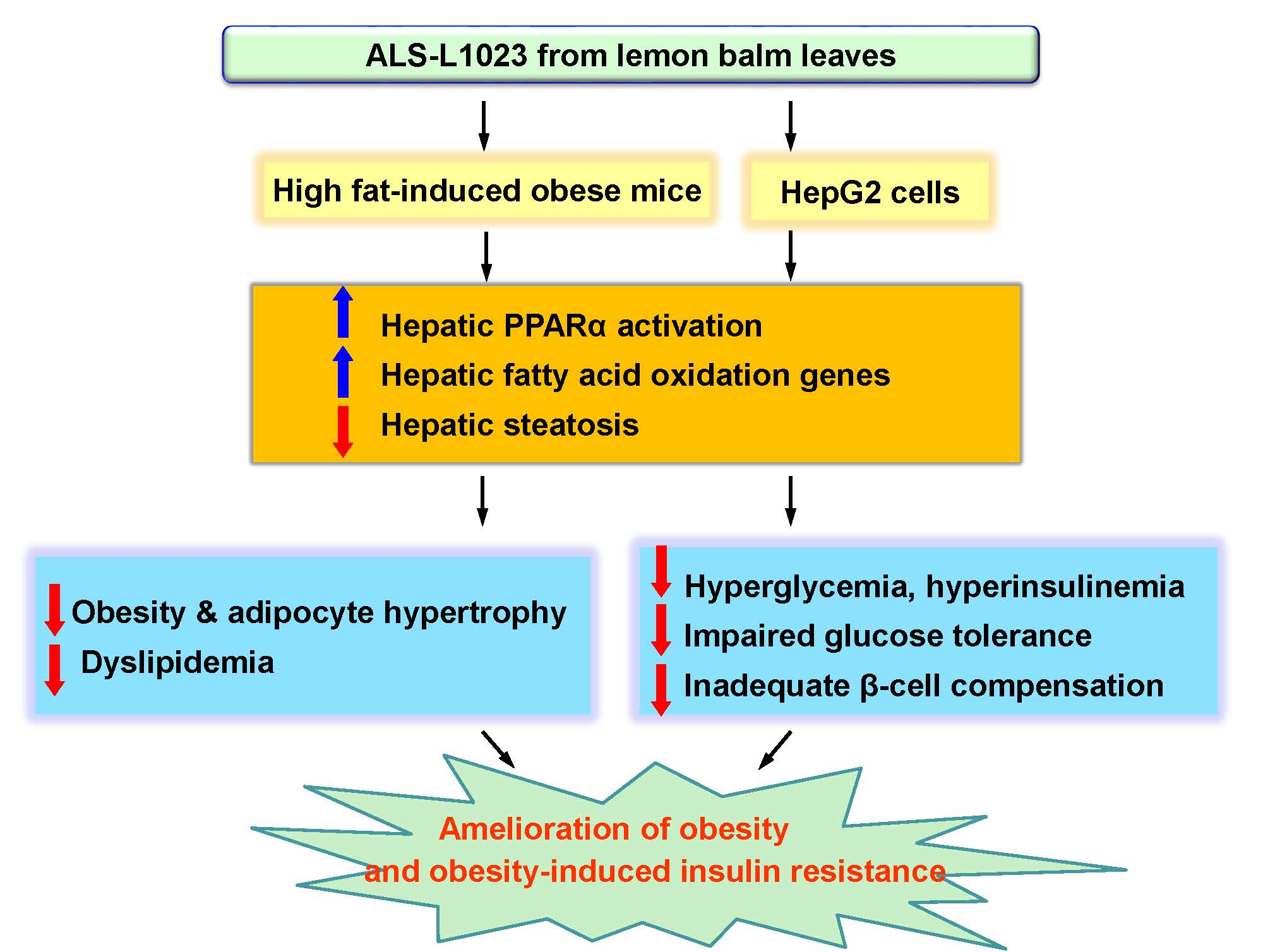
2.1. ALS-L1023 Reduces Weight Gain and Visceral Adipocyte Size in HFD-Fed Obese Mice

Mice fed an HFD supplemented with 0.4% ALS-L1023 had lower body weight gains after 12 weeks of treatment compared with obese HFD-Con mice (Figure 1A). ALS-L1023 significantly decreased total and visceral adipose tissue weights in obese mice (Figure 1B,C). This treatment resulted in a reduction of the average size of visceral adipocytes (Figure 1D,E). However, ALS-L1023 did not affect food intake in HFD-fed obese mice (Figure 1F). In pair-feeding experiments, HFD-ALS mice did not exhibit appetite effects (data not shown). In addition, ALS-L1023 did not exhibit any toxic effects.

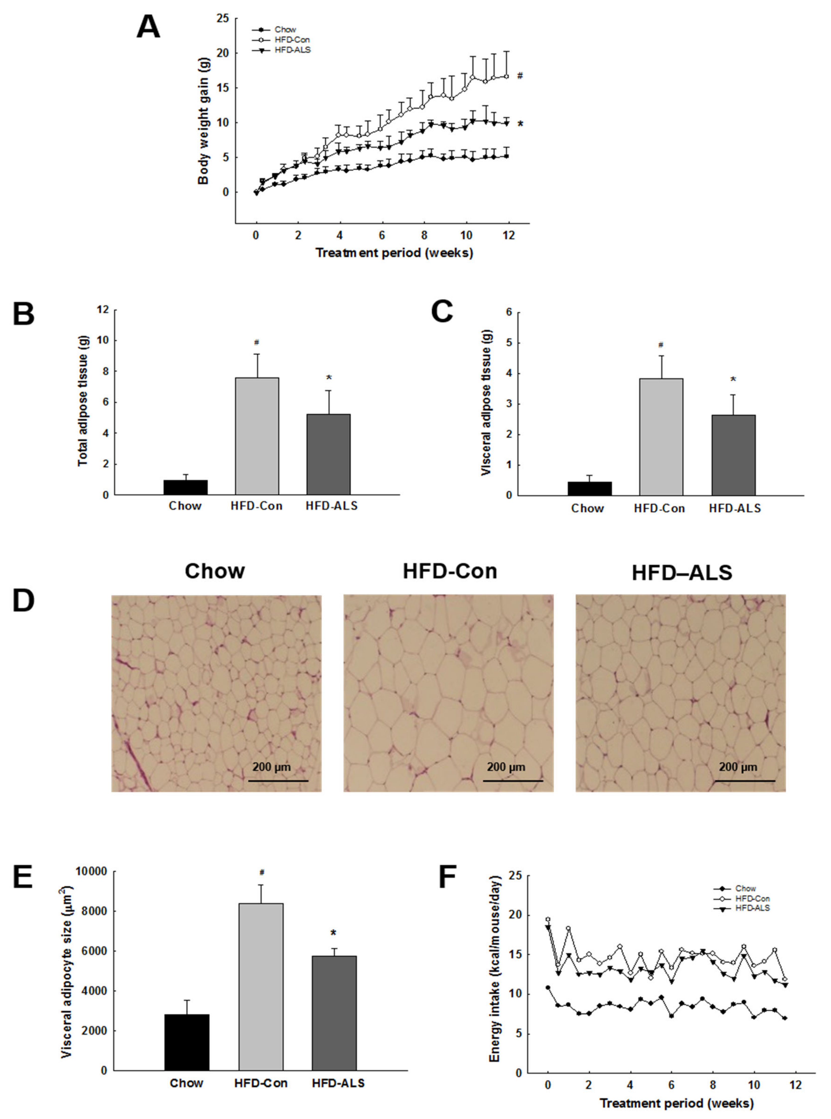
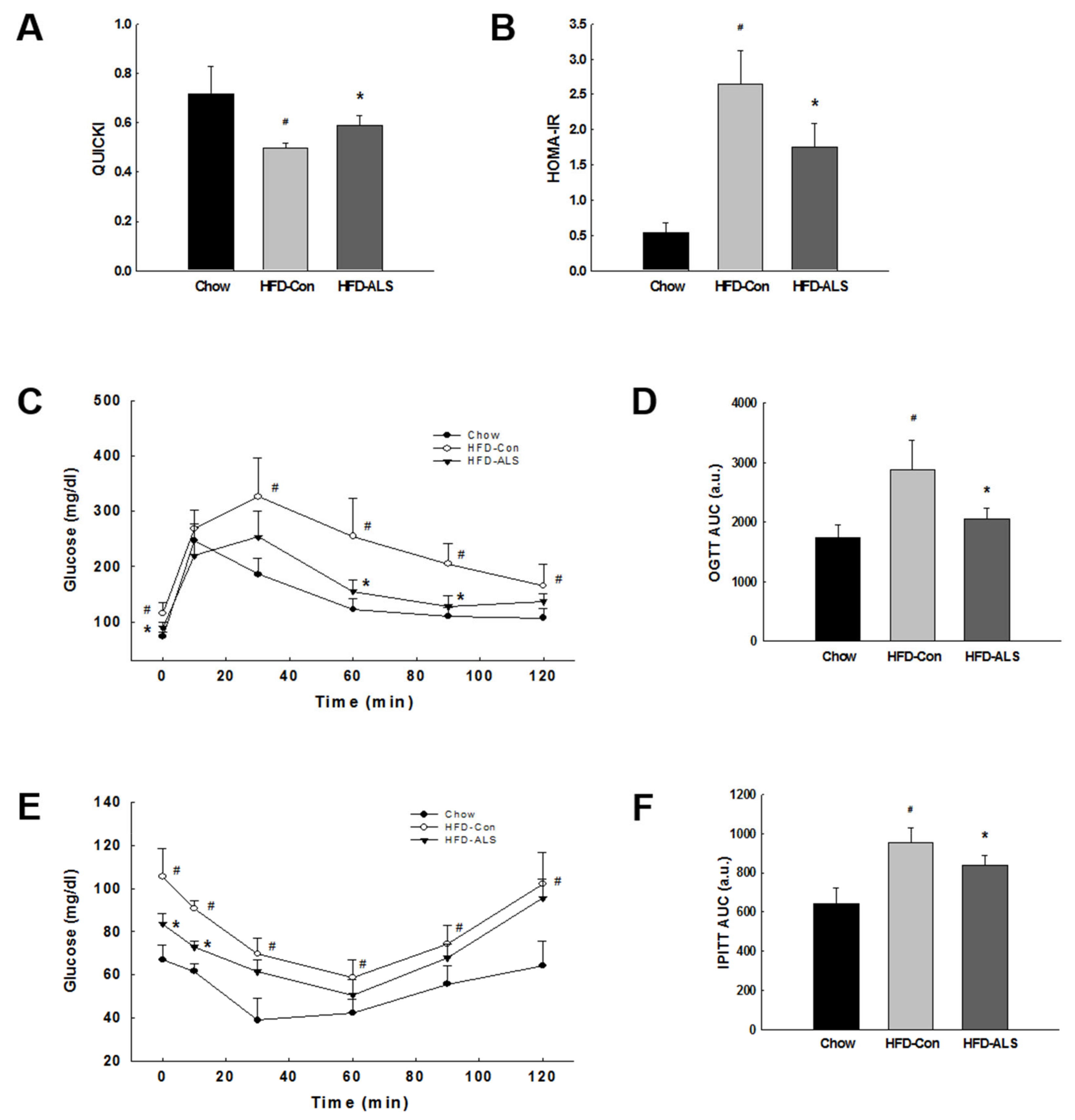
2.2. ALS-L1023 Lowers Elevated Glucose Levels and Increases Insulin Sensitivity in HFD-Fed Obese Mice

Consistent with the weight loss, ALS-L1023 treatment resulted in decreased serum triglycerides and free fatty acids in obese HFD-Con mice (Figure 2A,B). ALS-L1023 also reduced circulating concentrations of glucose and hemoglobin A1c (HbA1c) compared with obese mice. The glucose-lowering effects of ALS-L1023 were indicated by 23% and 10% reductions in glucose and HbA1c levels, respectively (Figure 2C,D). ALS-L1023 treatment reduced serum insulin levels by 36% in obese mice (Figure 2E).
The results of the quantitative insulin sensitivity check index (QUICKI) assessment, which is a well-known marker of insulin sensitivity, were increased in ALS-L1023-treated mice compared with obese HFD-Con mice (Figure 3A). Supplementation with ALS-L1023 resulted in lower homeostasis model assessment-estimated insulin resistance (HOMA-IR) scores; the HOMA-IR was used to test insulin resistance in obese mice (Figure 3B). Similarly, ALS-L1023 treatment resulted in significantly reduced blood glucose levels during the glucose and insulin tolerance tests in obese mice (Figure 3C–F).

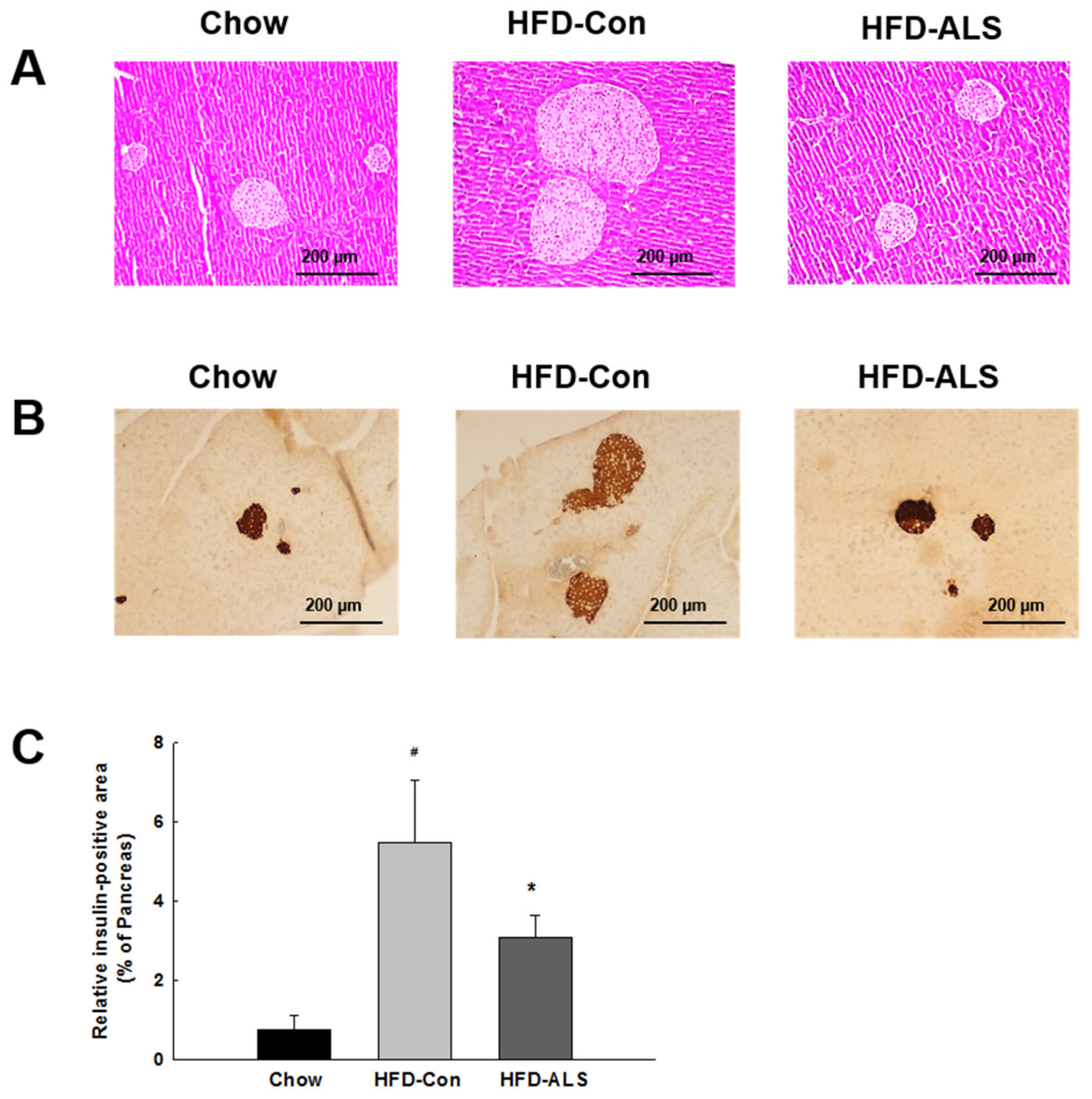
2.3. ALS-L1023 Normalizes Insulin-Positive β-Cell Mass in HFD-Fed Obese Mice

Examination of the hematoxylin and eosin (H&E)-stained pancreas sections revealed that pancreatic islet hypertrophy occurred in obese HFD-Con mice (Figure 4A). However, ALS-L1023 supplementation resulted in a significant decrease in the mean size of islets in HFD-Con mice although their sizes varied in HFD-ALS mice. HFD intake also caused expansion of β-cell mass, as indicated shown by a 621% increase in insulin-positive β-cell area (Figure 4B,C). Consistent with the decreased insulin secretion that occurred with ALS-L1023 supplementation, β-cell areas were reduced by 44% in HFD-ALS mice compared with HFD-Con mice (Figure 4C).

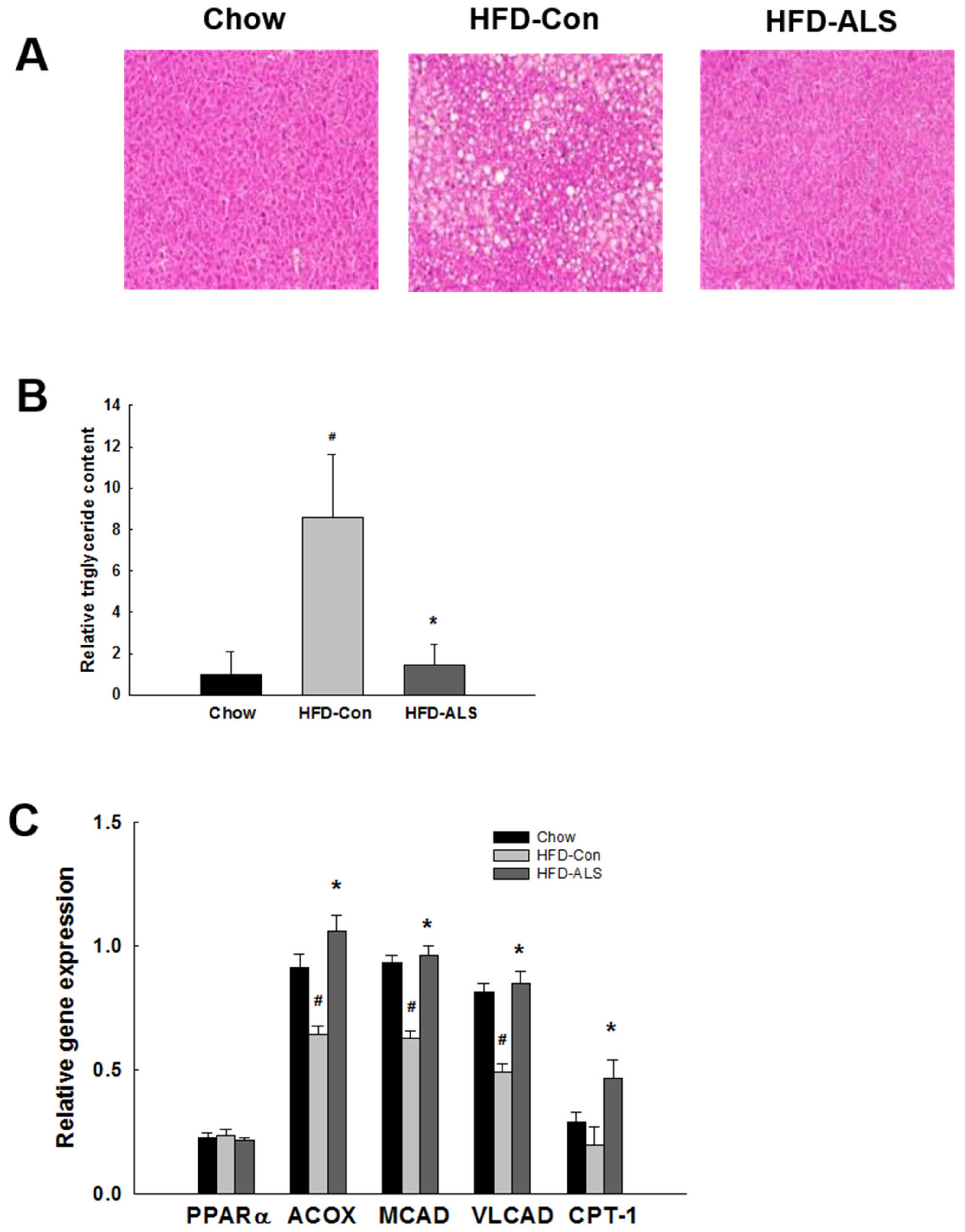
2.4. ALS-L1023 Inhibits Hepatic Steatosis and Increases Hepatic PPARα Target Gene Expression in HFD-Fed Obese Mice

To determine whether ALS-L1023 regulates hepatic steatosis, we examined intrahepatic triglyceride content following ALS-L1023 treatment. The HFD-Con mice had greater liver lipid accumulation compared with chow mice; ALS-L1023 supplementation completely eliminated the intrahepatic lipid droplets (Figure 5A,B). To evaluate whether the inhibitory effects of ALS-L1023 on hepatic lipid accumulation were caused by alterations in hepatic PPARα target gene expression, we measured mRNA levels of PPARα and its target genes (i.e., acyl-CoA oxidase (ACOX), medium-chain acyl-CoA dehydrogenase (MCAD), very long-chain acyl-CoA dehydrogenase (VLCAD), and carnitine palmitoyltransferase I (CPT-1)); these genes encode enzymes involved in fatty acid β-oxidation. Expression of these genes was higher in ALS-L1023-treated mice compared with untreated obese mice (Figure 5C). PPARα mRNA expression was not changed by ALS-L1023.

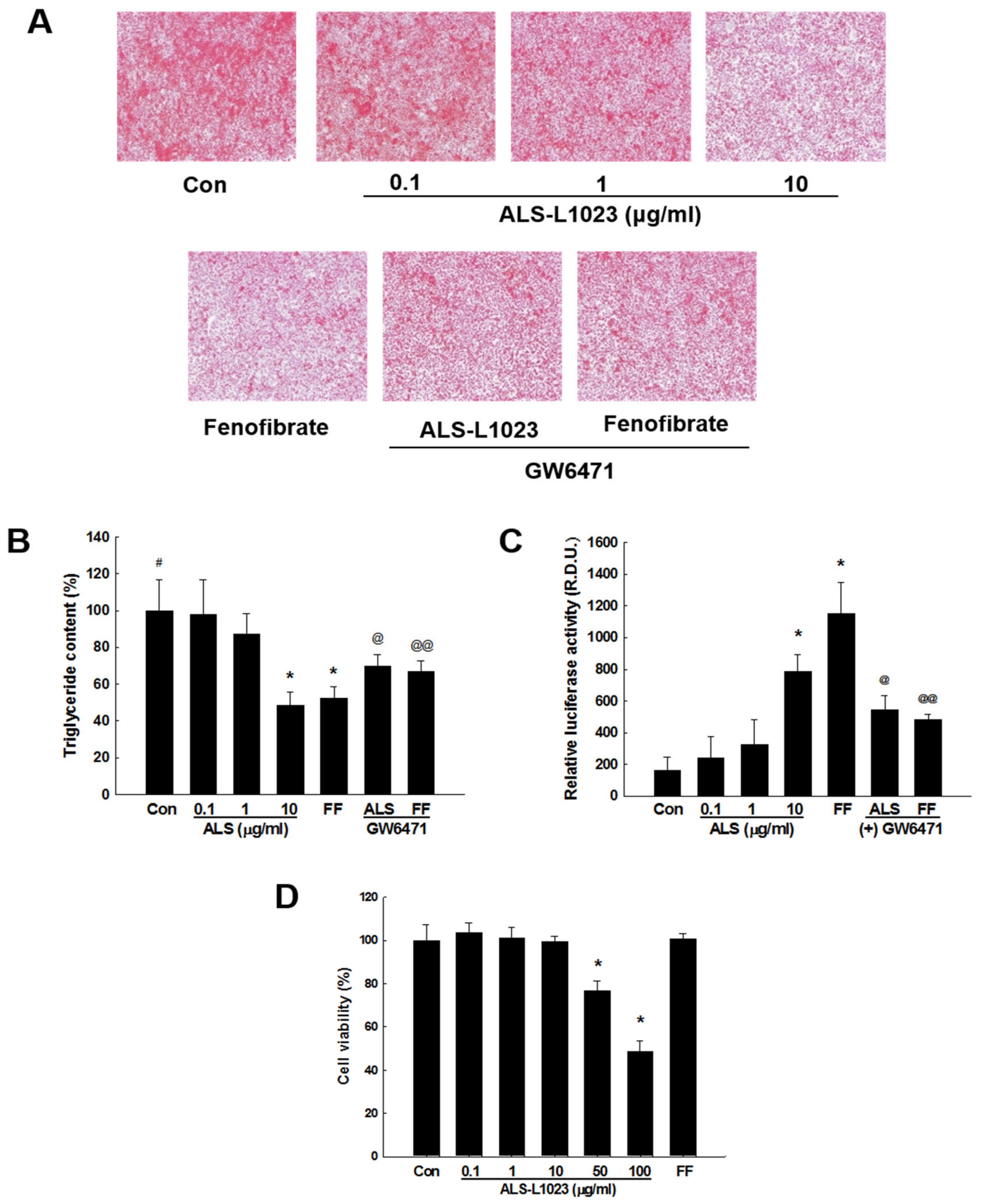
2.5. ALS-L1023 Decreases Lipid Accumulation and Increases PPARα Activity in HepG2 Cells

To investigate the ability of ALS-L1023 to suppress lipid accumulation in HepG2 cells, HepG2 cells were treated with a mixture of the free fatty acids, oleic acid, and palmitoleic acid. The results for Oil Red O-stained lipid droplets indicated that after treatment, HepG2 cells (Control) had significant lipid accumulation (Figure 6A,B). However, incubation of control cells with ALS-L1023 at a dose of 10 μg/mL resulted in decreased lipid accumulation. These inhibitory effects were similar to the effects of the PPARα activator fenofibrate. However, increased triglyceride droplets were observed when HepG2 cells were cotreated with ALS-L1023 and the PPARα antagonist GW6471 compared with ALS-L1023 alone. To further clarify whether ALS-L1023 modulates the PPARα promoter, HepG2 cells were co-transfected with pSG5-mPPARα and the luciferase reporter construct PPRE3-tk-luc. Treatment of transfected cells with ALS-L1023 increased PPARα reporter gene activity and the extent of luciferase activity in ALS-L1023-treated cells was similar to that in fenofibrate-treated cells (Figure 6C). However, concomitant treatment of ALS-L1023 and GW6471 reduced PPARα luciferase activity compared with ALS-L1023 alone. In addition, ALS-L1023 at doses between 0.1 and 10 μg/mL did not have an influence on HepG2 cell viability in 2,3-bis[2 -methoxy-4-nitro-5-sulfophenyl]-2H-tetrazolium-5-carboxanilide disodium salt (XTT) assays (Figure 6D).

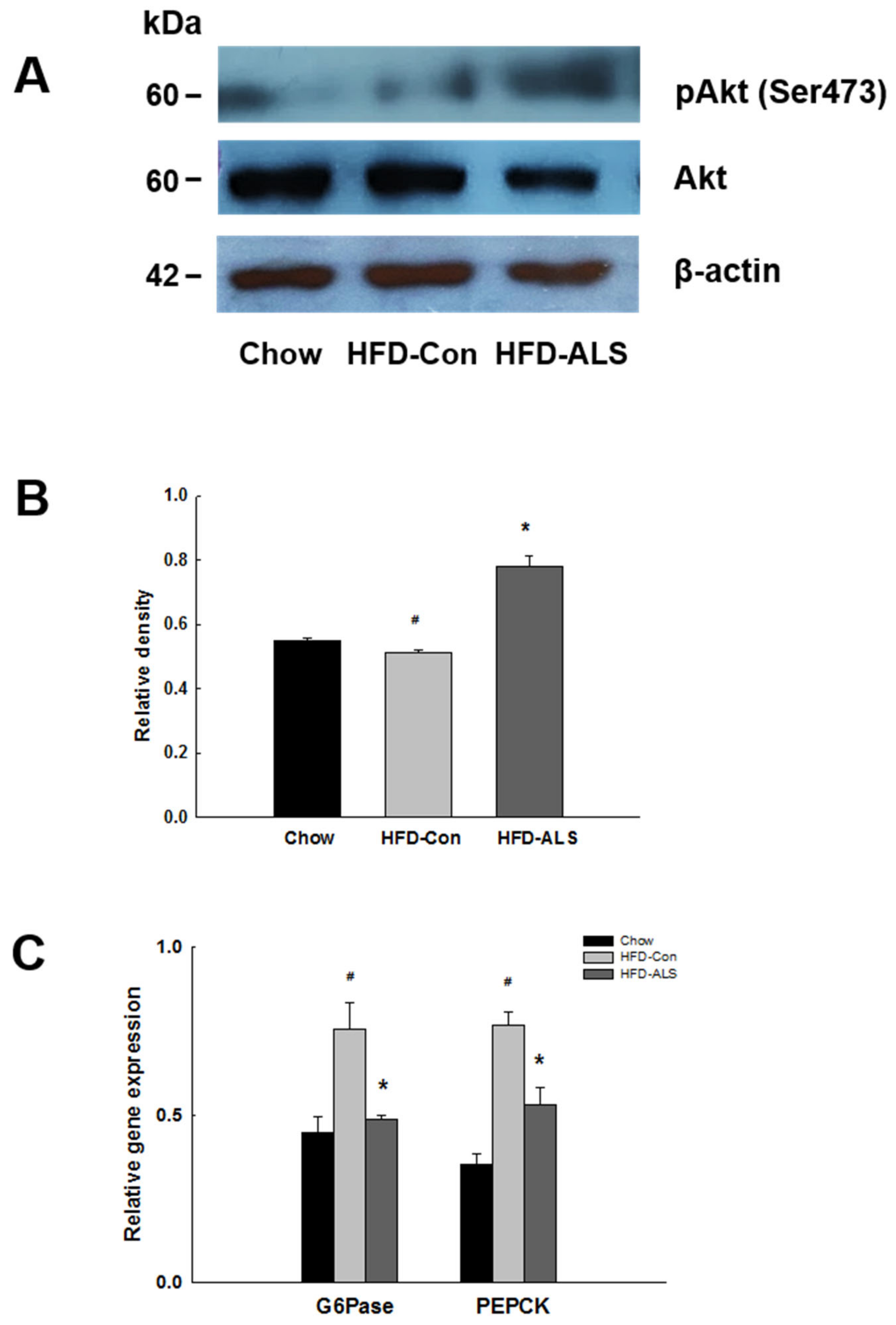
2.6. ALS-L1023 Activates Akt Phosphorylation and Decreases Expression of Gluconeogenesis Genes in HFD-Fed Obese Mice

Study results suggest that during hepatic insulin signaling, protein kinase B (Akt) phosphorylation suppresses hepatic glucose production via a reduction in the expression of gluconeogenic enzymes. To determine whether ALS-L1023 activates Akt under hepatic insulin-resistant conditions, we examined Akt phosphorylation in livers of obese mice. ALS-L1023 increased phosphorylated Akt (pAkt) levels in the livers of obese mice (Figure 7A,B). There was reduced expression of the gluconeogenic enzymes glucose 6-phosphatase (G6Pase) and phosphoenolpyruvate carboxykinase (PEPCK) in ALS-L1023-treated mice compared with untreated HFD-Con mice (Figure 7C).

3. Discussion

Several studies report that lipid accumulation in non-adipose tissues, such as liver and skeletal muscle, initiates the development of insulin resistance and T2D [28]. Since excess ectopic lipids in liver and skeletal muscle tissues interfere with insulin signaling, it may be possible that these tissues become insulin resistant and are responsible for elevated blood glucose levels. Similarly, impaired fatty acid oxidation can cause weight gain, insulin resistance, and the progression towards T2D. Consistent with the above-mentioned studies, current results indicate that ALS-L1023 treatment may induce weight loss and decrease hepatic lipid accumulation in part through hepatic PPARα activation and that reduced hepatic lipids seem to alleviate insulin resistance and hyperglycemia in ALS-L1023-treated obese mice.
Simultaneous treatment of mice with an HFD with ALS-L1023 decreased body weight gain, fat mass, and visceral adipocyte size compared with obese HFD mice. During ALS-L1023-induced weight reduction, food intake was not changed, showing that ALS-L1023 did not affect appetite. We had previously conducted animal studies using ALS-L1023 at doses of 0.1%, 0.25%, and 0.5% in HFD-fed obese rats and found that weight gains were substantially decreased by 0.5% ALS-L1023 (100 mg/kg/day) (unpublished data). According to the dose conversion from rat to mouse, 0.4% ALS-L1023 (200 mg/kg/day) was selected to supplement the HFD to enhance the effects of ALS-L1023 on obesity and related diseases. C57BL/6J mice (6–8 weeks of age) fed a diet containing 40–60% calories from fat for 8–16 weeks become overweight, obese, and develop impaired glucose tolerance and insulin resistance [29]. In the present study, C57BL/6J mice fed an HFD for 12 weeks had an approximately 8 times greater visceral fat mass than that of chow-fed mice. However, ALS-L1023 decreased body weight gain by 40%, visceral adipose tissue mass by 32%, and visceral adipocyte size by 22% compared with obese HFD mice. These results are consistent with the observation of reduced hepatic lipid accumulation after ALS-L1023 treatment, suggesting that ALS-L1023-induced increases in fat catabolism in livers may result in the prevention of weight gain and increased fat mobilization from visceral adipose tissue. Actually, ALS-L1023 treatment markedly reduced hepatic lipid accumulation, which may be due to increased fatty acid oxidation. The increased liver activity corresponded to a large reduction in adipose tissue mass and adipocyte size, which likely accounted for most of the body weight reduction.
The intake of HFD by C57BL/6J mice is a robust model of debilitated glucose tolerance and initial T2D [30]. These animals have indisputable insulin resistance and deficient β-cell compensation [3,31]. Consistent with the decreased visceral adipose tissue mass and adipocyte size, the elevated concentrations of fasting glucose and insulin in obese HFD-Con mice were significantly reduced following ALS-L1023 treatment. It has been reported that large adipocytes from obese rodents generate and secrete tumor necrosis factor α, leptin, and circulating lipids such as free fatty acids, which cause insulin resistance [32,33,34,35]. Thus, ALS-L1023 treatment may convert hypertrophic adipocytes into smaller adipocytes, leading to reduction of blood glucose and amelioration of insulin resistance [35,36].
Hypertrophy of pancreatic islets and enlargement of β-cell area were observed in HFD-fed C57BL/6J mice [37]. However, ALS-L1023 treatment decreased the mean size of islets and insulin-positive β-cell mass to the levels of chow mice, suggesting that ALS-L1023 treatment prevents islet hypertrophy and modulates the inadequate β-cell compensation due to insulin resistance. Interestingly, the effect of ALS-L1023 on the islet size varied, but almost all islet sizes decreased after ALS-L1023 treatment. These results were consistent with the reduction of insulin secretion and low glucose levels after ALS-L1023 treatment in HFD-Con mice. In insulin-resistant conditions, pancreatic islets increase the secretion of insulin to maintain normal blood glucose levels, which is called β-cell compensation [3]. Therefore, the alleviation of insulin resistance will reduce the size of islets and inhibit enlargement of β-cell mass. As mentioned earlier, ALS-L1023 may reduce weight gain and adipocyte size partly through hepatic lipid reduction via hepatic PPARα activation. Since excess hepatic lipids and visceral obesity are deeply associated with insulin resistance [11,38], their reductions by ALS-L1023 will suppress islet hypertrophy and β-cell expansion. It is possible that ALS-L1023 treatment makes liver and skeletal muscle tissues more sensitive to insulin.
The amount of intrahepatic triglycerides was prominent in HFD-Con mice whereas ALS-L1023 treatment markedly decreased hepatic lipid accumulation largely through fatty acid β-oxidation in mitochondria and peroxisomes [39,40]. We also observed similar results to the above in vivo data using HepG2 cells as a cellular model of hepatic steatosis. ALS-L1023 at a dose of 10 μg/mL caused a substantial decrease in lipid accumulation compared with control cells treated with a mixture of fatty acids. These suppressive effects of ALS-L1023 on excess fat deposition were comparable to a reduction by fenofibrate. Moreover, the inhibitory effects of ALS-L1023 on triglyceride droplets were reversed by GW6471 in HepG2 cells, suggesting that the actions of ALS-L0123 on hepatic steatosis may be mediated in part through PPARα. The presence of NAFLD has been related to a very high risk of hepatic insulin resistance, which results in the impaired fasting glucose and T2D [9,10]; it suggests that there is a close association between excess hepatic lipids and hepatic insulin resistance. Since present results indicate that ALS-L1023 reduces liver microvesicular and macrovesicular steatosis, it is conceivable that ALS-L1023 can alleviate glucose intolerance and insulin resistance through reduction of hepatic lipids.
To determine the actions of PPARα on ALS-L1023-mediated hepatic lipid reduction, we investigated the effects of ALS-L1023 on the expression of PPARα target genes involved in fatty acid β-oxidation in livers of HFD-fed mice. ALS-L1023 caused an increase in the expression of PPARα target enzymes (e.g., ACOX, MCAD, VLCAD, and CPT-1) in livers. In parallel with the in vivo data, transactivation assay results indicated that PPARα reporter gene activation was stimulated by ALS-L1023. ALS-L1023 treatment increased the expression of a PPRE-luciferase reporter gene in HepG2 cells. These stimulatory effects of ALS-L1023 were similar to those of fenofibrate. By contrast, GW6471 reversed ALS-L1023-induced increases in luciferase activity. This result suggests that ALS-L1023 can positively regulate the action of PPARα. PPARα promotes the catabolism of fatty acids by raising the transcriptional activation of enzymes critical for mitochondrial and peroxisomal β-oxidation [41,42,43]. Therefore, our results suggest that ALS-L1023 stimulation of hepatic expression of PPARα target enzymes decreases the hepatocellular fatty acid levels required for triglyceride production, which led to a reduction in hepatic steatosis.
To examine the molecular mechanism of the action of ALS-L1023 on lipid-induced hepatic insulin resistance, Akt activation was examined in livers of C57BL/6J mice following ALS-L1023 treatment. Akt is an important signaling molecule of insulin action. Akt is phosphorylated and activated under insulin-stimulated conditions, whereas its phosphorylation is suppressed when excess hepatic lipids are present [11]. Studies in mice and humans have revealed that hepatic diacylglycerol has a key role for activation of protein kinase Cε, resulting in the inhibition of insulin signaling, including the Akt impairment [44,45]. Diacylglycerol is derived from intracellular fatty acids; this process is regulated by a balance between fatty acid oxidation and synthesis. Interestingly, mice that are genetically defective in PPARα and long-chain acyl-CoA dehydrogenase exhibit decreased hepatic fatty acid oxidation and have a tendency to hepatic steatosis and insulin resistance [15,16]. Consistent with ALS-L1023-induced decreases in the availability of fatty acids through increasing the expression of PPARα-activated oxidizing enzymes, phosphorylated Akt protein levels were elevated in livers. Thus, ALS-L1023 may inhibit hepatic insulin resistance through Akt activation.
Under insulin-stimulated conditions, Akt activation inhibits liver glucose generation by decreasing the expression of gluconeogenesis enzymes (e.g., G6Pase and PEPCK) [46]. In parallel with the increased Akt phosphorylation, ALS-L1023 decreased G6Pase and PEPCK mRNA levels in livers of obese mice. Fasting glucose and HbA1c concentrations were decreased in ALS-L1023-treated mice. These results indicate that ALS-L1023 treatment increases Akt phosphorylation and subsequently decreases expression of gluconeogenic genes, leading to improved glucose metabolism.

4. Materials and Methods

4.1. Preparation of ALS-L1023

Activity-guided fractionation was used to manufacture ALS-L1023 from Melissa officinalis L. leaves, which were purchased from Alfred Galke GmbH, (Harz, Germany). Briefly, the dried Melissa leaves were exposed to aqueous ethanol and the resulting extract was filtered and concentrated. The concentrated ethanol extract was further fractionated using ethyl acetate, and the resulting fractions were concentrated and dried to obtain ALS-L1023 in a dried powder form. High-performance liquid chromatography was used to compare ALS-L1023 against two reference compounds (i.e., rosmarinic acid and caffeic acid) for standardization [27].

4.2. Animals and Treatments

Eight-week-old male wild-type C57BL/6J mice (n = 8/group) were randomly assigned to one of three treatment groups. Each group of mice was fed one of three diets for 12 weeks: a standard chow diet (chow, 10% kcal fat, Research Diets, New Brunswick, NJ, USA), an HFD (HFD-Con, 45% kcal fat, Research Diets), or an HFD supplemented with 0.4% (w/w) ALS-L1023 (HFD-ALS). Four grams ALS-L1023 powder was mixed with 1 kg of the HFD. The body weight of each animal was measured three times per day by a person blinded to the treatments. Food intake was determined by measuring the amounts of food consumed by the mice throughout the treatment period. All animal experiments were approved by the Institutional Animal Care and Use Committee of Mokwon University (permit number: NVRQS AEC-9) and followed National Research Council Guidelines.

4.3. Cell Culture, Treatment of Free Fatty Acids, and Analysis of Triglyceride Content

HepG2 cells were cultured in Dulbecco’s modified Eagle’s medium containing 10% fetal bovine serum (Invitrogen, Carlsbad, CA, USA). The cells were cultured until they reached 70–80% confluency (approximately 2 days). They were then exposed for 2 days to various concentrations of chemicals in the presence of a mixture of oleic acid and palmitoleic acid. Oleic acid and palmitoleic acid were dissolved in ethanol and isopropanol at concentrations of 0.3 mM and 0.15 mM, respectively. The cells were fixed with 10% formalin for 1 h and incubated with Oil Red O for 2 h at room temperature. Oil Red O-stained areas were measured using ImageJ software (National Institutes of Health, Bethesda, MD, USA).

4.4. In Vitro Cytotoxicity Test

HepG2 cells were plated on a 96 well plate at a density of 1 × 10⁴ cells/well and incubated for 24 h at 37 °C with culture medium in the presence of ALS-L1023 or fenofibrate. Cell viability was detected by an XTT assay using a Cell Proliferation Kit II (Roche, Basel, Switzerland).

4.5. Blood Analysis

After an 8 h fast on the last day of the study, blood was analyzed in mice fed a chow, an HFD-Con or an HFD-ALS for 12 weeks. Serum levels of triglycerides and free fatty acids were measured using an automatic blood chemical analyzer (CIBA Corning, Oberlin, OH). Levels of blood glucose and HbA1c were measured using the Accu-Chek Performa System (Roche, Basel, Switzerland) and NycoCard Reader II (Alere/Axis-Shield, Oslo, Norway), respectively. Oral glucose (2 g/kg body weight) and intraperitoneal insulin (0.75 units/kg body weight) tolerance tests were performed to determine blood glucose levels at selected time intervals. QUICKI values were calculated as: 1/(log (fasting insulin μU/mL) + log (fasting glucose mg/dL)). HOMA-IR was calculated via an online Oxford HOMA calculator (available at: www.dtu.ox.ac.uk) using the formula: (fasting insulin μU/mL × fasting glucose mg/dL)/405.

4.6. Histological Analysis

For H&E staining, tissues were fixed in 10% phosphate-buffered formalin for 1 day and processed in a routine manner to obtain paraffin sections. Tissue sections (5 μm thick) were cut and stained with H&E for microscopic examination. To quantify the sizes of visceral adipocytes, the H&E-stained sections were analyzed using an Image-Pro Plus analysis system (Media Cybernetics, Bethesda, MD, USA). Insulin-secreting β-cells were detected using a monoclonal mouse anti-insulin antibody (I2018; Sigma-Aldrich, St Louis, MO, USA). Sections of pancreas tissue (3 μm thick) were irradiated in a microwave oven for epitope retrieval. The sections were then incubated with the primary insulin antibody (1:1,400 dilution) and an anti-mouse IgG biotinylated secondary antibody (Vector Laboratories, Burlingame, CA, USA) using diaminobenzidine (Vector Laboratories) as the chromogen. Immunostained β-cell areas were analyzed using ImageJ software and relative insulin-positive areas were expressed as percentages of the total surveyed pancreatic area occupied by β cells.

4.7. Western Blot Analysis

Liver tissues were lysed in ice-cold lysis buffer (50 mM Tris-Cl (pH 8.0), 150 mM NaCl, 0.02% Sodium azide, and 1% Triton X-100) containing protease inhibitors (phenylmethylsulfonyl fluoride and aprotinin). The lysates were centrifuged at 12,000 rpm for 20 min at 4 °C, and the resulting supernatants (10 µg) were subjected to electrophoresis on 10% polyacrylamide gels. The separated proteins were transferred to polyvinylidene fluoride membranes (Millipore, Billerica, MA, USA). The membranes were incubated with primary rabbit antibodies of pAkt (1:1000 dilution) and Akt (1:1000 dilution) (Cell Signaling Technology, Danvers, MA, USA). After incubation with a goat anti-rabbit IgG secondary antibody (Cell Signaling Technology), the blots were measured using ImageJ software.

4.8. Quantitative Reverse Transcription Polymerase Chain Reaction (qRT-PCR)

Total RNA from liver tissues was prepared using Trizol reagent (Invitrogen) according to the manufacturer’s instructions followed by DNase treatment. The purity of total RNA extracted was assessed to the A260/A280 ratio of total RNA by spectrophotometer. The A260/A280 ratio of the extracted RNA was 1.8–2.0. The integrity of total RNA was assessed by agarose gel analysis. Total RNA on a gel showed a sharp, clear 28S and 18S rRNA bands, and the intensity of the 28S band was about twice that of the 18S band. Complementary DNA (cDNA) was synthesized from 2 μg total RNA using the TOPscriptTM DryMIX RT kit containing reverse transcriptase, RT buffer, a deoxyribonucleotide triphosphate mixture, RNase inhibitor, and oligo (dT)18 (Enzynomics, Seoul, Korea) in a final volume of 20 μL according to the manufacturer’s instructions. Briefly, RT reactions were incubated for 60 min at 50 °C and then inactivated by heating to 95 °C for 10 min. The genes of interest were amplified from the synthesized cDNA using BioFact TM 2X Real-Time PCR Master Mix (BioFact, Daejeon, Korea) and a Rotor-Gene 6000 system (Qiagen). The PCR conditions were as follows: 1 cycle of 95 °C for 15 min, followed by 50 cycles of 95 °C for 20 s, 58 °C for 15 s, and 72 °C for 20 s. The specificity of PCR products was determined by using a melt curve analysis. The identities of products were further investigated by 2% agarose gel electrophoresis analysis. Each qRT-PCR assay including a no template control and a minus reverse transcriptase control did not show amplification. Eight pairs of primers were designed using the PubMed primer designing tool (https://0-www-ncbi-nlm-nih-gov.brum.beds.ac.uk/tools/primer-blast/) and checked for specificity using BLAST (https://blast.ncbi.nlm.nih.gov/Blast.cgi). The PCR primers and amplification efficiencies are presented in Table 1. The relative expression levels were calculated as the ratios of the target gene cDNA to the β-actin cDNA.

4.9. Transient Transfection Assay

The expression vectors, pSG5-mPPARα and PPRE3-tk-luc reporter genes, were generously provided by Dr. Frank J. Gonzalez (National Cancer Institute, NIH, Bethesda, MD, USA). HepG2 cells were seeded in 6-well tissue culture plates (2 × 10⁴ cells/well) 24 h before transfection. Transfection was performed using 200 ng/well of each of the appropriate plasmids and Lipofectamine (Invitrogen). After 6 h, the culture medium was changed and the test compounds, ALS-L1023, fenofibrate or GW6471, were added. After incubation for 24 h in the presence of the aforementioned chemicals, the cells were assayed for luciferase and β-galactosidase activity (Promega, Madison, WI, USA).

4.10. Statistical Analysis
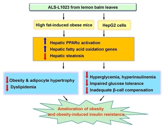
The results were expressed as mean ± standard deviation (SD) values. The statistical analysis was performed using analysis of variance followed by Turkey’s post-hoc tests. Statistical significance was defined as a p-value <0.05.

5. Conclusions

In conclusion, our data suggest that the lemon balm extract ALS-L1023 may inhibit obesity and insulin resistance in HFD-fed obese mice. This event may be mediated partly through inhibition of hepatic lipid accumulation via hepatic PPARα activation. In addition, these data indicate that ALS-L1023 can act as an activator of PPARα function. PPARα agonist treatment has been reported to reduce weight gain and improve glucose metabolism in insulin-resistant and diabetic rodents [38,47,48,49]. ALS-L1023 may thus be a useful drug to prevent and treat obesity, insulin resistance, and T2D. Further studies will be necessary to identify and characterize active components of ALS-L1023 for their effects on PPARα function for potential therapeutic uses.

Author Contributions

Conceived and designed the experiments: M.Y. and S.S.S.; Performed the experiments: D.L., Y.S., J.S.R., J.A., and S.J.; Analyzed the data: D.L., Y.S., J.S.R., J.A., S.J., S.S.S., and M.Y.; Wrote the paper: M.Y. and S.S.S. All authors have read and agreed to the published version of the manuscript.

Funding

This work was supported by the National Research Foundation (NRF) grant funded by the Korea Government (MSIP) (No. 2018R1D1A1B07042585) and the Convergence of Conventional Medicine and Traditional Korean Medicine R&D program funded by the Ministry of Health & Welfare through the Korea Health Industry Development Institute (KHIDI) (HI15C0075).

Conflicts of Interest

The authors declare no conflict of interest.

References

  1. Ahren, B.; Pacini, G. Insufficient islet compensation to insulin resistance vs. reduced glucose effectiveness in glucose-intolerant mice. Am. J. Physiol. Metab. 2002, 283, E738–E744. [Google Scholar] [CrossRef] [PubMed]
  2. Kahn, S.E. The relative contributions of insulin resistance and beta-cell dysfunction to the pathophysiology of Type 2 diabetes. Diabetology 2003, 46, 3–19. [Google Scholar] [CrossRef] [PubMed] [Green Version]
  3. Prentki, M.; Nolan, C.J. Islet β cell failure in type 2 diabetes. J. Clin. Investig. 2006, 116, 1802–1812. [Google Scholar] [CrossRef] [PubMed] [Green Version]
  4. Lara-Castro, C.; Garvey, W.T. Intracellular Lipid Accumulation in Liver and Muscle and the Insulin Resistance Syndrome. Endocrinol. Metab. Clin. North Am. 2008, 37, 841–856. [Google Scholar] [CrossRef] [PubMed] [Green Version]
  5. Krššák, M.; Petersen, K.F.; Dresner, A.; di Pietro, L.; Vogel, S.M.; Rothman, D.L.; Shulman, G.I.; Roden, M. Intramyocellular lipid concentrations are correlated with insulin sensitivity in humans: A 1 H NMR spectroscopy study. Diabetology 1999, 42, 113–116. [Google Scholar] [CrossRef] [PubMed] [Green Version]
  6. Kelley, D.E.; McKolanis, T.M.; Hegazi, R.A.F.; Kuller, L.H.; Kalhan, S.C. Fatty liver in type 2 diabetes mellitus: Relation to regional adiposity, fatty acids, and insulin resistance. Am. J. Physiol. Metab. 2003, 285, E906–E916. [Google Scholar] [CrossRef] [PubMed] [Green Version]
  7. Ryysy, L.; Hakkinen, A.M.; Goto, T.; Vehkavaara, S.; Westerbacka, J.; Halavaara, J.; Yki-Jarvinen, H. Hepatic fat content and insulin action on free fatty acids and glucose metabolism rather than insulin absorption are associated with insulin requirements during insulin therapy in type 2 diabetic patients. Diabetes 2000, 49, 749–758. [Google Scholar] [CrossRef] [Green Version]
  8. Tolman, K.G.; Fonseca, V.; Dalpiaz, A.; Tan, M.H. Spectrum of Liver Disease in Type 2 Diabetes and Management of Patients with Diabetes and Liver Disease. Diabetes Care 2007, 30, 734–743. [Google Scholar] [CrossRef] [Green Version]
  9. Petersen, K.F.; Dufour, S.; Feng, J.; Befroy, U.; Dziura, J.; Man, C.D.; Cobelli, C.; Shulman, G.I. Increased prevalence of insulin resistance and nonalcoholic fatty liver disease in Asian-Indian men. Proc. Natl. Acad. Sci. USA 2006, 103, 18273–18277. [Google Scholar] [CrossRef] [Green Version]
  10. Fabbrini, E.; Sullivan, S.; Klein, S. Obesity, and nonalcoholic fatty liver disease: Biochemical, metabolic, and clinical implications. Hepatology 2010, 51, 679–689. [Google Scholar] [CrossRef]
  11. Perry, R.J.; Samuel, V.T.; Petersen, K.F.; Shulman, G.I. The role of hepatic lipids in hepatic insulin resistance and type 2 diabetes. Nature 2014, 510, 84–91. [Google Scholar] [CrossRef] [PubMed]
  12. Kelley, D.E.; He, J.; Menshikova, E.V.; Ritov, V.B. Dysfunction of mitochondria in human skeletal muscle in type 2 diabetes. Diabetes 2002, 51, 2944–2950. [Google Scholar] [CrossRef] [PubMed] [Green Version]
  13. Petersen, K.F.; Dufour, S.; Befroy, U.; Garcia, R.; Shulman, G.I. Impaired mitochondrial activity in the insulin-resistant offspring of patients with type 2 diabetes. New Engl. J. Med. 2004, 350, 664–671. [Google Scholar] [CrossRef] [PubMed] [Green Version]
  14. Petersen, K.F.; Befroy, U.; Dufour, S.; Dziura, J.; Ariyan, C.; Rothman, U.L.; DiPietro, L.; Cline, G.W.; Shulman, G.I. Mitochondrial Dysfunction in the Elderly: Possible Role in Insulin Resistance. Science 2003, 300, 1140–1142. [Google Scholar] [CrossRef] [PubMed] [Green Version]
  15. Neschen, S.; Morino, K.; Dong, J.; Wang-Fischer, Y.; Cline, G.W.; Romanelli, A.J.; Rossbacher, J.C.; Moore, I.K.; Regittnig, W.; Munoz, D.S.; et al. n-3 Fatty Acids Preserve Insulin Sensitivity In Vivo in a Peroxisome Proliferator-Activated Receptor—Dependent Manner. Diabetes 2007, 56, 1034–1041. [Google Scholar] [CrossRef] [PubMed] [Green Version]
  16. Zhang, N.; Liu, Z.-X.; Choi, C.S.; Tian, L.; Kibbey, R.; Dong, J.; Cline, G.W.; Wood, P.A.; Shulman, G.I. Mitochondrial dysfunction due to long-chain Acyl-CoA dehydrogenase deficiency causes hepatic steatosis and hepatic insulin resistance. Proc. Natl. Acad. Sci. USA 2007, 104, 17075–17080. [Google Scholar] [CrossRef] [PubMed] [Green Version]
  17. Broskey, N.T.; Obanda, D.N.; Burton, J.; Cefalu, W.T.; Ravussin, E. Skeletal muscle ceramides and daily fat oxidation in obesity and diabetes. Metabolism 2018, 82, 118–123. [Google Scholar] [CrossRef]
  18. McGarry, J.D. Banting lecture 2001: Dysregulation of fatty acid metabolism in the etiology of type 2 diabetes. Diabetes 2002, 51, 7–18. [Google Scholar] [CrossRef] [Green Version]
  19. Yoon, M. The role of PPARα in lipid metabolism and obesity: Focusing on the effects of estrogen on PPARα actions. Pharmacol. Res. 2009, 60, 151–159. [Google Scholar] [CrossRef]
  20. Yoon, M. PPARα in Obesity: Sex Difference and Estrogen Involvement. PPAR Res. 2010, 2010, 1–16. [Google Scholar] [CrossRef] [Green Version]
  21. Jeong, S.; Kim, M.; Han, M.; Lee, H.; Ahn, J.; Kim, M.; Song, Y.-H.; Shin, C.; Nam, K.-H.; Kim, T.W.; et al. Fenofibrate prevents obesity and hypertriglyceridemia in low-density lipoprotein receptor-null mice. Metabolism 2004, 53, 607–613. [Google Scholar] [CrossRef] [PubMed]
  22. Jeong, S.; Yoon, M. Inhibition of the Actions of Peroxisome Proliferator-activated Receptor α on Obesity by Estrogen. Obesity 2007, 15, 1430–1440. [Google Scholar] [CrossRef] [PubMed]
  23. Blumenthal, M.; Goldberg, A.; Brinckmann, J. Herbal Medicine-Expanded Commission E. Mongraphs; Integrative Medicine Communications: Newton, MA, USA, 2000; pp. 230–232. [Google Scholar]
  24. Akhondzadeh, S.; Noroozian, M.; Mohammadi, M.; Ohadinia, S.; Jamshidi, A.H.; Khani, M. Melissa officinalis extract in the treatment of patients with mild to moderate Alzheimer’s disease: A double blind, randomized, placebo-controlled trial. J. Neuro. Neurosurg. Psychiatr. 2003, 6, 625–632. [Google Scholar] [CrossRef] [PubMed] [Green Version]
  25. Park, B.Y.; Lee, H.; Woo, S.; Yoon, M.; Kim, J.; Hong, Y.; Lee, H.S.; Park, E.K.; Hahm, J.C.; Kim, J.W.; et al. Reduction of Adipose Tissue Mass by the Angiogenesis Inhibitor ALS-L1023 from Melissa officinalis. PLoS ONE 2015, 10, e0141612. [Google Scholar] [CrossRef]
  26. Kim, J.; Lee, H.; Lim, J.; Oh, J.; Shin, S.S.; Yoon, M. The Angiogenesis Inhibitor ALS-L1023 from Lemon-Balm Leaves Attenuates High-Fat Diet-Induced Nonalcoholic Fatty Liver Disease through Regulating the Visceral Adipose-Tissue Function. Int. J. Mol. Sci. 2017, 18, 846. [Google Scholar] [CrossRef]
  27. Woo, S.; Yoon, M.; Kim, J.; Hong, Y.; Kim, M.-Y.; Shin, S.S.; Yoon, M. The anti-angiogenic herbal extract from Melissa officinalis inhibits adipogenesis in 3T3-L1 adipocytes and suppresses adipocyte hypertrophy in high fat diet-induced obese C57BL/6J mice. J. Ethnopharmacol. 2016, 178, 238–250. [Google Scholar] [CrossRef]
  28. Samuel, V.T.; Shulman, G.I. The pathogenesis of insulin resistance: Integrating signaling pathways and substrate flux. J. Clin. Investig. 2016, 126, 12–22. [Google Scholar] [CrossRef] [Green Version]
  29. Reuter, T.Y. Diet-induced models for obesity and type 2 diabetes. Drug Discov. Today Dis. Model. 2007, 4, 3–8. [Google Scholar] [CrossRef]
  30. Winzell, M.S.; Ahren, B. The high-fat diet-fed mouse: A model for studying mechanisms and treatment of impaired glucose tolerance and type 2 diabetes. Diabetes 2004, 53, S215–S219. [Google Scholar] [CrossRef] [Green Version]
  31. Winzell, M.S.; Holm, C.; Ahren, B. Downregulation of islet hormone-sensitive lipase during long-term high-fat feeding. Biochem. Biophys. Res. Commun. 2003, 304, 273–278. [Google Scholar] [CrossRef]
  32. Hotamisligil, G.; Shargill, N.; Spiegelman, B. Adipose expression of tumor necrosis factor-alpha: Direct role in obesity-linked insulin resistance. Science 1993, 259, 87–91. [Google Scholar] [CrossRef] [PubMed]
  33. Taylor, S.I.; Barr, V.; Reitman, M.L. Does Leptin Contribute to Diabetes Caused by Obesity? Science 1996, 274, 1151. [Google Scholar] [CrossRef] [PubMed]
  34. Boden, G. Role of fatty acids in the pathogenesis of insulin resistance and NIDDM. Diabetes 1997, 46, 3–10. [Google Scholar] [CrossRef] [PubMed]
  35. Okuno, A.; Tamemoto, H.; Tobe, K.; Ueki, K.; Mori, Y.; Iwamoto, K.; Umesono, K.; Akanuma, Y.; Fujiwara, T.; Horikoshi, H.; et al. Troglitazone increases the number of small adipocytes without the change of white adipose tissue mass in obese Zucker rats. J. Clin. Investig. 1998, 101, 1354–1361. [Google Scholar] [CrossRef]
  36. de Souza, C.J.; Eckhardt, M.; Gagen, K.; Dong, M.; Chen, W.; Laurent, D.; Burkey, B.F. Effects of pioglitazone on adipose tissue remodeling within the setting of obesity and insulin resistance. Diabetes 2001, 50, 1863–1871. [Google Scholar] [CrossRef] [Green Version]
  37. Ahrén, J.; Ahren, B.; Wierup, N. Increased? Cell volume in mice fed a high-fat diet: A dynamic study over 12 months. Islets 2010, 2, 353–356. [Google Scholar] [CrossRef] [Green Version]
  38. Jeong, S.; Yoon, M. Fenofibrate inhibits adipocyte hypertrophy and insulin resistance by activating adipose PPARalpha in high fat diet-induced obese mice. Exp. Mol. Med. 2009, 41, 397–405. [Google Scholar] [CrossRef] [Green Version]
  39. Lake, B.G. Mechanisms of hepatocarcinogenicity of peroxisome-proliferating drugs and chemicals. Annu. Rev. Pharmacol. Toxicol. 1995, 35, 483–507. [Google Scholar] [CrossRef]
  40. Minnich, A.; Tian, N.; Byan, L.; Bilder, G. A potent PPARalpha agonist stimulates mitochondrial fatty acid beta-oxidation in liver and skeletal muscle. Am. J. Physiol. Endocrinol. Metab. 2001, 280, E270–E279. [Google Scholar] [CrossRef] [Green Version]
  41. Staels, B.; Dallongeville, J.; Auwerx, J.; Schoonjans, K.; Leitersdorf, E.; Fruchart, J.C. Mechanism of action of fibrates on lipid and lipoprotein metabolism. Circulation 1998, 98, 2088–2093. [Google Scholar] [CrossRef] [Green Version]
  42. Jeong, S.; Han, M.; Lee, H.; Kim, M.; Kim, J.; Nicol, C.J.; Kim, B.H.; Choi, J.H.; Nam, K.-H.; Oh, G.T.; et al. Effects of fenofibrate on high-fat diet-induced body weight gain and adiposity in female 57BL/6J mice. Metabolism 2004, 53, 1284–1289. [Google Scholar] [CrossRef] [PubMed]
  43. Oh, K.S.; Kim, M.; Lee, J.; Kim, M.J.; Nam, Y.S.; Ham, J.E.; Kim, B.H.; Choi, J.H.; Nam, K.H.; Oh, G.T.; et al. Liver PPARalpha and UCP2 are involved in the regulation of obesity and lipid metabolism by swim training in genetically obese db/db mice. Biochem. Biophys. Res. Commun. 2006, 34, 1232–1239. [Google Scholar] [CrossRef] [PubMed]
  44. Samuel, V.T.; Liu, Z.X.; Wang, A.; Beddow, S.A.; Geisler, J.G.; Kahn, M.; Zhang, X.M.; Monia, B.P.; Bhanot, S.; Shulman, G.I. Inhibition of protein kinase Cepsilon prevents hepatic insulin resistance in nonalcoholic fatty liver disease. J. Clin. Invest. 2007, 117, 739–745. [Google Scholar] [CrossRef] [PubMed] [Green Version]
  45. Kumashiro, N.; Erion, D.M.; Zhang, N.; Kahn, M.; Beddow, S.A.; Chu, X.; Still, C.D.; Gerhard, G.S.; Han, X.; Dziura, J.; et al. Cellular mechanism of insulin resistance in nonalcoholic fatty liver disease. Proc. Natl. Acad. Sci. USA 2011, 108, 16381–16385. [Google Scholar] [CrossRef] [PubMed] [Green Version]
  46. Taniguchi, C.M.; Emanuelli, B.; Kahn, C.R. Critical nodes in signalling pathways: Insights into insulin action. Nat. Rev. Mol. Cell Boil. 2006, 7, 85–96. [Google Scholar] [CrossRef] [PubMed]
  47. Guerre-Millo, M.; Gervois, P.; Raspé, E.; Madsen, L.; Poulain, P.; Derudas, B.; Herbert, J.-M.; Winegar, D.A.; Willson, T.M.; Fruchart, J.-C.; et al. Peroxisome Proliferator-activated Receptor α Activators Improve Insulin Sensitivity and Reduce Adiposity. J. Boil. Chem. 2000, 275, 16638–16642. [Google Scholar] [CrossRef] [Green Version]
  48. Srivastava, R.A. Fenofibrate ameliorates diabetic and dyslipidemic profiles in KKAy mice partly via down-regulation of 11beta-HSD1, PEPCK and DGAT2. Comparison of PPARalpha, PPARgamma, and liver x receptor agonists. Eur. J. Pharmacol. 2009, 607, 258–263. [Google Scholar] [CrossRef]
  49. Nakasatomi, M.; Kim, H.; Arai, T.; Hirako, S.; Shioda, S.; Iizuka, Y.; Sakurai, K.; Matsumoto, A. Fish oil and fenofibrate inhibit pancreatic islet hypertrophy, and improve glucose and lipid metabolic dysfuntions with different ways in diabetic KK mice. Obes. Res. Clin. Pr. 2018, 12, 29–38. [Google Scholar] [CrossRef] [Green Version]
Figure 1. Effects of ALS-L1023 on body weight gain, total and visceral adipose tissue weights, visceral adipocyte size, and energy intake in high-fat diet (HFD)-fed obese C57BL/6J mice. Mice (n = 8/group) were fed a chow, an HFD-Con or an HFD-ALS for 12 weeks. (A) Body weight gains, (B) total adipose tissue weights, and (C) visceral adipose tissue weights. (D) Histology of visceral adipose tissue, (E) visceral adipocyte size, and (F) food consumption profiles. # p < 0.05 compared with chow. * p < 0.05 compared with HFD-Con.
Ijms 21 04256 g001
Figure 2. Effects of ALS-L1023 on levels of serum lipids, insulin, blood glucose, and hemoglobin A1c (HbA1c) in HFD-fed obese C57BL/6J mice. Mice (n = 8/group) were fed a chow, an HFD-Con or an HFD-ALS for 12 weeks. (A) Serum levels of triglycerides and (B) free fatty acids. (C) Fasting blood glucose and (D) HbA1c levels. (E) Serum insulin levels. # p < 0.05 compared to chow. * p < 0.05 compared to HFD-Con.
Ijms 21 04256 g002
Figure 3. Effects of ALS-L1023 on quantitative insulin sensitivity check index (QUICKI), homeostasis model assessment-estimated insulin resistance (HOMA-IR), oral glucose tolerance test (OGTT), and intraperitoneal insulin tolerance test (IPITT) in HFD-fed obese C57BL/6J mice. Mice (n = 8/group) were fed a chow, an HFD-Con or an HFD-ALS for 12 weeks. (A) QUICK and (B) HOMA-IR. (C) OGTT and (D) OGTT area under the curve (AUC). (E) IPITT and (F) IPITT AUC. # p < 0.05 compared to chow. * p < 0.05 compared to HFD-Con.
Ijms 21 04256 g003
Figure 4. Effects of ALS-L1023 on pancreatic morphology and insulin-positive β-cell area in HFD-fed obese C57BL/6J mice. Mice (n = 8/group) were fed a chow, an HFD-Con or an HFD-ALS for 12 weeks. (A) H&E-stained pancreas sections (original magnification, ×100). (B) Pancreas sections stained with an anti-insulin antibody (original magnification, ×100). (C) Relative insulin-secreting β-cell area. # p < 0.05 compared to chow. * p < 0.05 compared to HFD-Con.
Ijms 21 04256 g004
Figure 5. Effects of ALS-L1023 on hepatic steatosis and PPARα target gene expression in HFD-fed obese C57BL/6J mice. Mice (n = 8/group) were fed a chow, an HFD-Con or an HFD-ALS for 12 weeks. (A) H&E-stained liver sections (original magnification ×100). (B) Relative liver triglyceride content. (C) Relative expression of PPARα and its target genes in livers. # p < 0.05 compared to chow. * p < 0.05 compared to HFD-Con.
Ijms 21 04256 g005
Figure 6. Effects of ALS-L1023 on lipid accumulation and PPARα activation in HepG2 cells. HepG2 cells (n = 9/group) were treated with various concentrations of ALS-L1023 (ALS), 10 μM fenofibrate (FF), 10 μg/mL ALS plus 100 nM GW6471 or 10 μM FF plus 100 nM GW6471 in the presence of fatty acids. Control cells (Con) were treated with fatty acids. (A) Oil Red O-stained HepG2 cells. (B) Quantitative analysis of triglyceride content. (C) PPARα reporter gene expression. Values are expressed as relative luciferase units/β-galactosidase activity. (D) HepG2 cell viability by XTT assays. * p < 0.05 compared to Con. @ p < 0.05 compared with ALS. @@ p < 0.05 compared with FF.
Ijms 21 04256 g006
Figure 7. Effects of ALS-L1023 on protein kinase B (Akt) phosphorylation and expression of gluconeogenic enzymes in livers of HFD-fed obese C57BL/6J mice. Mice (n = 8/group) were fed a chow, an HFD-Con or an HFD-ALS for 12 weeks. (A) Western blots for pAkt and Akt. (B) Quantitative analysis, relative expression levels of pAkt to Akt. (C) Expression of gluconeogenic enzymes. # p < 0.05 compared to chow. * p < 0.05 compared to HFD-Con.
Ijms 21 04256 g007
Table 1. Primer sequences and amplification efficiencies for quantitative real-time PCR assays.
GenesGene Bank No.Primer SequencesSize (bp)Amplification Efficiency (%)
ACOXNM_015729.3Forward: 5′-GCCCAACTGTGACTTCCATT-3′11394
Reverse: 5′-GGCATGTAACCCGTAGCACT-3′
CPT-1NM_013495.2Forward: 5′-CAGCAGCAGGTGGAACTGT-3′9993
Reverse: 5′-GGAAACACCATAGCCGTCAT-3′
G6PaseNM_010493.2Forward: 5′-ACACCGACTACTACAGCAACAG-3′15199
Reverse: 5′-CCTCGAAAGATAGCAAGAGTAG-3′
MCADNM_007382.5Forward: 5′-TGATCAACGCGCACATTC-3′5398
Reverse: 5′-GAACGTTCCCAGGCCAAG-3′
PEPCKNM_011044.2Forward: 5′-CATATGCTGATCCTGGGCATAAC-3′16390
Reverse: 5′-CAAACTTCATCCAGGCAATGTC-3′
PPARαNM_011144.6Forward: 5′-GCAGCTCGTACAGGTCATCA-3′20291
Reverse: 5′-CTCTTCATCCCCAAGCGTAG-3′
VLCADNM_017366.3Forward: 5′-GCCCAGACACACAACCTTTG-3′9494
Reverse: 5′-CCGAGCCGACTGCATCTC-3′
β-actinNM_007393.5Forward: 5′-TACCACAGGCATTGTGATGG-3′19993
Reverse: 5′-TTTGATGTCACGCACGATTT-3′

