Review

Regulation of Drosophila Hematopoiesis in Lymph Gland: From a Developmental Signaling Point of View

1
Institute of Evolution & Marine Biodiversity, Ocean University of China, Qingdao 266003, China
2
College of Marine Life Sciences, Ocean University of China, Qingdao 266003, China
3
Fisheries College, Ocean University of China, Qingdao 266003, China
*
Authors to whom correspondence should be addressed.
Int. J. Mol. Sci. 2020, 21(15), 5246; https://0-doi-org.brum.beds.ac.uk/10.3390/ijms21155246
Submission received: 18 June 2020 / Revised: 22 July 2020 / Accepted: 22 July 2020 / Published: 24 July 2020
(This article belongs to the Section Molecular Biology)

Abstract

:
The Drosophila hematopoietic system is becoming increasingly attractive for its simple blood cell lineage and its developmental and functional parallels with the vertebrate system. As the dedicated organ for Drosophila larval hematopoiesis, the lymph gland harbors both multipotent stem-like progenitor cells and differentiated blood cells. The balance between progenitor maintenance and differentiation in the lymph gland must be precisely and tightly controlled. Multiple developmental signaling pathways, such as Notch, Hedgehog, and Wnt/Wingless, have been demonstrated to regulate the hematopoietic processes in the lymph gland. Focusing on blood cell maintenance and differentiation, this article summarizes the functions of several classic developmental signaling pathways for lymph gland growth and patterning, highlighting the important roles of developmental signaling during lymph gland development as well as Drosophila larval hematopoiesis.

1. Introduction

Developmental signaling pathways control the vast majority of cellular processes during animal development. Several important signals, such as Wnt/Wingless (Wg), Hedgehog (Hh), and Notch, are repeatedly used to accurately, timely, and specifically instruct tissue/organ development [1,2,3,4]. These pathways share similarities in the principles of signal transduction. In different developmental contexts, individual signaling may exhibit complexity in the composition of pathway components, transducing events, or cellular functions. Therefore, the characteristics and functions of these developmental signaling pathways should be carefully illustrated in a given organ or tissue.
The Drosophila lymph gland, a larval specialized hematopoietic organ, has emerged as an excellent model system to study hematopoiesis. The lymph gland is retained throughout larval development and dissociates during metamorphosis [5]. As a site of definitive hematopoiesis, the larval lymph gland harbors both stem-like hemocytic progenitor cells (prohemocytes) and differentiated blood cells. Coordinated cell signals are required in the specification and regulation of lymph gland hemocytes. In addition, the lymph gland is known to be a vital organ in the defense system of Drosophila larvae upon an immune stimulus, such as wasp parasitism, starvation, and injury, which has been thoroughly reviewed [6,7,8]. In this review, focusing on the developmental process of lymph glands under normal conditions, we summarize the current understanding of the blood cell type, function, and patterning in the lymph gland, particularly highlighting the physiological roles of several classic developmental signalings in the control of cell proliferation and lineage determination during Drosophila larval hematopoiesis.

2. Drosophila Blood Cell Types and Functions

The hemocytes in Drosophila participate in the immune response through antimicrobial peptide production, phagocytosis, melanization, and encapsulation. At least three distinct types of terminally differentiated blood cells are produced from Drosophila hematopoiesis: plasmatocytes, crystal cells, and lamellocytes [9,10,11,12].
The most abundant hemocyte type is the plasmatocyte, which is relatively small, 8–10 μm in diameter, accounting for ~90–95% of hemocytes. They mainly have phagocytic and antimicrobial functions, similar to mammalian macrophages. Additionally, the plasmatocytes secret extracellular matrix (ECM) proteins to facilitate tissue formation and remodeling [13,14,15].
Crystal cells are slightly larger than plasmatocytes, ~10–12 μm in size, constituting 2–5% of total blood cells. The crystal cell is thus named because it possesses crystalline inclusions of prophenoloxidase (PPO) enzymes, which mediate melanin formation. As a non-phagocytic blood cell type, crystal cells function to release these melanization-related proteases to promote the innate immune response and wound healing [16,17]. Crystal cells can be clearly visible by the Black cell (Bc) mutation [18], which causes premature melanization of the crystalline inclusions.
The lamellocyte is large, flat, and irregular-shaped with a diameter of 15–40 μm. They are specialized in mediating the encapsulation and killing of pathogens too large to be phagocytosed. Accordingly, they are rarely observed in larvae under healthy conditions, but massively produced in response to an immune stimulus, such as wasp parasitization, injury, or mechanical stress. Different from plasmatocytes and crystal cells, lamellocytes are only found in larvae but not in embryos or adults, even post-wasp infestation [6,16,19].

3. Overview of Drosophila Hematopoiesis from Embryos to Adults

Drosophila hematopoiesis occurs in at least two waves in the development from embryos to adults [7]. The first wave of blood cell production occurs during embryogenesis. A cluster of cells derived from procephalic mesoderm (head mesoderm) gives rise to hemocyte progenitors, which differentiate to plasmatocytes and crystal cells. The plasmatocytes migrate throughout the embryos, while the crystal cells remain in the anterior midgut region [9]. These embryo-derived hemocytes persist into the larvae as either circulating cells in the hemolymph or sessile hemocytes that aggregate in the segmentally repeated epidermal-muscular pockets under the larval cuticle [19,20,21,22,23].
The second wave of hematopoiesis takes place in the larval stage. In addition to embryo-derived sessile hemocyte proliferation [22], a specialized hematopoietic organ, the lymph gland, develops along the anterior cardiac tube (dorsal vessel). The lymph gland grows in size, and the number of hemocyte within it increases by ~100-fold during larval development [24]. Under normal conditions, prohemocytes in the lymph gland give rise to plasmatocytes and crystal cells, both of which are released into the circulation as the lymph gland is broken during metamorphosis [5]. Upon immune stress, lamellocytes are produced in the lymph gland [25,26]. Besides this, the embryo-derived sessile hemocytes and circulating plasmatocytes also contribute to the lamellocyte production upon immune induction [20,21,27,28].
Both embryo and lymph gland-derived blood cells contribute to the adult hemocyte population [29,30]. It has been found that the majority of adult blood cells accumulate in the respiratory epithelia and fat body, and their numbers continuously decrease during aging [30,31]. In contrast to the knowledge gained from extensive studies in embryos and larvae, adult hematopoiesis remains largely elusive. In particular, the capacity of adult flies to produce new blood cells is currently under debate. It had been believed for a long time that hematopoietic activity is absent in adult flies [19,31]. Recently, Ghosh et al. identified active hematopoietic hubs at the dorsal side of the abdomen to support the existence of active hematopoiesis at adulthood [11]. They reported that the progenitors in these hubs undergo differentiation to mature blood cells. However, this conclusion was robustly argued by a more recent publication [30], in which no sign of blood cell proliferation or production was observed in adult flies even after bacterial infection. To resolve these contradictions, more thorough investigations in the future are expected.

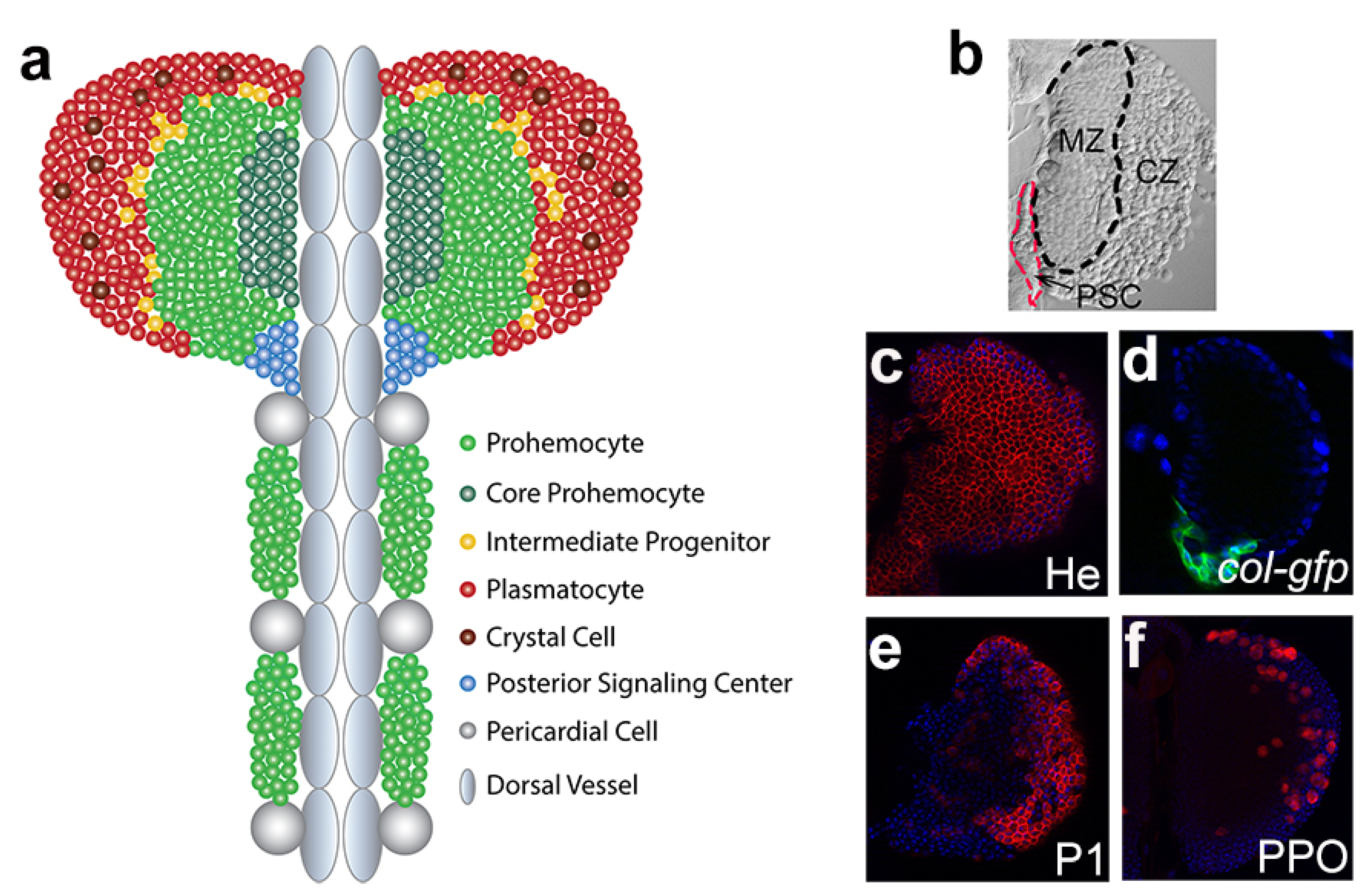
4. Drosophila Lymph Gland

The Drosophila lymph gland is located in the dorsal aspect of the larva in association with the dorsal vessel, Drosophila heart [32], and is derived from embryonic thoracic mesoderm [24]. In contrast to embryonic blood cells, lymph gland precursor cells in embryos quickly proliferate only and do not differentiate. During dorsal closure, these precursor cells migrate dorsally and are positioned to flank the dorsal vessel in the thoracic segments. In the late embryo, these cells constitute the lymph gland with a single pair of lobes. The lymph glands from first-instar and early-second-instar larvae are largely populated by undifferentiated hemocytes. The differentiated blood cells first appear in the lymph gland during the mid-to-late second instar. In the third instar, the lymph gland contains undifferentiated prohemocytes, differentiating hemocytes, and mature blood cells. At the onset of pupariation, the lymph gland dissociates and bursts open to release blood cells into circulation [5].

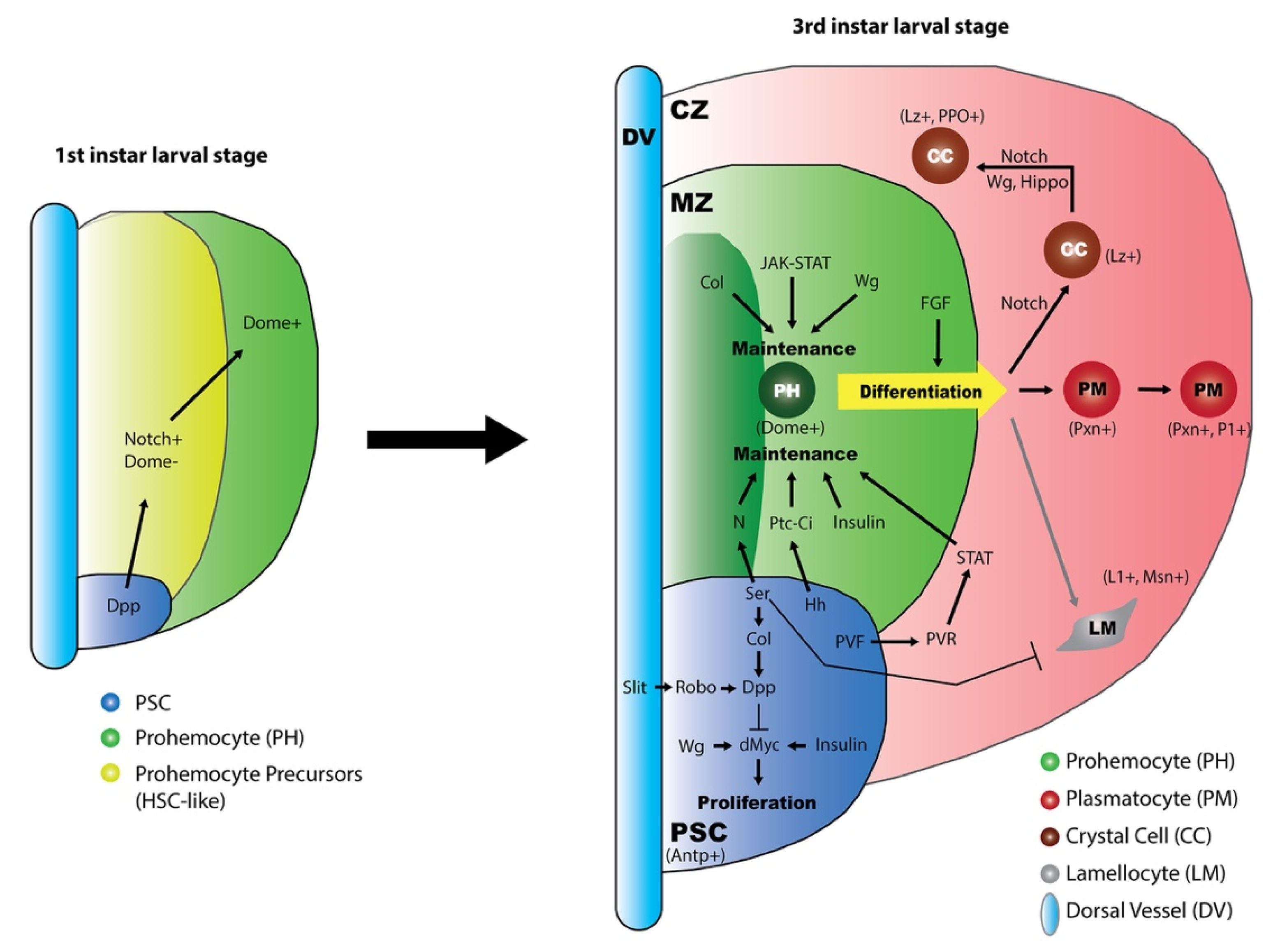
4.1. Lymph Gland Zones

Although lymph gland development is initiated in the embryo, the establishment of zones and the majority of hemocyte maturation take place in the third larval instar. The lymph glands are structurally composed of several pairs of lobes, each separated by pericardial cells [33]. The anterior-most lobe, called the primary lobe, is the largest in size. The differentiation of blood cells occurs mainly in the primary lobes, whereas the other posterior lobes consist of undifferentiated hemocyte progenitors.
When fully developed in third-instar larvae, the primary lobe can be divided into several zones based on cellular morphology, blood cell types, as well as distinct markers (Figure 1). A central zone, named the medullary zone (MZ), contains tightly packed blood cell progenitors (prohemocytes), which can be characterized by the expression of the JAK-STAT receptor domeless (dome). A periphery zone, termed the cortical zone (CZ), is rich in mature blood cells, which are loosely arranged and can be labeled by specific markers for plasmatocytes, crystal cells, and lamellocytes if there are any. Another crucial zone, called the posterior signaling center (PSC), is located at the posterior tip of the primary lobe and adjacent to MZ progenitors, specifically expressing the homeobox protein Antennapedia (Antp) [34]. Between MZ and CZ, there is an area termed the intermediate zone (IZ), where the cells undergo the transition from progenitors to specified blood cells, simultaneously expressing markers for both prohemocytes and early differentiated cells [24]. Recently, different subpopulations of Dome+ progenitors in the MZ were precisely defined with distinct markers. The most central progenitors that express Tep4 or collier (col, also known as knot) are defined as the Core progenitors [35], and the neighboring progenitors that lack Tep4 or col expression are named Distal progenitors [36].
During the lymph gland development, a small group of Dome-negative cells adjacent to the dorsal vessel were transiently observed in the first or second instar and identified as the founder cells of Dome+ prohemocytes [33,37]. This Dome-negative cell population in the first-instar lymph gland expresses several molecular markers that are associated with hematopoietic stem cells (HSCs) in vertebrates, and are multipotent to give rise to both progenitors and differentiated cells, and therefore has been termed HSCs [37]. Another clonal analysis also suggested the existence of HSCs in lymph glands [38]. Nevertheless, the important characteristics of stem cells, such as self-renewal or asymmetrical cell division, have not been clearly elucidated in these cells [37]. Thus, more evidence is expected to designate these cells as bona fide HSCs in Drosophila lymph glands.

4.2. Role of the PSC in the Lymph Gland

PSC cells exhibit high expression of Antp and the gene col, a Drosophila ortholog of mammalian Early B-cell Factor (EBF) [25,26,34], and produce multiple signaling molecules, such as Hh [34], Wg [39], Serrate (Ser, the ligand for Notch) [40], Decapentaplegic (Dpp) [41], and Pvf1 [a ligand for the platelet-derived growth factor (PDGF)] [42], all of which play critical roles in the maintenance of prohemocytes in the MZ during lymph gland hematopoiesis. Accordingly, the PSC has been established as an equivalent to the mammalian HSC niche. However, the requirement of PSC cells as well as the role of col in the PSC for progenitor maintenance have been challenged by recent studies [35,43], in which the genetic ablation of the PSC did not cause a loss of prohemocyte population. Later, additional studies repeatedly demonstrated that the signaling molecules sent out from the PSC, such as Hh and Ser, are indeed required to restrain hemocyte differentiation and maintain the MZ progenitors in an undifferentiated state [35,36,44], further supporting the critical role of the PSC as a hematopoietic niche. However, the role of col in the PSC has to be reconsidered. In contrast to PSC-expressing col, the col in the MZ has been revealed to act as an intrinsic regulator that controls the maintenance of prohemocytes, as knocking down col expression in the MZ led to a significant loss of prohemocytes and massive hemocyte differentiation [35,43], resembling the phenotype of a col mutant. In addition, it has been found that the MZ prohemocyte population is heterogeneous, containing both Col-positive and Col-negative cells [36,44]. The number of the Col-negative prohemocyte subpopulation is significantly reduced after PSC ablation, whereas the number of Col-positive prohemocytes is not affected by PSC manipulation [44]. The coexistence of PSC-dependent and PSC-independent subpopulations in the prohemocyte pool could be a clue to interpret the inconsistency reported for the role of PSC.
Besides their role in normal developmental conditions, the essential role of PSC cells in controlling the proper differentiation of lamellocytes from progenitors after wasp parasitization has been repeatedly implicated [25,43,45,46]. Interestingly, the production of lamellocytes upon mechanical stress, such as squeezing fly larvae with forceps, is independent of the PSC cellular domain [47].

5. Regulatory Signaling during Lymph Gland Development

Developmental signaling is a major regulatory mechanism in the control of organ development. Many major developmental signaling pathways play important roles in regulating blood cell proliferation, differentiation, and patterning during the development of lymph glands. In the following section, we focus on several classic developmental signals and review their functions during the lymph gland hematopoietic process.

5.1. Notch Signaling Instructs Crystal Cell Fate Determination

Notch signaling is evolutionarily conserved from flies to mammals and plays a fundamental role in cell fate determination. The core component in this pathway is the single-pass transmembrane protein Notch. In the canonical pathway, the extracellular domain of Notch interacts with the ligand protein Delta or Ser located at the neighboring cell membrane, which triggers the cleavage of the Notch protein to release the Notch intracellular domain (NICD). Then, the NICD enters the nucleus and facilitates transcription factor Suppressor of Hairless [Su(H)] to activate target gene expression [48]. Notch activation through Ser is required for blood cell lineage specification and maintenance in the lymph gland.
The best characterized function of Notch signaling during larval hematopoiesis is to determine the crystal cell fate. Notch expression is ubiquitous throughout the third-instar lymph gland primary lobe [36,49]. Loss-of-function mutations in Notch result in a drastically decreased number of crystal cells [24,49,50], suggesting that Notch signaling is required for crystal cell differentiation. It has been demonstrated that Ser, but not Delta, is the Notch ligand for the specification of crystal cells [25,40,50,51]. Actually, the Notch pathway is employed several times during crystal cell formation. First, in Distal progenitors of the MZ, Notch pathway activity promotes the cell fate of undifferentiated cells towards crystal cells but not plasmatocytes. Overexpression of Notch, Ser, or Su(H) in the MZ dramatically increases the crystal cell number and blocks plasmatocyte differentiation [36]. Second, Notch signaling specifies the crystal cell precursors in the IZ and CZ regions by activating the expression of lozenge (lz), a transcription factor essential for commitment to crystal cell fate [40]. The expression of lz is diminished in the Notch or Su(H) loss-of-function mutant [40]. Third, Lz cooperates with Notch signaling to selectively activate a combination of crystal cell-related target genes, such as klumpfuss and hindsight, to further lock the cell fate towards the crystal cell and prevent the cells from adopting other cell fates [52]. Fourth, non-canonical Notch activation via stabilization of the Notch receptor in the endocytic pathway mediated by Hif-α (Sima in fly) facilitates the maturation and survival of crystal cells, which is independent of the Ser ligand [53].
Ser is highly expressed in PSC cells [25,34,40]. Notch signaling in the PSC mediated by Ser is critical in controlling the transcription of col, which is very important for maintaining the PSC cell number and signaling [25,41]. Interfering with Notch signaling, either through the expression of a dominant-negative form of Ser in the PSC or in Notch loss-of-function mutants, results in nearly complete loss of col transcription in PSC cells [26]. Knocking down Notch or Ser expression in the PSC is also able to non-cell-autonomously induce the formation of lamellocytes without wasp infestation [49], suggesting an inhibitory role of Notch signaling in lamellocyte differentiation under physiological conditions. The loss of col in the PSC blocks the formation of lamellocytes, even after parasitization [25]. Therefore, the lamellocyte differentiation induced by inhibiting Notch signaling is likely irrelevant with col in the PSC.
The Notch pathway is required cell-autonomously to maintain the Core progenitor in an undifferentiated state [36]. Silencing of Notch or Su(H) provokes a reduction in Core progenitors and an increase in mature hemocytes. Ser is required at the PSC to activate Notch for Core progenitor maintenance.
In addition, Notch signaling has been identified as the signature marker for HSC-like cells in first-instar lymph glands [37]. These cells express several indicators for active Notch signaling, including Notch, Su(H), and Enhancer of split mβ [E(spl)mβ]. The expression of Notch signaling activity is lost from these cells in the second instar. The function of Notch signaling in these cells remains elusive.

5.2. Hh Signaling Maintains the Prohemocyte Potency

Hh signaling is a conserved pathway that regulates cell proliferation, migration, and differentiation during development. The pathway is initiated by the binding of Hh with its receptor Patched (Ptc). G-protein-coupled receptor Smoothened (Smo) is activated to prevent phosphorylation and proteolysis of the transcription factor Cubitus interruptus (Ci), which eventually translocates into the nucleus to switch on target gene expressions [54]. In the lymph gland, Hh signaling is crucial for the maintenance of blood cell progenitors in MZ.
It is known that Hh is produced from signal-producing cells and functions as a morphogen to activate cellular signal transduction in the signal-receiving cells. Accordingly, hh is specifically expressed in PSC cells of the lymph gland, whereas the receptor Ptc and downstream Smo and Ci are highly expressed in the neighboring MZ region [34,44]. Depletion of hh in the PSC has no effect on the PSC itself but results in the loss of the MZ and an increase in the differentiated hemocyte number [34]. Similarly, the lymph gland with a loss-of-function Ci or Smo, or the expression of ectopic Ptc, shows a reduction in the prohemocyte number in the MZ [34,55,56]. Conversely, activating Hh signaling in the MZ by overexpressing a dominant-negative form of Ptc, or by inhibiting Suppressor of Fused (Sufu), a negative regulator of Ci transcriptional activity, causes a strong increase in blood progenitor cells in parallel to a reduction in mature hemocytes [56]. In contrast to manipulating the pathway downstream components, misexpression of hh in the MZ is not sufficient to expand the progenitor population, suggesting a possibility that Hh protein in non-PSC cells cannot function properly to activate the pathway due to an unknown disturbing mechanism. Collectively, these results indicate that Hh signaling originates from PSC cells and functions in the MZ to maintain prohemocytes in a hematopoietic precursor state and prevent their differentiation. Actually, not all the prohemocytes in the MZ are responsive to Hh signaling. A recent study further clarified that a particular subpopulation of MZ prohemocytes with Col-negative expression is sensitive to Hh signaling, and Hh signaling is required to maintain this prohemocyte subpopulation [44].
Hh production in PSC cells is precisely controlled by a combination of positive and negative regulatory mechanisms [57]. The GATA factor Serpent (Srp) is essential for hh expression in the PSC. The transcriptional regulators Su(H) and U-shaped (Ush) prevent hh expression in non-PSC cells of the lymph gland. Unlike the sufu mutant, misexpression of hh in the MZ is not sufficient to expand the progenitor population [56,57]. Interestingly, the expression of ush in prohemocytes requires Hh signaling. hh RNAi in the PSC, Ci RNAi in the MZ, ptc overexpression in the MZ, or loss of smo all result in a significant reduction in ush expression [44]. Thus, the Ush and Hh pathways collaborate through a regulatory loop to restrict hh expression within the PSC and maintain prohemocyte potency. In addition, Smo genetically interacts with Ush to block lamellocyte differentiation under normal conditions. Double heterozygotes for ush/smo exhibit a significant increase in the number of lamellocytes compared with that in single heterozygote for either ush or smo [44].
The Hh signaling pathway has been extensively studied in the wing system, where col is one of the well-established responsive genes for Hh signaling [58]. Conversely, col expression in the lymph gland may not be subject to Hh regulation. In lymph glands, col is highly expressed in PSC cells and at a low level in the Core progenitor cells of the MZ area. The col in the MZ and PSC functions independently to regulate progenitor maintenance and the hemocyte differentiation rate, respectively [35]. The col expression in the PSC is regulated by the Notch/Ser signal and can be inhibited by Jumu [26,59]. Although it is still unclear how col expression in the MZ is regulated, it has been revealed that col expression in the MZ and its function in those cells are independent of hh expressed by PSC cells [35].

5.3. Wnt/Wg Signaling Modulates Hemocyte Proliferation and Differentiation

Wnt/Wg signaling has been implicated in multiple developmental processes of metazoans, including cell fate specification, cell proliferation, and maintenance. The signal activated by Wg binding with its receptor is transduced through a series of cellular events and eventually protects Armadillo (Arm, Drosophila β-catenin) from degradation and promotes its translocation into the nucleus to activate target gene transcription [1]. In Drosophila, there are two receptor proteins for Wg ligand: Frizzled (Fz) and Frizzled 2 (Fz2). It has been shown that Wg signaling via either Fz or Fz2 is required to regulate MZ development, whereas Wg signaling mediated by Fz2, but not Fz, is responsible for controlling PSC cell and crystal cell proliferation [39,60].
The Wg protein is expressed throughout the entire lymph gland, and its expression level in MZ cells is much higher than that in CZ cells. Arm shows a similar expression pattern in the lymph gland [39]. It has been evidenced that Wg signaling in the MZ is able to maintain the MZ integrity and block prohemocyte differentiation. Impaired Wg signaling not only results in the mislocalization of Dome+ cells in the cortical area of the lymph gland, possibly through the downregulation of the cell adhesion molecule E-cadherin, but also initiates hemocyte differentiation, thus increasing the number of IZ cells. Conversely, elevated Wg signaling activities in the MZ, by overexpressing arm or wg, can inhibit the differentiation of prohemocytes, thus resulting in enlarged MZ and defective CZ regions.
It is known that the maintenance of the MZ prohemocyte pool requires signals from the PSC. The Wg pathway is able to positively regulate PSC cell proliferation through the proto-oncogene dMyc [39,41]. In addition, the maturation of crystal cells, but not plasmatocytes, is regulated by Wg signaling in the CZ. Overexpression of wg in crystal cell precursors (Lz+) results in an increased number of mature crystal cells, whereas downregulated Wg signaling in Lz+ cells causes a decreased number of crystal cells, suggesting that Wg signaling is able to promote the maturation and proliferation of crystal cells [39].
The ECM protein Tiggrin (Tig) has been identified as a downstream effector of Wg signaling [60,61]. In lymph glands, Wg signal inhibits the expression of Tig. Therefore, the Tig protein is highly expressed in plasmatocytes in the CZ, where the Wg signal is low, but not in MZ and crystal cells, where Wg activity is high. Consistent with its expression pattern, Tig likely functions only in the maturation of plasmatocytes, but not crystal cells and prohemocytes, as the Tig mutant shows a prematuration of plasmatocytes, while Tig overexpression blocks the maturation of plasmatocytes.

5.4. JAK-STAT Signaling Inhibits Prohemocyte Differentiation

The JAK-STAT signal transduction cascade is named from its two major components: Janus Tyrosine Kinase (JAK) and Signal Transducers and Activators of Transcription (STAT). The binding between the cytokine ligand and its single-pass transmembrane receptor induces conformational changes in the receptor and therefore triggers the phosphorylation of JAK and recruitment of STAT transcription factors. In Drosophila, several related proteins have been functionally characterized [62]: three cytokine ligands, Unpaired1–3 (Upd1–3); one receptor, Dome; one JAK kinase, Hopscotch (Hop); and one STAT protein, Stat92E. It has been well established that either local or systemic JAK-STAT signaling is closely associated with lamellocyte formation during an immune response [63,64]. Besides this, the lymph gland under healthy conditions utilizes both JAK-dependent and JAK-independent STAT signalings to maintain prohemocyte potency [42,65,66].
The receptor Dome is highly expressed in the MZ as a common marker of prohemocyte [26,33]. Consistently, as a signaling readout, the Dome-MESO-LacZ reporter is highly expressed in the MZ [26,65], indicating that JAK-STAT signaling is active in the MZ cells. The hypomorphic mutants for dome or upd show premature differentiation of plasmatocytes in the primary lobes of lymph glands [67], suggesting that JAK-STAT signaling in the MZ is important for keeping prohemocytes in an undifferentiated state. Consistently, JAK-STAT signaling is required for the expression of ush, which maintains the prohemocyte potency in the MZ [65].
JAK-STAT signaling is also involved in blood cell proliferation and differentiation in lymph glands. Larvae with a hop gain-of-function mutation always show hypertrophic lymph glands [68]. Consistently, ectopic expression of hop in the CZ, but not in the MZ, led to hyperplasia of the lymph gland, which requires Stat92E, Dome, and Upd3 [69]. In lymph glands with either the hop hypermorphic mutation or hop overexpression, an increased number of lamellocytes was also observed, even without parasitization [68,69]. Together, these results suggest that JAK-STAT signaling possesses the ability to enhance blood cell proliferation and induce lamellocyte formation in lymph glands. However, under physiological conditions, the JAK-STAT pathway might not be required for CZ development, as knocking down hop expression by RNAi in CZ cells resulted in no significant changes [69].
In differentiating hemocytes, STAT also mediates an “equilibrium signal” to maintain prohemocyte potency, most likely independent of the Dome-JAK pathway [42,66]. Stat92E is expressed throughout the entire primary lobe of the lymph gland [70]. Knocking down stat92e by RNAi specifically in the CZ results in the loss of the prohemocyte pool and elevates the number of plasmatocytes and crystal cells, whereas Stat92E loss-of-function outside of the CZ has no effect on blood cell differentiation [42,70]. The whole animal stat92e mutant reveals a loss of the MZ but an enlarged population of differentiated hemocytes [26], which is likely due to the loss of Stat92E function in the CZ. Further studies showed that STAT in the CZ functions downstream of PDGF/VEGF-like Receptor (PVR) signaling, which is activated by its ligand PVF1 from PSC cells, to promote the expression of Adenosine Deaminase-related growth factor (ADGF) [42,66]. A complex of ARF1 (Ras small GTPase) and Asrij (an endocytic protein) in endosomes provides a scaffold for STAT activation [67,71]. This PVF-PVR-STAT signaling cascade promotes lymph gland growth in the early stage and later cell-non-autonomously maintain the quiescence state of progenitors in the MZ [42,66].

5.5. BMP/Dpp Signaling Restricts PSC Size

Bone morphogenetic proteins (BMP) belong to the Transforming growth factor beta (TGFβ) superfamily. The BMP signal pathway is initiated by the BMP ligand binding with a type II receptor, which recruits and phosphorylates a type I receptor. Then, the type I receptor phosphorylates the SMAD protein and eventually activates target gene expressions in the nucleus. In Drosophila, there are two BMP ligands, Decapentaplegic (Dpp) and Glass bottom boat (Gbb); two type I receptors, Thickveins (Tkv) and Saxophone (Sax); two type II receptors, Wishful thinking (Wit) and Punt; one SMAD protein, Mother against Dpp (Mad); and multiple target genes, such as daughters against Dpp (dad) [72]. It has been demonstrated that BMP signal transduction is mainly mediated by the binding of the ligand Dpp to Tkv/Wit receptors in Drosophila lymph glands [41].
Dpp signaling is specifically activated in the PSC and is required for controlling its size during Drosophila larval hematopoiesis [41]. The activity of the BMP/Dpp signaling pathway can be reflected by the phosphorylation of the Mad protein and the expression of target genes. In wild type lymph glands, a high level of the phosphorylated Mad protein and target gene dad expression are both detected specifically in PSC cells, indicating that Dpp signaling is active in the niche. Blocking Dpp signaling either by using dpp, tkv, or wit mutants or by overexpressing a dominant-negative form of tkv significantly increases the number of PSC cells, suggesting that Dpp signaling negatively regulates PSC cell numbers. As PSC maintains prohemocytes, the loss of Dpp signaling in PSC cells results in a correlatively enlarged prohemocyte pool and impaired hemocyte differentiation. Furthermore, Dpp signaling maintains the low number of PSC cells by repressing dMyc expression in the PSC. The epistatic analysis indicated that Dpp signaling antagonizes Wg activity in the control of PSC cell numbers. Dpp signaling in the PSC is dependent on the local expression of col, as Dpp signal activity is lost when col expression is repressed in the PSC cells. In addition, Dpp signaling in the PSC can be enhanced by the Robo signal, which is activated by the glycoprotein Slit secreted from the dorsal vessel [73].
In addition to the cell-autonomous function of controlling PSC size, the Dpp signal is active in the PSC during the first instar larval stage and serves as a niche signal to maintain HSC-like cells, which are the founder cells of Dome+ prohemocytes in the MZ [37]. The phosphorylated Mad protein is enriched in these HSC-like cells. The knockdown of dpp in the PSC during the first instar stage or depletion of Mad in these HSC-like cells results in the loss of these HSC-like cells, and eventually drastically reduces the number of prohemocytes and the overall size of the lymph gland in the third instar larval stage. Notably, the PSC cell number did not change upon these manipulations, suggesting that the Dpp signal activity in the early larval PSC is not required for the development of the PSC itself.
Besides the signals mentioned above, many other pathways also play important roles during normal lymph gland development. The Fibroblast Growth Factor (FGF) ligands, Pyramus and Thisbe, and FGF receptor, Heartless, are expressed at high level in Dome+ progenitors, and have been found to facilitate blood cell differentiation in lymph glands [74]. Inhibition of FGF signaling in these prohemocytes by overexpressing a dominant-negative form of heartless, thisbe RNAi, or pyramus RNAi enlarges the MZ prohemocyte pool and decreases the differentiated hemocyte population. On the other hand, the enforced expression of FGF ligands or receptor results in a loss of Dome+ progenitor cells and an increased proportion of differentiated hemocytes in the primary lobes of lymph glands. This FGF-induced differentiation of prohemocytes also takes place in the second lobes, which normally harbor undifferentiated hemocytes. All these results indicate that FGF signaling is required for hemocyte differentiation and also sufficient to induce progenitor cell differentiation. The Hippo pathway, an evolutionarily conserved regulator of organ growth and cell fate determination, is known to regulate hematopoiesis by restricting hemocyte proliferation and differentiation in Drosophila lymph glands. Lymph glands lacking the downstream Hippo pathway kinase Warts are larger in size and filled with differentiated cells as a result of hemocyte overproliferation and premature differentiation [75,76]. The key effector of the Hippo pathway is the protein Yorkie, which is directly phosphorylated by Warts. Yorkie functions as a transcription co-activator, but its phosphorylation by Warts blocks its function by locking it in the cytoplasm. Yorkie and its binding partner, the transcription factor Scalloped, function together to regulate crystal cell formation in a cell-autonomous manner by transcriptionally activating lz expression in crystal cell progenitors [75] or in a non-cell-autonomous manner by promoting Ser expression in a subset of cortical area cells to instruct the neighboring Notch-positive cells to commit to the crystal cell fate [51,77]. Insulin signaling and downstream Target of Rapamycin (TOR) signaling are also known to stimulate cell proliferation via dMyc in the PSC [78,79,80,81,82], and are essential for progenitor maintenance [78,83,84].

6. Concluding Remarks

Drosophila melanogaster is a classic model organism that has made important contributions to genetic research. Recently, the Drosophila lymph gland has emerged as an excellent model system to study hematopoiesis. Undifferentiated progenitors and multiple types of differentiated blood cells coexist in the lymph gland, which allows researchers to address many questions regarding cell lineage and the signals that mediate blood cell progenitor maintenance, proliferation, and differentiation. Many classic development signaling pathways have been demonstrated to play vital roles during lymph gland hematopoiesis (Figure 2). In brief, the PSC in the lymph gland provides signal molecules, such as Hh, to activate signaling pathways in the neighboring MZ. The progenitors in the MZ exhibit high activities of local signals, such as Wg, JAK/STAT, and Col. These PSC-derived and MZ local signals, together with the equilibrium signals from differentiating cells in the CZ, such as PVR and downstream STAT, collaborate to maintain the progenitor cells in a quiescent state and/or regulate their differentiation. The differentiating blood cells require Notch signaling, which cooperates with Wg signaling and Lz, to develop towards crystal cell fate. To date, huge progress has been achieved in understanding the functions of developmental signaling pathways during larval hematopoiesis; however, their cross-regulation remains largely undefined.
Signaling events occur at precise locations and times to properly control animal development. Determining the spatiotemporal activity and function of developmental signals is critical for understanding the specific developmental process. During larval lymph gland hematopoiesis, many developmental signaling pathways play roles in specific areas and/or at particular times. For example, in the PSC, Dpp functions as a niche signal to maintain HSC-like cells during the early larval stage and controls the PSC cell number in the late larval stage. Instead, the role as a niche signal for MZ progenitor cell maintenance is taken by Hh signaling in the third-instar lymph gland. In the past decades, studies in the Drosophila lymph gland have benefited a lot from powerful experimental tools, such as versatile genetic approaches and lineage-tracing methods. Recently, many new techniques in Drosophila have been reported for monitoring and perturbing signaling molecules, such as the kinase translocation reporter (KTR)-based sensors, which enable the live-cell measurement of kinase activity at a single-cell level [85], and optogenetics and imaging techniques, which have been successfully utilized to study the function of cell signaling [86,87,88] or modulate cell contractility during embryogenesis [89]. These new techniques are promising for their application in future studies to address specific questions relevant to the spatiotemporal control of lymph gland hematopoiesis.
It is known that Drosophila hematopoiesis employs many conserved molecular strategies. For example, the Drosophila plasmatocytes are considered homologous to macrophages in humans, and the crystal cells and lamellocytes in Drosophila have functional similarities to platelets and giant cells in humans. Strikingly, many signaling pathways and transcription factors that control Drosophila hematopoiesis play comparable functions in mammals. Therefore, fully understanding the principles that control Drosophila hematopoiesis will definitely shed light on human hematopoiesis in further studies.

Funding

This research was funded by the grants from the Marine S&T Fund of Shandong Province for Pilot National Laboratory for Marine Science and Technology (Qingdao) (2018SDKJ0406-2), the National Natural Science Foundation of China (31701274, 31970506), and the Fundamental Research Funds for Central Universities, China (201612010, 201822016).

Acknowledgments

We would like to thank Ando I. (Biological Research Center of the Hungarian Academy of Sciences) and Asano T. (Tokyo Metropolitan University, Japan) for antibodies, Crozatier M. (CNRS/Université Toulouse III and Centre de Biologie Intégrative, France) for fly strains, and Rui Lv (Ocean University of China) for assistance with the manuscript preparation. We apologize to those authors whose important work was accidentally not cited.

Conflicts of Interest

The authors declare no conflict of interest.

References

  1. Bejsovec, A. Wingless Signaling: A Genetic Journey from Morphogenesis to Metastasis. Genetics 2018, 208, 1311–1336. [Google Scholar] [CrossRef] [Green Version]
  2. Kong, J.H.; Siebold, C.; Rohatgi, R. Biochemical mechanisms of vertebrate hedgehog signaling. Development 2019, 146. [Google Scholar] [CrossRef] [PubMed] [Green Version]
  3. Siebel, C.; Lendahl, U. Notch Signaling in Development, Tissue Homeostasis, and Disease. Physiol. Rev. 2017, 97, 1235–1294. [Google Scholar] [CrossRef] [PubMed] [Green Version]
  4. Massagué, J. TGFβ signalling in context. Nat. Rev. Mol. Cell Biol. 2012, 13, 616–630. [Google Scholar] [CrossRef]
  5. Grigorian, M.; Mandal, L.; Hartenstein, V. Hematopoiesis at the onset of metamorphosis: Terminal differentiation and dissociation of the Drosophila lymph gland. Dev. Genes Evol. 2011, 221, 121–131. [Google Scholar] [CrossRef] [Green Version]
  6. Letourneau, M.; Lapraz, F.; Sharma, A.; Vanzo, N.; Waltzer, L.; Crozatier, M. Drosophila hematopoiesis under normal conditions and in response to immune stress. FEBS Lett. 2016, 590, 4034–4051. [Google Scholar] [CrossRef]
  7. Banerjee, U.; Girard, J.R.; Goins, L.M.; Spratford, C.M. Drosophila as a Genetic Model for Hematopoiesis. Genetics 2019, 211, 367–417. [Google Scholar] [CrossRef] [Green Version]
  8. Shim, J. Drosophila blood as a model system for stress sensing mechanisms. BMB Rep. 2015, 48, 223–228. [Google Scholar] [CrossRef] [Green Version]
  9. Lebestky, T.; Chang, T.; Hartenstein, V.; Banerjee, U. Specification of Drosophila hematopoietic lineage by conserved transcription factors. Science 2000, 288, 146–149. [Google Scholar] [CrossRef]
  10. Hartenstein, V. Blood cells and blood cell development in the animal kingdom. Annu. Rev. Cell Dev. Biol. 2006, 22, 677–712. [Google Scholar] [CrossRef] [Green Version]
  11. Ghosh, S.; Singh, A.; Mandal, S.; Mandal, L. Active hematopoietic hubs in Drosophila adults generate hemocytes and contribute to immune response. Dev. Cell 2015, 33, 478–488. [Google Scholar] [CrossRef] [PubMed] [Green Version]
  12. Leitão, A.B.; Sucena, É. Drosophila sessile hemocyte clusters are true hematopoietic tissues that regulate larval blood cell differentiation. eLife 2015, 4. [Google Scholar] [CrossRef] [PubMed] [Green Version]
  13. Bunt, S.; Hooley, C.; Hu, N.; Scahill, C.; Weavers, H.; Skaer, H. Hemocyte-secreted type IV collagen enhances BMP signaling to guide renal tubule morphogenesis in Drosophila. Dev. Cell 2010, 19, 296–306. [Google Scholar] [CrossRef] [PubMed] [Green Version]
  14. Olofsson, B.; Page, D.T. Condensation of the central nervous system in embryonic Drosophila is inhibited by blocking hemocyte migration or neural activity. Dev. Biol. 2005, 279, 233–243. [Google Scholar] [CrossRef] [PubMed] [Green Version]
  15. Van De Bor, V.; Zimniak, G.; Papone, L.; Cerezo, D.; Malbouyres, M.; Juan, T.; Ruggiero, F.; Noselli, S. Companion Blood Cells Control Ovarian Stem Cell Niche Microenvironment and Homeostasis. Cell Rep. 2015, 13, 546–560. [Google Scholar] [CrossRef] [PubMed] [Green Version]
  16. Dudzic, J.P.; Kondo, S.; Ueda, R.; Bergman, C.M.; Lemaitre, B. Drosophila innate immunity: Regional and functional specialization of prophenoloxidases. BMC Biol. 2015, 13. [Google Scholar] [CrossRef] [PubMed] [Green Version]
  17. Binggeli, O.; Neyen, C.; Poidevin, M.; Lemaitre, B. Prophenoloxidase activation is required for survival to microbial infections in Drosophila. PLoS Pathog. 2014, 10, e1004067. [Google Scholar] [CrossRef]
  18. Gajewski, K.M.; Sorrentino, R.P.; Lee, J.H.; Zhang, Q.; Russell, M.; Schulz, R.A. Identification of a crystal cell-specific enhancer of the black cells prophenoloxidase gene in Drosophila. Genesis 2007, 45, 200–207. [Google Scholar] [CrossRef]
  19. Lanot, R.; Zachary, D.; Holder, F.; Meister, M. Postembryonic Hematopoiesis in Drosophila. Dev. Biol. 2001, 230, 243–257. [Google Scholar] [CrossRef] [Green Version]
  20. Markus, R.; Laurinyecz, B.; Kurucz, E.; Honti, V.; Bajusz, I.; Sipos, B.; Somogyi, K.; Kronhamn, J.; Hultmark, D.; Ando, I. Sessile hemocytes as a hematopoietic compartment in Drosophila melanogaster. Proc. Acad. Natl. Sci. USA 2009, 106, 4805–4809. [Google Scholar] [CrossRef] [Green Version]
  21. Honti, V.; Csordas, G.; Markus, R.; Kurucz, E.; Jankovics, F.; Ando, I. Cell lineage tracing reveals the plasticity of the hemocyte lineages and of the hematopoietic compartments in Drosophila melanogaster. Mol. Immunol. 2010, 47, 1997–2004. [Google Scholar] [CrossRef] [PubMed] [Green Version]
  22. Makhijani, K.; Alexander, B.; Tanaka, T.; Rulifson, E.; Bruckner, K. The peripheral nervous system supports blood cell homing and survival in the Drosophila larva. Development 2011, 138, 5379–5391. [Google Scholar] [CrossRef] [PubMed] [Green Version]
  23. Makhijani, K.; Alexander, B.; Rao, D.; Petraki, S.; Herboso, L.; Kukar, K.; Batool, I.; Wachner, S.; Gold, K.S.; Wong, C.; et al. Regulation of Drosophila hematopoietic sites by Activin-beta from active sensory neurons. Nat. Commun. 2017, 8, 15990. [Google Scholar] [CrossRef] [PubMed] [Green Version]
  24. Krzemien, J.; Oyallon, J.; Crozatier, M.; Vincent, A. Hematopoietic progenitors and hemocyte lineages in the Drosophila lymph gland. Dev. Biol. 2010, 346, 310–319. [Google Scholar] [CrossRef] [PubMed] [Green Version]
  25. Crozatier, M.; Ubeda, J.M.; Vincent, A.; Meister, M. Cellular immune response to parasitization in Drosophila requires the EBF orthologue collier. PLoS Biol. 2004, 2, E196. [Google Scholar] [CrossRef] [Green Version]
  26. Krzemien, J.; Dubois, L.; Makki, R.; Meister, M.; Vincent, A.; Crozatier, M. Control of blood cell homeostasis in Drosophila larvae by the posterior signalling centre. Nature 2007, 446, 325–328. [Google Scholar] [CrossRef]
  27. Stofanko, M.; Kwon, S.Y.; Badenhorst, P. Lineage Tracing of Lamellocytes Demonstrates Drosophila Macrophage Plasticity. PLoS ONE 2010, 5, e14051. [Google Scholar] [CrossRef]
  28. Anderl, I.; Vesala, L.; Ihalainen, T.O.; Vanha-aho, L.-M.; Andó, I.; Rämet, M.; Hultmark, D. Transdifferentiation and Proliferation in Two Distinct Hemocyte Lineages in Drosophila melanogaster Larvae after Wasp Infection. PLoS Pathog. 2016, 12, e1005746. [Google Scholar] [CrossRef]
  29. Holz, A.; Bossinger, B.; Strasser, T.; Janning, W.; Klapper, R. The two origins of hemocytes in Drosophila. Development 2003, 130, 4955–4962. [Google Scholar] [CrossRef] [Green Version]
  30. Sanchez Bosch, P.; Makhijani, K.; Herboso, L.; Gold, K.S.; Baginsky, R.; Woodcock, K.J.; Alexander, B.; Kukar, K.; Corcoran, S.; Jacobs, T.; et al. Adult Drosophila Lack Hematopoiesis but Rely on a Blood Cell Reservoir at the Respiratory Epithelia to Relay Infection Signals to Surrounding Tissues. Dev. Cell 2019, 51, 787–803.e5. [Google Scholar] [CrossRef]
  31. Mackenzie, D.K.; Bussiere, L.F.; Tinsley, M.C. Senescence of the cellular immune response in Drosophila melanogaster. Exp. Gerontol. 2011, 46, 853–859. [Google Scholar] [CrossRef] [PubMed]
  32. El Shatoury, H.H. The structure of the lymph glands of Drosophila larvae. Wilhelm Roux Arch. Entwickl. Mech. Org. 1955, 147, 489–495. [Google Scholar] [CrossRef]
  33. Jung, S.H.; Evans, C.J.; Uemura, C.; Banerjee, U. The Drosophila lymph gland as a developmental model of hematopoiesis. Development 2005, 132, 2521–2533. [Google Scholar] [CrossRef] [Green Version]
  34. Mandal, L.; Martinez-Agosto, J.A.; Evans, C.J.; Hartenstein, V.; Banerjee, U. A Hedgehog- and Antennapedia-dependent niche maintains Drosophila haematopoietic precursors. Nature 2007, 446, 320–324. [Google Scholar] [CrossRef]
  35. Oyallon, J.; Vanzo, N.; Krzemien, J.; Morin-Poulard, I.; Vincent, A.; Crozatier, M. Two Independent Functions of Collier/Early B Cell Factor in the Control of Drosophila Blood Cell Homeostasis. PLoS ONE 2016, 11, e0148978. [Google Scholar] [CrossRef] [PubMed]
  36. Blanco-Obregon, D.; Katz, M.J.; Durrieu, L.; Gandara, L.; Wappner, P. Context-specific functions of Notch in Drosophila blood cell progenitors. Dev. Biol. 2020. [Google Scholar] [CrossRef] [PubMed]
  37. Dey, N.S.; Ramesh, P.; Chugh, M.; Mandal, S.; Mandal, L. Dpp dependent Hematopoietic stem cells give rise to Hh dependent blood progenitors in larval lymph gland of Drosophila. elife 2016, 5. [Google Scholar] [CrossRef]
  38. Minakhina, S.; Steward, R. Hematopoietic stem cells in Drosophila. Development 2010, 137, 27–31. [Google Scholar] [CrossRef] [PubMed] [Green Version]
  39. Sinenko, S.A.; Mandal, L.; Martinez-Agosto, J.A.; Banerjee, U. Dual role of wingless signaling in stem-like hematopoietic precursor maintenance in Drosophila. Dev. Cell 2009, 16, 756–763. [Google Scholar] [CrossRef] [Green Version]
  40. Lebestky, T.; Jung, S.H.; Banerjee, U. A Serrate-expressing signaling center controls Drosophila hematopoiesis. Genes Dev. 2003, 17, 348–353. [Google Scholar] [CrossRef] [Green Version]
  41. Pennetier, D.; Oyallon, J.; Morin-Poulard, I.; Dejean, S.; Vincent, A.; Crozatier, M. Size control of the Drosophila hematopoietic niche by bone morphogenetic protein signaling reveals parallels with mammals. Proc. Natl. Acad. Sci. USA 2012, 109, 3389–3394. [Google Scholar] [CrossRef] [PubMed] [Green Version]
  42. Mondal, B.C.; Mukherjee, T.; Mandal, L.; Evans, C.J.; Sinenko, S.A.; Martinez-Agosto, J.A.; Banerjee, U. Interaction between differentiating cell- and niche-derived signals in hematopoietic progenitor maintenance. Cell 2011, 147, 1589–1600. [Google Scholar] [CrossRef] [PubMed] [Green Version]
  43. Benmimoun, B.; Polesello, C.; Haenlin, M.; Waltzer, L. The EBF transcription factor Collier directly promotes Drosophila blood cell progenitor maintenance independently of the niche. Proc. Natl. Acad. Sci. USA 2015, 112, 9052–9057. [Google Scholar] [CrossRef] [PubMed] [Green Version]
  44. Baldeosingh, R.; Gao, H.; Wu, X.; Fossett, N. Hedgehog signaling from the Posterior Signaling Center maintains U-shaped expression and a prohemocyte population in Drosophila. Dev. Biol. 2018, 441, 132–145. [Google Scholar] [CrossRef] [PubMed]
  45. Sinenko, S.A.; Shim, J.; Banerjee, U. Oxidative stress in the haematopoietic niche regulates the cellular immune response in Drosophila. EMBO Rep. 2011, 13, 83–89. [Google Scholar] [CrossRef] [Green Version]
  46. Louradour, I.; Sharma, A.; Morin-Poulard, I.; Letourneau, M.; Vincent, A.; Crozatier, M.; Vanzo, N. Reactive oxygen species-dependent Toll/NF-kappaB activation in the Drosophila hematopoietic niche confers resistance to wasp parasitism. elife 2017, 6. [Google Scholar] [CrossRef]
  47. Tokusumi, Y.; Tokusumi, T.; Schulz, R.A. Mechanical stress to Drosophila larvae stimulates a cellular immune response through the JAK/STAT signaling pathway. Biochem. Biophys. Res. Commun. 2018, 502, 415–421. [Google Scholar] [CrossRef]
  48. Kopan, R.; Ilagan, M.X. The canonical Notch signaling pathway: Unfolding the activation mechanism. Cell 2009, 137, 216–233. [Google Scholar] [CrossRef] [Green Version]
  49. Small, C.; Ramroop, J.; Otazo, M.; Huang, L.H.; Saleque, S.; Govind, S. An unexpected link between notch signaling and ROS in restricting the differentiation of hematopoietic progenitors in Drosophila. Genetics 2014, 197, 471–483. [Google Scholar] [CrossRef] [Green Version]
  50. Duvic, B.; Hoffmann, J.A.; Meister, M.; Royet, J. Notch signaling controls lineage specification during Drosophila larval hematopoiesis. Curr. Biol. 2002, 12, 1923–1927. [Google Scholar] [CrossRef] [Green Version]
  51. Ferguson, G.B.; Martinez-Agosto, J.A. Yorkie and Scalloped signaling regulates Notch-dependent lineage specification during Drosophila hematopoiesis. Curr. Biol. 2014, 24, 2665–2672. [Google Scholar] [CrossRef] [PubMed] [Green Version]
  52. Terriente-Felix, A.; Li, J.; Collins, S.; Mulligan, A.; Reekie, I.; Bernard, F.; Krejci, A.; Bray, S. Notch cooperates with Lozenge/Runx to lock haemocytes into a differentiation programme. Development 2013, 140, 926–937. [Google Scholar] [CrossRef] [PubMed] [Green Version]
  53. Mukherjee, T.; Kim, W.S.; Mandal, L.; Banerjee, U. Interaction between Notch and Hif-alpha in development and survival of Drosophila blood cells. Science 2011, 332, 1210–1213. [Google Scholar] [CrossRef] [PubMed] [Green Version]
  54. Zhao, L.; Wang, L.; Chi, C.; Lan, W.; Su, Y. The emerging roles of phosphatases in Hedgehog pathway. Cell Commun. Signal. 2017, 15, 35. [Google Scholar] [CrossRef] [Green Version]
  55. Giordani, G.; Barraco, M.; Giangrande, A.; Martinelli, G.; Guadagnuolo, V.; Simonetti, G.; Perini, G.; Bernardoni, R. The human Smoothened inhibitor PF-04449913 induces exit from quiescence and loss of multipotent Drosophila hematopoietic progenitor cells. Oncotarget 2016, 7, 55313–55327. [Google Scholar] [CrossRef] [Green Version]
  56. Tokusumi, T.; Tokusumi, Y.; Schulz, R.A. The mir-7 and bag of marbles genes regulate Hedgehog pathway signaling in blood cell progenitors in Drosophila larval lymph glands. Genesis 2018, 56, e23210. [Google Scholar] [CrossRef]
  57. Tokusumi, Y.; Tokusumi, T.; Stoller-Conrad, J.; Schulz, R.A. Serpent, suppressor of hairless and U-shaped are crucial regulators of hedgehog niche expression and prohemocyte maintenance during Drosophila larval hematopoiesis. Development 2010, 137, 3561–3568. [Google Scholar] [CrossRef] [Green Version]
  58. Hooper, J.E.; Scott, M.P. Communicating with Hedgehogs. Nat. Rev. Mol. Cell Biol. 2005, 6, 306–317. [Google Scholar] [CrossRef]
  59. Hao, Y.; Jin, L.H. Dual role for Jumu in the control of hematopoietic progenitors in the Drosophila lymph gland. elife 2017, 6. [Google Scholar] [CrossRef]
  60. Zhang, C.U.; Blauwkamp, T.A.; Burby, P.E.; Cadigan, K.M. Wnt-mediated repression via bipartite DNA recognition by TCF in the Drosophila hematopoietic system. PLoS Genet. 2014, 10, e1004509. [Google Scholar] [CrossRef] [Green Version]
  61. Zhang, C.U.; Cadigan, K.M. The matrix protein Tiggrin regulates plasmatocyte maturation in Drosophila larva. Development 2017, 144, 2415–2427. [Google Scholar] [CrossRef] [PubMed] [Green Version]
  62. Trivedi, S.; Starz-Gaiano, M. Drosophila Jak/STAT Signaling: Regulation and Relevance in Human Cancer and Metastasis. Int. J. Mol. Sci. 2018, 19, 4056. [Google Scholar] [CrossRef] [PubMed] [Green Version]
  63. Morin-Poulard, I.; Vincent, A.; Crozatier, M. The Drosophila JAK-STAT pathway in blood cell formation and immunity. JAKSTAT 2013, 2, e25700. [Google Scholar] [CrossRef] [Green Version]
  64. Yang, H.; Kronhamn, J.; Ekstrom, J.O.; Korkut, G.G.; Hultmark, D. JAK/STAT signaling in Drosophila muscles controls the cellular immune response against parasitoid infection. EMBO Rep. 2015, 16, 1664–1672. [Google Scholar] [CrossRef] [PubMed]
  65. Gao, H.; Wu, X.; Fossett, N. Upregulation of the Drosophila Friend of GATA gene U-shaped by JAK/STAT signaling maintains lymph gland prohemocyte potency. Mol. Cell Biol. 2009, 29, 6086–6096. [Google Scholar] [CrossRef] [Green Version]
  66. Ferguson, G.B.; Martinez-Agosto, J.A. The TEAD family transcription factor Scalloped regulates blood progenitor maintenance and proliferation in Drosophila through PDGF/VEGFR receptor (Pvr) signaling. Dev. Biol. 2017, 425, 21–32. [Google Scholar] [CrossRef]
  67. Sinha, A.; Khadilkar, R.J.; Vinay, K.S.; Roychowdhury Sinha, A.; Inamdar, M.S. Conserved regulation of the Jak/STAT pathway by the endosomal protein asrij maintains stem cell potency. Cell Rep. 2013, 4, 649–658. [Google Scholar] [CrossRef] [Green Version]
  68. Sorrentino, R.P.; Tokusumi, T.; Schulz, R.A. The Friend of GATA protein U-shaped functions as a hematopoietic tumor suppressor in Drosophila. Dev. Biol. 2007, 311, 311–323. [Google Scholar] [CrossRef] [Green Version]
  69. Terriente-Félix, A.; Pérez, L.; Bray, S.J.; Nebreda, A.R.; Milán, M. A Drosophila model of myeloproliferative neoplasm reveals a feed-forward loop in the JAK pathway mediated by p38 MAPK signalling. Dis. Mod. Mech. 2017, 10, 399–407. [Google Scholar] [CrossRef] [Green Version]
  70. Minakhina, S.; Tan, W.; Steward, R. JAK/STAT and the GATA factor Pannier control hemocyte maturation and differentiation in Drosophila. Dev. Biol. 2011, 352, 308–316. [Google Scholar] [CrossRef]
  71. Khadilkar, R.J.; Rodrigues, D.; Mote, R.D.; Sinha, A.R.; Kulkarni, V.; Magadi, S.S.; Inamdar, M.S. ARF1-GTP regulates Asrij to provide endocytic control of Drosophila blood cell homeostasis. Proc. Natl. Acad. Sci. USA 2014, 111, 4898–4903. [Google Scholar] [CrossRef] [PubMed] [Green Version]
  72. Hamaratoglu, F.; Affolter, M.; Pyrowolakis, G. Dpp/BMP signaling in flies: From molecules to biology. Semin. Cell Dev. Biol. 2014, 32, 128–136. [Google Scholar] [CrossRef] [PubMed] [Green Version]
  73. Morin-Poulard, I.; Sharma, A.; Louradour, I.; Vanzo, N.; Vincent, A.; Crozatier, M. Vascular control of the Drosophila haematopoietic microenvironment by Slit/Robo signalling. Nat. Commun. 2016, 7, 11634. [Google Scholar] [CrossRef] [Green Version]
  74. Dragojlovic-Munther, M.; Martinez-Agosto, J.A. Extracellular matrix-modulated Heartless signaling in Drosophila blood progenitors regulates their differentiation via a Ras/ETS/FOG pathway and target of rapamycin function. Dev. Biol. 2013, 384, 313–330. [Google Scholar] [CrossRef] [Green Version]
  75. Milton, C.C.; Grusche, F.A.; Degoutin, J.L.; Yu, E.; Dai, Q.; Lai, E.C.; Harvey, K.F. The Hippo pathway regulates hematopoiesis in Drosophila melanogaster. Curr. Biol. 2014, 24, 2673–2680. [Google Scholar] [CrossRef] [Green Version]
  76. Khadilkar, R.J.; Tanentzapf, G. Septate junction components control Drosophila hematopoiesis through the Hippo pathway. Development 2019, 146. [Google Scholar] [CrossRef] [Green Version]
  77. Ferguson, G.B.; Martinez-Agosto, J.A. Kicking it up a Notch for the best in show: Scalloped leads Yorkie into the haematopoietic arena. Fly (Austin) 2014, 8, 206–217. [Google Scholar] [CrossRef] [Green Version]
  78. Benmimoun, B.; Polesello, C.; Waltzer, L.; Haenlin, M. Dual role for Insulin/TOR signaling in the control of hematopoietic progenitor maintenance in Drosophila. Development 2012, 139, 1713–1717. [Google Scholar] [CrossRef] [Green Version]
  79. Tokusumi, Y.; Tokusumi, T.; Shoue, D.A.; Schulz, R.A. Gene Regulatory Networks Controlling Hematopoietic Progenitor Niche Cell Production and Differentiation in the Drosophila Lymph Gland. PLoS ONE 2012, 7, e41604. [Google Scholar] [CrossRef] [Green Version]
  80. Lam, V.; Tokusumi, T.; Tokusumi, Y.; Schulz, R.A. bantam miRNA is important for Drosophila blood cell homeostasis and a regulator of proliferation in the hematopoietic progenitor niche. Biochem. Biophys. Res. Commun. 2014, 453, 467–472. [Google Scholar] [CrossRef] [Green Version]
  81. Tokusumi, T.; Tokusumi, Y.; Hopkins, D.W.; Schulz, R.A. Bag of Marbles controls the size and organization of the Drosophila hematopoietic niche through interactions with the Insulin-like growth factor pathway and Retinoblastoma-family protein. Development 2015, 142, 2261–2267. [Google Scholar] [CrossRef] [PubMed] [Green Version]
  82. Kaur, H.; Sharma, S.K.; Mandal, S.; Mandal, L. Lar maintains the homeostasis of the hematopoietic organ in Drosophila by regulating insulin signaling in the niche. Development 2019, 146, dev178202. [Google Scholar] [CrossRef] [PubMed]
  83. Shim, J.; Mukherjee, T.; Banerjee, U. Direct sensing of systemic and nutritional signals by haematopoietic progenitors in Drosophila. Nat. Cell Biol. 2012, 14, 394–400. [Google Scholar] [CrossRef] [PubMed] [Green Version]
  84. Dragojlovic-Munther, M.; Martinez-Agosto, J.A. Multifaceted roles of PTEN and TSC orchestrate growth and differentiation of Drosophila blood progenitors. Development 2012, 139, 3752–3763. [Google Scholar] [CrossRef] [Green Version]
  85. Kudo, T.; Jeknic, S.; Macklin, D.N.; Akhter, S.; Hughey, J.J.; Regot, S.; Covert, M.W. Live-cell measurements of kinase activity in single cells using translocation reporters. Nat. Protoc. 2018, 13, 155–169. [Google Scholar] [CrossRef]
  86. Viswanathan, R.; Necakov, A.; Trylinski, M.; Harish, R.K.; Krueger, D.; Esposito, E.; Schweisguth, F.; Neveu, P.; De Renzis, S. Optogenetic inhibition of Delta reveals digital Notch signalling output during tissue differentiation. EMBO Rep. 2019, 20, e47999. [Google Scholar] [CrossRef]
  87. Johnson, H.E.; Goyal, Y.; Pannucci, N.L.; Schupbach, T.; Shvartsman, S.Y.; Toettcher, J.E. The Spatiotemporal Limits of Developmental Erk Signaling. Dev. Cell 2017, 40, 185–192. [Google Scholar] [CrossRef] [Green Version]
  88. Johnson, H.E.; Toettcher, J.E. Signaling Dynamics Control Cell Fate in the Early Drosophila Embryo. Dev. Cell 2019, 48, 361–370.e3. [Google Scholar] [CrossRef] [Green Version]
  89. Guglielmi, G.; Barry, J.D.; Huber, W.; De Renzis, S. An Optogenetic Method to Modulate Cell Contractility during Tissue Morphogenesis. Dev. Cell 2015, 35, 646–660. [Google Scholar] [CrossRef] [Green Version]
Figure 1. The lymph gland at the third instar larval stage. (a) A schematic diagram of the third-instar lymph gland. (b) A DIC image shows the morphological features of a primary lobe of the third-instar lymph gland. The black and red dashed lines outline the medullary zone (MZ) and posterior signaling center (PSC), respectively. The remaining area of the lobe is the cortical zone (CZ). (cf) The immunofluorescence revealed by antibodies against Hemese (He), P1 antigen, or PPO, indicates pan hemocytes, plasmatocytes, and crystal cells, respectively, in red. The col-gfp transgene marks PSC in green. DAPI staining is blue.
Ijms 21 05246 g001
Figure 2. The regulation of lymph gland development by developmental signalings.
Ijms 21 05246 g002

