Article

DoRWA3 from Dendrobium officinale Plays an Essential Role in Acetylation of Polysaccharides

1
Key Laboratory of South China Agricultural Plant Molecular Analysis and Genetic Improvement, South China Botanical Garden, Chinese Academy of Sciences, Guangzhou 510650, China
2
College of Life Sciences, University of Chinese Academy of Sciences, Beijing 100049, China
3
Independent researcher, P.O. Box 7, Miki-cho Post Office, Ikenobe 3011-2, Miki-cho, Kita-gun, Kagawa-ken 761-0799, Japan
*
Author to whom correspondence should be addressed.
Int. J. Mol. Sci. 2020, 21(17), 6250; https://doi.org/10.3390/ijms21176250
Submission received: 7 August 2020 / Revised: 24 August 2020 / Accepted: 24 August 2020 / Published: 28 August 2020
(This article belongs to the Special Issue Orchid Biochemistry 2.0)

Abstract

:
The acetylation or deacetylation of polysaccharides can influence their physical properties and biological activities. One main constituent of the edible medicinal orchid, Dendrobium officinale, is water-soluble polysaccharides (WSPs) with substituted O-acetyl groups. Both O-acetyl groups and WSPs show a similar trend in different organs, but the genes coding for enzymes that transfer acetyl groups to WSPs have not been identified. In this study, we report that REDUCED WALL ACETYLATION (RWA) proteins may act as acetyltransferases. Three DoRWA genes were identified, cloned, and sequenced. They were sensitive to abscisic acid (ABA), but there were no differences in germination rate and root length between wild type and 35S::DoRWA3 transgenic lines under ABA stress. Three DoRWA proteins were localized in the endoplasmic reticulum. DoRWA3 had relatively stronger transcript levels in organs where acetyl groups accumulated than DoRWA1 and DoRWA2, was co-expressed with polysaccharides synthetic genes, so it was considered as a candidate acetyltransferase gene. The level of acetylation of polysaccharides increased significantly in the seeds, leaves and stems of three 35S::DoRWA3 transgenic lines compared to wild type plants. These results indicate that DoRWA3 can transfer acetyl groups to polysaccharides and is a candidate protein to improve the biological activity of other edible and medicinal plants.

1. Introduction

Polysaccharides, which are extracted from many edible and medicinal plants, have been widely used in food, cosmetics and pharmaceutical industries due to their therapeutic properties and low toxicity [1,2]. The functional properties of polysaccharides depend on several structural parameters, particularly the composition of monosaccharides, molecular weight and functional groups [3]. Acetyl groups, which are substituted at the backbone or sidechain of polysaccharides, can expose more hydroxyl groups in water, thus influence the solubility, gelation, surface structure and other physical properties of polysaccharides [4,5,6]. Furthermore, in plants, the deacetylation or acetylation of polysaccharides can affect their molecular weight, structure and conformation, and thus influence their biological activity, conferring various activities (antibacterial, antibiofilm, antioxidant, anticoagulant and immunoregulatory) [7,8].
Thus far, three different protein families have been shown to be involved in the O-acetylation of polysaccharides, REDUCED WALL ACETYLATION (RWA), ALTERED XYLOGLUCAN9 (AXY9), and TRICHOME BIREFRINGENCE LIKE (TBL), with 4, 1 and 46 members in Arabidopsis thaliana [9,10,11,12,13]. The single mutant rwa2 showed an indistinguishable phenotype and had a 17% lower degree of acetylation (DA) compared with the wild type (WT) [10] while the quadruple mutant rwa1rwa2rwa3rwa4 displayed a severely dwarfed phenotype and 63% lower DA in rosette leaves [14]. Similarly, RWA genes in hybrid aspen downregulated the acetylation of wood, including xylan and xyloglucan, by 15–20%, but this did not affect the height or stem diameter of plants significantly [15]. A single mutant axy9.2 in A. thaliana had smaller leaves and 35% less DA in rosette leaves [11]. The single knockout mutant tbl27 showed 14% lower DA of xyloglucan, while the double mutant rwa2tbl27 showed as much as 24% lower DA in rosette leaves [12]. Although the biosynthetic pathway for O-acetylation of polysaccharides is fairly clear, very little is known about the mechanism of O-acetylation in edible and medicinal plants.
Dendrobium officinale Kimura et Migo could be a good model species to address these research challenges. First, as a traditional Chinese medicine (TCM), the in vivo and in vitro biological activities of water-soluble polysaccharides (WSPs), which are the major medicinal ingredients of D. officinale, have antioxidant, antitumor, antidiabetic, anti-inflammation, and immunomodulating activities [16]. Secondly, WSPs contain mannose, glucose and acetyl groups substituted at the O-2 or O-3 site of mannosyl residues [17], and the primary structure, such as mannose, β-(1→4)-Man linkage and acetyl groups, mainly contribute to the bioactivity of WSPs [18]. Finally, the O-acetyl content accounts for as much as 2.9% (w/w, dry weight) of the polysaccharides [19]. Despite this, until now, the mechanism of O-acetylation in D. officinale had not yet been reported.
This article focuses on D. officinale RWA genes. Bioinformatics tools were used to obtain basic information about DoRWA genes such as gene structure, cis-elements and conserved domains. The expression levels of three DoRWA genes in different organs and developmental stages, and in response to abiotic stresses, were also assessed. Three DoRWA proteins were transformed into the protoplasts of A. thaliana with a localization marker to assess the localization of these proteins. Most importantly, 35S::DoRWA3 transgenic lines were constructed to verify the biological functions of DoRWA3. The exploration of RWA genes in this orchid would facilitate the targeting of the genes coding for acetyltransferase.

2. Results

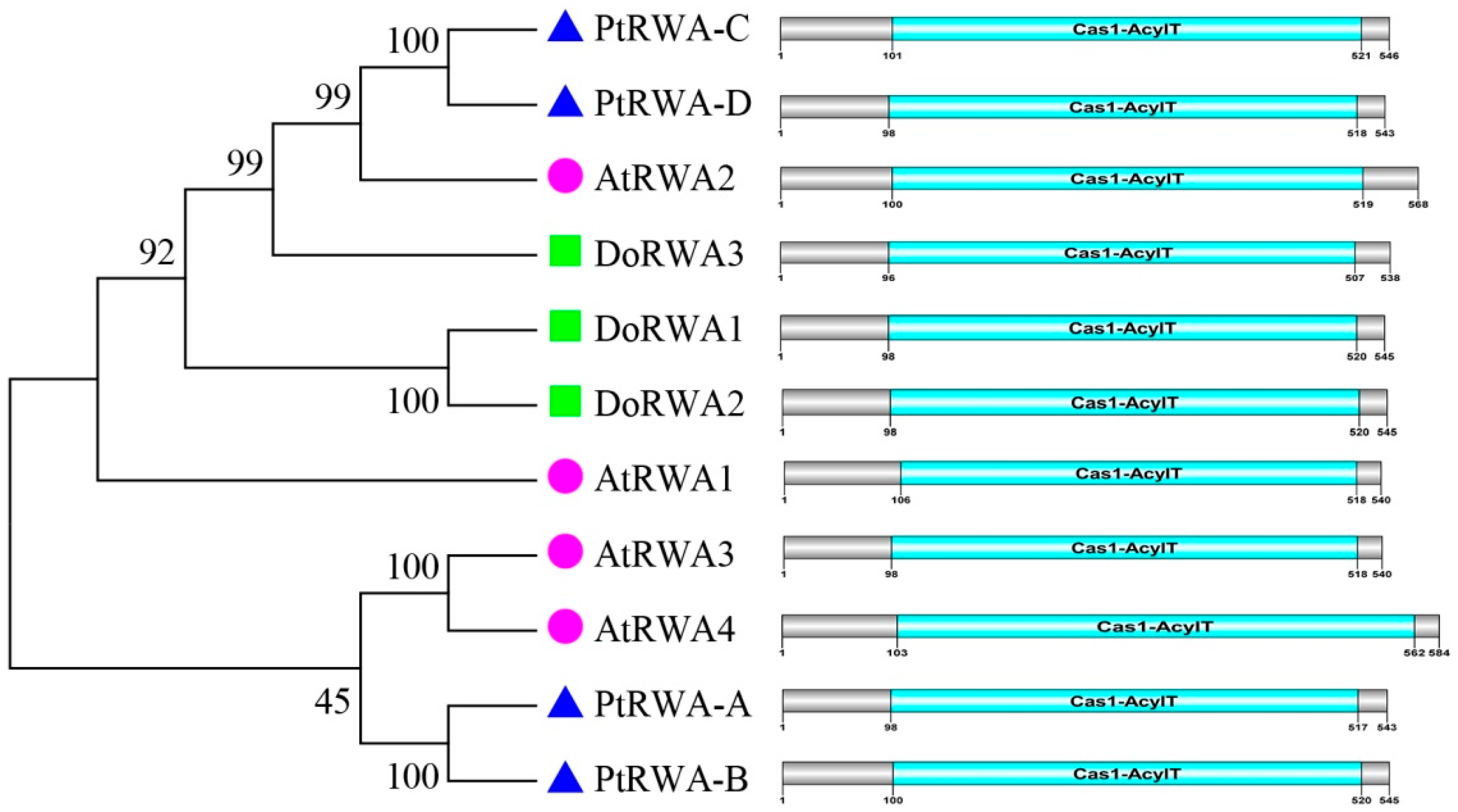
2.1. Isolation and Sequence Analysis of the DoRWA Genes

Three DoRWA genes, named DoRWA1, DoRWA2 and DoRWA3, were identified in the D. officinale genome [20]. Their open reading frames (ORFs) were 1638, 1638 and 1617 bp long, encoding 545, 545 and 538 aa. The molecular weights (MWs) of the three genes were 63.825, 63.780 and 63.634 Da, and their isoelectric points (pIs) were 8.84, 9.01 and 8.94. The ORF sequences of three genes were submitted to NCBI with the accession numbers MT199223, MT199224 and MT199225. BlastP results revealed that DoRWA1 had 99% similarity with RWA1 of Dendrobium catenatum (XP_020674439.1) and 92% similarity with RWA1 of Phalaenopsis equestris (XP_020584729.1); DoRWA2 had 99% similarity with RWA1 of D. catenatum (XP_028548609.1) and 92% similarity with RWA3L of P. equestris (XP_020577546.1); DoRWA3 had 99% similarity with RWA4 of D. catenatum (XP_020684246.1) and 84% similarity with RWA4L of P. equestris (XP_020573535.1).

2.2. Bioinformatics of the DoRWA Genes

The RWA protein sequences from three plants were aligned (Figure S1). The highest similarity of these proteins was 71.59%, indicating that RWA was considerably conserved in these plants. These proteins had the same domain Cas1-AcylT (411–459 aa), accounting for 73.77–78.60% of the full length (538–584 aa) (Figure 1). The exon–intron structures and length of the three genes varied. DoRWA1 (37 kb) and DoRWA2 (14 kb) had a similar gene structure and contained 16 exons and 15 introns, while DoRWA3 (6 kb) had 15 exons and 14 introns (Figure S2). The protein sequences of the three plants were also used to construct a phylogenetic tree using the Neighbor-Joining (N-J) method. DoRWA1 and DoRWA2 were clustered as one branch, and DoRWA3 was clustered with AtRWA2, PtRWA-C and PtRWA-D (Figure 1). Using the homologous A. thaliana protein, RWA2 was used to conduct a protein–protein interaction network analysis in which RWA2 was correlated with other TBL proteins [12,13] (Figure 2), such as pectin O-acetyltransferase TBR [21], xylan O-aceryltransferase TBL3 and TBL31 [22], and xyloglucan O-acetyltransferase TBL27 [23], indicating that DoRWA2 and DoTBL proteins were active in the same metabolic pathway.

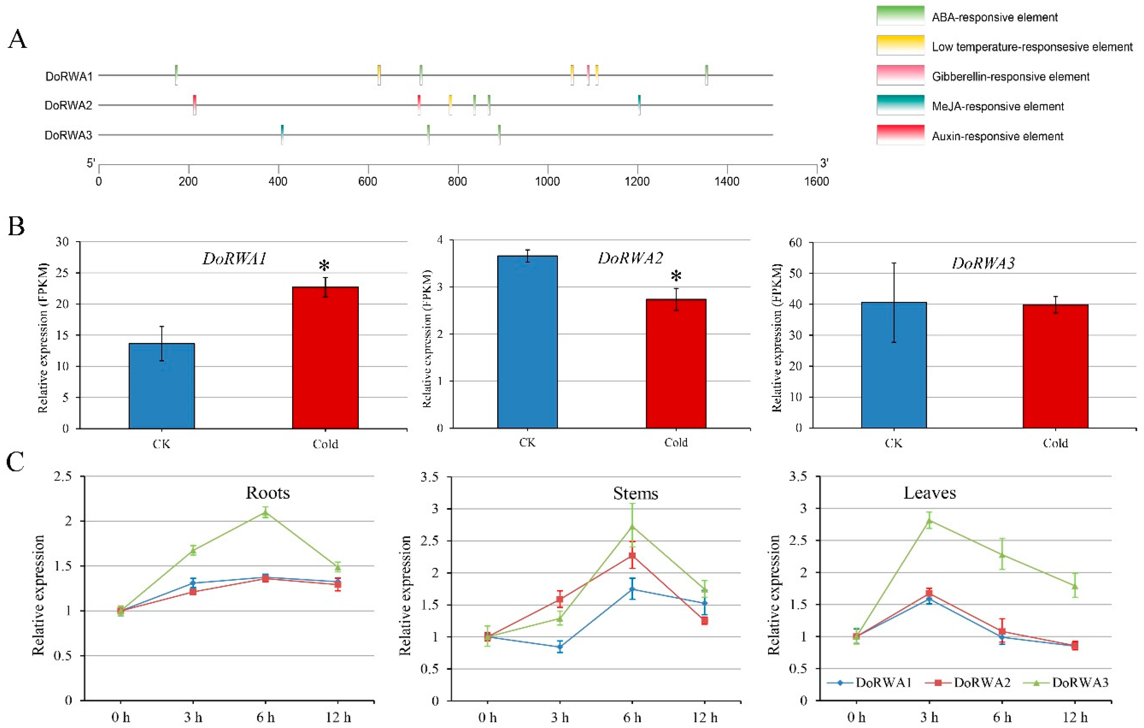
2.3. Analysis of cis-Elements, and Expression Patterns of DoRWA Genes under Cold and ABA Treatments

The cis-elements of the three genes contained hormone-responsive elements (methyl jasmonate, abscisic acid (ABA), auxin, gibberellin) and abiotic stress-responsive elements (low temperature) (Figure 3A). All three DoRWA genes had the ABA-responsive element while DoRWA1 and DoRWA2 (but not DoRWA3) had a low temperature-responsive element. To verify these predictions, the expression patterns of the three DoRWA genes in response to cold and ABA were assessed. In the cold treatment, the relative expression level (fragments per kilobase per million, FPKM) of DoRWA1 was upregulated, DoRWA2 was downregulated, but DoRWA3 showed no difference (Figure 3B). In the ABA treatment, the transcript levels of the three genes were upregulated at first, then peaked, but were finally downregulated in three organs (roots, stems, leaves), with peak expression at 6 h in roots and stems, and at 3 h in leaves (Figure 3C).

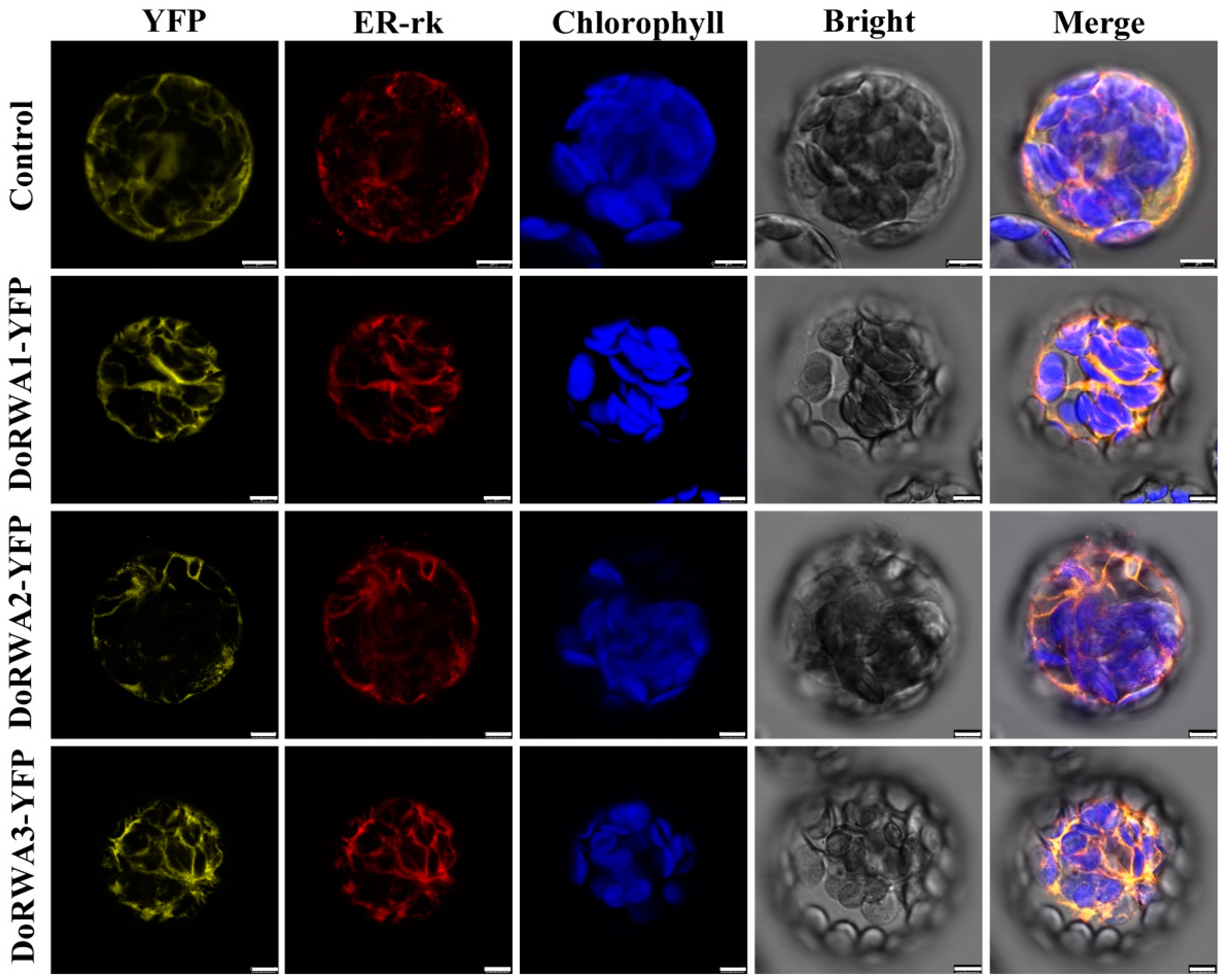
2.4. Cellular Localization of DoRWA Proteins

The three DoRWA proteins (DoRWA1, DoRWA2, DoRWA3) had 10, 10 and 11 transmembrane helices, which indicated they may be localized in a membranous organelle (Figure S3A,B,D). The fluorescent signals showed that all three DoRWA proteins were co-localized with the ER-rk (Figure 4), which is also localized in the endoplasmic reticulum (ER) [24]. Furthermore, the YFP fluorescence of DoRWA3 was not localized in the Golgi apparatus (GA) (Figure S3D). These results indicate that the ER plays an important role in the acetylation of polysaccharides.

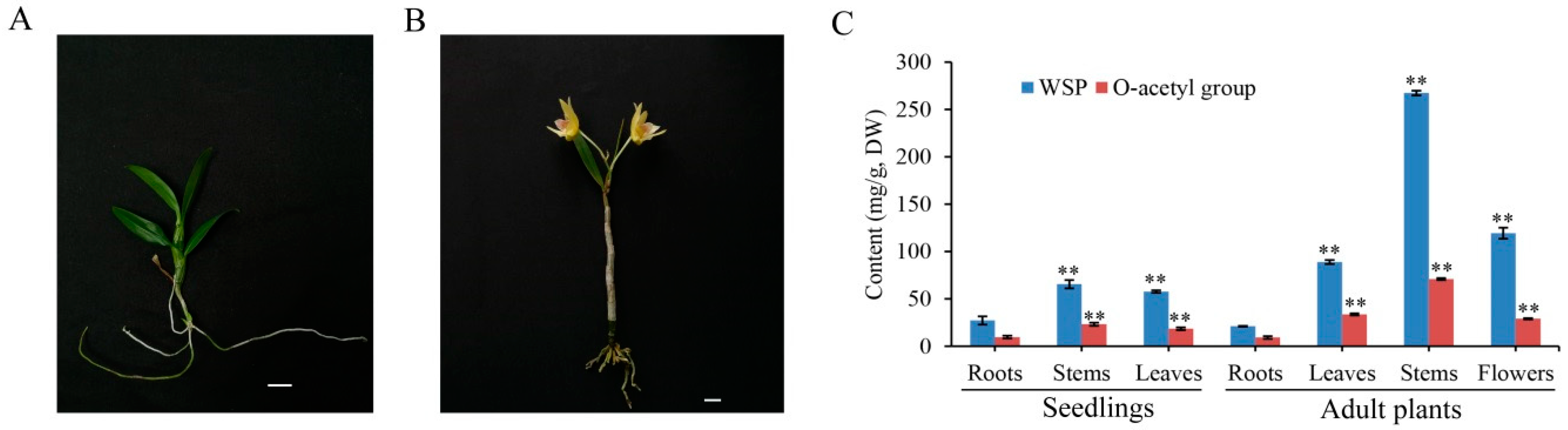
2.5. WSPs and O-Acetyl Groups Mainly Accumulated in the Stems of D. officinale

WSPs and O-acetyl groups showed a similar trend, accumulating the most in stems, followed by flowers and leaves, and the least in roots of seedlings and adult plants (Figure 5). The content of WSPs in stems was 65.55 mg/g in seedlings and 267.48 mg/g in adult plants. The content of O-acetyl groups of WSPs in stems was 23.22 mg/g in seedlings and 70.96 mg/g in adult plants.

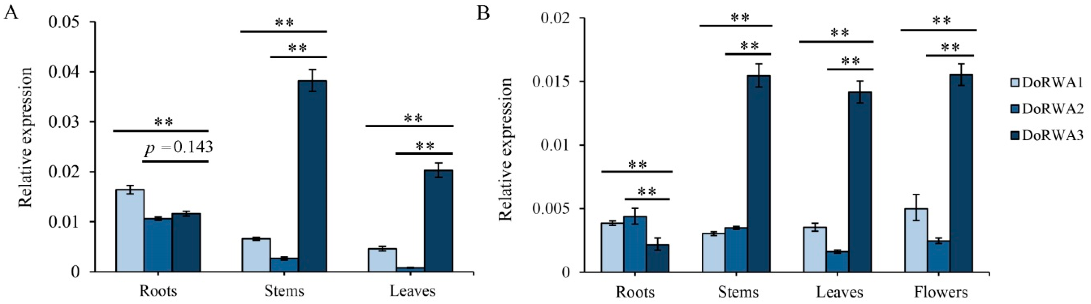
2.6. Expression Patterns of DoRWA Genes in Different Organs of Seedlings and Adult Plants

In D. officinale seedlings, the expression profiles of DoRWA1 and DoRWA2 were similar, with higher transcript levels in roots than in stems and leaves, while the relative expression of DoRWA3 was higher in stems than in roots and leaves (Figure 6A). In adult plants, the expression levels of DoRWA1 and DoRWA2 showed fewer differences in several organs, but the expression of DoRWA3 was relatively higher in stems, leaves and flowers where acetyl groups accumulated (Figure 6B). The mRNA ratio of DoRWA3 was 5.80- and 14.41-fold higher than DoRWA1 and DoRWA2 in stems, and 4.42- and 25.94-fold higher than in leaves of seedlings (Figure 6A). Similar results were also found in adult plants (Figure 6B).

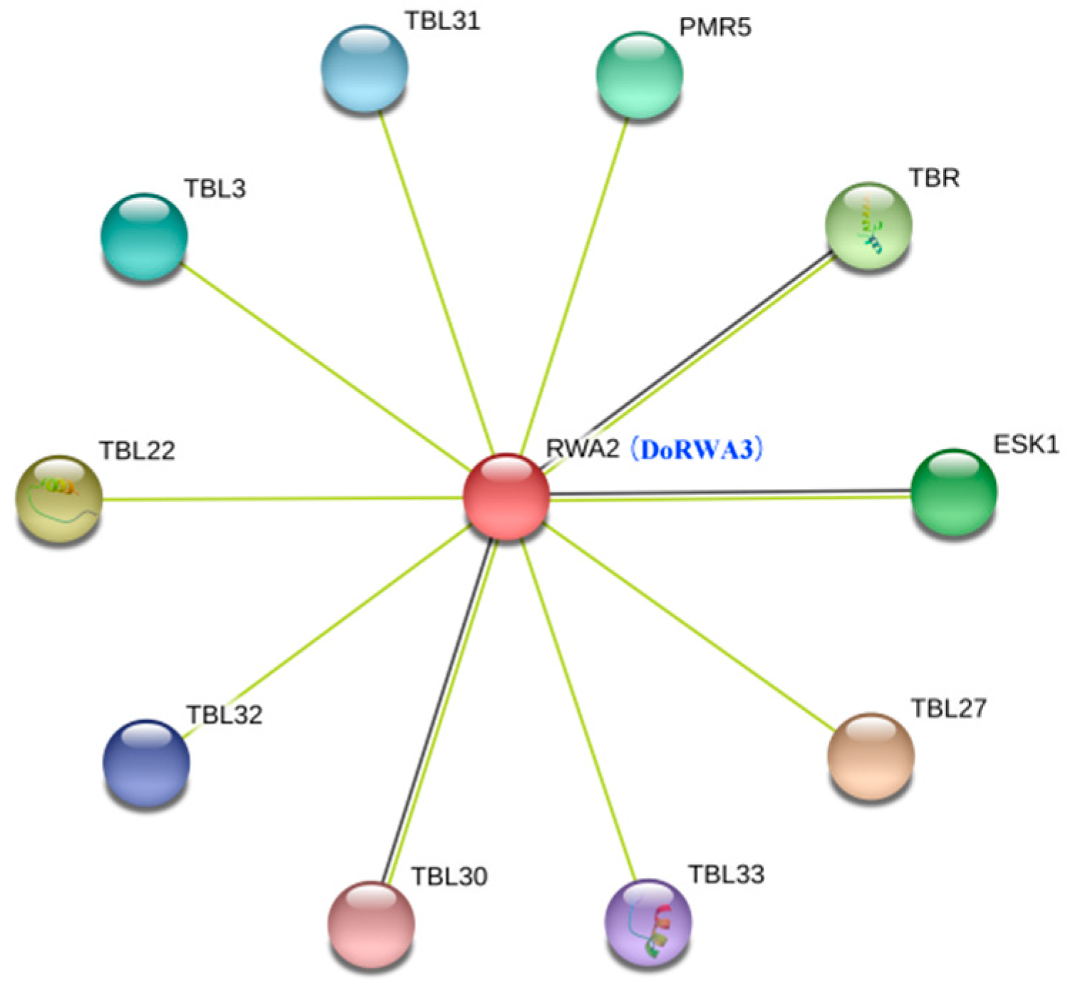
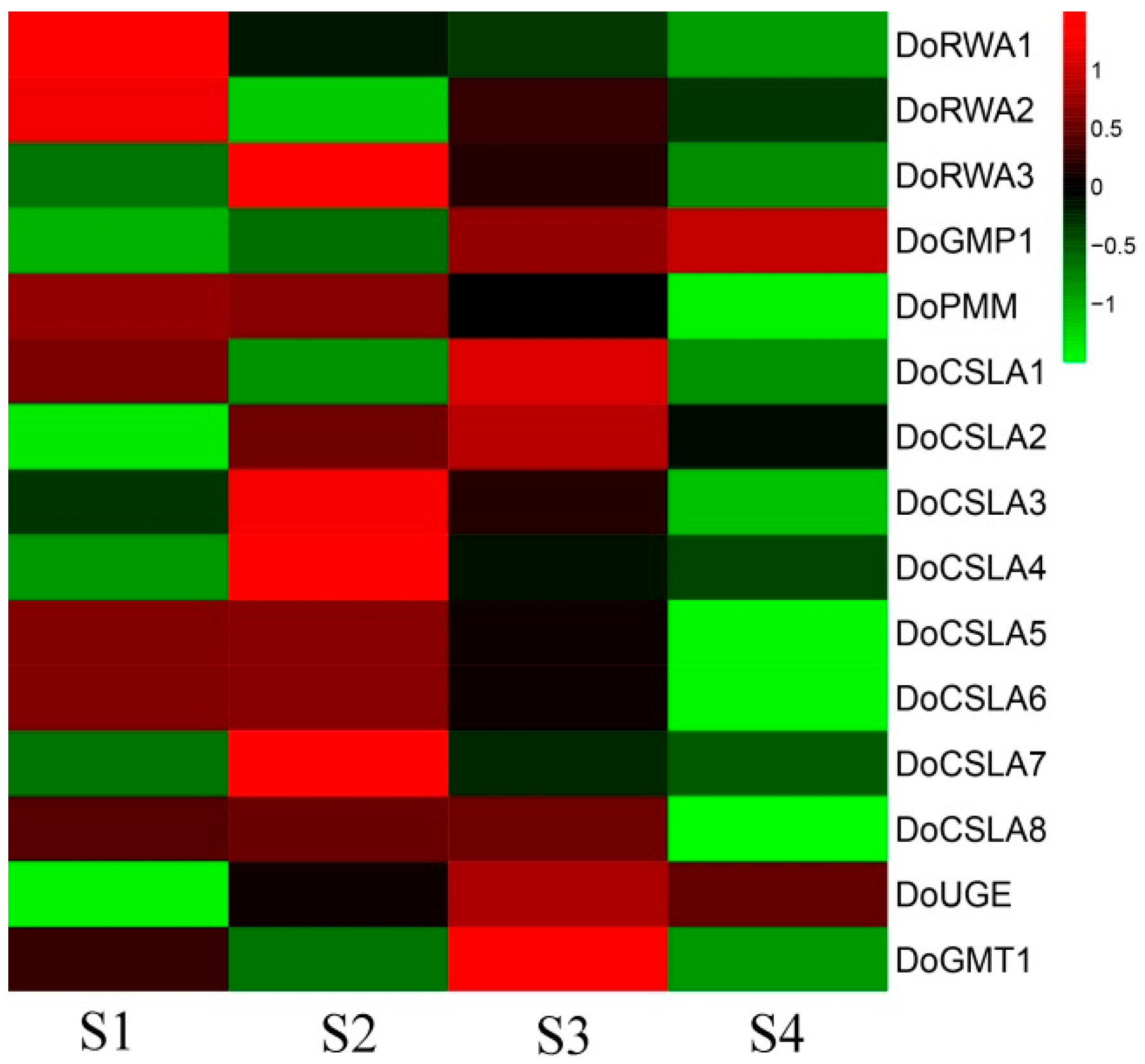
2.7. Co-Expression of DoRWA3 with Synthetic Genes of Polysaccharides

In the four developmental stages, the content of WSPs increased from S1 to S3, decreased from S3 to S4, and peaked at S3 [25]. The transcript levels of key genes related to polysaccharides in D. officinale, such as cellulose synthesis-like (CSL) [25], GDP-mannose pyrophosphorylation (GMP) [26], UDP glucose 4-epimerase (UGE) [27] and GDP-mannose transporter (GMT) [28] peaked at S2 or S3, corresponding to trends in the content of WSPs (Figure 5C). The expression profile of DoRWA3 peaked at S2, and showed a close association with the key genes described above (Figure 7), This indicates that DoRWA3 may be a candidate gene responsible for coding the enzyme that transfers acetyl groups to WSPs.

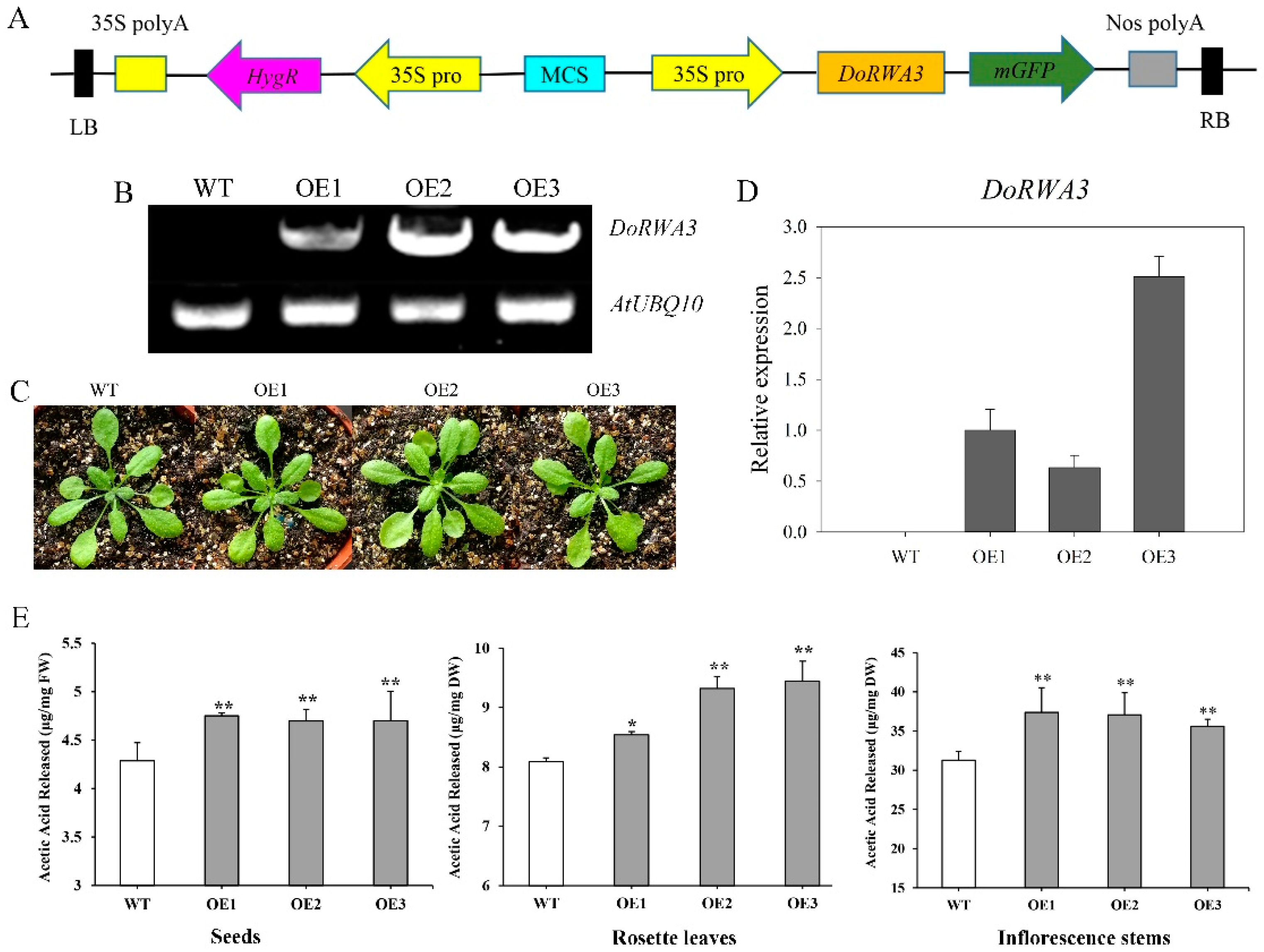
2.8. DoRWA3 Overexpression Increased the Acetylation Level of Polysaccharides in A. thaliana

The semi-quantitative PCR and qRT-PCR results (Figure 8B,D) indicated that DoRWA3 was successfully inserted into the A. thaliana genome and could be transcribed normally. There were no differences in the phenotype (color, size, flowering time, etc.) between WT and the three overexpression (OE) transgenic lines (Figure 8C). Simultaneously, the transcript level of four AtRWA genes in WT and three OE transgenic lines were also tested. There were no differences in the relative expression levels of AtRWA1, AtRWA3, and AtRWA4 between WT and transgenic lines, but the expression level of AtRWA2 was lower in the three transgenic lines compared with WT (Figure 9).
In seeds, the content of released acetic acid was about 1.11-, 1.10- and 1.10-fold higher in the three transgenic OE lines than in WT (Figure 8E). The corresponding values were 1.06-, 1.15- and 1.17-fold higher in rosette leaves (Figure 8E) and 1.20-, 1.19- and 1.14-fold higher in inflorescence stems (Figure 8E). The exogenously inserted DoRWA3 gene increased the level of acetylation of polysaccharides in seeds, leaves and stems of transgenic A. thaliana by 10–11%, 6–17%, and 14–20%, respectively. The released acetic acid of stems (31–38 mg/g) was higher than leaves (8–9.5 mg/g) and seeds (4–5 mg/g), indicating that stems accumulated more O-acetyl groups in polysaccharides of A. thaliana, relative to D. officinale.

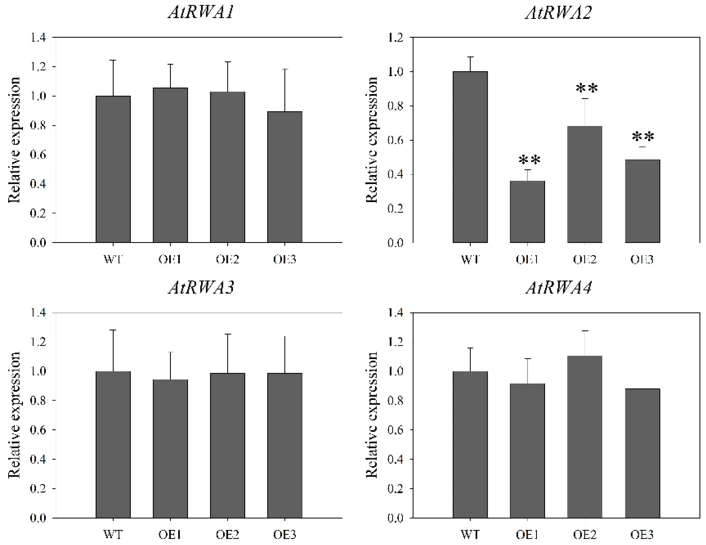
2.9. ABA Sensitivity Was Not Affected by Constitutive Expression of DoRWA3

The germination rate of WT and transgenic lines was almost 100% (Figure S4A,C). ABA treatment reduced root length of all plants: in WT, root length decreased from 5.95 cm (control) to 5.11 cm (2 μM ABA), but there were no significant (p > 0.05) differences between the WT and transgenic plants (Figure S4D). These findings indicate that ABA sensitivity may not be affected by the exogenous DoRWA3.

3. Discussion

Acetyl groups affect the biological activities of polysaccharides in many edible and medicinal plants, such as Cyclocarya paliurus [29], Dendrobium huoshanense [30], D. officinale [31] and Plantago asiatica [32], Amorphophallus konjac [33] and Aloe vera [34]. Konjac glucomannan (KGM), which is extracted from the corm of A. konjac [35], consisting of β-1,4-linked mannose and glucose residues (molar ratio: 1.6:1) with substituted acetyl groups. Deacetylated KGM has less health benefits than KGM [36]. Acemannan extracted from A. vera leaves contains the β-1,4-linked mannose and glucose (molar ratio: 3:1) with substituted acetyl groups [37]. After treating with alkaline to remove acetyl groups, the solubility, hydrophilicity and bioactivities of acemannan are reduced [5].
Cell wall acetylation has been shown to play broad roles in plant abiotic and biotic responses. rwa2-1 and rwa2-3 mutants showed enhanced resistance to the fungus Botrytis cinerea [10]. Some genes related to hormones and oxidative stress, such as auxin, ABA, jasmonic acid (JA), cytokinin (CK), light, cold and drought are down or upregulated in the transcriptome of mutant rwa2-3 compared with the WT, but no difference in the phenotype (growth and root length) was observed when the WT and rwa2-3 were treated with hormones (auxin, ABA, JA and CK) [38]. In this study, many cis-elements in the promoter region of DoRWA genes were associated with hormones and abiotic stress (Figure 3A). qRT-PCR proved that DoRWA genes were responsive to ABA (Figure 3C), but there were no differences in germination rate and root length between A. thaliana WT and OE transgenic lines when treated with ABA (Figure S4), indicating that DoRWA3 might not participate in the ABA-dependent signaling pathway.
The expression levels of four RWA genes in A. thaliana varied in different organs, but they all had relatively higher expression levels in inflorescence stems than in leaves and flowers, while AtRWA2 expression was also relatively higher in leaves than in flowers [10]. The expression levels of four RWA genes in Populus tremula varied in different organs (seeds, roots, leaves, buds and flowers) and treatment (drought): PtRWA-A was always expressed more than PtRWA-B, and PtRWA-C more than PtRWA-D [15]. In different organs, DoRWA1 and DoRWA2 showed the highest mRNA ratio in roots where the content of acetyl groups was lowest compared with other organs (Figure 6A), most likely because they had similar gene structures and conserved domains (Figure 1 and Figure S2); DoRWA3 had relatively higher expression levels in organs where acetyl groups accumulated, namely stems, leaves and flowers (Figure 6). The mRNA ratio of DoRWA3 showed significantly higher expression levels than DoRWA1 and DoRWA2 in stems, leaves and flowers (Figure 6). In four developmental stages, DoRWA3, but not DoRWA1 and DoRWA2, was co-expressed with the synthetic genes of WSPs (Figure 7). Thus, we hypothesize that DoRWA3 is a key gene coding for an acetyltransferase, while DoRWA1 and DoRWA2 may be redundant.
The AtRWA2 protein was localized in the ER or GA in Nicotiana benthamiana leaves or carrot protoplasts [9,10], showing species specificity. Since polysaccharides are synthesized in the GA and the related GDP-mannose transporter protein is also localized in the GA [28], it was expected that the DoRWA proteins would also be localized in the GA. However, they were localized in the ER (Figure 4), indicating that ER may be responsible for the upstream acetylation of polysaccharides.
The phenotype (growth and morphology) of a single mutant was not different from the WT [10], while triple and quadruple rwa mutants showed a severely dwarfed phenotype [14]. Similarly, the three 35S::DoRWA3 transgenic lines were indistinguishable from the WT (Figure 8C). This suggests that a single rwa mutant or DoRWA3 OE transgenic lines have no effect on the growth of A. thaliana.
A minor reduction in the acetylation level was detected in the inflorescence stems of single mutants rwa1, rwa2, rwa3 and rwa4 [9], indicating that the four AtRWA genes may influence the level of cell wall acetylation in A. thaliana. To verify if these four AtRWA genes participate in increasing the acetylation level in cell wall polymers, their transcript levels were detected in WT and three OE transgenic lines. The expression patterns of AtRWA1, AtRWA3 and AtRWA4 were similar, while the expression profile of AtRWA2 was lower in the three transgenic lines compared with the WT (Figure 9), indicating that the four AtRWA genes did not play a vital role in increasing the acetylation level of polysaccharides in OE transgenic lines.
The level of acetylation of cell wall residues from leaves was significantly reduced in a single mutant rwa2-1 compared with WT, while only a small difference was found in the stems [10]. The quadruple mutant rwa1rwa2rwa3rwa4 showed the largest reduction of acetylation level, about a 63% decrease [14]. In our study, the content of acetic acid increased significantly in the three transgenic lines compared with the WT in three organs: 6–17% in leaves, 10–11% in seeds and 14–20% in stems (Figure 8E). These findings indicate that exogenous DoRWA3 could increase the acetylation level of polysaccharides in A. thaliana. DoRWA3 could be considered as a candidate gene to improve the biological activities of polysaccharides in other edible and medicinal plants.

4. Materials and Methods

4.1. Plant Materials and Hormone Treatment

The young seedlings of D. officinale were cultured on half-strength (macro- and micronutrients) Murashige and Skoog (MS) medium [39], containing 0.5% activated carbon, 2% sucrose, and 0.5% agar (pH 5.7). Adult D. officinale plants were planted in a ground bark substrate in a greenhouse of South China Botanical Garden (Guangzhou, Guangdong, China) under ambient conditions. The seeds of A. thaliana Columbia (Col-0) were placed at 4 °C and continual darkness for 3 d, then transferred to a substrate containing nutritive soil and vermiculite (v/v, 2:1) under a controlled environment (80% humidity, 22 °C, 16-h photoperiod). For the ABA treatment, 10-month-old plantlets of D. officinale were treated with 100 μM ABA for 0, 3, 6 and 12 h, 0 h was regarded as the control group. The roots, stems, leaves, and flowers of young seedlings and adult plants were collected, frozen in nitrogen liquid, and stored at −80 °C.

4.2. RNA Extraction, cDNA Synthesis and qRT-PCR Analysis

Total RNA was extracted by an sodium dodecyl sulfate (SDS) method [40]. Briefly, 0.2 g of fresh sample was ground into a powder with liquid nitrogen. Extraction buffer (100 mM Tris-HCl, pH = 8.0; 50 mM EDTA, pH = 8.0; 500 mM NaCl; 1% SDS; 4% β-mercaptoethanol) was added and vortexed. After centrifuging at 12,000 rpm for 5 min, 1/3 (v/v) KAC (pH 4.8, 5 mol/L) was added to the solution to remove polysaccharides. After centrifuging at 12,000 rpm for 10 min, the supernatant was washed with chloroform:isoamyl alcohol (v/v, 24:1) and precipitated by 100% isopropanol. The precipitate was washed twice with 75% ethanol, dissolved in RNAase-free water, and stored at −80 °C. Any contaminating DNA was removed by Recombinant DNAase I (TaKaRa Bio Inc., Dalian, China) following the manufacturer’s protocol.
Purified RNA was reverse transcribed to cDNA by the GoScriptTM Reverse Transcription System Protocol (Promega, Madison, WI, USA) according to the manufacturer’s instructions. cDNA was further used for gene cloning and qRT-PCR analysis. For qRT-PCR analysis, the iTaqTM Univeral SYBR® Green Supermix (Bio-Rad Laboratories Co. Ltd., Hercules, CA, USA) was used as the polymerase in the following reaction system: stage 1 (95 °C for 2 min); stage 2 (40 cycles of 95 °C for 15 s, 60 °C for 1 min); stage 3 (95 °C for 15 s, 60 °C for 1 min, 95 °C for 15 s, 60 °C for 15 s). Actin (JX294908) and EF-1α gene [41] from D. officinale, and Actin2 (At3g18780), UBC (At5g25760) and PP2AA3 (At1g13320) [10] from A. thaliana were used as reference genes. The primers designed for qRT-PCR analysis are listed in Table S1. The 2−ΔΔCT method [42] was used to calculate the relative expression levels of different genes. All treatments were sampled as three biological and technical replicates.

4.3. Identification and Cloning of DoRWA Genes

Four A. thaliana RWA proteins were downloaded from TAIR (https://www.arabidopsis.org/): AtRWA1 (At5g46340), AtRWA2 (At3g06550), AtRWA3 (At2g34410) and AtRWA4 (At1g29890) [10]. They were used as queries to search for homologous proteins in the D. officinale protein database [19] using Bioedit software [43]. All putative D. officinale RWA proteins were further identified by BlastP in NCBI (https://blast.ncbi.nlm.nih.gov/Blast.cgi) to discard any repeated proteins or proteins without the conserved domains of the RWA family. The identified RWA genes were used to design the specific primers for gene cloning.
RWA genes were cloned from cDNA using KOD FX polymerase (Toyobo Co. Ltd., Osaka, Japan) with the following protocol: stage 1 (94 °C for 3 min); stage 2 (40 cycles of 98 °C for 10 s, 55 °C for 30 s, 72 °C for 2 min); stage 3 (72 °C for 10 min). PCR products were separated on a 1% agarose gel, purified by a DNA Gel Extraction Kit (Dongsheng Co. Ltd., Guangzhou, China), linked to the PMD-18T vector (TaKaRa Bio Inc.), sequenced by Beijing Genome Institute (Shenzhen, Guangdong, China), then submitted to NCBI. The protein sequences of the three genes were submitted to ExPASy (https://web.expasy.org/protparam/) to calculate MWs and theoretical pIs. The primers designed for cloning the three DoRWA genes are listed in Table S1.

4.4. Bioinformatics Analysis

The RWA protein sequences from D. officinale, A. thaliana and Populus trichocarpa were initially aligned with DNAMAN 7.0 software (Lynnon Biosoft Crop., San Ramon, CA., USA). To complete a phylogenetic analysis, all RWA proteins from these three plants were further aligned by MUSCLE (https://www.ebi.ac.uk/Tools/msa/muscle/), then were used to construct a Neighbor-Joining (N-J) tree built in MEGA 7.0 software [44] with the following parameters: 1000 bootstrap replications; pair deletion. Protein domain structures were drawn in DOG software [45]. Gene structure, including exons, introns, 5′-UTRs and 3′-UTRs of DoRWA genes, were obtained from the D. officinale gff database [20], then submitted to GSDS version 2.0 [46] to draw the exon-intron structure. The promoter region (from 0 to −1500 bp) of DoRWA genes were obtained from the D. officinale scaffold database [20], submitted to PlantCare [47] to discover all cis-elements, then the type and number of cis-elements were assessed and submitted to TBtools software [48]. STRING Version 11.0 was used to analyze the protein–protein association networks [49].

4.5. Protoplast Isolation and Subcellular Localization of DoRWA Proteins

Protoplasts were isolated following the protocol described by Schapire et al. [50]. At first, the enzyme solution was made as follows: 1.5% (w/v) cellulase R-10 (Yakult Pharmaceutical Industry Co. Ltd., Tokyo, Japan), 0.3% (w/v) macerozyme R-10 (Yakult Pharmaceutical Industry), 20 mM KCl (Mackline, Shanghai, China), 20 mM MES (pH 5.7; Sigma-Aldrich, St. Louis, MO, USA), and 0.4 M mannitol (Mackline). The enzyme solution was warmed to 55 °C for 10 min, then 10 mM CaCl2 (Sigma-Aldrich) and 0.1% (w/v) bovine serum albumin (Sigma-Aldrich) were added after the solution had cooled down. Next, the lower epidermal surface cell layer was peeled off young leaves of 4- to 5-week-old A. thaliana plants by autoclave tape, then leaves were treated with the enzyme solution at 25 °C for 2 h (50 rpm). The resulting harvested protoplasts were washed twice by W5 solution containing 154.5 mM NaCl (Mackline), 125 mM CaCl2, 5 mM KCl, 2 mM MES (pH 5.7) and 5 mM glucose (Aladdin, Shanghai, China). Finally, protoplasts were gently resuspended in MMG solution that contained 0.4 mM mannitol, 15 mM MgCl2 and 4 mM MES (pH 5.7).
To predict transmembrane helices, the sequences of three DoRWA proteins (DoRWA1, DoRWA2, DoRWA3) sequences were submitted to the TMHMM Server v. 2.0 (http://www.cbs.dtu.dk/services/TMHMM/). The full-length coding sequences of the three DoRWA genes (stop codon was removed) were inserted into the pSAT6-EYFP-N1 vector [51] at the NcoI site using the In-fusion® HD Cloning Kit (TaKaRa Bio Inc.). Since AtRWA2 was localized in the GA [9] or ER [10], to assess in which organelle DoRWA proteins were localized, recombinant protein combined with GA or ER localization marker [24] were transformed into leaf mesophyll protoplasts of 4- to 5-week-old A. thaliana plants using PEG-mediated transformation [52]. After maintaining protoplasts at 22 °C for 16 h in the dark, fluorescence signals were visualized with a Leica TCS SP8 STED 3x microscope (Leica Camera AG, Solms, Germany). The primers designed for pSAT6-EYFP-N1-DoRWAs construction are listed in Table S1.

4.6. Content of Water-Soluble Polysaccharides and O-Acetyl Groups in Different Organs

The content of WSPs in different organs (roots, stems, leaves) was measured according to He et al. [25]. The O-acetyl groups of WSPs were detected by a modified colorimetric method, as described by Gudlavalleti et al. [53]. Briefly, 0.1 g of each organ was weighed accurately, added to 25 mL of purified water, then warmed at 80 °C for 2 h. After centrifuging at 8000 rpm for 10 min, 1 mL of supernatant was added to 2 mL of freshly formulated alkaline hydroxylamine (mixture of 2 mol/L of hydroxylamine hydrochloride and 3.5 mol/L sodium hydroxide (v/v, 1:1)), then vortexed immediately. After 4 min, 1 mL of 4 mol/L hydrochloric acid and 1 mL of 0.37 mol/L ferrous chloride–hydrochlolic acid were added. Absorbance of the mixture was measured with an ultraviolet spectrophotometer (UV-1800PC; AOE Instruments Co. Ltd., Shanghai, China) at 540 nm. The acetylcholine chloride was used as the standard. In the control, hydrochloric acid was added before the formulated alkaline hydroxylamine.

4.7. RNA-Seq Expression Analysis at Four Developmental Stages and under Cold Stress

To develop the expression profiles of DoRWA genes and key genes related to the synthesis of WSPs at four developmental stages (S1–S4: WSP content was upregulated from S1–S3, peaked at S3, and downregulated at S4) [25], four raw reads (SRR1917040, SRR1917041, SRR1917042, SRR1917043) [25] corresponding to the S1, S2, S3 and S4 stage, were mapped to the transcriptome sequence database of D. officinale [54]. From the mapped database [24], the reads per kilobase per million reads (RPKM) [55] values of three DoRWA genes and WSP synthetic genes at S1–S4 stage were first downloaded then were log-transformed to render data suitable for heatmap analysis. To obtain the expression patterns of DoRWA genes under cold stress, six raw reads (SRR3210613, SRR3210621, SRR3210626, SRR3210630, SRR3210635, SRR3210636) [56] were downloaded from NCBI, then mapped to the D. officinale genome sequence database [20]. The FPKM value was used for gene expression analysis.

4.8. Generation of 35S::DoRWA3 Transgenic Lines

The full-length coding sequence of DoRWA3 (stop codon was removed) was linked with the pCAMBIA1302 vector at the NcoI site using the In-fusion® HD Cloning Kit (TaKaRa Bio Inc.). Recombinant plasmid was transformed into Agrobacterium tumefaciens EHA105. Inflorescences of 5- to 6-week-old WT plants were transfected by A. tumefaciens with the floral dip method [57]. Three homologous OE lines (OE1, OE2, OE3) were screened on half-strength MS (1/2 MS) medium containing 25 μg/mL hygromycin B (Roche Holding AG, Basel, Switzerland). The primers designed for the pCAMBIA1302-DoRWA3 construction are listed in Table S1.

4.9. Semi-Quantitative RT-PCR

Total RNAs from the leaves of 1-month-old WT and three OE transgenic lines were extracted and purified as described above. The semi-quantitative PCR reaction was catalyzed by KOD FX polymerase (Toyobo Co. Ltd.) with the following protocol: stage 1 (94 °C for 3 min); stage 2 (40 cycles of 98 °C for 10 s, 56 °C for 30 s, 72 °C for 2 min); stage 3 (72 °C for 10 min). UBQ10 (At4g05320) from A. thaliana was used as the control. PCR products were visualized on a 1% agrose gel under ultraviolet light. The primers designed for semi-quantitative PCR are listed in Table S1.

4.10. Cell Wall Preparation and Determination of Acetyl Esters

The rosette leaves and inflorescence stems of 6-week-old plants, and seeds of WT and three 35S::DoRWA3 transgenic lines, were collected then dried at 80 °C for 12 h (seeds were naturally air-dried for 2 weeks). Samples were ground into power for cell wall extraction.
The alcohol-insoluble residues (AIR) of samples were extracted according to Harholt et al. [58]. Briefly, 30 mg of sample was weighed accurately, washed with 1 mL of 96% ethanol, then kept at 70 °C for 30 min to deactivate enzymes. After centrifuging the mixture at 10,000 g for 5 min, the supernatant was removed, 1 mL of 70% ethanol was added, and the mixture was vortexed. The pellet was washed with 70% ethanol, then centrifuged at 10,000 g for 5 min, and this was repeated until the solution became colorless. Finally, the precipitate was washed in 1 mL of 100% acetone, vortexed immediately, placed at room temperature (RT) for 10 min, then centrifuged at 10,000 g for 5 min. The supernatant was discarded and the pellet was oven-dried at 50 °C until constant weight.
AIR (4 mg) of different samples were accurately weighed, saponified by 400 μL of 1 mol/L NaOH, then centrifuged at 150 rpm overnight (at 28 °C). The solution was neutralized with 400 μL of 1 mol/L HCl, then centrifuged at 12,000 rpm for 10 min. The released acetic acid content in the supernatant was determined by using the Acetic Acid Assay Kit (Megazyme, Wicklow, Ireland) as described by Gill et al. [59]. Briefly, 40 μL of supernatant of different samples was added into a UV- capable 96-well plate, then diluted with 64 μL of ddH2O. An amount of 42 μL of mixture (Soulution 1 and Solution 2; v/v, 2.5:1) was transferred into each sample, mixed and incubated at RT for 3 min. Absorbance of the mixture was read at 340 nm (A0). An amount of 12 μL of 10-fold diluted solution 3 was added to the wells, mixed and incubated at RT for 4 min, then read at 340 nm (A1). Finally, 12 μL of 10-fold diluted Solution 4 was added to the plate, mixed thoroughly and incubated at RT for 12 min, then read at 340 nm (A2). Solution 5 (acetic acid solution) served as the standard. In the control, 40 μL of supernatant was replaced by 40 μL of ddH2O. A0, A1 and A2 values were used to calculate ΔA according to the manufacturer’s recommendations.

4.11. ABA Treatment and Phenotype Assay

To study the effect of ABA on germination rates, the seeds (48 seeds per sample) of WT and three OE transgenic lines (OE1, OE2 and OE3) were sown on the 1/2 MS medium containing 0 or 1 μM ABA for 4 d. For the root length assay, seedlings growing on 1/2 MS medium for 3 d were transplanted to 1/2 MS medium supplemented with 1 or 2 μM ABA. After vertical culture for 7 d, root length was assessed using ImageJ software (http://rsbweb.nih.gov/ij/).

4.12. Statistical Analysis

All data were plotted in Excel 2013 (Microsoft Inc., Redmond, WA, USA) and Sigmaplot 12.0 (Systat Software Inc., San Jose, CA, USA). Data were analyzed by one-way analysis of variance (ANOVA) and means separated by Duncan’s multiple range test (DMRT) (p < 0.05, p < 0.01) in SPSS version 22.0 software (IBM Corp., Armonk, NY, USA).

5. Conclusions

Three DoRWA genes, named DoRWA1, DoRWA2 and DoRWA3, were cloned from the medicinal orchid, D. officinale. Phylogenetic analysis revealed that DoRWA3 was clustered with the identified acetyltransferase genes (i.e., AtRWA2, PtRWA-C, PtRWA-D) into one branch. Interestingly, the cis-elements of the three DoRWA genes had the ABA-responsive element and their expression patterns were sensitive to ABA treatment. The results of subcellular localization showed that the three DoRWA proteins were localized in the ER, and not in the GA. The O-acetyl groups shared a similar trend as WSPs in different organs. qRT-PCR and RNA-seq results showed that DoRWA3 was mainly expressed in the organs where the O-acetyl groups accumulated, displaying significantly higher expression than DoRWA1 and DoRWA2 in different organs, except for roots. DoRWA3 was co-expressed with key genes related to the synthesis of WSPs, so it is regarded as a candidate gene that codes for an acetyltransferase. The acetylation level of polysaccharides in seeds, leaves and stems of the three A. thaliana OE transgenic lines was significantly higher than in WT, indicating that DoRWA3 has a similar function as AtRWA2.

Supplementary Materials

Supplementary materials can be found at https://0-www-mdpi-com.brum.beds.ac.uk/1422-0067/21/17/6250/s1. Figure S1: Alignment of amino acid sequences of RWA proteins in Dendrobium officinale, Arabidopsis thaliana and Populus trichocarpa; Figure S2: Gene structure of three DoRWA genes. Figure S3: Predictions of subcellular localization of DoRWA1 (A), DoRWA2 (B), DoRWA3 (C) and subcellular localization of DoRWA3-YFP with Golgi apparatus localization marker G-rk (D); Figure S4: Comparison of germination rate and root length between wild type (WT) and overexpression (OE) transgenic plants; Table S1: Primers designed for PCR.

Author Contributions

Conceptualization, J.D., C.S. and J.A.T.d.S.; methodology, C.S.; software, C.S., C.H., Z.Y.; validation, C.Z., H.W. and M.Z.; formal analysis, C.S.; investigation, C.S.; resources, J.D.; data curation, J.D.; writing—original draft preparation, C.S. and J.A.T.d.S.; writing—review and editing, C.S. and J.A.T.d.S.; visualization, C.S. and J.A.T.d.S.; supervision, J.D.; project administration, J.D.; funding acquisition, J.D. All authors have read and agreed to the published version of the manuscript.

Funding

This research was funded by the project “Cultivation of new varieties of Dendrobium officinale in Guangdong Province“, grant number Y334041001.

Acknowledgments

The authors are grateful to Rufang Deng for assistance with subcellular localization analysis. We also thank the editor and reviewers for providing suggestions that improved the quality of this manuscript.

Conflicts of Interest

The authors declare no conflict of interest.

Abbreviations

ABAAbscisic acid
AIRAlcohol-insoluble residue
Axy9Altered xyloglucan9
CSLCellulose synthesis-like
CKCytokinin
DADegree of acetylation
DMRTDuncan’s multiple range test
EREndoplasmic reticulum
FPKMFragments per kilobase per million
GMPGDP-mannose pyrophosphorylation
GMTGDP-mannose transporter
GAGolgi apparatus
JAJasmonic acid
KGMKonjac glucomannan
MWMolecular weight
MSMurashige and Skoog
N-JNeighbor-Joining
ORFOpen reading frame
OEOverexpression
PMMPhosphomannomutase
pIIsoelectric Points
RPKMReads per kilobase per million
RTRoom temperature
TCMTraditional Chinese medicine
qRT-PCRQuantitative real time polymerase chain reaction
RWAReduced wall acetylation
TBLTrichome birefringence-like
UGEUDP glucose 4-epimerase
WSPWater-soluble polysaccharide

References

  1. Zhang, Z.S.; Wang, X.W.; Yu, S.C.; Yin, L.; Zhao, M.X.; Han, Z.P. Synthesized oversulfated and acetylated derivatives of polysaccharide extracted from Enteromorpha linza and their potential antioxidant activity. Int. J. Biol. Macromol. 2011, 49, 1012–1015. [Google Scholar] [CrossRef] [PubMed]
  2. Song, Y.; Yang, Y.; Zhang, Y.Y.; Duan, L.S.; Zhou, C.L.; Ni, Y.Y.; Liao, X.J.; Li, Q.H.; Hu, X.S. Effect of acetylation on antioxidant and cytoprotective activity of polysaccharides isolated from pumpkin (Cucurbita pepo, lady godiva). Carbohyd. Polym. 2013, 98, 686–691. [Google Scholar] [CrossRef] [PubMed]
  3. Chen, Y.; Zhang, H.; Wang, Y.X.; Nie, S.P.; Li, C.; Xie, M.Y. Acetylation and carboxymethylation of the polysaccharide from Ganoderma atrum and their antioxidant and immunomodulating activities. Food Chem. 2014, 156, 279–288. [Google Scholar] [CrossRef] [PubMed]
  4. Chen, F.; Huang, G.L. Preparation and immunological activity of polysaccharides and their derivatives. Int. J. Biol. Macromol. 2018, 12, 211–216. [Google Scholar] [CrossRef] [PubMed]
  5. Chokboribal, J.; Tachaboonyakiat, W.; Sangvanich, P.; Ruangpornvisuti, V.; Jettanacheawchankit, S.; Thunyakitposal, P. Deacetylation affects the physical properties and bioactivity of acemannan, an extracted polysaccharide from Aloe vera. Carbohyd. Polym. 2015, 133, 556–566. [Google Scholar] [CrossRef] [PubMed]
  6. Du, X.Z.; Li, J.; Chen, J.; Li, B. Effect of degree of deacetylation on physicochemical and gelation properties of konjac glucomannan. Food Res. Int. 2012, 46, 270–278. [Google Scholar] [CrossRef]
  7. Salah, F.; Ghoul, Y.E.; Mahdhi, A.; Majdoub, H.; Jarroux, N.; Sakli, F. Effect of the deacetylation degree on the antibacterial and antibiofilm activity of acemannan from Aloe vera. Ind. Crop. Prod. 2017, 103, 13–18. [Google Scholar] [CrossRef]
  8. Huang, S.Y.; Chen, F.; Cheng, H.; Huang, G.L. Modification and application of polysaccharide from traditional Chinese medicine such as Dendrobium officinale. Int. J. Biol. Macromol. 2020, 157, 385–393. [Google Scholar] [CrossRef]
  9. Lee, C.H.; Teng, Q.H.; Zhong, R.Q.; Ye, Z.H. The four Arabidopsis REDUCED WALL ACETYLATION genes are expressed in secondary wall-containing cells and required for the acetylation of xylan. Plant Cell Physiol. 2011, 52, 1289–1301. [Google Scholar] [CrossRef]
  10. Manabe, Y.; Nafisi, M.; Verhertbruggen, Y.; Orfila, C.; Gille, S.; Rautengarten, C.; Cherk, C.; Marcus, S.E.; Somerville, S.; Pauly, M.; et al. Loss-of-function mutation of REDUCED WALL ACETYLATION2 in Arabidopsis leads to reduced cell wall acetylation and increased resistance to Botrytis cinerea. Plant Physiol. 2011, 155, 1068–1078. [Google Scholar] [CrossRef] [Green Version]
  11. Schultink, A.; Naylor, D.; Dama, M.; Pauly, M. The role of the plant-specific ALTERED XYLOGLUCAN9 protein in Arabidopsis cell wall polysaccharide O-acetylation. Plant Physiol. 2015, 167, 1271–1283. [Google Scholar] [CrossRef] [PubMed] [Green Version]
  12. Gill, S.; Souza, A.D.; Xiong, G.Y.; Benz, M.; Cheng, K.; Schultink, A.; Reca, I.B.; Pauly, M. O-acetylation of Arabidopsis hemicellulose xyloglucan requires AXY4 or AXY4L, proteins with a TBL and DUF231 Domain. Plant Cell 2011, 23, 4041–4053. [Google Scholar] [CrossRef] [PubMed] [Green Version]
  13. Bischoff, V.; Nita, S.; Neumetzler, L.; Schindelasch, D.; Urbain, A.; Eshed, R.; Persson, S.; Delmer, D.; Scheible, W.R. TRICHOME BIREFRINGENCE and its homolog AT5G01360 encode plant-specific DUF231 proteins required for cellulose biosynthesis in Arabidopsis. Plant Physiol. 2010, 153, 590–602. [Google Scholar] [CrossRef] [PubMed] [Green Version]
  14. Manabe, Y.; Verhertbruggen, Y.; Gill, S.; Harholt, J.; Chong, S.L.; Pawar, P.M.A.; Mellerowicz, E.J.; Tenkanen, M.; Cheng, K.; Pauly, M.; et al. Reduced wall acetylation proteins play vital and distinct roles in cell wall O-acetylation in Arabidopsis. Plant Physiol. 2013, 163, 1107–1117. [Google Scholar] [CrossRef] [PubMed] [Green Version]
  15. Pawar, P.M.A.; Ratke, C.; Balasubramanian, V.K.; Chong, S.L.; Gandla, M.L.; Adriasola, M.; Sparrman, T.; Hedenström, M.; Szwaj, K.; Derba-Maceluch, M.; et al. Downregulation of RWA genes in hybrid aspen affects xylan acetylation and wood saccharification. New Phytol. 2017, 214, 1491–1505. [Google Scholar] [CrossRef] [Green Version]
  16. Teixeira da Silva, J.A.; Ng, T.B. The medicinal and pharmaceutical importance of Dendrobium species. Appl. Microbiol. Biotechnol. 2017, 101, 2227–2239. [Google Scholar] [CrossRef]
  17. Xing, X.H.; Cui, S.W.; Nie, S.P.; Phillips, G.O. Study on Dendrobium officinale O-acetyl-glucomannan (Dendronan®): Part II. Fine structure of O-acetylated residues. Carbohyd. Polym. 2015, 117, 422–433. [Google Scholar] [CrossRef]
  18. Huang, X.J.; Nie, S.P.; Cai, H.L.; Zhang, G.Y.; Cui, S.W.; Xie, M.Y.; Phillips, G.O. Study on Dendrobium officinale O-acetyl-glucomannan (Dendronan®): Part IV. Immunomogulatory activity in vivo. J. Funct. Foods 2015, 15, 525–532. [Google Scholar] [CrossRef]
  19. Wei, W.; Feng, L.; Bao, W.R.; Ma, D.L.; Leung, C.H.; Nie, S.P.; Han, Q.B. (2016) Structure characterization and immunomodulating effects of polysaccharides isolated from Dendrobium officinale. J. Agric. Food Chem. 2016, 64, 881–889. [Google Scholar] [CrossRef]
  20. Zhang, G.Q.; Xu, Q.; Bian, C.; Tsai, W.C.; Yeh, C.M.; Liu, K.W.; Youshida, K.; Zhang, L.S.; Chang, S.B.; Chen, F.; et al. The Dendrobium catenatum Lindl. genome sequence provides insights into polysaccharide synthase, floral development and adaptive evolution. Sci. Rep. 2016, 6, 19029. [Google Scholar] [CrossRef] [Green Version]
  21. Sinclair, S.A.; Larue, C.; Bonk, L.; Khan, A.; Michel, H.C.; Stein, R.J.; Grolimund, D.; Begerow, D.; Neumann, U.; Haydon, M.J.; et al. Etiolated seedlings development requires repression of photomorphogenesis by a small cell-wall-derived dark signal. Curr. Biol. 2017, 27, 1–16. [Google Scholar] [CrossRef] [PubMed] [Green Version]
  22. Yuan, Y.X.; Teng, Q.; Zhong, R.Q.; Ye, Z.H. TBL3 and TBL31, two Arabidopsis DUF231 domain proteins, are required for 3-O-monoacetylation of xylan. Plant Cell Physiol. 2016, 57, 35–45. [Google Scholar] [CrossRef] [PubMed] [Green Version]
  23. Zhu, X.F.; Sun, Y.; Zhang, B.C.; Mansoori, N.; Wan, J.X.; Liu, Y.; Wang, Z.W.; Shi, Y.Z.; Zhou, Y.H.; Zheng, S.J. TRICHOME BIREFRINGENCE-LIKE27 affects aluminum sensitivity by modulating the O-acetylation of xyloglucan and aluminum-binding capacity in Arabidopsis. Plant Physiol. 2014, 166, 181–189. [Google Scholar] [CrossRef] [PubMed] [Green Version]
  24. Nelson, B.K.; Cai, X.; Nebenführ, A. A multicolored set of in vivo organelle markers for co-localization studies in Arabidopsis and other plants. Plant J. 2007, 51, 1126–1136. [Google Scholar] [CrossRef]
  25. He, C.M.; Zhang, J.X.; Liu, X.C.; Zeng, S.J.; Wu, K.L.; Yu, Z.M.; Wang, X.J.; Teixeira da Silva, J.A.; Lin, Z.J.; Duan, J. Identification of genes involved in biosynthesis of mannan polysaccharides in Dendrobium officinale by RNA-seq analysis. Plant Mol. Biol. 2015, 88, 219–231. [Google Scholar] [CrossRef]
  26. He, C.M.; Yu, Z.M.; Teixeira da Silva, J.A.; Zhang, J.X.; Liu, X.C.; Wang, X.J.; Zhang, X.H.; Zeng, S.J.; Wu, K.L.; Tan, J.W.; et al. DoGMP1 from Dendrobium officinale contributes to mannose content of water-soluble polysaccharides and plays a role in salt stress response. Sci. Rep. 2017, 7, 41040. [Google Scholar] [CrossRef] [Green Version]
  27. Yu, Z.M.; He, C.M.; Teixeira da Silva, J.A.; Zhang, G.H.; Dong, W.; Luo, J.P.; Duan, J. Molecular cloning and functional analysis of DoUGE related to water-soluble polysaccharides from Dendrobium officinale with enhanced abiotic stress tolerance. Plant Cell Tissue Organ Cult. 2017, 131, 579–599. [Google Scholar] [CrossRef]
  28. Yu, Z.M.; He, C.M.; Teixeira da Silva, J.A.; Luo, J.P.; Yang, Z.Y.; Duan, J. The GDP-mannose transporter gene (DoGMT) from Dendrobium officinale is critical for mannan biosynthesis in plant growth and development. Plant Sci. 2018, 277, 43–54. [Google Scholar] [CrossRef]
  29. Liu, X.; Xie, J.H.; Jia, S.; Huang, L.X.; Wang, Z.J.; Li, C.; Xie, M.Y. Immunomodulatory effects of an acetylated Cyclocarya paliurus polysaccharide on murine macrophages RAW264.7. Int. J. Biol. Macromol. 2017, 98, 576–581. [Google Scholar] [CrossRef]
  30. Hsieh, Y.Y.; Chien, C.; Liao, S.K.S.; Liao, S.F.; Huang, W.T.; Yang, W.B.; Lin, C.C.; Cheng, T.J.R.; Chang, C.C.; Fang, J.M.; et al. Structure and bioactivity of polysaccharides in medicinal plant Dendrobium huoshanense. Bioorgan. Med. Chem. 2008, 16, 6054–6068. [Google Scholar] [CrossRef]
  31. Tong, W.; Yu, Q.; Li, H.; Cui, W.W.; Nie, S.P. Chemical modification and immunoregulatory activity of polysaccharides from Dendrobium officinale. Food Sci. 2017, 38, 155–160. [Google Scholar]
  32. Jiang, L.M.; Nie, S.P.; Huang, D.F.; Fu, Z.H.; Xie, M.Y. Acetylation modification improves immunoregulatory effect of polysaccharide from seeds of Plantago asiatica L. J. Chem. 2018, 2018, 3082026. [Google Scholar] [CrossRef] [Green Version]
  33. Li, J.; Ye, T.; Wu, X.F.; Chen, J.; Wang, S.S.; Lin, L.F.; Li, B. Preparation and characterization of heterogeneous deacetylated konjac glucomannan. Food Hydrocoll. 2014, 40, 9–15. [Google Scholar] [CrossRef]
  34. Javed, S.; Rahman, A. Aloe vera gel in food, health products and cosmetics industry. In Studies in Natural Products and Chemistry; Rahman, A.U., Ed.; Elsevier Science & Technology: London, UK, 2014; Volume 41, pp. 261–285. [Google Scholar]
  35. Nishinari, K.; Williams, P.A.; Phillips, G.O. Review of the physicochemical characteristics and properties of konjac mannan. Food Hydrocoll. 1992, 6, 199–222. [Google Scholar] [CrossRef]
  36. Li, M.Y.; Feng, G.P.; Wang, H.; Yang, R.L.; Xu, Z.L.; Sun, Y.M. Deacetylated konjac glucomannan is less effective in reducing dietary-induced hyperlipidemia and hepatic steatosis in C57BL/6 mice. J. Agric. Food Chem. 2017, 65, 1556–1565. [Google Scholar] [CrossRef] [PubMed]
  37. Humman, J.H. Composition and applications of Aloe vera leaf gel. Molecules 2008, 13, 1599–1616. [Google Scholar] [CrossRef] [Green Version]
  38. Nafisi, M.; Stranne, M.; Fimognari, L.; Atwell, S.; Martens, H.J.; Pedas, P.R.; Hansen, S.F.; Nawrath, C.; Scheller, H.V.; Kliebenstein, D.J.; et al. Acetylation of cell wall is required for structural integrity of the leaf surface and exerts a global impact on plant stress responses. Front. Plant Sci. 2015, 6, 550. [Google Scholar] [CrossRef]
  39. Murashige, T.; Skoog, F. A revised medium for rapid growth and bioassays with tobacco tissue cultures. Physiol. Plant 1962, 15, 473–497. [Google Scholar] [CrossRef]
  40. Chattopadhyay, N.; Kher, R.; Godbole, M. Inexpensive SDS/phenol method for RNA extraction from tissues. Biotechniques 1993, 15, 24–26. [Google Scholar]
  41. Livak, K.J.; Schmittgen, T.D. Analysis of relative gene expression data using real-time quantitative PCR and 2−ΔΔCT method. Methods 2001, 25, 402–408. [Google Scholar] [CrossRef]
  42. Zhang, G.; Zhao, M.M.; Zhang, D.W.; Guo, S.X. Reference gene selection for real-time quantitative PCR analysis of Dendrobium officinale. Chin. Pharm. J. 2013, 48, 1664–1668. [Google Scholar]
  43. Altschul, S.F.; Madden, T.L.; Schäffer, A.A.; Zhang, J.H.; Zhang, Z.; Miller, W.; Lipman, D.J. Gapped BLAST and PSI-BLAST: A new generation of protein database search programs. Nucl. Acids Res. 1997, 25, 3389–3402. [Google Scholar] [CrossRef] [PubMed] [Green Version]
  44. Kumar, S.; Stecher, G.; Tamura, K. MEGA7: Molecular evolutionary genetics analysis version 7.0 for bigger datasets. Mol. Biol. Evol. 2016, 33, 1870–1874. [Google Scholar] [CrossRef] [PubMed] [Green Version]
  45. Ren, J.; Wen, L.P.; Gao, X.J.; Jin, C.J.; Xue, Y.; Yao, X.B. DOG1.0: Illustrator of protein domain structures. Cell Res. 2009, 19, 271–273. [Google Scholar] [CrossRef]
  46. Hu, B.; Jin, J.P.; Guo, A.Y.; Zhang, H.; Luo, J.C.; Gao, G. GSDS2.0: An upgraded gene feature visualization server. Bioinformatics 2015, 31, 1296–1297. [Google Scholar] [CrossRef] [Green Version]
  47. Lescot, M.; Déhais, P.; Thijs, G.; Marchal, K.; Moreau, Y.; Peer, Y.V.D.; Rouzé, P.; Rombauts, S. PlantCARE, a database of plant cis-acting regulatory elements and a portal to tools for in silico analysis of promoter sequences. Nucl. Acids Res. 2002, 30, 325–327. [Google Scholar] [CrossRef]
  48. Chen, C.J.; Chen, H.; Zhang, Y.; Thomas, H.R.; Frank, M.H.; He, Y.H.; Xia, R. TBtools—An integrative toolkit developed for interactive of big biological data. Mol. Plant 2020, 13, 1194–1202. [Google Scholar] [CrossRef]
  49. Szklarczyk, D.; Gable, A.L.; Lyon, D.; Junge, A.; Wyder, S.; Cepas, J.H.; Simonovic, M.; Doncheva, N.T.; Morris, J.H.; Bork, P.; et al. STRING V11: Protein-protein association networks with increased coverage, supporting functional discovery in genome-wide experimental datasets. Nucl. Acids Res. 2019, 47, 607–613. [Google Scholar] [CrossRef] [Green Version]
  50. Schapire, A.L.; Lois, L.M. A simplified and rapid method for the isolation and transfection of Arabidopsis leaf mesophyll protoplasts for large-scale applications. In Methods in Molecular Biology; Walker, J.M., Clifton, N.J., Eds.; Humana Press: New York, NY, USA, 2016; pp. 79–88. [Google Scholar]
  51. Citovsky, V.; Lee, L.Y.; Vyas, S.; Glick, E.; Chen, M.E.; Vainstein, A.; Gafni, Y.; Gelvin, S.B.; Tzfira, T. Subcellular localization of interacting proteins by biomolecular fluorescence complementation in planta. J. Mol. Biol. 2006, 362, 1120–1131. [Google Scholar] [CrossRef]
  52. Yoo, S.D.; Cho, Y.H.; Sheen, J. Arabidopsis mesophyll protoplasts: A versatile cell system for transient gene expression analysis. Nat. Protoc. 2007, 2, 1565–1572. [Google Scholar] [CrossRef] [Green Version]
  53. Gudlavalleti, S.K.; Datta, A.K.; Tzeng, Y.L.; Noble, C.; Carlson, R.W.; Stephens, D.S. The Neisseria meningitides serogroup a capsular polysaccharide O-3 and O-4 acetyltransferase. J. Biol. Chem. 2004, 279, 42765–42773. [Google Scholar] [CrossRef] [PubMed] [Green Version]
  54. Zhang, J.X.; He, C.M.; Wu, K.L.; Teixeira da Silva, J.A.; Zeng, S.J.; Zhang, X.H.; Yu, Z.M.; Xia, H.Q.; Duan, J. Transcriptome analysis of Dendrobium officinale and its application to the identification of genes associated with polysaccharide synthesis. Front. Plant. Sci. 2016, 7, 5. [Google Scholar] [CrossRef] [PubMed] [Green Version]
  55. Mortazavi, A.; Williams, B.A.; McCue, K.; Schaeffer, L.; Wold, B. Mapping and quantifying mammalian transcriptomes by RNA-seq. Nat. Methods 2008, 5, 621–628. [Google Scholar] [CrossRef] [PubMed]
  56. Wu, Z.G.; Jiang, W.; Chen, S.L.; Mantri, N.; Tao, Z.M.; Jiang, C.X. Insights from the cold transcriptome and metabolome of Dendrobium officinale: Global reprogramming of metabolic and gene regulation networks during cold accumulation. Front. Plant Sci. 2016, 7, 1653. [Google Scholar] [CrossRef] [PubMed] [Green Version]
  57. Clough, S.J.; Bent, A.F. Floral dip: A simplified method for Agrobacterium-mediated transformation for Arabidopsis thaliana. Plant. J. 1998, 16, 735–743. [Google Scholar] [CrossRef] [PubMed] [Green Version]
  58. Harholt, J.; Jensen, J.K.; Sørensen, S.O.; Orfila, C.; Pauly, M.; Scheller, H.V. ARABINAN DEFICIENT 1 is a putative arabinosyltransferase involved in biosynthesis of pectic arabinan in Arabidopsis. Plant Physiol. 2006, 140, 49–58. [Google Scholar] [CrossRef] [PubMed] [Green Version]
  59. Gill, S.; Cheng, K.; Skinner, M.E.; Liepman, A.H.; Wilkerson, C.G.; Pauly, M. Deep sequencing of voodoo lily (Amorphophallus konjac): An approach to identify relevant genes involved in the synthesis of the hemicellulose glucomannan. Planta 2011, 234, 515–526. [Google Scholar] [CrossRef] [Green Version]
Figure 1. Phylogenetic tree and conserved domain analysis of REDUCED WALL ACETYLATION (RWA) proteins in Dendrobium officinale, Arabidopsis thaliana and Populus trichocarpa. The phylogenetic tree was constructed by the neighbor-joining (N-J) method in MEGA 7.0 software. Domains were drawn by DOG2.0 software. The gene IDs of RWA proteins are: DoRWA1 (MT199223), DoRWA2 (MT199224), DoRWA3 (MT199225), AtRWA1 (At5g46340), AtRWA2 (At3g06550), AtRWA3 (At2g34410), AtRWA4 (At1g29890), PtRWA-A (Potri.001g352300), PtRWA-B (Potri.011g079400), PtRWA-C (Potri.010g148500), and PtRWA-D (Potri.008g102300). The blue area indicates the Cas1-AcylT conserved domain.
Ijms 21 06250 g001
Figure 2. Protein–protein association networks of DoRWA3 using RWA2, a homologous Arabidopsis thaliana protein. Yellow lines represent “textmining” and black lines represent “co-expression”.
Ijms 21 06250 g002
Figure 3. Analysis of cis-elements and relative expression levels of three DoRWA genes under cold stress (4 °C) and abscisic acid (ABA), (100 μM) treatment. (A) Analysis of cis-elements in the promoter region of three DoRWA genes. Different colors represent different cis-elements. (B) Relative expression level of three DoRWA genes under CK (20 °C) and cold stress (4 °C) using the FPKM value. (C) Relative expression level of three DoRWA genes in the ABA treatment. The transcript level of DoRWA genes at 0 h was set as 1. Each data bar represents mean ± SD (n = 3). * indicates p < 0.05 between the expression level of three DoRWA genes in CK and cold treatment according to Duncan’s multiple range test (DMRT).
Ijms 21 06250 g003
Figure 4. Subcellular localization of YFP, DoRWA1-YFP, DoRWA2-YFP and DoRWA3-YFP with the endoplasmic reticulum localization marker ER-rk in the Arabidopsis thaliana mesophyll protoplasts. Bar = 5 μm.
Ijms 21 06250 g004
Figure 5. The metabolic accumulation of water-soluble polysaccharides (WSPs) and O-acetyl groups in different organs of Dendrobium officinale. (A) Ten-month-old seedling. Bar = 1 cm. (B) Adult plant. Bar = 1 cm. (C) The content of WSPs and related O-acetyl groups in different organs of seedlings and adult plants. Each data bar represents the mean ± SD (n = 3). ** indicates p < 0.01 between the content of WSPs and O-acetyl groups in the roots of seedlings and in other organs of seedlings and adult plants according to DMRT.
Ijms 21 06250 g005
Figure 6. Expression profiles of DoRWA genes in different organs. Relative expression levels of three DoRWA genes in different organs of (A) seedlings and (B) adult plants. In A and B, EF-1α served as the control. Each data bar represents the mean ± SD (n = 3). ** indicates p < 0.01 between the expression level of DoRWA3 and DoRWA1 (DoRWA2) according to DMRT.
Ijms 21 06250 g006
Figure 7. Expression profiles of three DoRWA genes and other polysaccharide-related genes in four developmental stages. RPKM values of different genes were log-transformed (log of mean RPKM).
Ijms 21 06250 g007
Figure 8. Overexpression of DoRWA3 increased the acetylation level of polysaccharides in A. thaliana. (A) The pCAMBIA1302 vector used for A. thaliana transformation. (B) Semi–quantitative PCR of the DoRWA3 gene in wild type (WT) and three overexpression (OE) transgenic (35S::DoRWA3) lines: OE1, OE2 and OE3. (C) Phenotype of the WT and three OE transgenic lines. (D) qRT-PCR analysis of DoRWA3 in the WT and three OE transgenic lines. (E) The content of released acetic acid in different organs of the WT and three OE transgenic lines. The transcript level of DoRWA3 in OE1 was set as 1. FW, fresh weight; DW, dry weight. Data bars represent the mean ± SD (n = 3). * and ** indicate p < 0.05 and p < 0.01 between the WT and OE transgenic lines according to DMRT.
Ijms 21 06250 g008
Figure 9. Relative expression levels of four AtRWA genes in the wild type (WT) and three overexpression (OE) 35S::DoRWA3 transgenic lines (OE1, OE2, OE3). The transcript level of AtRWA genes in the WT was set as 1. Each data bar represents mean ± SD (n = 3). ** indicate p < 0.01 between the transcript levels of three DoRWA genes WT and OE transgenic lines according to DMRT.
Ijms 21 06250 g009

