Article

Induction of a Specific Humoral Immune Response by Nasal Delivery of Bcla2ctd of Clostridioides difficile

by
Ana Raquel Maia
1,
Rodrigo Reyes-Ramírez
2,3,
Marjorie Pizarro-Guajardo
2,3,
Anella Saggese
1,
Pablo Castro-Córdova
2,3,
Rachele Isticato
1,
Ezio Ricca
1,
Daniel Paredes-Sabja
2,3,* and
Loredana Baccigalupi
4,*
1
Dipartimento di Biologia, Università di Napoli Federico II, Via Cinthia 26, 80126 Napoli, Italy
2
Microbiota-Host Interactions and Clostridia Research Group, Departamento de Ciencias Biológicas, Facultad de Ciencias de la Vida, Universidad Andrés Bello, Avenida Republica 330, 8370186 Santiago, Chile
3
Millennium Nucleus in the Biology of Intestinal Microbiota, Avenida Republica 330, 8370186 Santiago, Chile
4
Dipartimento di Medicina Molecolare e Biotecnologie Mediche, Università di Napoli Federico II, Via Pansini 5, 80131 Napoli, Italy
*
Authors to whom correspondence should be addressed.
Int. J. Mol. Sci. 2020, 21(4), 1277; https://0-doi-org.brum.beds.ac.uk/10.3390/ijms21041277
Submission received: 23 December 2019 / Revised: 21 January 2020 / Accepted: 10 February 2020 / Published: 14 February 2020
(This article belongs to the Special Issue Bacterial Spores)

Abstract

:
Clostridioides difficile, formerly known as Clostridium difficile, is a spore-forming bacterium considered as the most common cause of nosocomial infections in developed countries. The spore of C. difficile is involved in the transmission of the pathogen and in its first interaction with the host; therefore, a therapeutic approach able to control C. difficile spores would improve the clearance of the infection. The C-terminal (CTD) end of BclA2, a spore surface protein of C. difficile responsible of the interaction with the host intestinal cells, was selected as a putative mucosal antigen. The BclA2 fragment, BclA2CTD, was purified and used to nasally immunize mice both as a free protein and after adsorption to the spore of Bacillus subtilis, a well-established mucosal delivery vehicle. While the adsorption to spores increased the in vitro stability of BclA2CTD, in vivo both free and spore-adsorbed BclA2CTD were able to induce a similar, specific humoral immune response in a murine model. Although in the experimental conditions utilized the immune response was not protective, the induction of specific IgG indicates that free or spore-bound BclA2CTD could act as a putative mucosal antigen targeting C. difficile spores.

Graphical Abstract

1. Introduction

Clostridioides difficile is a Gram-positive, spore-forming, and obligate anaerobe gastrointestinal bacterium, currently considered the most common cause of hospital-acquired infectious diarrhea in the developed world [1]. C. difficile spores are transmitted by the oro-fecal route and, once in the gut, interact with the intestinal cells of the host and persist in the spore form. In the gut, the spores can germinate and colonize the intestine when the conditions are favorable, i.e., when the number of other intestinal bacteria is reduced, for example, by an antibiotic treatment. Germination-derived vegetative cells of C. difficile then produce virulence factors, such as the TcdA and TcdB toxins, that induce a strong immune response and cause C. difficile infection (CDI) [2]. CDI produces a spectrum of clinical responses that can range from asymptomatic colonization to mild or severe diarrhea, pseudomembranous colitis, toxic megacolon, bowel perforation, sepsis, and possible death. In the last twenty years, the severity of CDI increased worldwide due to the emergence of new hyper-virulent strains such as R20291 [3]. These strains are resistant to broad-spectrum antibiotics and contain mutations in a negative regulator of the expression of the TcdA and TcdB toxins, thus increasing toxin production [4] and host mortality rate [5].
The antibiotics vancomycin and metronidazole have been the first choices of treatment for CDI for about 30 years [6]. Nowadays, in some CDI cases the patients do not respond to the antibiotics while in some other cases they undergo recurrence of the infection after a first episode [7]. Recurrence of CDI can affect up to 35% of the patients and largely increases after a second and a third episode [8]. The reduced efficacy of antibiotics has stimulated the development of new potential therapeutic options. Vaccines developed to target C. difficile toxins [9,10] have not been successful so far, while fecal microbiota transplantation [11,12] and probiotic therapies [13], although very promising, are still under investigation. Since C. difficile spores have an essential role in the transmission of the pathogen and in its first interaction with the intestinal cells, new anti-CDI treatments must focus on the spore and on its interaction with the host cells.
C. difficile spores are structurally similar to those of other spore formers and are characterized by a dehydrated cytoplasm (core) surrounded by protective layers, the peptidoglycan-like cortex, the proteinaceous coat and the outermost exosporium, rich in glycoproteins [14]. The BclA family of collagen-like glycoproteins are homogeneously distributed in the exosporium of C. difficile spores. Two members of the family, BclA2 and BclA3, have been identified as responsible of the formation of hair-like projections on the spore surface of the hypervirulent strain R20291 [15]. Both BclA2 and BclA3 are involved in the interaction with intestinal epithelial cells [15] and, therefore, are potential targets of new anti-CDI treatments. BclA2 protein of C. difficile R20291 is a 47.8 kDa protein, organized into three domains: (i) an N-terminal domain (NTD) anchored to the exosporium; (ii) a collagen-like domain; (iii) and a C-terminal domain (CTD) exposed to the exterior [16].
In this study, the BclA2 protein of C. difficile R20291 was evaluated as a potential antigen to be used to develop a mucosal vaccination strategy against CDI. The 131 amino acid CTD domain of BclA2 (BclACTD) was selected as a candidate mucosal antigen. BclA2CTD was displayed on the surface of Bacillus subtilis spores, a well-characterized mucosal vaccine delivery system [17,18], to increase its stability and favor its delivery. Free and spore-adsorbed BclA2CTD were nasally administered to mice and tested for specific anti-BclA2CTD immune response.

2. Results

2.1. Purified Bcla2ctd Has Low Stability

First, we used an in silico approach, based on the analysis of the physicochemical properties of amino acid residues and their frequency of occurrence in experimentally known segmental epitopes in the amino acid sequence of BclA2 (Kolaskar and Tongaonkar Antigenicity Method) [19]. The 131 amino acid residues corresponding to the C-terminal end of BclA2 (BclA2CTD) was identified as a putative antigenic domain for the high score as B cell epitope [19] and as T cell MHC-I and MHC-II epitope [20,21] (Figure 1). Therefore, His-tagged BclA2CTD was overexpressed in Escherichia coli BL21(DE3) and purified by affinity chromatography with Ni-sepharose columns as described in the Methods section. Next, to evaluate the stability of BclA2CTD, 800 ng of the purified protein suspended in phosphate buffer (PBS) pH 7.0 was incubated for 48 h either at −20, +4, or +25 °C and compared to the same amount of protein stored in the same buffer at −80 °C by dot blotting experiments with anti-His antibody (Figure 2A). Results of the densitometric analysis of the dot blot (Table S1) were plotted and showed that while storage at −20 °C caused only a minimal decrease in the amount of protein detected by the antibody, storage at 4 and 25 °C caused over 30% and 80% decrease, respectively (Figure 2B).

2.2. Bcla2ctd Is Efficiently Displayed on B. Subtilis Spores

In order to increase the stability of BclA2CTD, the antigen was adsorbed on B. subtilis spores, as previously reported for other antigens [22,23]. Purified BclA2CTD (4 µg) was incubated with purified spores (2 × 10⁹) of the B. subtilis strain PY79 [24] as schematically shown in Figure 3A. The reaction mixture was then fractionated by centrifugation and pellet (containing spores and spore-bound BclA2CTD) and supernatant (containing unbound, free BclA2CTD) fractions analyzed. Spores in the pellet fraction were used to extract surface proteins that were then analyzed by Western blotting with anti-6xHis antibody. As shown in Figure 3B, BclA2CTD was extracted from B. subtilis spores adsorbed with the purified protein, indicating that BclA2CTD was adsorbed to purified spores. The supernatant fraction of the adsorption reaction was then analyzed by dot blotting with anti-6xHis antibody to measure the amount of BclA2CTD left unbound and evaluate the efficiency of adsorption (Figure 3C), as previously reported [25]. In parallel, the same amount of purified BclA2CTD used in the adsorption reaction (4 µg) was incubated for 1 h in PBS at pH 3.5 and analyzed by dot blotting with anti-6xHis antibody, showing no degradation under those conditions (Figure 3C). Results of the densitometric analysis of the dot blot (Table S2) were plotted and showed that 0.4% of the BclA2CTD used in the adsorption reaction was left unbound, indicating that more than 99% (corresponding to 3.9 µg) of BclA2CTD was adsorbed to B. subtilis spores (Figure 3D). These results are consistent with previously reported efficiency of adsorption for other proteins and antigens [23].

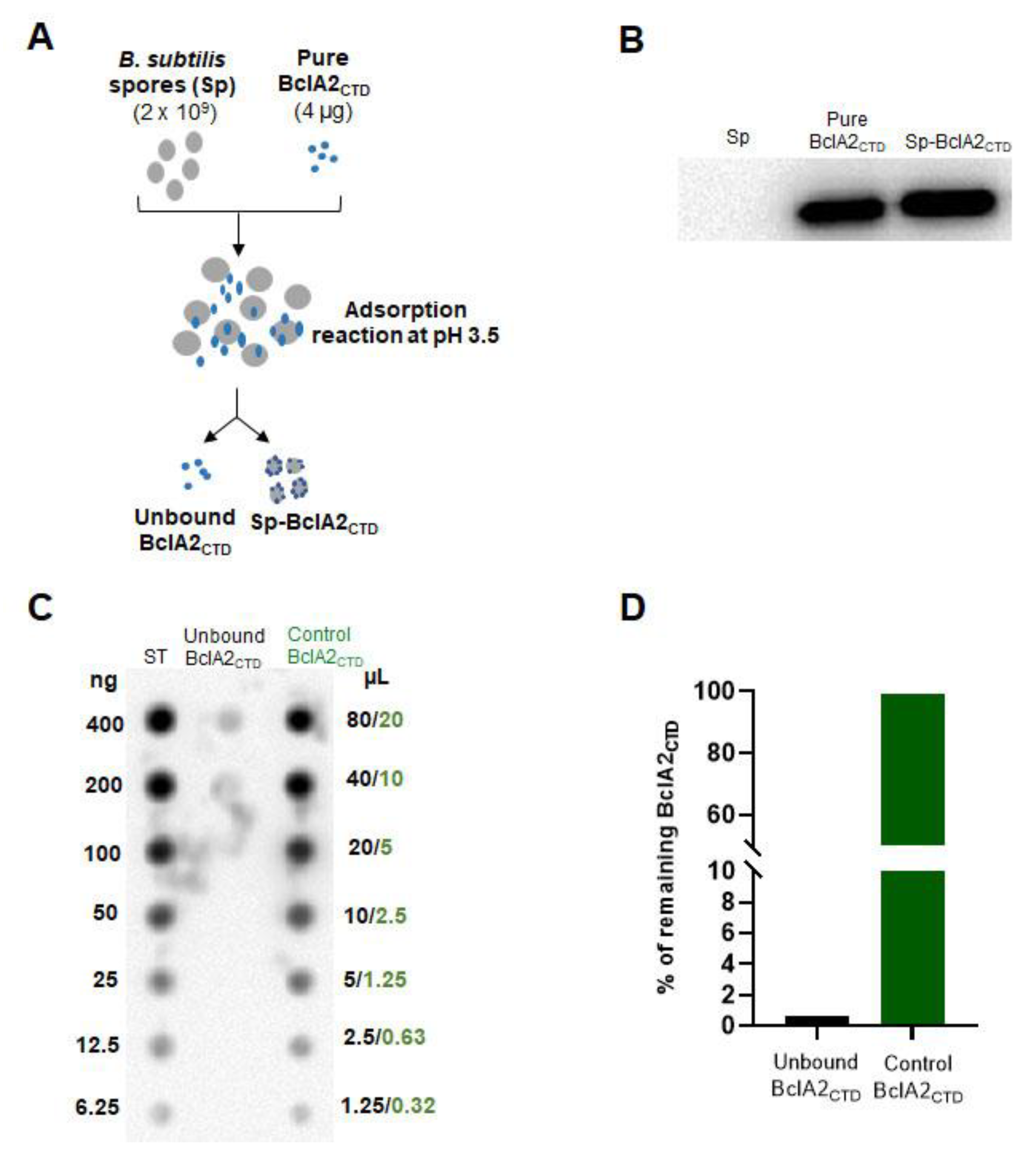
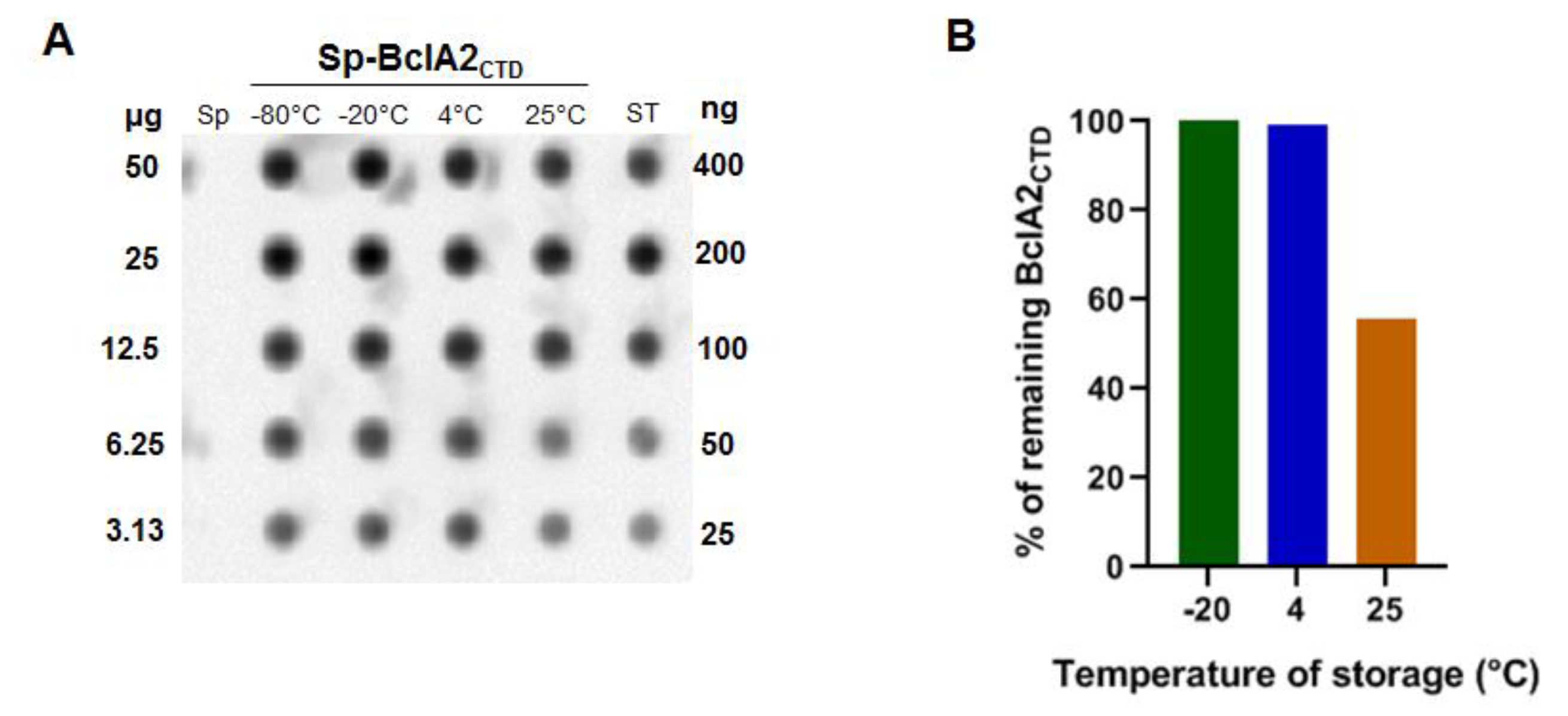
2.3. Bcla2ctd Is Stabilized by the Adsorption on B. Subtilis Spores

To evaluate the stability of spore-adsorbed BclA2CTD, 2 × 10⁹ spores displaying 4 µg of BclA2CTD were centrifuged and incubated as pellets for 48 h either at −20, +4, or +25 °C. Pellets incubated at −80 °C were used as control. The spore surface proteins were then extracted and analysed by dot blot experiments with anti-6xHis antibody, and the amount of BclA2CTD was compared to known amounts of pure protein stored at −80 °C immediately after its purification as standard (ST) (Figure 4A). Results of the densitometric analysis of the dot blot (Table S3) were plotted and showed that the amount of BclA2CTD recognized by the antibody was not decreased after incubation at −20 or +4 °C; however, a 50% decrease in BclA2CTD recognition was observed after incubation at +25 °C (Figure 4B).
The comparison of the results of Figure 2 and Figure 4 clearly indicate that upon adsorption to spores the heat stability of BclA2CTD was increased, suggesting that the binding to the spore protects the antigen from degradation.

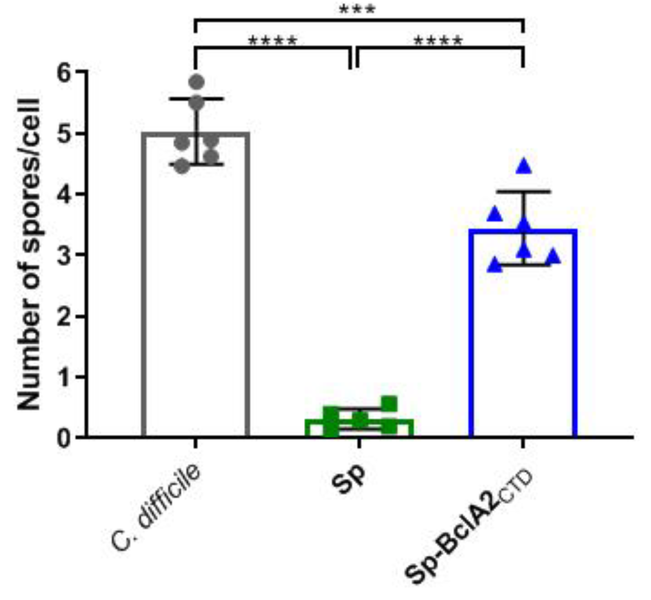
2.4. Adsorption of Bcla2ctd Increases Spore Adherence to Caco-2 Cells

The adherence of B. subtilis spores adsorbed with BclA2CTD to differentiated Caco-2 cells was evaluated and compared to the adherence pattern of C. difficile R20291 and B. subtilis spores. While the adherence of R20291 spores reached 5 spores/cell, B. subtilis spores had an average adherence of less than 1 spore/cell (Figure 5). The adherence of B. subtilis spores to Caco-2 cells was significantly (p < 0.0001) increased upon adsorption of BclA2CTD (Figure 5), suggesting that BclA2CTD contributes to adherence of the spores to intestinal epithelial cells.

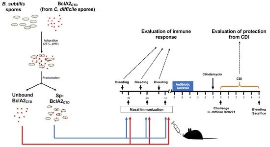
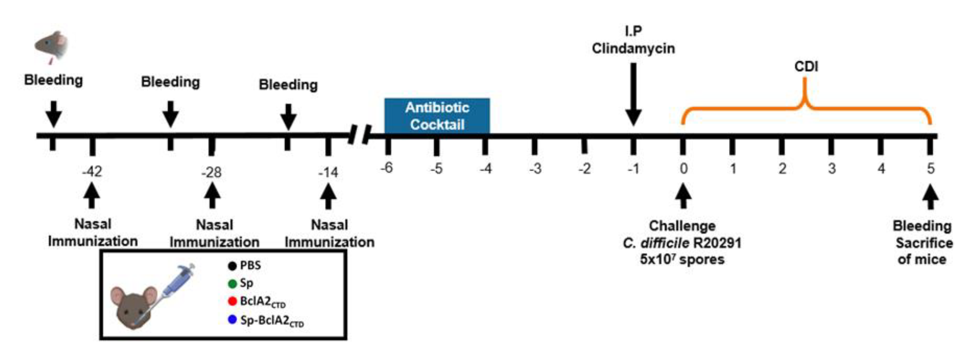
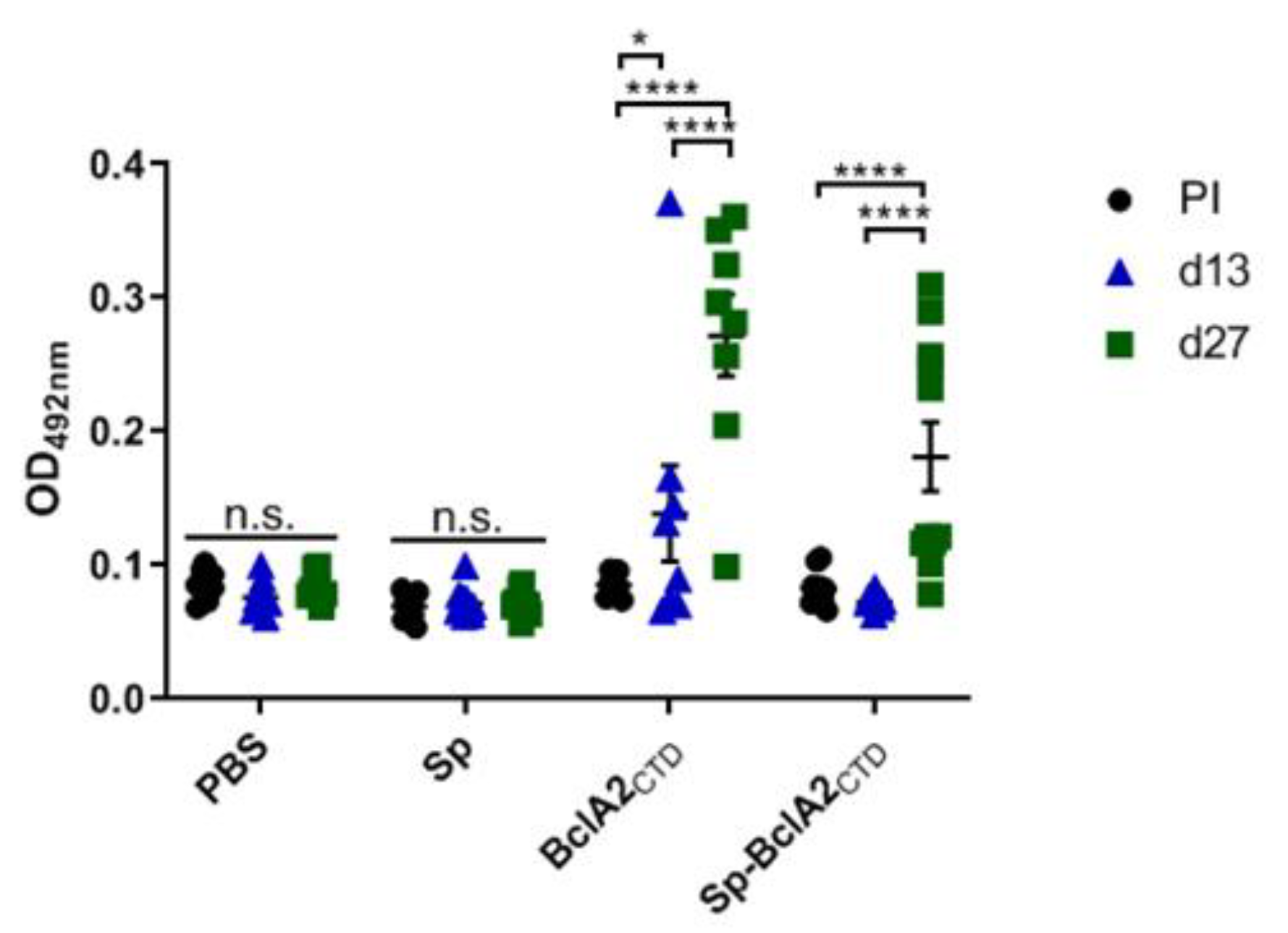
2.5. Intranasal Immunization

B. subtilis spores presenting BclA2CTD on their surface, and the pure BclA2CTD peptide, were used for mucosal immunization experiments in mice. In particular, the animals were assigned into four experimental groups (n = 11), according to the immunization regimen: (i) PBS; (ii) 2 × 10⁹ B. subtilis PY79 spores (Sp); (iii) 2 × 10⁹ B. subtilis spores adsorbed with BclA2CTD (Sp-BclA2CTD); and (iv) 4 μg of purified BclA2CTD. The animals were nasally immunized 42, 28, and 14 d before the challenge with C. difficile R20291. Animal serum was collected one day before each immunization and on the time of sacrifice (Figure 6). The production of specific anti-BclA2CTD IgG was assessed by ELISA (Figure 7).
Animals treated with purified BclA2CTD showed a significant increase of IgG titers mainly after the second immunization (p < 0.0001). The same result was observed for animals immunized with Sp-BclA2CTD (p < 0.0001), suggesting that the spore-bound antigen is correctly assembled and able to induce a response. However, no differences were observed between the purified and the spore-bound antigen, indicating that Bacillus spores did not have an adjuvant effect.
To explore the pattern of recognition of C. difficile spores by the raised antibodies, spores of C. difficile R20291 were incubated with the serum of mice immunized with purified BclA2CTD, Sp-BclA2CTD, or PBS and stained with secondary anti-mouse IgG Alexa 488 conjugate and then analysed by fluorescence microscopy (Figure 8A). No immunofluorescence reactivity was observed in serum of mice immunized with PBS (control). By contrast, significant immunofluorescence was evidenced by antibodies produced by immunizing mice with purified BclA2CTD and spore-adsorbed BclA2CTD (average fluorescence intensity was 88- and 89-fold higher than the control, respectively) (Figure 8B). Altogether, these results indicate that nasal immunization with either purified free BclA2CTD or spore-bound BclA2CTD was capable of inducing BclA2CTD-specific antibody production.

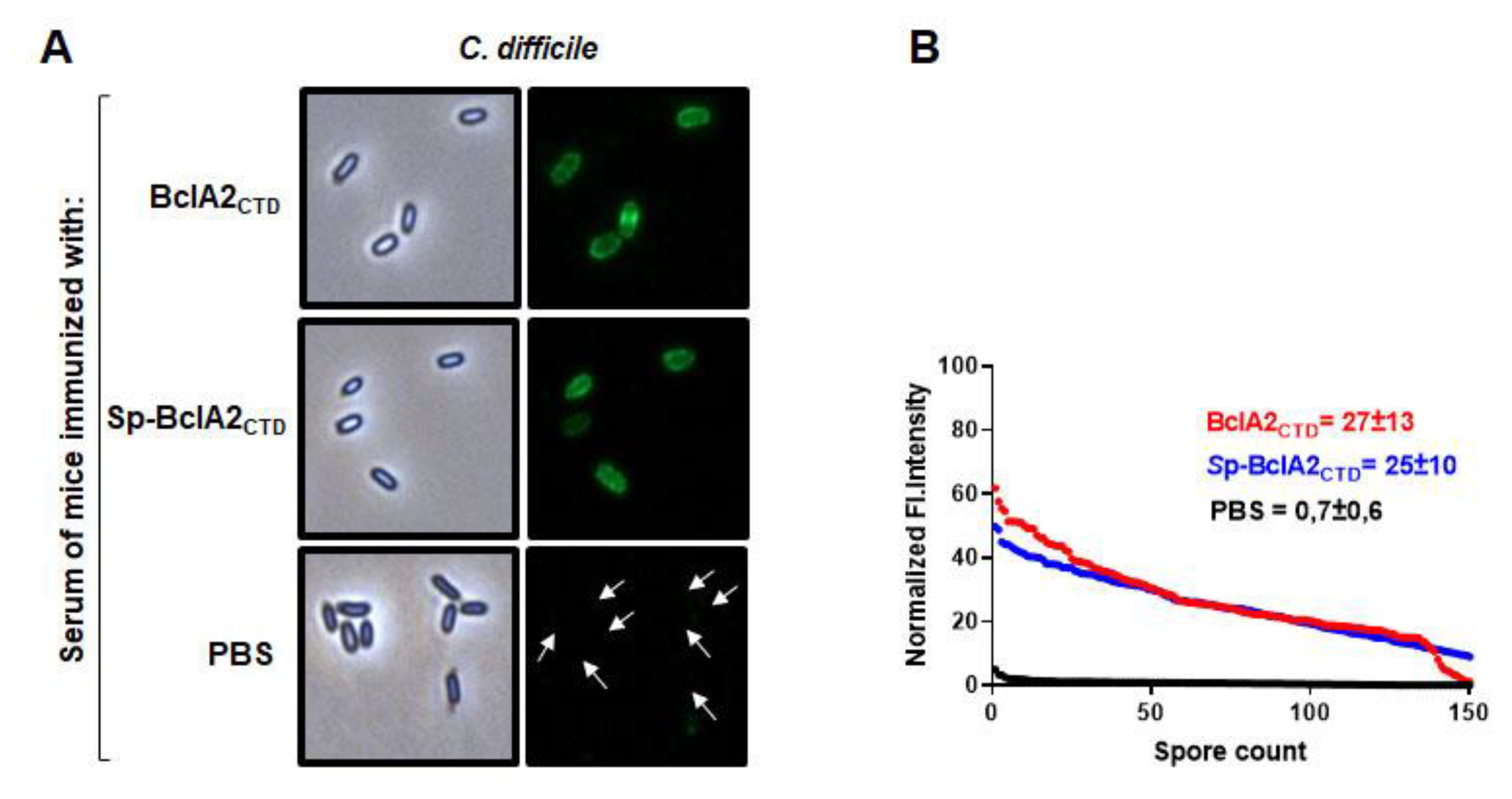
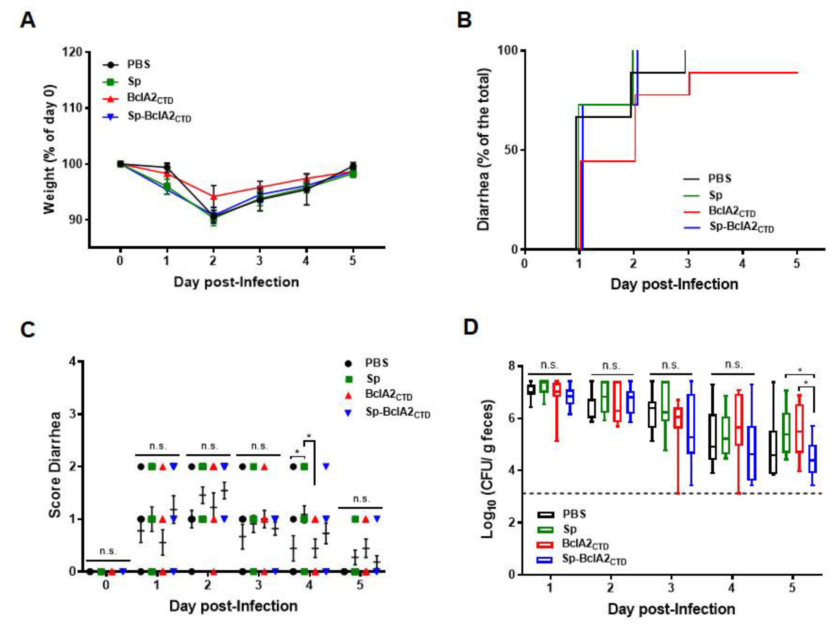
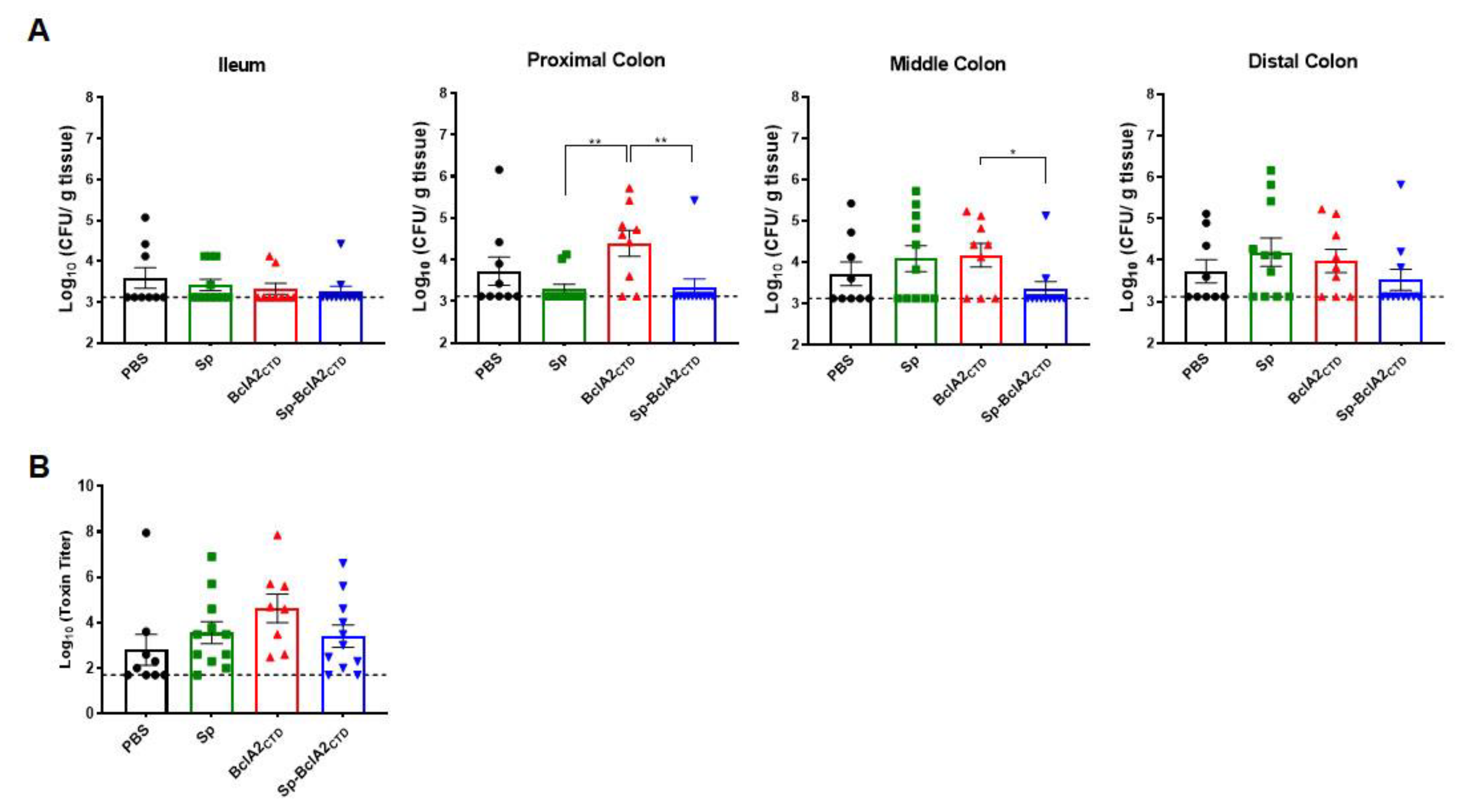
2.6. Effect of Nasal- Bcla2ctd Immunization Against C. Difficile R20291 Infection

Next, we assessed whether nasal immunization of mice protected against a C. difficile challenge. Weight loss and presence of diarrhea after infection, characterized by high spore-load in feces and colonic tissues, are expected symptoms of CDI. We measured weight loss progression over the five days after challenge and assigned a score of diarrhea according to its severity. Results demonstrate that nasal immunizations did not halt the weight loss in the subsequent 5 d after infection (Figure 9A). However, despite not being statistically significant, the animals immunized with purified BclA2CTD showed a delayed onset of diarrhea, contrary to the other groups in which more than 50% of the animals already had diarrhea at day 1 after the infection (Figure 9B). Moreover, when comparing the score of diarrhea between groups (Figure 9C), it is possible to observe that on day 1 after infection, the group of animals immunized with purified BclA2CTD had a reduced score of diarrhea even though not statistically significant. Interestingly, on day 3 after infection, the animals immunized with adsorbed spores showed a reduction in C. difficile spore load in feces (Figure 9D), which became significant on day 5 after infection (p = 0.0215 and p = 0.0306 when compared with Sp and purified free BclA2CTD).
No significant differences between groups in C. difficile spore-load in the ileum or distal colon were observed (Figure 10A). However, animals immunized with Sp-BclA2CTD showed a slight decrease in spore-load in the proximal and middle colon in comparison with the other groups. This reduction became significant in comparison with the animals immunized with purified BclA2CTD (p = 0.0041 for proximal colon and p = 0.0324 for middle colon). However, there were no significant differences between groups on the cytotoxic titers of the cecal content (Figure 10B), meaning that the immunizations did not halt spore colonization inside the cecum.

3. Discussion

In recent years, the emergence of antibiotic-resistant, hypervirulent strains has urged the scientific community to develop new therapeutic strategies to fight C. difficile infection (CDI) [26,27]. New treatments based on probiotics or fecal transplants, although very promising, are still at an experimental stage, while vaccines targeting C. difficile cells [28] or toxins [29] have been so far unsuccessful. Since CDI is transmitted by spores that are also responsible of the initial interaction with the host, alternative vaccination strategies have to focus on the spore and on the surface proteins involved in the interaction with the intestinal epithelial cells. Most C. difficile genomes have a pseudogenized version of bclA1, leading to the expression of a 48 aa polypeptide sequence of the amino-terminal domain that is localized to the exosporium layer of C. difficile spores [16]. In this context, recent work has shown that intraperitoneal immunization with the spore surface protein BclA1 induced a strong, specific immune response but failed to provide protective immunity [30]. Two additional collagen-like exosporium proteins are also encoded in C. difficile genomes, BclA2 and BclA3, which to date have not been evaluated as vaccine candidates. In this study, a fragment of the spore surface protein BclA2 (BclA2CTD) was identified as a potential antigen and tested as a mucosal vaccine.
The mucosal surfaces of the host are the most common route of entry used by pathogens, including C. difficile; therefore, it is important for a vaccine to elicit an immune response at the mucosal surfaces. However, only few mucosal vaccines are currently licensed, mostly because of the low immunogenicity of mucosal antigens and to antigen degradation during storage or at the mucosal surfaces [31]. One strategy that we used to avoid or reduce antigen degradation was to display BclA2CTD on B. subtilis spores, a mucosal vaccine delivery system previously tested with other antigens [22,23]. Our data demonstrate that BclA2CTD was very efficiently displayed in B. subtilis spores, and the interaction with the spore surface layers reduced BclA2CTD degradation during storage, as previously reported for other antigens and enzymes [32]. However, in spite of the low in vitro stability of the free BclA2CTD with respect to the spore-bound antigen, both forms of BclA2CTD induced similarly strong humoral immune responses when administered to mice by the nasal route.
Upon immunizations, mice were subjected to a challenge with the epidemically relevant C. difficile strain R20291. Although no obvious protective effects were observed on mice, the C. difficile spore load in feces showed a tendency to decrease three days after infection in animals immunized with the spores displaying BclA2CTD. In addition, these same groups of animals also showed a reduction of spore load in the proximal and middle colon, possibly a consequence of C. difficile spore opsonization by anti-BclA2CTD antibodies. A similar lack of protection efficacy was previously observed upon immunization with the BclA1 and SleC, but not with the cysteine-rich proteins CdeC and CdeM [30], suggesting that the collagen-like proteins BclA1 and SleC are not good vaccine candidates. A possible explanation of the lack of clear signs of protection observed in our results is that the dose of antigen might not have been high enough to reduce CDI symptoms and C. difficile spore germination inside the mice cecum. Indeed, works using B. subtilis spores adsorbed with heterologous antigens that induced a protective effect in nasally immunized mice applied a higher dose regimen and frequency than we did in this work. Plus, some works have also used the same immunization regimen and induced both humoral and cellular immune responses. However, they aimed to have a stronger effect in the nasal mucosa and not in the gut, since they used Mycobacterium tuberculosis antigens or H5N1 virions [33,34].
Additional in vivo experiments with higher doses of antigen and/or using the oral route of immunization would be needed to address this point. Even though no protection against a challenge was observed, the identification of an efficient antigen for C. difficile spores, active when administered by the mucosal route, is a promising result that could open to a new vaccination strategy against CDI.

4. Materials and Methods

4.1. Bacterial Strains and Spore Purification

B. subtilis PY79 [24] was used for the adsorption reaction. The hypervirulent C. difficile strain R20291 was used for the challenge experiment. E. coli strain BL21 (DE3) (ThermoFisher Scientific, Waltham MA, USA) was used for BclA2CTD overexpression.
Sporulation of B. subtilis was induced by the exhaustion method [35]. Briefly, after 35 h of growth in Difco Sporulation (DS) medium at 37 °C with vigorous shaking, spores were collected, washed, and purified. The purification was performed using KCl 1M, lysozyme 10mM, NaCl 1M, SDS 0.05%, and several washes with water.
C. difficile spores were purified as described elsewhere [36]. Briefly, 1:100 dilution of C. difficile overnight culture was plated onto a 70:30 medium (63 g Bacto peptone (BD Difco), 3.5 g proteose peptone (BD Difco), 0.7 g ammonium sulfate, 1.06 g Tris Base, 11.1 g brain heart infusion extract (BD Difco), and 1.5 g yeast extract (BD Difco) for 1 L) and incubated under anaerobic conditions for 7 d at 37 °C [37]. Upon incubation, the surface of the plates was scraped up with ice-cold sterile water, and the spores were gently washed five times with ice-cold sterile water in the micro centrifuge at 14,000 rpm for 5 min. After, the spores were loaded onto a 50% Nycodenz solution and centrifuged once again in the same conditions previously described. The pellet was then washed five times with ice-cold sterile water to remove Nycodenz remnants. The spores were quantified in the Neubauer chamber, and the volume was adjusted at 5 × 10⁹ spores per mL.

4.2. BclA2CTD over-Production and Purification

The coding sequence of the BclA2 C-terminal domain (CTD) was amplified using the chromosomal DNA of C. difficile R20291 (396bp) as a template and oligonucleotides BclA2CTDsense (ggtaccccatggggatccGCAGCAAACAATGCACAATTTACAG, in lower case are the recognition sites for KpnI, NcoI, and BamHI restriction enzymes) and BclA2CTDanti (tctagactgcagCTATTGTATTCTATAAACTGATACATAC, in lower case are the recognition sites for XbaI and PstI restriction enzymes) to prime the reaction. Amplified DNA was cloned in pGEMT-easy (Promega, Madison, Wisconsin, USA). Subsequently, the gene of interest was cleaved with BamHI/PstI restriction enzymes and inserted in-frame to the sequence coding for an N-terminal polyhistidine tag in the expression vector pRSETA (Invitrogen) previously digested with the same enzymes. Expression of the recombinant gene was controlled by a T7 promoter inducible by lactose. The recombinant plasmid was used to transform competent cells of the E. coli strain BL21(DE3). The recombinant strain was grown for 16 h at 37 °C in auto-induction medium [38] to express the heterologous protein. The His-tagged BclA2CTD protein was purified under native conditions using the His-Trap column (eluted with 500mM of imidazole) as recommended by the manufacturer (GE Healthcare Life Science, Merk, Darmstardt, Germany). The purified protein was desalted and concentrated with the Centricon cut-off 10kDa (Merck, Millipore, Darmstardt, Germany). The purity of the protein was verified by SDS-page and Western blot using anti-His antibodies.

4.3. Adsorption Reaction, Stability, and Production of Spores for Animal Immunization

Purified BclA2CTD (4 µg) was added to a suspension of 2 × 10⁹ B. subtilis spores in 0.15 M PBS pH 3.5 at 25 °C in a final volume of 200 µL. After 1 h of incubation, free unbound BclA2CTD and spore-adsorbed BclA2CTD (Sp-BclA2CTD) were separated by centrifugation and analysed by dot and Western blotting. The stability of free and spore-adsorbed BclA2CTD was assessed by exposing purified BclA2CTD (suspended in phosphate buffer pH 7) and the BclA2CTD-adsorbed spores at different temperatures before analysis.
For mice immunization, 144 µg of purified BclA2CTD was incubated with a suspension of 7.4 × 10¹⁰ B. subtilis spores in 0.15 M PBS pH 3.5 at 25 °C in a final volume of 8 mL. A total of 2 × 10⁹ spores were used for Western and dot blot analyses, and the remaining spores were aliquoted and stored at −80 °C for mice immunization.

4.4. Western and Dot-Blot Analyses

A total of 2 × 10⁹ B. subtilis spores or pure BclA2CTD were suspended in extraction buffer 2x [25] incubated at 100 °C for 7 min. Proteins were loaded on SDS-PAGE gel (15%), electro-transferred to nitrocellulose filters (Amersham Pharmacia Biotech, Milano, Italy), and used for Western blot analysis. For the quantitative evaluation of the amount of BclA2CTD, serial dilutions of purified BclA2CTD, of the supernatant of the adsorption reaction or of protein extracted from spores displaying BclA2CTD, were analysed by dot blot experiments. Protein extraction from the spore coat was obtained by incubating 2 × 10⁹ Sp-BclA2CTD in SDS 10%, DTT 1 M, and Tris-HCl pH 6.8 1.5 M 1 h at 65 °C (200 μL final volume), 2 min at 4 °C, and posterior centrifugation. Protein quantification on the dots was obtained by exposing the filters to ECL-prime (Amersham Pharmacia Biotech), and then, using Quantity One 1-D Analysis Software (Bio-Rad, Segrate, Milano, Italy), the dots were subjected to densitometric analysis. Anti-His antibodies were used both in Western and dot blot assays.

4.5. Adherence to Caco-2 Cells

An existing stock of Caco-2 cells in the Microbiota–Host Interaction and Clostridia Research Group at the Universidad Andrés Bello was routinely grown at 37 °C with 5% CO2 in Dulbecco’s modified Eagle’s minimal essential medium (DMEM) (HyClone, GE Healthcare Life Science, Merk, Darmstardt, Germany), supplemented with 10% (vol/vol) fetal bovine serum (FBS) (HyClone), penicillin (100 U/mL), and streptomycin (100 µg/mL). Spore adherence of C. difficile R20291, B. subtilis PY79, and B. subtilis adsorbed with BclA2CTD to Caco-2 cell line was measured as previously described [39]. Briefly, Caco-2 cells were seeded onto glass coverslips in 24-well plates (4 × 10⁵ cells per well), and to obtain differentiated Caco-2 cells, cells were cultured for 8 d post-confluence, changing the medium every other day, using previously described methods [40]. Monolayers were infected with 2.5 × 10⁷ spores in 200 μL of culture medium without FBS. Cells infected with spores were incubated under aerobic conditions 3 h at 37 °C. After incubation, the unbound spores were removed by washing three times with DPBS. In order to count the number of cells, the nuclei were stained with Hoechst stain (Sigma-Aldrich, St. Louis, MO, USA) 1:1000 in PBS 10 min. After several washes with PBS and H2O, the coverslips were dried 15 min at 37 °C, mounted using Dako Fluorescence Mounting medium (Dako, North America, Carpinteria, CA, USA), and sealed with nail polish. Samples were analysed with an Olympus BX53 microscope. The number of spores and cells were counted, and the adherence was represented as number of spores/cell. The experiment was conducted in triplicate.

4.6. Animals

Pathogen-free male or female C57BL/6 mice (age 8–12 w) were obtained from a breeding colony at Facultad de Ciencias Biologicas Universidad Andres Bello (Santiago, Chile), established with animals purchased from Jackson Laboratories. Mice handling and experimental protocols were performed according to Animal ethic committee of the Faculty of Life Sciences of the Universidad Andrés Bello (Protocol number 0035/2018, project identification code: Fondef ID18/10230; approval date: 22nd january 2018; approval act code 0035/2018).
Mice were exposed to a 12-h cycle of light and darkness, and water, bedding, and cages were previously autoclaved.

4.7. Immunization Regimen in Mice

Mice were randomly assigned to four experimental groups (11 animals in each group) according to the type of immunization received. The mice were intranasally immunized on days 0, 14, and 28 after the beginning of the experiment with 20 μL (10 µL per nostril) of PBS pH 7, 2 × 10⁹ spores of B. subtilis PY79, 2 × 10⁹ B. subtilis PY79 spores adsorbed with BclA2CTD (Sp- BclA2CTD), or 4μg of pure BclA2CTD. The day before each immunization and on the day of the sacrifice (on 47th day), the blood was collected.

4.8. Animal Infection Model

Six days before infection with C. difficile spores, mice were exposed to an antibiotic cocktail with kanamycin (40 mg/kg body weight; Sigma-Aldrich, USA.), gentamicin (3.5 mg/kg body weight; Sigma-Aldrich, USA.), colistin (4.2 mg/kg body weight; Sigma-Aldrich, USA.), metronidazole (21.5 mg/kg body weight; Sigma-Aldrich, USA.), and vancomycin (40 mg/kg body weight; Sigma-Aldrich, USA.) for 3 d by oral administration. The antibiotic treatment was followed by intraperitoneal administration of a single dose of clindamycin (10 mg/kg) 1 d before C. difficile infection [41]. All animals were infected oro-gastrically with 100 μL of PBS containing 5 × 10⁷ spores of C. difficile strain R20291. From this point, all procedures were made inside a biosafe cabinet to contain spore-mediated transmission. Each mouse was individually housed in strerile cages, and mice had ad libitum access to food and water.
Mice conditions were daily monitored according to a scoring system. Presence and morphology of diarrhea was classified as follows: (i) normal stool (score = 0); (ii) colour/consistency change (score = 1); (iii) presence of wet tail or mucosa (score = 2); (iv) liquid stools (score = 3). A score higher than 1 was considered as diarrhea [42]. Other clinical symptoms as variations of weight, physical aspect (i.e., abnormal/hunched gait, piloerection), spontaneous behaviour (i.e., lethargy, inactivity or lack of mobility), and emaciation were monitored as described [43]. Moribund mice or mice displaying overt signs of disease were sacrificed. At the time of euthanasia, ileum, proximal, median, and distal colon were collected as well as the cecum content.

4.9. Quantification of Spores from Feces and Colon Samples

Fecal samples were collected daily and stored at −20 °C until spore quantification. Ten microliters of PBS was added for each mg of stool, mixed, and incubated for 30 min at room temperature. Then, 50 µL of absolute ethanol (Sigma-Aldrich) was added to 50 µL of feces and incubated for 30 min at room temperature. Samples were serially diluted and plated onto selective medium supplemented with taurocholate (0.1% w/v), cefoxitin (16 μg/mL), and l-cycloserine (250 μg/mL) (TCCFA plates). The plates were incubated anaerobically at 37 °C for 48 h, the C. difficile colonies were counted, and the results were expressed as the log10 (CFU/g of faeces). Proximal, median, and distal colon were collected from mice upon sacrifice and washed with PBS with a syringe. Then, they were resuspended and homogenized with 2.5 µL of PBS for each mg of tissue. Upon incubation at room temperature with absolute ethanol and serially diluted, they were plated onto TCCFA plates. The plates were incubated anaerobically at 37 °C for 48 h. Finally, the colony count was expressed as the log10 (CFU/g of tissue).

4.10. Evaluation of Bcla2ctd-Specific Igg Levels in Mice Serum

The blood collected the day before each immunization and at the time of sacrifice was incubated at 37 °C for 30 min and posteriorly centrifuged at 5000 rpm for 20 min at 4 °C. The supernatant containing the serum fraction was stored at −20 °C until use. To assess the production of IgG against BclA2CTD, an enzyme-linked immunosorbent assay (ELISA) was performed. Pure BclA2CTD was coated onto 96-well plates at 100ng/well overnight at 4 °C. Plates were blocked with PBS–0.05% Tween-20 (PBS-T) containing 2% BSA for 1 h at 37°C. After several washes, the wells were next incubated with 1:100 of animal serum (in 1% BSA in PBS-T). The plates were incubated 2 h at 37 °C. After the removal of non-adherent IgG by several washes, the plates were incubated with secondary antibody anti-mouse HRP for 1 h at 37 °C. Finally, the colorimetric reaction was initiated upon the addition of 50 µL of reaction buffer containing 0.05 M citric acid, 0.1 M disodiumhydrogen phosphate, 2 mg/mL of o-phenlyendiamine (Sigma-Aldrich, USA.), and 0.015% of H2O2 (Merck, Germany). The reaction was stopped after 20 min with 25 µL of 4.5 N of H2SO4, and absorbance was measured at 492 nm. The experiment was performed in duplicate.

4.11. Immunofluorescence Analysis

Spores of C. difficile R20291 were fixed for 20 min with 3% paraformaldehyde (pH 7.4) on cover glass slides previously coated with poly-l-lysine. Coverslips were washed three times with PBS, blocked 1 h with 1% BSA, and further incubated at room temperature 1 h with 1:100 of serum from an immunized mouse with PBS, Sp-BclA2CTD, or purified BclA2CTD (in PBS BSA 1%). The coverslips were rinsed three times with PBS and incubated 1 h at room temperature with 1:400 anti-mouse conjugated with Alexa 488 (ThermoFisher Scientific, Invitrogen, Waltham MA, USA) in PBS BSA 1% and washed three times with PBS and one time with sterile water. Finally, after drying 30 min at room temperature, the coverslips were mounted with Dako Fluorescence Mounting medium (Dako, North America), sealed with nail polish, and then analysed with Olympus BX53 fluorescence microscope. The fluorescence images were obtained with 30 ms of exposition. ImageJ software was used to quantify the fluorescence signal, as previously reported [25].

4.12. Cytotoxicity Assay

The toxicity of the cecum content was determined using Vero cells [44]. Briefly, 96-well flat-bottom microtiter plates were seeded with 105 Vero cells/well. Mice cecum contents were weighted and then mixed with PBS (10 μL of PBS per mg of cecum content) and centrifuged (14,000 rpm 5 min). The supernatant, previously sterilized with a filter, was serially diluted in DMEM supplemented with FBS 10% and penicillum streptomycin 1%, and then 100 μL of each dilution was added to wells containing Vero cells. The plates were incubated 16 h at 37 °C, and then cell rounding was analysed. For each series of dilutions for one cecum content, the highest dilution that produced at least 80% of cell rounding under X200 magnification was considered.

4.13. Statistical Analysis

Prism 8 (GraphPad Software, Inc., San Diego, CA, USA) was used for statistical analysis. Normality was assessed by Shapiro–Wilk tests. For populations that did not follow a normal distribution, significance between groups was assessed by Mann–Whitney unpaired t-tests. Comparative analysis between groups was performed by analysis of variance with Turkey’s multiple comparison test for populations that followed a normal distribution. A p-value of ≤0.05 was accepted as the level of statistical significance.

5. Conclusions

Final goal of this work is to develop a new vaccination strategy against Clostridium difficile spores, the cellular form of the pathogen involved in the transmission of the pathogen and in its first interaction with the host. To this aim, we identified the C-terminal domain of the exosporium protein BclA2 (BclA2CTD) of C. difficile as an effective mucosal antigen. To increase antigen stability and facilitate its delivery, BclA2CTD was efficiently exposed on B. subtilis spores and observed that the interaction with the spore protected the antigen from degradation during the storage. Both free and spore-adsorbed BclA2CTD were used to nasally immunize mice and were both able to induce a specific humoral immune response.

Supplementary Materials

Supplementary materials can be found at https://0-www-mdpi-com.brum.beds.ac.uk/1422-0067/21/4/1277/s1.

Author Contributions

Conceptualization, E.R., D.P.-S., and L.B.; Formal analysis, A.R.M., R.R.-R., M.P.-G., and P.C.-C.; Funding acquisition, D.P.-S. and L.B.; Investigation, A.R.M.; Methodology, A.R.M.; Resources, E.R., D.P.-S., and L.B.; Supervision, E.R., D.P.-S., and L.B; Validation, A.R.M., A.S., and R.I.; Writing—original draft, A.R.M., E.R., D.P.-S., and L.B.; Writing—review & editing, E.R., D.P.-S., and L.B. All authors have read and agreed to the published version of the manuscript.

Funding

This research was funded by “Finanziamento di Ricerca di Ateneo” to L.B, project title “SP-LAY: Bacterial spores as live platform for proteins display”; Fondo Nacional de Ciencia y Tecnología de Chile (FONDECYT Grant 1191601); Millennium Science Initiative of the Ministry of Economy, Development and Tourism to D.P-S. Support was also provided by a grant from Fondo de Fomento al Desarrollo Científico y Tecnológico (FONDEF) ID18|10230 to M.P-G and D.P-S.

Conflicts of Interest

The authors declare no conflicts of interest.

Abbreviations

CDIClostridioides difficile infection
BclABacillus collagen-like protein of anthracis
NTDN-terminal domain
CTDC-terminal domain
BclA2CTDC-terminal domain of BclA2
STStandard
PBSPhosphate-buffered saline
SpSpores of B. subtilis
Sp-BclA2CTDSpores of B. subtilis adsorbed with BclA2CTD
ANOVAAnalysis of variance
ELISAEnzyme-linked immunosorbent assay
IgGImmunoglobulin G
I.PIntraperitoneal
WtWild-type
PIPre-Immune
d13Day 13
d27Day 27
ODOptical density
Fl.IFluorescence intensity
CFUColony forming unit

References

  1. Kelly, C.P.; Pothoulakis, C.; LaMont, J.T. Clostridium difficile colitis. N. Engl. J. Med. 1994, 330, 257–262. [Google Scholar] [CrossRef] [PubMed]
  2. Heinlen, L.; Ballard, J.D. Clostridium difficile infection. Am. J. Med. Sci. 2010, 340, 247–252. [Google Scholar] [CrossRef] [PubMed]
  3. McDonald, L.C.; Killgore, G.E.; Thompson, A.; Owens, R.C., Jr.; Kazakova, S.V.; Sambol, S.P.; Johnson, S.; Gerding, D.N. An epidemic, toxin gene-variant strain of Clostridium difficile. N. Engl. J. Med. 2005, 353, 2433–2441. [Google Scholar] [CrossRef] [PubMed] [Green Version]
  4. Warny, M.; Pepin, J.; Fang, A.; Killgore, G.; Thompson, A.; Brazier, J.; Frost, E.; McDonald, L.C. Toxin production by an emerging strain of Clostridium difficile associated with outbreaks of severe disease in North America and Europe. Lancet 2005, 366, 1079–1084. [Google Scholar] [CrossRef]
  5. Hookman, P.; Barkin, J.S. Clostridium difficile associated infection, diarrhea and colitis. World J. Gastroenterol. 2009, 15, 1554–1580. [Google Scholar] [CrossRef] [PubMed]
  6. Cornely, O.A. Current and emerging management options for Clostridium difficile infection: What is the role of fidaxomicin? Clin. Microbiol. Infect. 2012, 18 (Suppl. 6), 28–35. [Google Scholar] [CrossRef] [Green Version]
  7. Valiente, E.; Dawson, L.F.; Cairns, M.D.; Stabler, R.A.; Wren, B.W. Emergence of new PCR ribotypes from the hypervirulent Clostridium difficile 027 lineage. J. Med. Microbiol. 2012, 61, 49–56. [Google Scholar] [CrossRef]
  8. McFarland, L.V.; Elmer, G.W.; Surawicz, C.M. Breaking the cycle: Treatment strategies for 163 cases of recurrent Clostridium difficile disease. Am. J. Gastroenterol. 2002, 97, 1769–1775. [Google Scholar] [CrossRef]
  9. Feher, C.; Soriano, A.; Mensa, J. A review of experimental and off-label therapies for Clostridium difficile infection. Infect. Dis. 2017, 6, 1–35. [Google Scholar] [CrossRef] [Green Version]
  10. Goldberg, E.J.; Bhalodia, S.; Jacob, S.; Patel, H.; Trinh, K.V.; Varghese, B.; Yang, J.; Young, S.R.; Raffa, R.B. Clostridium difficile infection: A brief update on emerging therapies. Am. J. Health. Syst. Pharm. 2015, 72, 1007–1012. [Google Scholar] [CrossRef]
  11. Leffler, D.A.; Lamont, J.T. Clostridium difficile infection. N. Engl. J. Med. 2015, 372, 1539–1548. [Google Scholar] [CrossRef] [PubMed] [Green Version]
  12. Khan, M.Y.; Dirweesh, A.; Khurshid, T.; Siddiqui, W.J. Comparing fecal microbiota transplantation to standard-of-care treatment for recurrent Clostridium difficile infection: A systematic review and meta-analysis. Eur. J. Gastroenterol. Hepatol. 2018, 30, 1309–1317. [Google Scholar] [CrossRef] [PubMed]
  13. Maziade, P.J.; Pereira, P.; Goldstein, E.J. A Decade of Experience in Primary Prevention of Clostridium difficile Infection at a Community Hospital Using the Probiotic Combination Lactobacillus acidophilus CL1285, Lactobacillus casei LBC80R, and Lactobacillus rhamnosus CLR2 (Bio-K+). Clin. Infect. Dis. 2015, 60 (Suppl. 2), S144–S147. [Google Scholar] [CrossRef]
  14. Diaz-Gonzalez, F.; Milano, M.; Olguin-Araneda, V.; Pizarro-Cerda, J.; Castro-Cordova, P.; Tzeng, S.C.; Maier, C.S.; Sarker, M.R.; Paredes-Sabja, D. Protein composition of the outermost exosporium-like layer of Clostridium difficile 630 spores. J. Proteom. 2015, 123, 1–13. [Google Scholar] [CrossRef] [PubMed]
  15. Mora-Uribe, P.; Miranda-Cardenas, C.; Castro-Cordova, P.; Gil, F.; Calderon, I.; Fuentes, J.A.; Rodas, P.I.; Banawas, S.; Sarker, M.R.; Paredes-Sabja, D. Characterization of the Adherence of Clostridium difficile Spores: The Integrity of the Outermost Layer Affects Adherence Properties of Spores of the Epidemic Strain R20291 to Components of the Intestinal Mucosa. Front. Cell. Infect. Microbiol. 2016, 6, 99. [Google Scholar] [CrossRef] [PubMed] [Green Version]
  16. Pizarro-Guajardo, M.; Olguin-Araneda, V.; Barra-Carrasco, J.; Brito-Silva, C.; Sarker, M.R.; Paredes-Sabja, D. Characterization of the collagen-like exosporium protein, BclA1, of Clostridium difficile spores. Anaerobe 2014, 25, 18–30. [Google Scholar] [CrossRef]
  17. Ricca, E.; Baccigalupi, L.; Cangiano, G.; De Felice, M.; Isticato, R. Mucosal vaccine delivery by non-recombinant spores of Bacillus subtilis. Microb. Cell. Fact. 2014, 13, 115. [Google Scholar] [CrossRef] [Green Version]
  18. Isticato, R.; Ricca, E. Spore Surface Display. Microbiol. Spectr. 2014, 2. [Google Scholar] [CrossRef]
  19. Kolaskar, A.S.; Tongaonkar, P.C. A semi-empirical method for prediction of antigenic determinants on protein antigens. Febs. Lett. 1990, 276, 172–174. [Google Scholar] [CrossRef] [Green Version]
  20. Wang, P.; Sidney, J.; Dow, C.; Mothe, B.; Sette, A.; Peters, B. A systematic assessment of MHC class II peptide binding predictions and evaluation of a consensus approach. PLOS. Comput. Biol. 2008, 4, e1000048. [Google Scholar] [CrossRef] [Green Version]
  21. Moutaftsi, M.; Peters, B.; Pasquetto, V.; Tscharke, D.C.; Sidney, J.; Bui, H.H.; Grey, H.; Sette, A. A consensus epitope prediction approach identifies the breadth of murine T(CD8+)-cell responses to vaccinia virus. Nat. Biotechnol. 2006, 24, 817–819. [Google Scholar] [CrossRef] [PubMed]
  22. Huang, J.M.; Hong, H.A.; Van Tong, H.; Hoang, T.H.; Brisson, A.; Cutting, S.M. Mucosal delivery of antigens using adsorption to bacterial spores. Vaccine 2010, 28, 1021–1030. [Google Scholar] [CrossRef] [PubMed]
  23. Isticato, R.; Sirec, T.; Treppiccione, L.; Maurano, F.; De Felice, M.; Rossi, M.; Ricca, E. Non-recombinant display of the B subunit of the heat labile toxin of Escherichia coli on wild type and mutant spores of Bacillus subtilis. Microb. Cell. Fact. 2013, 12, 98. [Google Scholar] [CrossRef] [PubMed] [Green Version]
  24. Youngman, P.; Perkins, J.B.; Losick, R. Construction of a cloning site near one end of Tn917 into which foreign DNA may be inserted without affecting transposition in Bacillus subtilis or expression of the transposon-borne erm gene. Plasmid 1984, 12, 1–9. [Google Scholar] [CrossRef]
  25. Isticato, R.; Ricca, E.; Baccigalupi, L. Spore Adsorption as a Nonrecombinant Display System for Enzymes and Antigens. J. Vis. Exp. 2019. [Google Scholar] [CrossRef]
  26. Barkin, J.A.; Sussman, D.A.; Fifadara, N.; Barkin, J.S. Clostridium difficile Infection and Patient-Specific Antimicrobial Resistance Testing Reveals a High Metronidazole Resistance Rate. Dig. Dis. Sci. 2017, 62, 1035–1042. [Google Scholar] [CrossRef]
  27. Johnson, S.; Sanchez, J.L.; Gerding, D.N. Metronidazole resistance in Clostridium difficile. Clin. Infect. Dis. 2000, 31, 625–626. [Google Scholar] [CrossRef]
  28. Potocki, W.; Negri, A.; Peszynska-Sularz, G.; Hinc, K.; Obuchowski, M.; Iwanicki, A. The combination of recombinant and non-recombinant Bacillus subtilis spore display technology for presentation of antigen and adjuvant on single spore. Microb. Cell. Fact. 2017, 16, 151. [Google Scholar] [CrossRef] [Green Version]
  29. Permpoonpattana, P.; Hong, H.A.; Phetcharaburanin, J.; Huang, J.M.; Cook, J.; Fairweather, N.F.; Cutting, S.M. Immunization with Bacillus spores expressing toxin A peptide repeats protects against infection with Clostridium difficile strains producing toxins A and B. Infect. Immun. 2011, 79, 2295–2302. [Google Scholar] [CrossRef] [Green Version]
  30. Ghose, C.; Eugenis, I.; Edwards, A.N.; Sun, X.; McBride, S.M.; Ho, D.D. Immunogenicity and protective efficacy of Clostridium difficile spore proteins. Anaerobe 2016, 37, 85–95. [Google Scholar] [CrossRef] [Green Version]
  31. Kim, S.H.; Jang, Y.S. The development of mucosal vaccines for both mucosal and systemic immune induction and the roles played by adjuvants. Clin. Exp. Vaccine. Res. 2017, 6, 15–21. [Google Scholar] [CrossRef] [PubMed]
  32. Sirec, T.; Strazzulli, A.; Isticato, R.; De Felice, M.; Moracci, M.; Ricca, E. Adsorption of beta-galactosidase of Alicyclobacillus acidocaldarius on wild type and mutants spores of Bacillus subtilis. Microb. Cell. Fact. 2012, 11, 100. [Google Scholar] [CrossRef] [PubMed] [Green Version]
  33. Song, M.; Hong, H.A.; Huang, J.M.; Colenutt, C.; Khang, D.D.; Nguyen, T.V.; Park, S.M.; Shim, B.S.; Song, H.H.; Cheon, I.S.; et al. Killed Bacillus subtilis spores as a mucosal adjuvant for an H5N1 vaccine. Vaccine 2012, 30, 3266–3277. [Google Scholar] [CrossRef] [PubMed]
  34. Reljic, R.; Sibley, L.; Huang, J.M.; Pepponi, I.; Hoppe, A.; Hong, H.A.; Cutting, S.M. Mucosal vaccination against tuberculosis using inert bioparticles. Infect. Immun. 2013, 81, 4071–4080. [Google Scholar] [CrossRef] [Green Version]
  35. Harwood, C.R.; Cutting, S.M. Molecular Biological Methods for Bacillus; Wiley: Chichester, NY, USA, 1990; p. xxxv. 581p. [Google Scholar]
  36. Calderon-Romero, P.; Castro-Cordova, P.; Reyes-Ramirez, R.; Milano-Cespedes, M.; Guerrero-Araya, E.; Pizarro-Guajardo, M.; Olguin-Araneda, V.; Gil, F.; Paredes-Sabja, D. Clostridium difficile exosporium cysteine-rich proteins are essential for the morphogenesis of the exosporium layer, spore resistance, and affect C. difficile pathogenesis. Plos. Pathog. 2018, 14, e1007199. [Google Scholar] [CrossRef]
  37. Edwards, A.N.; McBride, S.M. Isolating and Purifying Clostridium difficile Spores. Methods. Mol. Biol. 2016, 1476, 117–128. [Google Scholar] [CrossRef] [Green Version]
  38. Isticato, R.; Pelosi, A.; De Felice, M.; Ricca, E. CotE binds to CotC and CotU and mediates their interaction during spore coat formation in Bacillus. subtilis. J. Bacteriol. 2010, 192, 949–954. [Google Scholar] [CrossRef] [Green Version]
  39. Paredes-Sabja, D.; Sarker, M.R. Adherence of Clostridium difficile spores to Caco-2 cells in culture. J. Med. Microbiol. 2012, 61, 1208–1218. [Google Scholar] [CrossRef]
  40. Chantret, I.; Rodolosse, A.; Barbat, A.; Dussaulx, E.; Brot-Laroche, E.; Zweibaum, A.; Rousset, M. Differential expression of sucrase-isomaltase in clones isolated from early and late passages of the cell line Caco-2: Evidence for glucose-dependent negative regulation. J. Cell. Sci. 1994, 107 Pt 1, 213–225. [Google Scholar]
  41. Chen, X.; Katchar, K.; Goldsmith, J.D.; Nanthakumar, N.; Cheknis, A.; Gerding, D.N.; Kelly, C.P. A mouse model of Clostridium difficile-associated disease. Gastroenterology 2008, 135, 1984–1992. [Google Scholar] [CrossRef]
  42. Warren, C.A.; van Opstal, E.J.; Riggins, M.S.; Li, Y.; Moore, J.H.; Kolling, G.L.; Guerrant, R.L.; Hoffman, P.S. Vancomycin treatment’s association with delayed intestinal tissue injury, clostridial overgrowth, and recurrence of Clostridium difficile infection in mice. Antimicrob. Agents. Chemother. 2013, 57, 689–696. [Google Scholar] [CrossRef] [PubMed] [Green Version]
  43. Deakin, L.J.; Clare, S.; Fagan, R.P.; Dawson, L.F.; Pickard, D.J.; West, M.R.; Wren, B.W.; Fairweather, N.F.; Dougan, G.; Lawley, T.D. The Clostridium difficile spo0A gene is a persistence and transmission factor. Infect. Immun. 2012, 80, 2704–2711. [Google Scholar] [CrossRef] [PubMed] [Green Version]
  44. Theriot, C.M.; Koumpouras, C.C.; Carlson, P.E.; Bergin, I.I.; Aronoff, D.M.; Young, V.B. Cefoperazone-treated mice as an experimental platform to assess differential virulence of Clostridium difficile strains. Gut. Microbes. 2011, 2, 326–334. [Google Scholar] [CrossRef] [PubMed] [Green Version]
Figure 1. In silico analysis of BclA2 from Clostridioides difficile R20291. (A) Analysis of the B cell epitope propensity score (Kolaskar & Tongaonkar Antigenicity Method from Immune Epitope Database) of BclA2. The X- and Y-axes represent the sequence position and antigenic propensity score, respectively. The threshold value was generated by default by Immune Epitope Database (http://tools.iedb.org/bcell/). The regions above the threshold are antigenic. (B) Amino acid sequence of the 131 residues at the C-terminal end of BclA2 identified as a potential antigenic region and indicated as BclA2CTD by Kolaskar & Tongaonkar Antigenicity Method.
Ijms 21 01277 g001
Figure 2. Stability of purified BclA2CTD. (A) Dot blotting of purified BclA2CTD stored for 2 d at −20, +4, or at +25 °C with anti-6x His antibody. The protein stored at −80 °C right after purification (ST) was considered as standard. Loaded volumes of purified protein (μL) and known amount of the standard (ng) are indicated. (B) Plot of the densitometric analysis of dot blot experiment of panel A, considering the amount of protein stored at −80 °C as 100%.
Ijms 21 01277 g002
Figure 3. Adsorption of BclA2CTD on Bacillus subtilis spores. (A) Schematic representation of the adsorption reaction. (B) Western blotting of spore-extracted proteins after the adsorption reaction with anti-6xHis antibody. B. subtilis spores (Sp) and pure BclA2CTD were used as negative and positive controls, respectively. (C) Dot blot analysis of the supernatant of the adsorption reaction. Loaded volumes (μL) of the supernatant fraction (unbound BclA2CTD) and of the pure protein incubated in the same condition of adsorption (control BclA2CTD) are indicated in black and green, respectively. Known amounts (ng) of the pure protein incubated at −80 °C (ST) were used as standard. (D) Plot of the densitometric analysis of the dot blot experiment of panel C, considering the amount of protein stored at −80 °C as 100%.
Ijms 21 01277 g003
Figure 4. Stability of spore-adsorbed BclA2CTD. (A) Dot blot of the protein extracted from spores adsorbed with BclA2CTD stored for 2 d at −80, −20, +4, or +25 °C, using an anti-6xHis antibody. Proteins extracted from B. subtilis spores (sp) were used as negative control. Known amounts of the pure protein stored at −80 °C immediately after its purification (ST) were used as controls (right). (B) Plot of the densitometry analysis of dot blot experiment of panel A, considering the amount of protein extracted from spores stored at −80 °C as 100%.
Ijms 21 01277 g004
Figure 5. Adherence of spores to human intestinal cells. Caco-2 cells were differentiated for 8 d and infected with spores of C. difficile R20291, B. subtilis (Sp), and B. subtilis spores adsorbed with BclA2CTD (Sp-BclA2CTD). The number of adhered spores and cells was counted in 10 microscopic fields on phase-contrast microscopy. The data represent the mean of three independent experiments, and the error bars are the standard error of the mean. The groups were compared with one-way ANOVA Turkey’s multiple comparison test, and statistical differences (p < 0.05) are indicated by asterisks.
Ijms 21 01277 g005
Figure 6. Overview of the experimental design schematics for the prevention of C. difficile infection in a murine model. C57BL/6 mice were nasally immunized 3 times (42, 28, and 14 d before challenge with C. difficile R20291 spores) with PBS, spores of B. subtilis PY79 (Sp), pure BclA2CTD, or spores of B. subtilis adsorbed with BclA2CTD (Sp-BclA2CTD). Prior to the infection, the animals were submitted to an antibiotic cocktail (4 to 6 d before challenge) and clindamycin administration (1 d before challenge). On day 0 mice were infected with 5 x 10⁷ spores of C. difficile R20291 and were monitored for C. difficile infection (CDI) symptoms from day 0 to day 5. Serum was collected one day before each immunization as well as on the day of sacrifice (days 0, 13, 27, and 47).
Ijms 21 01277 g006
Figure 7. Immunogenicity of BclA2CTD protein and Sp-BclA2CTD in mice after nasal immunizations. IgG anti-BclA2CTD levels were measured by ELISA in the serum of mice on days 0 (pre-immune serum, PI), 13 (d13), and 27 (d27) after the beginning of the experiment (one day before each immunization). Results are reported as optical density (OD) units at 492nm. The geometric mean (± standard error of the mean) for each group is shown. IgG titers of each group were compared between d0 (PI), d13, and d27 with one-way ANOVA Turkey’s multiple comparison tests, and statistical differences (p < 0.05) are indicated by asterisks.
Ijms 21 01277 g007
Figure 8. Recognition of C. difficile spores by the serum of animals immunized with purified BclA2CTD and Sp-BclA2CTD. (A) Spores of C. difficile R20291 were incubated 1 h with serum (1:100) of mice immunized with BclA2CTD and Sp-BclA2CTD or PBS, as indicated. (B) The immunofluorescence micrographs are depicted in fluorescence intensity (FI.Intensity) profiles provided by fluorescence microscopy images using ImageJ. The values shown in the graphs are the average ± standard error of the fluorescence intensity from 150 spores. The secondary antibody is anti-mouse conjugated with Alexa 488.
Ijms 21 01277 g008
Figure 9. Challenge experiments. C57BL/6 mice were nasally immunized with PBS, spores of B. subtilis (Sp), Sp-BclA2CTD, and purified BclA2CTD (9 to 11 animals each group) and challenged with C. difficile R20291 spores. Mice were monitored in the following 5 d after infection for (A) weight loss, presented as the relative % of the weight to the day of infection (day 0); (B) time of occurrence of diarrhea, presented as the relative % of diarrhea in a group to the total mice; (C) score of diarrhea per day; and (D) number of C. difficile spores in feces (represented as log10 CFU/g of feces). Differences between groups were assessed by two-way ANOVA Turkey’s multiple comparison tests, and statistical differences (p < 0.05) are indicated by asterisks. The bars are the geometric mean ± standard error of the mean.
Ijms 21 01277 g009
Figure 10. Challenge experiments on colonic tissues. (A) The loads of C. difficile spores in the ileum, proximal colon, middle colon and distal colon were evaluated upon sacrifice as log10 CFU/g of tissue. (B) The cecum content toxicity was measured and represented as log10 toxin titer. The differences between groups are evaluated with Mann–Whitney tests between groups for each tissue and one-way ANOVA Turkey’s multiple comparison tests for toxin titers. Statistical differences (p < 0.05) are indicated by asterisks The bars are the geometric mean mean ± standard error of the mean.
Ijms 21 01277 g010

