Article

Identification and Characterization of the First Virulent Phages, Including a Novel Jumbo Virus, Infecting Ochrobactrum spp.

1
Department of Environmental Microbiology and Biotechnology, Institute of Microbiology, Faculty of Biology, University of Warsaw, Miecznikowa 1, 02-096 Warsaw, Poland
2
Department of Molecular Virology, Institute of Microbiology, Faculty of Biology, University of Warsaw, Miecznikowa 1, 02-096 Warsaw, Poland
*
Author to whom correspondence should be addressed.
These authors contributed equally.
Int. J. Mol. Sci. 2020, 21(6), 2096; https://0-doi-org.brum.beds.ac.uk/10.3390/ijms21062096
Submission received: 14 February 2020 / Revised: 14 March 2020 / Accepted: 16 March 2020 / Published: 18 March 2020
(This article belongs to the Special Issue Bacteriophage—Molecular Studies)

Abstract

:
The Ochrobactrum genus consists of an extensive repertoire of biotechnologically valuable bacterial strains but also opportunistic pathogens. In our previous study, a novel strain, Ochrobactrum sp. POC9, which enhances biogas production in wastewater treatment plants (WWTPs) was identified and thoroughly characterized. Despite an insightful analysis of that bacterium, its susceptibility to bacteriophages present in WWTPs has not been evaluated. Using raw sewage sample from WWTP and applying the enrichment method, two virulent phages, vB_OspM_OC and vB_OspP_OH, which infect the POC9 strain, were isolated. These are the first virulent phages infecting Ochrobactrum spp. identified so far. Both phages were subjected to thorough functional and genomic analyses, which allowed classification of the vB_OspM_OC virus as a novel jumbo phage, with a genome size of over 227 kb. This phage encodes DNA methyltransferase, which mimics the specificity of cell cycle regulated CcrM methylase, a component of the epigenetic regulatory circuits in Alphaproteobacteria. In this study, an analysis of the overall diversity of Ochrobactrum-specific (pro)phages retrieved from databases and extracted in silico from bacterial genomes was also performed. Complex genome mining allowed us to build similarity networks to compare 281 Ochrobactrum-specific viruses. Analyses of the obtained networks revealed a high diversity of Ochrobactrum phages and their dissimilarity to the viruses infecting other bacteria.

Graphical Abstract

1. Introduction

Ochrobactrum spp. are non-fermenting, aerobic, Gram-negative bacteria of the Alphaproteobacteria class, which are frequently isolated from a variety of environmental (water, soil, and plants) and clinical (animals and human) samples [1,2,3,4,5,6,7,8]. Many strains originating from various natural and anthropogenically-shaped environments, such as wastewater or polluted soil, exhibit an extensive repertoire of enzymatic properties, which are attractive from a biotechnological perspective [9,10,11]. The reported metabolic properties include the ability to utilize many pollutants (e.g., pesticides, herbicides, and dimethylamine) and the biotransformation of toxic metal ions [12,13,14,15]. It is important to mention that some strains of Ochrobactrum spp. (e.g., representatives of O. anthropii and O. intermedium) are recognized as opportunistic pathogens. In these cases, bacteria are isolated mostly from immunocompromised patients, as pathogens colonizing respiratory tract and wounds. These are mostly opportunistic infections, including central venous catheter-associated bloodstream infections, prosthetic valve endocarditis, septic arthritis, osteomyelitis, and peritonitis [16,17,18,19,20]. Hence, to prevent using pathogenic or multi-resistant strains, the application of Ochrobactrum spp. in bioremediation or industry ought to be preceded by a careful and insightful analysis of their metabolic properties and preferentially their genomic sequences.
In 2017, the first two active lysogenic phages of Ochrobactrum spp. were described: a myovirus POA1180 and a podovirus POI1126 [21]. These phages were isolated together with a set of other viruses identified during an extensive screening of 125 Ochrobactrum isolates [21]. The performed induction experiment was successful for 60% of strains and resulted in identification of the morphologically diverse Caudovirales representatives. This diversity was also reflected in the host range of the induced prophages. A detailed analysis of the PAO1180 phage (originating from O. anthropii POA1180) showed high similarity to the prophage previously identified in the Brucella strain 10RB9215 isolated from an African bullfrog [22]. The annotation of the PAO1180 genome (GenBank acc. no. KX669658) revealed 58 additional genes, which are presumably relevant to its host’s metabolism (i.e., conferring resistance to chromium, mediating the uptake of sulfate or encoding predicted NAD(P) transhydrogenase). The second phage, POI1126, originating from O. intermedium POI1126, exhibited similarities to the Sinorhizobium meliloti phage PCB5, the Erwinia pyrifoliae phage PEp14, and the Burkholderia cenocepacia phages DC1, Bcep22, and BcepIL02. Interestingly, the POI1126 phage genome was nearly identical to the unnamed plasmid of the O. intermedium strain LMG 3301, which suggests that this phage appears in a plasmid-like form in both strains [21].
To our best knowledge, we are the first to identify two virulent phages of Ochrobactrum spp. For their identification, the previously characterized strain Ochrobactrum sp. POC9 (which efficiently enhances the utilization of sewage sludge, thereby increasing the effectiveness of biogas production) was used [11,23]. This strain was isolated from the wastewater treatment plant (WWTP) “Czajka” in Warsaw (Poland). It is a biofilm-forming and metal-resistant strain with lipolytic, proteolytic, cellulolytic, and amylolytic activities useful during the aerobic hydrolysis of complex organic compounds, such as lipids, proteins, and polysaccharides [11]. This study initially aimed to determine whether there are viruses in WWTPs that may act as natural bactericidal agents for the POC9 strain, which may limit its efficiency as a biogas production enhancer.
Both phages identified in this study, vB_OspP_OH and vB_OspM_OC, were isolated from a waste sample originating from WWTP “Wolomin” (Poland) and were subjected to further genomic and functional analyses. Moreover, a set of lysogenic phages was in silico identified in genomes of sequenced Ochrobactrum spp., and then a complex comparative genomic analysis of all known Ochrobactrum viruses was performed. This offered new insights into the overall diversity of Ochrobactrum phages.

2. Results and Discussion

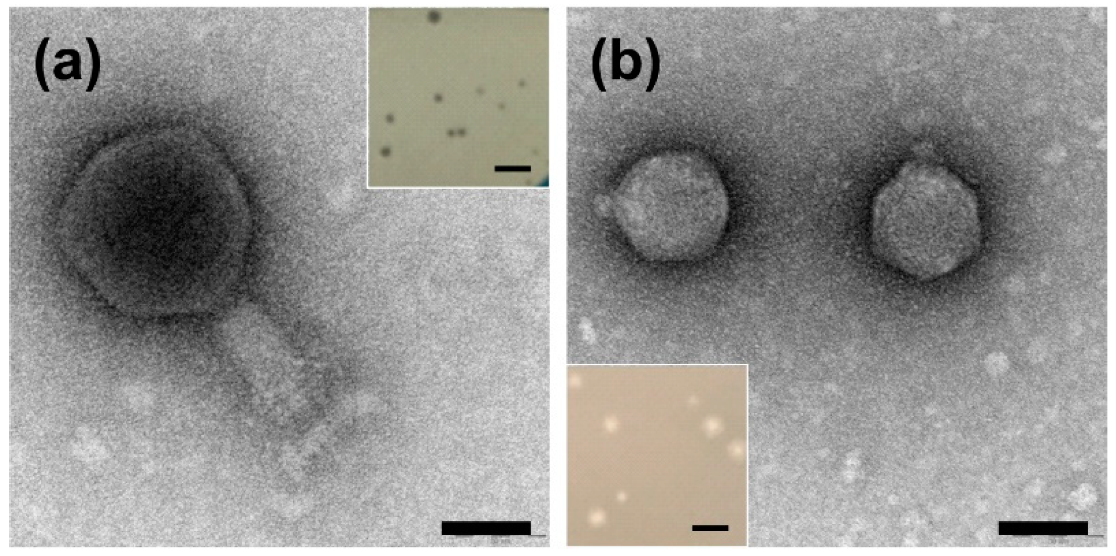
2.1. Identification and Characterization of the Plaque and Virion Morphologies of Ochrobactrum-Specific Phages

The phages that are able to infect Ochrobactrum sp. POC9 were isolated from a wastewater sample via the standard enrichment method [24]. After preparation of the first double layer agar plate presenting plaques, we noticed that two distinct plaque morphologies were present. Three rounds of phage propagation from a single plaque allowed us to obtain pure cultures of two different phages, initially named OC and OH, with clear plaques characterized by distinct morphologies (Figure 1). The plaques of the OC phage were smaller, albeit of irregular size in comparison to those from the OH virus. The OH phage created bigger plaques with an additional, very small (around 0.2–0.5 mm), halo zone.
Virion visualization and further morphology-based classification of the phages were carried out with the use of transmission electron microscopy (TEM). The TEM analysis of purified OC and OH phages revealed that they belong to the Myoviridae and Podoviridae families, respectively (Figure 1). Hence, according to the current naming conventions, the phages were named vB_OspM_OC and vB_OspP_OH, respectively [25]. A detailed characterization of the vB_OspM_OC and vB_OspP_OH virions’ morphologies is presented in Table 1.

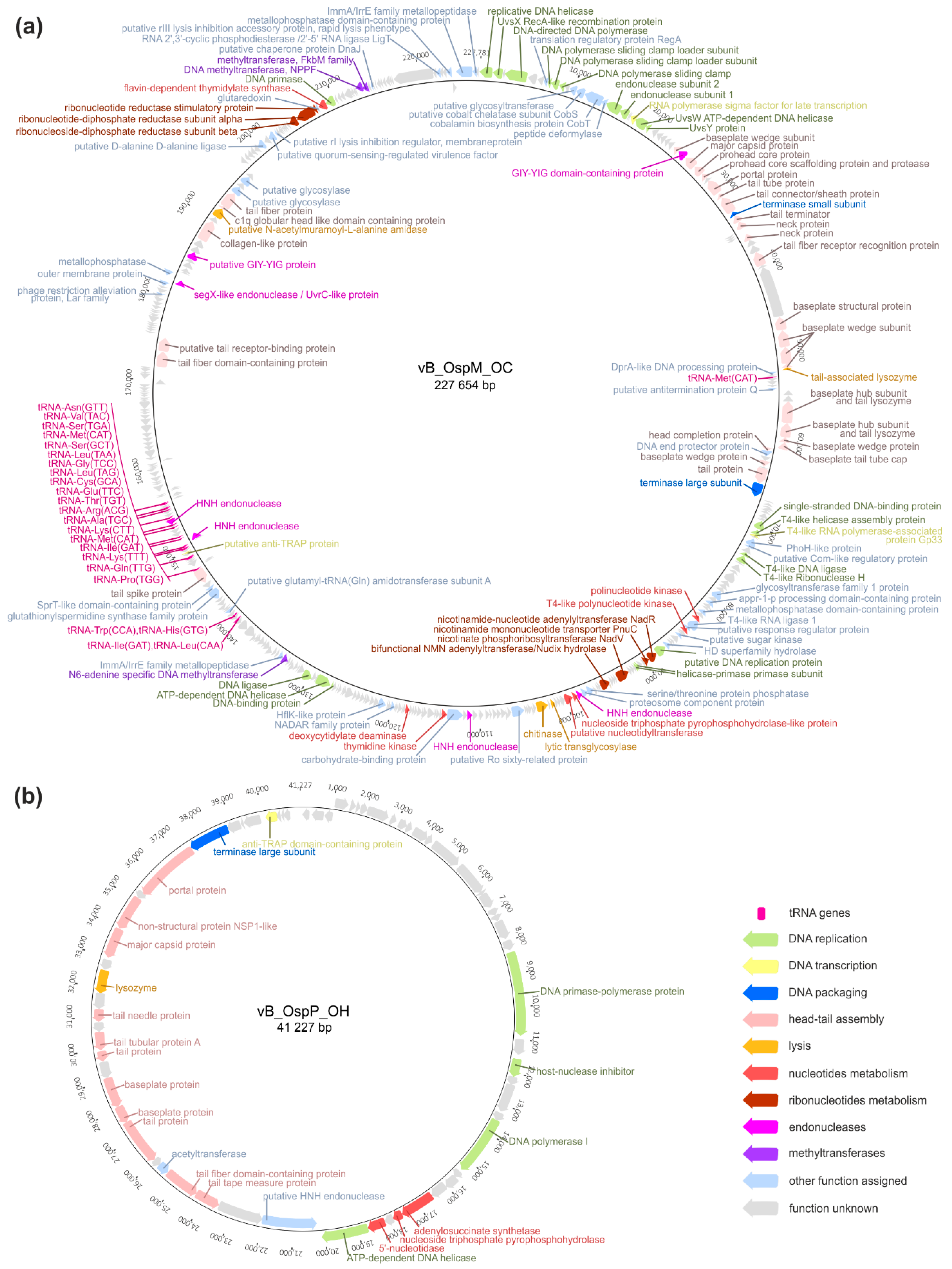
2.2. Genomic Analysis of the vB_OspM_OC and vB_OspP_OH Phages

The vB_OspM_OC phage has a dsDNA circularly permuted genome of 227,654 bp and has a %GC content of 37.7%, which is 18% lower than the %GC content of the host POC9 genome. Such low %GC content is typical for T-even phages, particularly Enterobacteria T4 phages, with 35.3% GC content, while the E. coli (its host) %GC content is around 50% [26]. The annotation of the vB_OspM_OC genome revealed the presence of 390 putative genes and 24 tRNA genes encoding 17 different tRNAs. However, despite careful manual annotation, the predicted functions could only be assigned to 113 (29%) of the genes.
As mentioned above, the vB_OspM_OC phage carries 24 tRNA genes which cover all amino acids except of aspartic acid (Asp), phenylalanine (Phe), selenocysteine (Sec) and tyrosine (Tyr). The analysis of the vB_OspM_OC codon usage shows that, when considering all proteins being expressed, the composition of tRNAs encoded by vB_OspM_OC could cover nearly 40% of codons used during the translation. Moreover, this set of tRNAs does not seem to be correlated with low %GC content of the vB_OspM_OC genome. Interestingly, it was found, that there are only 42 (20%) or 94 (44%) of jumbo phages carrying at least 10 or only one tRNA gene, respectively. Therefore, the presence of the numerous tRNA genes is not common in jumbo phages and, as we speculate, in the case of vB_OspM_OC it may have an impact on the efficacy of its infection and propagation within the host cell.
The vB_OspM_OC phage has a large genome; based on the NCBI Genome database as of October 19th 2019, there were 214 phages with genomes over 200 kb, and 137 phages had genomes bigger than vB_OspM_OC. The size of a phage genome above 200 kb is an informal threshold for classifying phages as giant (so called ‘jumbo’) phages. Moreover, the phages within that group are supposed to encode the proteins responsible for DNA replication, nucleotide metabolism, several proteins for host-cell lysis, and paralogous genes encoded across different gene clusters [27,28]. Since vB_OspM_OC possesses most of these features, which are described in detail below, it can be considered a novel representative of the jumbo phages (Figure 2a).
It was also indicated that vB_OspM_OC encodes proteins that show homology with the proteins encoded by the genomes of T4-like phages. Comparative analyses revealed that vB_OspM_OC shares its core genome with this group of viruses and can thus be conceived as a novel member of the group of T4-like phages (Supplementary Table S1).
An annotation of the vB_OspM_OC genome revealed the presence of at least 21 genes encoding proteins potentially involved in replication. For 15 of them, direct counterparts of the T4 phage were identified [26,29]. Among these genes are proteins that build the replisome, such as DNA polymerase (PhiOC_p004), DNA polymerase sliding clamp (PhiOC_p010), and its subunits (PhiOC_p008-9), as well as DNA helicase (PhiOC_p001), DNA primase (PhiOC_p384), and single-stranded DNA-binding protein (PhiOC_p084). Moreover, RNase H (PhiOC_p092) and DNA ligase (PhiOC_p091) are involved in the removal of the initiator RNA primers of Okazaki fragments, and the ligation of those primers during lagging-strand DNA synthesis have also been indicated [26,30]. Potentially, vB_OspM_OC’s modes of initiating replication are similar to those of the T4 phage and are origin- and recombination-dependent [26]. This origin-dependent replication may be initiated by the predicted DNA replication protein (PhiOC_p118), which shows sequence similarity to the putative DNA replication factor of Shewanella amazonensis (PDB acc. no. 3BOS_B; 99.45% probability using HHPred) and the DNA polymerase III subunit beta of Eschericia coli O157:H7 (PDB acc. no. 5X06_G; 99.42% probability). Within this protein, AAA+-type ATPase containing the peptidase M41 domain (KOG0731; 99.35% probability) was identified based on HHpred searches. However, the exact sites involved in the priming of vB_OspM_OC DNA synthesis were not determined. The latter type of replication may be conducted by a set of DNA recombination repair proteins, such as UvsWXY homologs (PhiOC_021, PhiOC_p002, and PhiOC_022), like in the T4 phage [26,29]. The resulting DNA concatemers are required to be resolved before or during packaging, which could be conducted by endonuclease encoded by the phiOC_p016-17 genes, which are homologs of the T4 genes, gp46 and gp47.
The vB_OspM_OC also encodes at least seven genes that are potentially involved in pyrimidine nucleotide metabolism. It is possible that the predicted deoxycytidylate deaminase (PhiOH_p179; EC 3.5.4.12) converts deoxycytidylic acid (dCMP) to deoxyuridylic acid (dUMP), which can be further converted into thymidylic acid (dTMP) via flavin-dependent thymidylate synthase (PhiOC_p383; EC 2.1.1.148). Moreover, the presence of thymidine kinase (PhiOC_p167; EC 2.7.1.21) possibly helps to catalyze the conversion of (deoxy)thymidine into dTMP. Moreover, a gene encoding the nucleoside triphosphate pyrophosphohydrolase-like protein was also identified (PhiOC_p139; EC 3.6.1.9). This protein may convert nucleoside triphosphate (dNTP) into a nucleotide (dNMP) and diphosphate (PPi). Moreover, genes encoding two polynucleotide kinases (PhiOC_p107 and PhiOC_p110) and a putative nucleotidyltransferase (PhiOC_p140) were also identified, which together take part in the nucleotide salvage pathway [30,31].
Another feature typical for jumbo phages is nicotinamide adenine dinucleotide (NAD+) metabolism. The vB_OspM_OC phage encodes at least seven proteins that are potentially involved in that process. The presence of bifunctional NMN adenylyltransferase (NAmPRTase)/Nudix hydrolase (NatV; PhiOC_p132; EC 2.4.2.12/EC 3.6.1.22) and nicotinate phosphoribosyltransferase (NadV; PhiOC_p128; EC 6.3.4.21) possibly allows the vB_OspM_OC phage to convert nicotamide to nicotamide D-ribonucleotide and nicotinate into nicotinate D-ribonucleotide, respectively. Both of these ribonucleotides can be further transformed into NAD+ or deamino-NAD+, respectively, via nicotinamide–nicleotide adenylyltransferase (NadR; PhiOC_p119; EC 2.7.7.1). The Nudix hydrolase domain of NatV may also help reverse the last reaction and transform deamino-NAD+ into a nicotamide D-ribonucleotide. The presence and activity of NatV and NatD encoded by the Vibrio phage KVP40 were recently shown to be sufficient for the NAD+ salvage pathway [32]. Moreover, it was shown that at least 169 different bacteriophages encode the NadV-like NAmPRTase. However, only 50 documented phages encode a bifunctional NatV-like enzyme, and all of these also contain a nadV gene in their genomes [30]. Amongst them, 21 phages have genomes with sizes above 200 kb, including the previously mentioned KVP40 phage. Therefore, the vB_OspM_OC phage could be another example of a giant phage extending the group of viruses with both NatV and NatD. However, their activity requires experimental validation during future studies. The vB_OspM_OC phage also encodes the predicted nicotinatinamide mononucleotide transporter PnuC (PhiOC_p120), which is involved in the NAD+ biosynthesis pathway. Furthermore, three other genes encode ribonucleotide-diphosphate reductase (rNDP; PhiOC_p379 and PhiOC_p380; EC 1.17.4.1) and ribonucleotide reductase (RNR; PhiOC_p381; EC 1.17.4.2), which possibly catalyze the formation of deoxyribonucleotides from ribonucleotides and can further be used in the synthesis of DNA [31].
The vB_OspM_OC virion is relatively large, and 33 out of 113 proteins with recognized functions are possibly involved in its formation. Eleven of these proteins were identified as baseplate and/or wedge proteins that may correspond to the firm and complex structure of the baseplate shown on the micrographs (Figure 1a). Within four of these structural proteins, the domains involved in host cell lysis were also identified, including the tail-associated lysozymes in PhiOC_p064 and PhiOC_p065, the glycoside hydrolase domain in PhiOC_p326 (annotated as a putative tail receptor-binding protein), and endo-N-acetylneuraminidase in a tail fiber protein (PhiOC_p358). The repertoire of lysing enzymes of vB_OspM_OC is extended by the putative N-acetylmuramoyl-L-alanine amidase (PhiOC_p356), lytic transglycosylase (PhiOC_p145), and chitinase (PhiOC_p147), of which the latter two were not located within structural gene clusters.
Interestingly, within the vB_OspM_OC genome, a predicted N6-adenine (m6A) DNA methyltransferase gene (PhiOC_p213) was found. As shown previously, viruses infecting Alphaproteobacteria often have DNA methyltransferases (MTases) that exhibit GANTC (methylated nucleotide is underlined) specificity, just as the host-encoded cell cycle regulated MTase CcrM [33,34,35]. To determine whether GANTC sequences are substrates for PhiOC_p213, we digested the pET_PhiOC_p213 plasmid DNA isolated from IPTG-induced and non-induced E. coli cultures with the HinfI restriction enzyme (specificity GANTC, inhibited by m6A methylation). The DNA isolated from the induced cultures was resistant to cleavage by HinfI but sensitive to cleavage with the various enzymes (e.g., Csp6I and Hin1II) used as controls. The pET_PhiOC_p213 DNA isolated from the non-induced cultures was susceptible to all restriction enzymes used, including HinfI.
DNA MTases, which are related to PhiOC_p213, are encoded within several other phages, including the Silicibacter phage DSS3-P1 (GenBank acc. no. AET42337.1) and the Loktanella phage pCB2051-A (GenBank acc. no. AGH31487.1), with a ~45% protein sequence identity. It is worth mentioning that, in contrast to previously characterized phage-encoded MTases with GANTC specificity (such as PhiLM21_p027 [35], phi2LM21_p21, and phi3LM21_p21 [33], which do not share sequence similarities with the host-encoded CcrM enzymes), PhiOC_p213 exhibits 30.49% identity with CcrM of Ochrobactrum sp. POC9 (GenBank acc. no. PWU77214.1) and shows 29% identity with for example CcrM of O. pseudogrignonense K8 (GenBank acc. no. ANG95423.1), for which methylation specificity was shown by a PacBio analysis (as found in REBASE: http://rebase.neb.com/cgi-bin/pacbioget?19606). This sequence conservation is limited to the N-terminal methyltransferase domain of CcrM, since a C-terminal nonspecific DNA-binding domain (~80 residue segment) is present only in bacterial CcrM MTases (Supplementary Figure S1).
The co-occurrence of phage- and host-encoded enzymes with the same sequence specificity mimicking the host strategies in DNA methylation by viruses is a known phenomenon discovered not only in Alphaproteobacteria [33] but also in Gammaproteobacteria. The dam-like activity (the methylation of GATC to Gm6ATC in Gammaproteobacteria) of phage MTases has been shown for, e.g., P1 [36], VT-2 [37] and the T-even coliphages [38], but only DamT2 and DamT4 are homologs of the cellular (E. coli in this case) Dam enzyme [39], like the above mentioned PhiOC_p213 and CcrM of Ochrobactrum sp. POC9. The potential functions of the maintenance of both, the specificity of the T4-like phage and the bacterial CcrM (in Alphaproteobacteria) and Dam MTases (in Gammaproteobacteria), as well as the conservation of the amino acid sequences of these proteins in virus–host biology, remain to be elucidated.
The second isolated phage, vB_OspP_OH, has a dsDNA genome, with a size of 41,227 bp. In contrast to vB_OspM_OC, its %GC content is 55.2%, which is nearly the same as the content of the host strain. Within its genome, 65 potential protein encoding genes were identified, of which 23 (35.4%) had their functions predicted. The vB_OspP_OH phage seems to be unique, showing only limited similarities with the viruses recovered from the environmental samples collected by Mizuno and coworkers, (i.e., the uncultured Mediterranean phage uvMED [40]).
Amongst the predicted proteins of vB_OspP_OH, there are three involved in phage genome replication, including a potential bifunctional DNA primase-polymerase protein (PhiOH_p22), DNA polymerase I (PhiOH_p28), and DNA helicase (PhiOH_p36) (Figure 2b). The sensitive searches with HHpred allowed identification within the PhiOH_p22 protein the primase-polymerase domain (cd04859; 99.4% confidence) in its N terminal part (positions 39–217) and the phage- or plasmid-associated DNA primase domain (100% confidence) on its C terminus (positions 452–813).
Within the genome of vB_OspP_OH, three genes encoding proteins potentially involved in nucleotide metabolism were found (Figure 2b). These proteins are adenylosuccinate synthase (PhiOH_p32, EC 6.3.4.4), adenosine triphosphate pyrophosphatase (PhiOH_p33, EC 3.6.1.8), and 5′-nucleotidase (PhiOH_p35, EC 3.1.3.5). Their presence potentially allows vB_OspP_OH to transform nucleotides into nucleosides, resulting in the salvage of (pyro)phosphate groups for further reuse in dNTP synthesis during phage genome replication [31].
The virion structure of vB_OspP_OH allows its classification as a podovirus. Within the vB_OspP_OH genome, 10 genes encoding structural proteins were identified (Figure 2b). These include: (i) a portal protein (PhiOH_p57), (ii) a scaffolding protein (PhiOH_p55), (iii) a major capsid protein (PhiOH_p54), (iv) a tail tubular protein A (phiOH_p48), (v) a minor tail protein (phiOH_p47), (vi) a tail fiber protein (PhiOH_p45), (vii) a tail protein (PhiOH_p43), (viii) a tail fiber protein (PhiOH_p42), (ix) a tail-tape measure protein (PhiOH_p42), and (x) a tail tape measure protein (PhiOH_p39). Within the last protein, the peptidoglycan hydrolase (EtgA) domain was identified using HHpred. This protein could be responsible for the presence of the plaque halo effect observed for this phage (Figure 1b).
The assumed endolysin of vB_OspP_OH (PhiOH_p52) shows the highest similarity to the Ochrobactrum phage POI1126 secretion activator protein (APU92960.1, 43.3% sequence identity). An analysis of the domains of PhiOH_p52 and APU92960.1 revealed the presence of the glycosyl hydrolase 108 domain (pfam05838, e-value below 1e-30), which acts as a lysozyme [41].

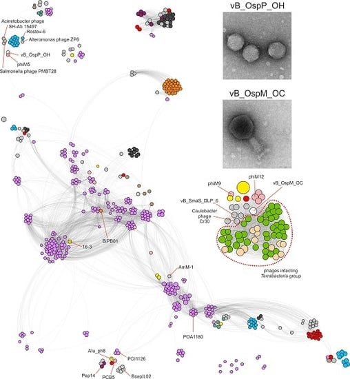
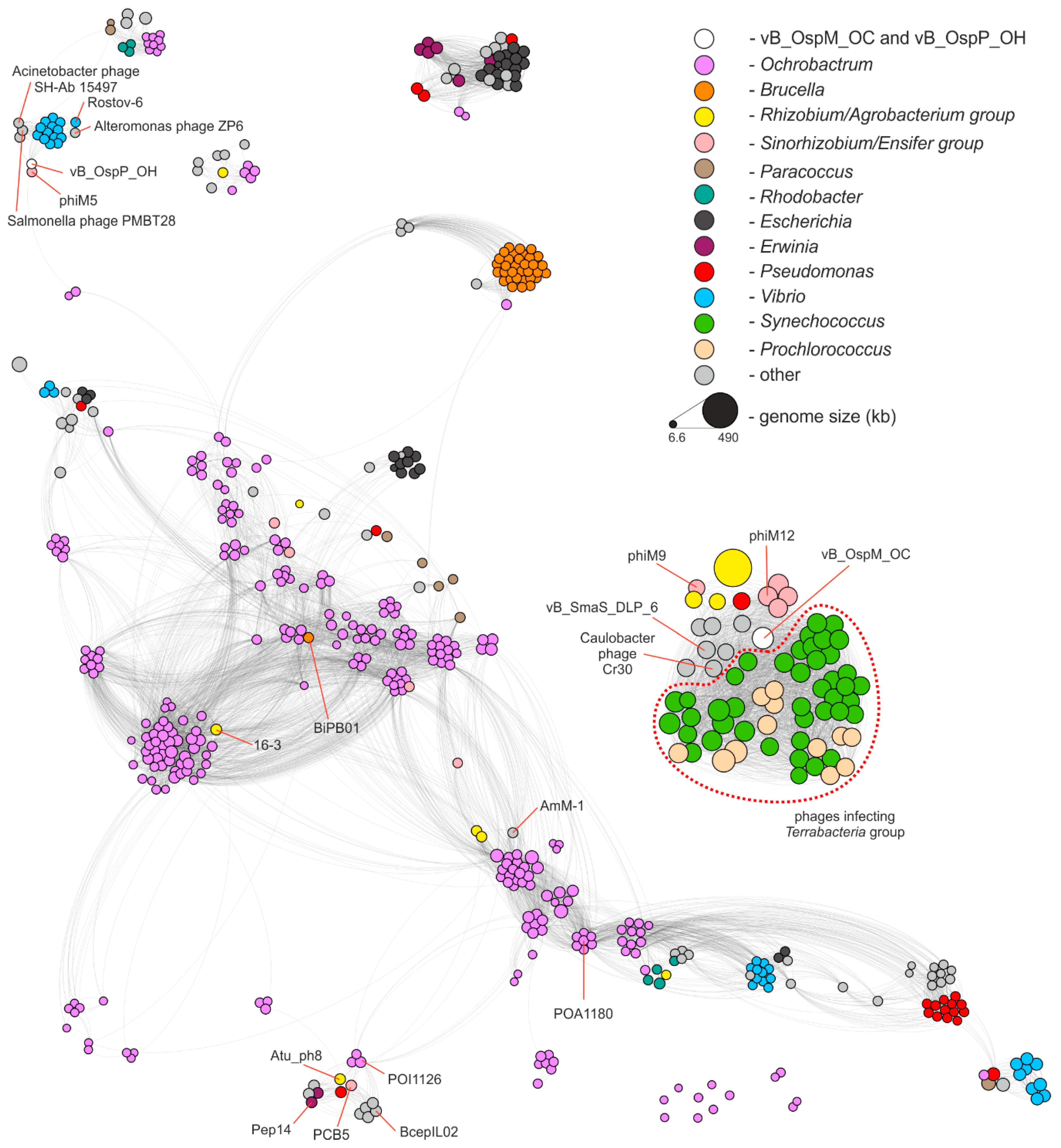
2.3. Comparative Genomics of Ochrobactrum Phages

Both phages characterized in this study are distinct from the viruses identified so far, with a protein sequence identity not exceeding 70% for vB_OspM_OC (PhiOC_p379) or 59% for vB_OspP_OH (PhiOH_p53), when at least a 75% sequence coverage was considered. To perform a complex analysis of the diversity of Ochrobactrum phages, the PhiSpy v3.4 tool (trained with the complete Ochrobactrum spp. genomes and a draft genome of Ochrobactrum sp. POC9, in which the prophage regions were indicated manually) was applied. This enabled us to significantly extend the knowledge in this field since, in the previous works by Jackel et al. [21,42,43] a total of 66 Ochrobactrum (pro)phages were indicated, while in our analysis, 277 prophages were identified within 104 out of the 113 analyzed genomes (Supplementary Table S2). Moreover, by applying the vConTACT v2 tool [44], we compared vB_OspM_OC, vB_OspP_OH, and all identified Ochrobactrum prophages with the ProcaryoticViralRefSeq85-Merged database resources and the 12,817 bacteriophages present in the NCBI Genome database (as of 10 July 2019). This resulted in the identification of 294 phages similar to Ochrobactrum spp. (pro)phages. A comparison of all those (pro)phages is presented in the form of a similarity network composed of 575 (pro)phages in total (Figure 3).
The distribution of (pro)phages (represented by nodes) in the network allows for more precise insight into Ochrobactrum (pro)phages diversity. It was shown that 198 (70.5%) Ochrobactrum (pro)phages shared similarity to the phages infecting other Proteobacteria (223 (pro)phages in the network), which is representatives of the Terrabacteria group (54) or of phages recovered from the metagenomic samples for which hosts were not assigned (17). Amongst the Proteobacteria (pro)phages grouped together with Ochrobactrum phages, 89 viruses infected Alphaproteobacteria, 15 Betaproteobacteria, and 115 Gammaproteobacteria. As for the Terrabacteria group, these phages were recognized as infecting agents only for cyanobacteria, representing the Synechoccoccus (42 viruses) and Prochlorococcus (12) genera. It was also shown that 75 (26.7%) Ochrobactrum viruses shared homologous proteins exclusively with other Ochrobactrum prophages, and 8 viruses formed orphan nodes, which exemplifies the uniqueness of these (pro)phages.
This global analysis also allowed us to determine the relatives of the vB_OspM_OC and vB_OspP_OH phages. vB_OspM_OC is related to large, mostly T4-like, phages. Its highest similarity was observed to be with myoviruses (i.e., the Sinorhizobium phages phiN3, phiM7, phiM12, and phiM19), Stenotrophomonas phage vB_SmaS_DLP_6, and Caulobacter phage Cr30 (GenBank acc. nos.: NC_028945.1, NC_041929.1, NC_027204.1, KR052481.1, KU682439.2, and NC_025422.1, respectively) [45,46]. Interestingly, vB_OspM_OC seems to also be related to the phages infecting the Terrabacteria group of bacteria. This may suggest their common origin.
vB_OspP_OH created a separate cluster with another podovirus, the genomically highly mosaic Sinorhizobium meliloti phage phiM5 [45]. Direct comparison of these phages (Supplementary Figure S2) indicated that both viruses encode homologous structural proteins. However, other functionally equivalent proteins, like bifunctional DNA primase/polymerase, were also found. Interestingly, vB_OspP_OH also showed similarities to the group of Vibrio podoviruses (e.g., phiVC8, Rostov-6) and the Alteromonas phage ZP6, as well as two siphoviruses the Salmonella phage PMBT28 and the Acinetobacter phage SH-Ab 15497 (GenBank acc. nos.: NC_027118.1, MH105773.1, MK203850.1, MG641885.1, and MG674163.1, respectively) [47,48].

2.4. Functional Characterization of vB_OspM_OC and vB_OspP_OH

2.4.1. Host Range Analysis

Various bacterial strains (29 in total), including the reference ones (Agrobacterium tumefaciens LBA288, Escherichia coli C600, E. coli DH5α, E. coli K-12, E. coli BR825, Paracoccus alcaliphilus JCM 7364, Paracoccus aminophilus JCM 7686, Pseudomonas aeruginosa PAO1161, and Variovorax paradoxus EPS), the isolates from wastewater treatment plants (Bacillus sp. LPOC3, Bacillus sp. LPSUB4, Brevundimonas sp. POC21, Klebsiella sp. POC16, Lysinibacillus sp. LPSUB15, Rummelibacillus sp. POC4, and Stenotrophomonas sp. POC10), and other environmental isolates (Achromobacter sp. LM16R, Brevundimonas sp. LM18, Brevundimonas sp. LPMIX5, Ensifer sp. M14, Janthinobacterium sp. M1_6, Janthinobacterium sp. M1_18, Janthinobacterium sp. M2_12, Janthinobacterium sp. W1_1, Ochrobactrum sp. LM19, Pseudomonas sp. LM7, Psychrobacter sp. DAB_AL32B, Sinorhizobium sp. LM21, and Sphingomonas sp. WLOD2_3), were used in the host range assay analysis. The host range and lytic activity of the vB_OspM_OC and vB_OspP_OH phages were assessed using spot test assays. Even though various bacteria representing the 18 genera were used in the assay, we observed clear zones only on the plates with Ochrobactrum sp. POC9 and Ochrobactrum sp. LM19. The comparison of plaques obtained for LM19 and POC9 showed no differences in the number of plaques (when performing a spot test assay using a series of various concentrations of phages) and in their morphology. Both analyzed phages produced plaques on both Ochrobactrum species. This suggests that both phages are specific to Ochrobactrum spp.

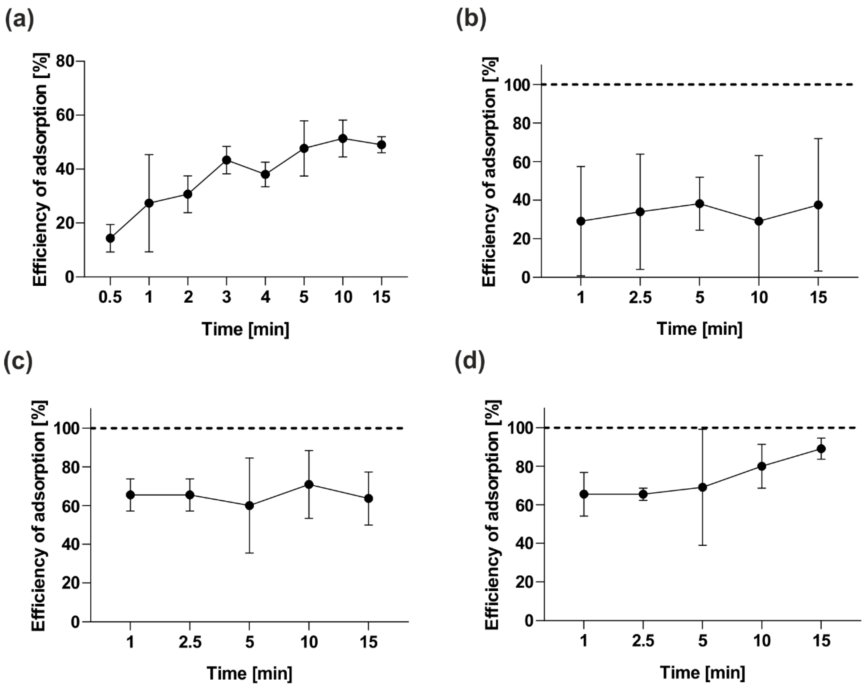
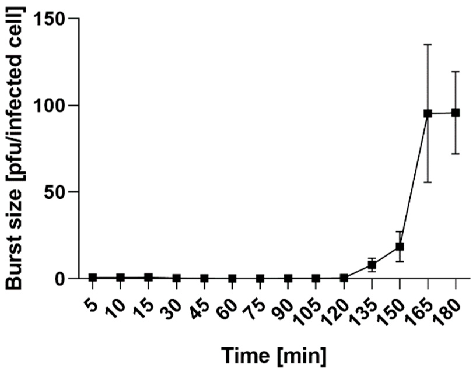
2.4.2. Adsorption Assay and One-Step Growth Curve

The efficiency of phage development depends on two main steps: the phage’s adsorption to the bacteria and its development inside the host cell. Adsorption assays of vB_OspM_OC and vB_OspP_OH were performed to analyze the kinetics of their adsorption to Ochrobactrum sp. POC9. First, we carried out an analysis with the use of bacteria after overnight culturing. The results of that experiment (Figure 4a,b) revealed that 25% of both phages attached to the host cells within 1 min. However, after 5 min, an increase of the adsorbed phages to around 50% was observed only in the case of vB_OspP_OH. The attachment efficiency of vB_OspM_OC was stable, and the amount was around 25% during the whole experiment. Since vB_OspM_OC adsorbed inefficiently to the bacteria from the overnight culture, the adsorption assay for this phage was repeated using actively growing bacteria (Figure 4c,d). We observed that 65% of the vB_OspM_OC attached to the host cells within 1 min in both experimental variants (with initial OD600 = 0.2 and OD600 = 0.4). Moreover, around 90% of the vB_OspM_OC phages were adsorbed into the host cells in 15 min, but only when a bacterial culture of OD600 = 0.4 was used. Otherwise, the attachment efficiency of vB_OspM_OC was stable, and the amount was around 65% during the whole experiment.
The phage growth parameters were determined with the use of a one-step growth curve assay. The latent period of vB_OspP_OH is 135 ± 5 min, and the burst size of one lytic cycle is approximately 95 ± 10 pfu per infected cell (Figure 5). The vB_OspM_OC phage was more difficult to analyze, and, despite several repeats of the one-step growth experiment, we were unable to obtain conclusive and replicable results (Supplementary Figure S3). Therefore, the latent period and burst size were not determined.
We speculate, that this ‘problematic’ propagation of the vB_OspM_OC phage could be explained by its complex development strategies. As determined previously, the vB_OspM_OC belongs to the T4-like group of viruses, and its genome may contain genes encoding proteins that enable diverse modes of phage development, such as the T4 phage. It was shown that T4 can develop by applying alternative mechanisms, such as lysis inhibition (LIN) [49] or pseudolysogeny [50]. It is possible that various (possibly not yet described) development strategies may also be characteristic for vB_OspM_OC, especially since the functions of most proteins that it encodes remain still unknown. An alternative, speculative explanation could be the activity of a phage infection defensive mechanism in Ochrobactrum sp. POC9. The manual annotation of the POC9 prophage regions identified an abortive infection protein (GenBank acc. no. WP_109988358.1) encoded within vB_OspX_pp134 (Supplementary Table S2). This kind of defensive mechanism allows successful phage entry but interrupts one of the essential processes in the phage lifecycle, which prevents the release of functional virions. This induces infected bacterial cell death and protects the bacterial population from further infections [51,52].

2.4.3. Killing Assay

To assess the killing activity of vB_OspM_OC and vB_OspP_OH on Ochrobactrum sp. POC9, bacterial growth was monitored by measuring the optical densities of OD600 after phage infection. To evaluate the efficiency of phage development, various concentrations of phages were used for infection (from 102 to 1010 pfu/mL, which corresponds to MOI approximately from 0.0000001 to 10) (Supplementary Figure S4).
Both phages were able to cause lysis of the liquid bacterial culture, especially when the phage lysates with the highest concentrations (108–1010 pfu/mL) were used (Supplementary Figure S4). In these cases, the optical density of the cultures decreased at approximately 100–400 min post-infection. When lower concentrations of phages were used (≤107 pfu/mL), a decrease in optical density was observed (except of 102 and 103 pfu/mL for vB_OspM_OC), albeit at a longer time after infection (Supplementary Figure S4).

2.4.4. Stability of the vB_OspM_OC and vB_OspP_OH Phages

Storage, transfer, and downstream experiments with the phages require information about their stability. vB_OspM_OC seems to be temperature-sensitive and exhibited a decrease of its phage titer to approximately 50% after incubation at 43 °C for 15 min (Figure 6a). After incubation for an hour at all chosen temperatures, there was almost a complete reduction in the vB_OspM_OC titer (Figure 6a). In contrast, vB_OspP_OH seems to be more temperature-resistant (Figure 6d). It was observed that incubation of vB_OspP_OH at 43 °C for 15 min does not influence the phage titer. However, a longer incubation at 43 °C results in the inactivation of approximately 85% of phages. Only around 18% of vB_OspP_OH particles were able to survive at 60 °C (Figure 6d).
Both phages appeared to be stable at pH values from 7 to 8 (Figure 6b,e). However, vB_OspM_OC showed higher stability at a higher pH (pH from 9 to 10) (Figure 6b), whereas vB_OspP_OH showed higher stability at lower pH values (pH from 5 to 6) (Figure 6e).
An analysis of phage particle stability after UV light treatment showed that both phages are UV-sensitive (Figure 6c,f). However, vB_OspP_OH showed higher resistance in comparison to vB_OspM_OC (Figure 6f). The plaques of vB_OspP_OH was observed even after 30 min of exposure, while vB_OspM_OC was completely inactivated (no plaques were observed) after 20 min of UV exposition (Figure 6c).

3. Materials and Methods

3.1. Bacterial Strains, Plasmids and Culture Conditions

The following bacterial strains were used: Achromobacter sp. LM16R [53], Agrobacterium tumefaciens LBA288 [54], Bacillus sp. LPOC3 (laboratory collection), Bacillus sp. LPSUB4 (laboratory collection), Brevundimonas sp. LM18 [53], Brevundimonas sp. LPMIX5 [23], Brevundimonas sp. POC21 (laboratory collection), Ensifer sp. M14 [55], E. coli C600 (laboratory collection), E. coli DH5α [56], E. coli K-12 (laboratory collection), Janthinobacterium sp. M1_6 (laboratory collection), Janthinobacterium sp. M1_18 (laboratory collection), Janthinobacterium sp. M2_12 (laboratory collection), Janthinobacterium sp. W1_1 (laboratory collection), Klebsiella sp. POC16 (laboratory collection), Lysinibacillus sp. LPSUB15 (laboratory collection), Ochrobactrum sp. LM19 [53], Ochrobactrum sp. POC9 [11], Paracoccus alcaliphilus JCM 7364 [57], Paracoccus aminophilus JCM 7686 [58], Pseudomonas aeruginosa PAO1161 [59], Pseudomonas sp. LM7 [60], Psychrobacter sp. DAB_AL32B [61], Rummelibacillus sp. POC4 [23], Sinorhizobium sp. LM21 [53], Sphingomonas sp. WLOD2_3 (laboratory collection), Stenotrophomonas sp. POC10 (laboratory collection), and Variovorax paradoxus EPS [62]. These strains were grown on the LB medium at 37 °C (E. coli) and 30 °C (all other strains). The medium was solidified by the addition of 1.5% (w/v) agar. Where necessary, the medium was supplemented with X-gal, IPTG, and kanamycin (50 μg/mL). Plasmid pET30a was used for cloning of the DNA methyltransferase gene.

3.2. DNA Manipulations and Introduction of Plasmid DNA into Bacterial Cells

Standard DNA manipulations were performed as described by Sambrook and Russell (2001) [63]. Plasmids constructed in this study were introduced into E. coli ER2566 by chemical transformation [64].

3.3. Cloning, Overexpression, Purification, and Testing of Putative DNA MTase Activities

The predicted DNA MTase gene identified within the vB_OspM_OC phage was amplified by PCR using a Thermo Scientific™ Phusion™ High-Fidelity DNA Polymerase (Thermo Fisher Scientific, Inc., Waltham, MA, USA) and following the primer pairs OcFnde (5′-GTTGTTCATATGGAAAATTTGACACTTTTTAATGGTAAC-3′) and OcRxho (5′-GTTGTTAATAAAAATAGGCGTTAGCTCACC-3′). DNA amplification was performed using a Mastercycler (Eppendorf, Hamburg, Germany). Each thermocycle started with an initial denaturation at 95 °C for 1 min followed by 30 cycles of denaturation at 98 °C for 10 s, annealing at 66 °C for 20 s, extension at 72 °C for 30 s, and finished with a final extension at 72 °C for 2 min. The PCR product (after purification) was digested with NdeI and XhoI and ligated with a pET30a vector cut with the same enzymes. The recombinant proteins were expressed in the E. coli ER2566. The protein expression and restriction enzyme digestion protection assays for revealing the sequence specificity of the tested MTase were performed as previously described [33,65].

3.4. Bacteriophages Isolation

Both phages, vB_OspP_OH and vB_OspM_OC, were isolated from a wastewater sample collected from a sewage treatment plant in Wołomin (Wołomin, Masovian Voivodship, Poland) on 25 August 2018. Phage isolation was performed according to the previously described procedure [24]. After the isolation, three rounds of propagation from each individual phage plaque were carried out to prepare a lysate of the pure phage strain.

3.5. Transmission Electron Microscopy (TEM)

Transmission electron microscopic images of the phage virions were obtained using a TEM LIBRA 120 (Zeiss, Oberkochen, Germany) microscope (HT = 120 kV, LaB6 cathode). Samples were prepared by applying a 10 μL droplet of phage suspension to thin, carbon-coated copper grids (400 mesh), followed by immersing the grids in 1% uranyl acetate for contrasting. Then, the samples were left to dry at room temperature. The visualization of the phages was performed at the Laboratory of Theory and Applications of Electrodes, Faculty of Chemistry, University of Warsaw, Poland.

3.6. Host Range Analysis

To determine the host range of the vB_OspM_OC and vB_OspP_OH phages, 2.5 μL of each phage lysate was spotted on double layer agar plates with various tested bacteria in the upper layer. The appearance of the plaques was observed after an overnight incubation of the plates at 30 °C.

3.7. Adsorption Kinetics Assay of vB_OspM_OC and vB_OspP_OH

The adsorption assay was conducted as described previously [66]. Briefly, 1 mL of an overnight or logarithmic Ochrobactrum sp. POC9 culture was spiked with 10 µL of the phage suspension (around 109 pfu/mL). The mixture was incubated without shaking at 37 °C. After 1, 2.5, 5, 10, and 15 min, 100 µL aliquots were withdrawn and centrifuged (5000× g, 1 min, RT) to deposit the phage-adsorbed cells as sediment. The titer of the remaining free phages was determined by supernatant titration on double-layer agar plates. The initial number of phages (100% of the phages used) was determined by adding an appropriate volume of the vB_OspM_OC or vB_OspP_OH phage lysate to a medium without bacteria, followed by titration. The number of adsorbed phages was determined via the decrease in PFUs in the supernatant relative to the initial number of phages. The data were obtained from three independent experiments.

3.8. Killing Assay

For the host culture collapse studies, liquid Ochrobactrum sp. POC9 cultures were infected with vB_OspM_OC or vB_OspP_OH phage lysates. Briefly, an overnight culture of bacteria was refreshed in an LB medium and allowed to grow until reaching an OD600 of 0.25. Then, the culture was infected with the vB_OspM_OC or vB_OspP_OH phage lysate at various concentrations ranging from 102 to 1010 pfu/mL. The OD600 measurement was made every 15 min for 24 h. Measurements were carried out with the use of an Infinite 200 PRO multimode plate reader and microplate reader software v 1.8 2010 (Tecan, Männedorf, Switzerland). The data were obtained from three independent experiments.

3.9. One-Step Growth Curve

For a one-step growth analysis, 25 mL of LB medium was inoculated with an overnight culture of Ochrobactrum sp. POC9 (50:1, v/v). The culture was grown at 37 °C with shaking (150 rpm) until OD600 = 0.2 and then infected with the vB_OspP_OH phage at MOI of 0.1 and incubated without shaking at 37 °C for 5 min. Next, 1 mL of the infected bacteria was centrifuged at 5000 rpm for 1 min at 37 °C. The resulting pellet was washed and re-suspended in 1 mL of fresh LB broth. Then, 25 µL of infected cells was transferred to a fresh LB broth and incubated at 37 °C with shaking (150 rpm). At appropriate time points, the PFUs (300 µL of samples treated with 300 µL of chloroform) were calculated. The samples for estimation of the number of infection centers (ICs) and the burst size presented as pfu/IC were analyzed as described previously [49]. The data were obtained from three independent experiments.

3.10. Bacteriophage Stability at Various Values of pH and Temperature

For pH stability testing, 10 µL of the vB_OspM_OC or vB_OspP_OH phage lysate (1 × 10⁹ pfu/mL) was mixed with 990 µL LB broth in a series of tubes, each with a different pH (adjusted using NaOH or HCl), and incubated for 10, 30, or 60 min at room temperature. For temperature stability testing, 10 µL of the phage lysate (1 × 10⁹ pfu/mL) was mixed with 990 µL LB broth in a series of tubes, and the samples were incubated at 43, 60, or 80 °C for 15, 30, or 60 min. At each of these time-points, the number of phages (plaque forming units, PFUs) was calculated. The control phage samples were incubated at pH 7 and room temperature (22 °C), respectively. The data were obtained from three independent experiments.

3.11. Bacteriophage Resistance to UV Light

For UV resistance testing, 10 µL of the vB_OspM_OC or vB_OspP_OH phage lysate (1 × 10⁹ pfu/mL) was mixed with 990 µL of LB broth. The mixture was then spotted onto a polystyrene Petri dish and exposed to UV-C light (UVC lamp, Philips TUV-30-W-245 nm Lamp, type No. 57413-P/40) from a distance of 50 cm for 1, 2.5, 5, 10, 15, 25, and 30 min. A control sample was incubated on a laboratory bench at room temperature. The data were obtained from three independent experiments.

3.12. DNA Sequencing

DNA sequencing was performed using an Illumina MiSeq instrument (Illumina, San Diego, CA, USA) in the paired-end mode using a v3 chemistry kit (Illumina). The obtained sequence reads were filtered for quality using fastp v0.19.5 with a window size of 10 bps moving from 5′ to 3′ and removing bps with a quality lower than 15, polyX at the 3′ end, and the reads of lengths lower than 150 bps [67]. Afterwards, the reads were assembled using SPAdes v3.11.1 with default parameters [68]. The presence of redundant ends in the assembled contigs was verified by remapping the reads against the assembled genomes using bwa mem v0.7.17-r1188, processed afterwards with samtools v1.7 and viewed in the Integrative Genome Viewer v2.6.2 [69,70,71]. Circulation of the genomes was validated by capillary sequencing of the PCR products using an ABI3730xl DNA Analyser (Applied Biosystems, Waltham, MA, USA).

3.13. Detection of Prophages in Bacterial Genomes and Classification of Phages

On October 19th 2019, 125 genomes of Ochrobactrum spp. were retrieved from the National Center for Biotechnology Information (NCBI) genome browser (Supplementary Table S2). Twelve genomes were removed from the analysis due to the lack of annotations. From among the remaining 113, six complete genomes (O. anthropi strains ATTC 49188, OAB and T16R-87, O. pseudogrigronense K8, O. pituitosum AA2, and O. quorumnocens A44) and a draft genome of Ochrobactrum sp. POC9 were manually inspected for the presence of the prophage regions (Supplementary Table S2). These were indicated and provided as additional training sets for PhiSpy v3.4 [42,43]. Then, retrained PhiSpy was used for the screening of prophage regions within the remaining 106 draft genomes of Ochrobactrum spp. The prophage was distinguished when at least 20 phage-like genes were found in the particular DNA region. Identification of the prophages was additionally validated by searching the pVOG database HMM profiles against the extracted regions using hmmsearch v3.1b2 [72] and applying cut-offs for sequence e-value—1e-10 and domain e-value—1e-5, followed by manual inspection [72,73].
The taxonomic assignment of all (pro)phages was conducted based on the results of vConTACT v2 with the use of the ProcaryoticViralRefSeq85-Merged database applying DIAMOND v0.9.24 for protein clustering and ClusterOne v1.0 for protein and viral cluster analyses [44,74,75].

3.14. Genome Annotation

The manual annotation of the analyzed phage genomes was conducted using Artemis software [76]. First, possible genes were indicated using PHANOTATE [77]. The annotation was then based on homology searches performed with BLAST programs against the NR database, including domain searches with CD-Search and the PRIAM database [78,79,80]. Moreover, the determination of potential protein functions was also based on the searches performed on the HHpred server using HHpred or HMMER tools against the PDB_mmCIF70_11_Oct, SCOPe70_2.07, COG_KOG_v.1.0, and NCBI_Conserved_Domains (CD)_v3.16 or the nr50_1_Oct databases [81]. Putative tRNA genes were identified with the tRNAScan-SE v2.0 and ARAGORN programs [82,83].

3.15. Comparative Genomics

Genomes of the isolated phages were compared with prophage regions indicated in the Ochrobactrum spp. genomes and the phage genomes from NCBI genome browser (accessed on 10 July 2019) using protein-based similarity network constructed with vConTACT v2. During the program run, ProkaryoticViralRefSeq85-Merged was used as the reference database. The phages present in that database were removed from the set of genomes downloaded from the NCBI to avoid redundancy. In total, 10629 phage genomes were analyzed using the same settings for vConTACT as described in Section 3.11. The resulting network was filtered using a Python script and only the nodes reflecting the phages connected to (pro)phages infecting Ochrobactrum spp. and edges reflecting the similarity between the two phages connecting those nodes were retained. The resulting network was visualized in Gephi v0.92 using ForceAtlas 2 and Noverlap layouts to arrange the nodes in a two-dimensional space [84,85].

3.16. Nucleotide Sequence Accession Numbers

The nucleotide sequences of the vB_OspM_OC and vB_OspP_OH phages have been deposited in the GenBank (NCBI) database with the accession numbers MT028491 and MT028492, respectively.

4. Conclusions

In this study, the biological and genomic properties of two novel phages infecting Ochrobactrum sp. POC9, the strain enhancing the reduction of organic biomass and biogas production in WWTPs, was characterized. The identified phages, vB_OspM_OC (myovirus) and vB_OspP_OH (podovirus), are the first (to the best of our knowledge) virulent phages observed to infect Ochrobactrum spp. so far. The vB_OspM_OC virus was recognized as a representative of giant (jumbo) phages. Moreover, vB_OspM_OC encodes a CcrM-like DNA methyltransferase, which constitutes another example of the molecular mimicry between the phage-encoded DNA methylases and host-specific regulatory MTase in Alphaproteobacteria.
In this study, a thorough identification of the prophages in Ochrobactrum spp. genomes combined with a complex comparative genomic analysis was also performed. This analysis discovered 277 prophages within 104 out of 113 analyzed genomes of Ochrobactrum spp. Moreover, by applying similarity networking, it was shown that that nearly 30% of Ochrobactrum viruses shared homologies exclusively with other Ochrobactrum prophages or formed orphan nodes, which exemplifies the uniqueness of this (pro)phages. This global analysis also revealed the relatives of the vB_OspM_OC and vB_OspP_OH phages. It was shown that vB_OspM_OC is related to giant T4-like phages, while vB_OspP_OH created a separate sub-cluster with another podovirus (Sinorhizobium meliloti phage phiM5) and showed some similarities to the Vibrio and Alteromonas podoviruses, as well as the Salmonella and Acinetobacter siphoviruses.
Moreover, in this study, bacteriophages were recognized as another factor besides chemical (e.g., metal ions) and physical (e.g., pH or temperature) agents that influence bacterial cultures applied to environmental biotechnologies. The presence of phages may be a critical, or at least important, factor interfering in the overall effectiveness of bioaugmentation and thus the success of the whole biotechnological process.

Supplementary Materials

Supplementary materials can be found at https://0-www-mdpi-com.brum.beds.ac.uk/1422-0067/21/6/2096/s1. Figure S1: Multisequence alignment of CcrM and CcrM-like DNA methyltransferases. Figure S2: Genomic comparison of Sinorhizobium phage phiM5 and vB_OspP_OH. Figure S3: One-step growth curves of the vB_OspM_OC phage. Figure S4: Bacterial culture collapse as the effect of phage infection. Table S1: The comparison of vB_OspM_OC phage-encoded proteins with T4 and T4-like phages using protein BLAST (1e-5 e-value threshold). Table S2: Prophages identified within Ochrobactrum spp. genomes.

Author Contributions

Conceptualization, P.D., P.G. and L.D.; Data curation, P.D., P.G. and L.D.; Funding acquisition, P.D., P.G. and L.D.; Investigation, P.D., P.G., M.S. and M.R.; Methodology, P.D., P.G. and M.R.; Project administration, P.G. and L.D.; Resources, P.G. and L.D.; Software, P.D.; Supervision, P.G. and L.D.; Validation, P.D., P.G. and L.D.; Visualization, P.D. and P.G.; Writing—original draft, P.D., P.G., M.R. and L.D.; Writing—review & editing, P.D., P.G. and L.D. All authors have read and agreed to the published version of the manuscript.

Funding

This research was funded by the Ministry of Science and Higher Education, Poland (program “Iuventus Plus” realized in 2015–2016), grant number IP2014 009073 and National Science Centre, Poland, grant number 2016/23/B/NZ6/02537. The APC was funded by University of Warsaw (Poland).

Acknowledgments

We thank Jan Gawor from the DNA Sequencing and Oligonucleotide Synthesis Laboratory, IBB Polish Academy of Science for his technical assistance during plasmid sequencing. Library construction and plasmid genome assembly were carried out at the DNA Sequencing and Oligonucleotide Synthesis Laboratory IBB PAN using the CePT infrastructure financed by the European Union—the European Regional Development Fund (Innovative economy 2007–13, Agreement POIG.02.02.00-14-024/08-00). We thank Pawel Bacal for his help with TEM imaging.

Conflicts of Interest

The authors declare no conflict of interest. The funders had no role in the design of the study; in the collection, analyses, or interpretation of data; in the writing of the manuscript, or in the decision to publish the results.

References

  1. Thomas, R.; Janardhanan, A.; Varghese, R.T.; Soniya, E.V.; Mathew, J.; Radhakrishnan, E.K. Antibacterial properties of silver nanoparticles synthesized by marine Ochrobactrum sp. Braz. J. Microbiol. 2014, 45, 1221–1227. [Google Scholar] [CrossRef] [PubMed] [Green Version]
  2. Mishra, S.K.; Khan, M.H.; Misra, S.; Dixit, V.K.; Khare, P.; Srivastava, S.; Chauhan, P.S. Characterisation of Pseudomonas spp. and Ochrobactrum sp. isolated from volcanic soil. Antonie Leeuwenhoek Int. J. Gen. Mol. Microbiol. 2017, 110, 253–270. [Google Scholar] [CrossRef] [PubMed]
  3. Sipahutar, M.K.; Vangnai, A.S. Role of plant growth-promoting Ochrobactrum sp. MC22 on triclocarban degradation and toxicity mitigation to legume plants. J. Hazard. Mater. 2017, 329, 38–48. [Google Scholar] [CrossRef] [PubMed]
  4. Kampfer, P.; Huber, B.; Busse, H.J.; Scholz, H.C.; Tomaso, H.; Hotzel, H.; Melzer, F. Ochrobactrum pecoris sp. nov., isolated from farm animals. Int. J. Syst. Evol. Microbiol. 2011, 61, 2278–2283. [Google Scholar] [CrossRef] [PubMed]
  5. Bathe, S.; Achouak, W.; Hartmann, A.; Heulin, T.; Schloter, M.; Lebuhn, M. Genetic and phenotypic microdiversity of Ochrobactrum spp. FEMS Microbiol. Ecol. 2006, 56, 272–280. [Google Scholar] [CrossRef] [Green Version]
  6. Alonso, C.A.; Kwabugge, Y.A.; Anyanwu, M.U.; Torres, C.; Chah, K.F. Diversity of Ochrobactrum species in food animals, antibiotic resistance phenotypes and polymorphisms in the blaOCH gene. FEMS Microbiol. Lett. 2017, 364. [Google Scholar] [CrossRef]
  7. Chudasama, K.S.; Thaker, V.S. Genome sequence of Ochrobactrum anthropi strain SUBG007, a plant pathogen and potential xenobiotic compounds degradation bacterium. Genom. Data 2017, 11, 116–117. [Google Scholar] [CrossRef]
  8. Bhattacharya, M.; Biswas, D.; Sana, S.; Datta, S. Biodegradation of waste lubricants by a newly isolated Ochrobactrum sp. C1. 3 Biotech 2015, 5, 807–817. [Google Scholar] [CrossRef] [Green Version]
  9. Katsivela, E.; Moore, E.R.B.; Kalogerakis, N. Biodegradation of aliphatic and aromatic hydrocarbons: Specificity among bacteria isolated from refinery waste sludge. Water Air Soil Pollut. 2003, 3, 103–115. [Google Scholar] [CrossRef]
  10. Arulazhagan, P.; Vasudevan, N. Biodegradation of polycyclic aromatic hydrocarbons by a halotolerant bacterial strain Ochrobactrum sp. VA1. Mar. Pollut. Bull. 2011, 62, 388–394. [Google Scholar] [CrossRef]
  11. Poszytek, K.; Karczewska-Golec, J.; Ciok, A.; Decewicz, P.; Dziurzynski, M.; Gorecki, A.; Jakusz, G.; Krucon, T.; Lomza, P.; Romaniuk, K.; et al. Genome-guided characterization of Ochrobactrum sp. POC9 enhancing sewage sludge utilization - biotechnological potential and biosafety considerations. Int. J. Environ. Res. Public Health 2018, 15, 1501. [Google Scholar] [CrossRef] [PubMed] [Green Version]
  12. Pujar, N.K.; Laad, S.; Premakshi, H.G.; Pattar, S.V.; Mirjankar, M.; Kamanavalli, C.M. Biodegradation of phenmedipham by novel Ochrobactrum anthropi NC-1. 3 Biotech 2019, 9. [Google Scholar] [CrossRef] [PubMed]
  13. Jiang, B.; Zhang, N.; Xing, Y.; Lian, L.; Chen, Y.; Zhang, D.; Li, G.; Sun, G.; Song, Y. Microbial degradation of organophosphorus pesticides: Novel degraders, kinetics, functional genes, and genotoxicity assessment. Environ. Sci. Pollut. Res. 2019. [Google Scholar] [CrossRef] [PubMed]
  14. Chen, D.; Liu, S.J.; Du, W. Chemotactic screening of imidazolinone-degrading bacteria by microfluidic SlipChip. J. Hazard. Mater. 2019, 512–519. [Google Scholar] [CrossRef] [PubMed]
  15. Veeranagouda, Y.; Emmanuel Paul, P.V.; Gorla, P.; Siddavattam, D.; Karegoudar, T.B. Complete mineralisation of dimethylformamide by Ochrobactrum sp. DGVK1 isolated from the soil samples collected from the coalmine leftovers. Appl. Microbiol. Biotechnol. 2006, 71, 369–375. [Google Scholar] [CrossRef] [PubMed]
  16. Moller, L.V.; Arends, J.P.; Harmsen, H.J.; Talens, A.; Terpstra, P.; Slooff, M.J. Ochrobactrum intermedium infection after liver transplantation. J. Clin. Microbiol. 1999, 37, 241–244. [Google Scholar] [CrossRef] [Green Version]
  17. Menezes, F.G.; Abreu, M.G.; Kawagoe, J.Y.; Warth, A.N.; Deutsch, A.D.; Dornaus, M.F.; Martino, M.D.; Correa, L. Ochrobactrum anthropi bacteremia in a preterm infant with cystic fibrosis. Braz. J. Microbiol. 2014, 45, 559–561. [Google Scholar] [CrossRef] [Green Version]
  18. Galanakis, E.; Bitsori, M.; Samonis, G.; Christidou, A.; Georgiladakis, A.; Sbyrakis, S.; Tselentis, Y. Ochrobactrum anthropi bacteraemia in immunocompetent children. Scand. J. Infect. Dis. 2002, 34, 800–803. [Google Scholar] [CrossRef]
  19. Mahmood, M.S.; Sarwari, A.R.; Khan, M.A.; Sophie, Z.; Khan, E.; Sami, S. Infective endocarditis and septic embolization with Ochrobactrum anthropi: Case report and review of literature. J. Infect. 2000, 40, 287–290. [Google Scholar] [CrossRef]
  20. Jelveh, N.; Cunha, B.A. Ochrobactrum anthropi bacteremia. Hear. Lung J. Acute Crit. Care 1999, 28, 145–146. [Google Scholar] [CrossRef]
  21. Jäckel, C.; Hertwig, S.; Scholz, H.C.; Nockler, K.; Reetz, J.; Hammerl, J.A. Prevalence, host range, and comparative genomic analysis of temperate Ochrobactrum phages. Front. Microbiol. 2017, 8. [Google Scholar] [CrossRef] [Green Version]
  22. Scholz, H.C.; Mühldorfer, K.; Shilton, C.; Benedict, S.; Whatmore, A.M.; Blom, J.; Eisenberg, T. The change of a medically important genus: Worldwide occurrence of genetically diverse novel Brucella species in exotic frogs. PLoS ONE 2016, 11, e0168872. [Google Scholar] [CrossRef] [PubMed] [Green Version]
  23. Poszytek, K.; Karczewska-Golec, J.; Dziurzynski, M.; Stepkowska-Kowalska, O.; Gorecki, A.; Decewicz, P.; Dziewit, L.; Drewniak, L. Genome-wide and functional view of proteolytic and lipolytic bacteria for efficient biogas production through enhanced sewage sludge hydrolysis. Molecules 2019, 24, 2624. [Google Scholar] [CrossRef] [PubMed] [Green Version]
  24. Van Twest, R.; Kropinski, A.M. Bacteriophage enrichment from water and soil. Methods Mol. Biol. 2009, 501, 15–21. [Google Scholar] [CrossRef] [PubMed]
  25. Adriaenssens, E.M.; Rodney Brister, J. How to name and classify your phage: An informal guide. Viruses 2017, 9, 70. [Google Scholar] [CrossRef] [Green Version]
  26. Miller, E.S.; Kutter, E.; Mosig, G.; Arisaka, F.; Kunisawa, T.; Ruger, W. Bacteriophage T4 Genome. Microbiol. Mol. Biol. Rev. 2003, 67, 86–156. [Google Scholar] [CrossRef] [Green Version]
  27. Yuan, Y.; Gao, M. Jumbo bacteriophages: An overview. Front. Microbiol. 2017, 8. [Google Scholar] [CrossRef] [Green Version]
  28. Hendrix, R.W. Jumbo bacteriophages. Curr. Top. Microbiol. Immunol. 2009, 328, 229–240. [Google Scholar] [CrossRef]
  29. Kreuzer, K.N.; Brister, J.R. Initiation of bacteriophage T4 DNA replication and replication fork dynamics: A review in the Virology Journal series on bacteriophage T4 and its relatives. Virol. J. 2010, 7. [Google Scholar] [CrossRef] [Green Version]
  30. Lee, J.Y.; Li, Z.; Miller, E.S. Vibrio phage KVP40 encodes a functional NAD+ salvage pathway. J. Bacteriol. 2017, 199. [Google Scholar] [CrossRef] [Green Version]
  31. Wood, E.J. Fundamentals of biochemistry: Life at the molecular level (Third Edition) by Voet, D., Voet, J. and Pratt. C.W. Biochem. Mol. Biol. Educ. 2008, 36, 319–320. [Google Scholar] [CrossRef]
  32. Miller, E.S.; Heidelberg, J.F.; Eisen, J.A.; Nelson, W.C.; Durkin, A.S.; Ciecko, A.; Feldblyum, T.V.; White, O.; Paulsen, I.T.; Nierman, W.C.; et al. Complete genome sequence of the broad-host-range vibriophage KVP40: Comparative genomics of a T4-related bacteriophage. J. Bacteriol. 2003, 185, 5220–5233. [Google Scholar] [CrossRef] [PubMed] [Green Version]
  33. Decewicz, P.; Radlinska, M.; Dziewit, L. Characterization of Sinorhizobium sp. LM21 prophages and virus-encoded DNA methyltransferases in the light of comparative genomic analyses of the sinorhizobial virome. Viruses 2017, 9, 161. [Google Scholar] [CrossRef] [PubMed]
  34. Decewicz, P.; Dziewit, L.; Golec, P.; Kozlowska, P.; Bartosik, D.; Radlinska, M. Characterization of the virome of Paracoccus spp. (Alphaproteobacteria) by combined in silico and in vivo approaches. Sci. Rep. 2019, 9, 7899. [Google Scholar] [CrossRef]
  35. Dziewit, L.; Oscik, K.; Bartosik, D.; Radlinska, M. Molecular characterization of a novel temperate Sinorhizobium bacteriophage, ΦLM21, encoding DNA methyltransferase with CcrM like specificity. J. Virol. 2014, 88, 13111–13124. [Google Scholar] [CrossRef] [Green Version]
  36. Coulby, J.N.; Sternberg, N.L. Characterization of the phage P1 dam gene. Gene 1988, 74, 191. [Google Scholar] [CrossRef]
  37. Radlinska, M.; Bujnicki, J.M. Cloning of enterohemorrhagic Escherichia coli phage VT-2 dam methyltransferase. Acta Microbiol. Pol. 2001, 50, 161–167. [Google Scholar]
  38. Scherzer, E.; Auer, B.; Schweiger, M. Identification, purification, and characterization of Escherichia coli virus T1 DNA methyltransferase. J. Biol. Chem. 1987, 262, 15225–15231. [Google Scholar]
  39. Hattman, S.; Wilkinson, J.; Swinton, D.; Schlagman, S.; Macdonald, P.M.; Mosig, G. Common evolutionary origin of the phage T4 dam and host Escherichia coli dam DNA-adenine methyltransferase genes. J. Bacteriol. 1985, 164, 932–937. [Google Scholar] [CrossRef] [Green Version]
  40. Mizuno, C.M.; Rodriguez-Valera, F.; Kimes, N.E.; Ghai, R. Expanding the marine virosphere using metagenomics. PLoS Genet. 2013, 9. [Google Scholar] [CrossRef]
  41. Stojković, E.A.; Rothman-Denes, L.B. Coliphage N4 N-acetylmuramidase defines a new family of murein hydrolases. J. Mol. Biol. 2007, 366, 406–419. [Google Scholar] [CrossRef] [PubMed]
  42. Edwards, R.; Katelyn, D.P.; Daniel, S. Linsalrob/PhiSpy: Version 3.4; Zenodo: Geneva, Switzerland, 2019. [Google Scholar] [CrossRef]
  43. Akhter, S.; Aziz, R.K.; Edwards, R.A. PhiSpy: A novel algorithm for finding prophages in bacterial genomes that combines similarity- and composition-based strategies. Nucleic Acids Res. 2012, 40, e126. [Google Scholar] [CrossRef] [PubMed]
  44. Bin Jang, H.; Bolduc, B.; Zablocki, O.; Kuhn, J.H.; Roux, S.; Adriaenssens, E.M.; Brister, J.R.; Kropinski, A.M.; Krupovic, M.; Lavigne, R.; et al. Taxonomic assignment of uncultivated prokaryotic virus genomes is enabled by gene-sharing networks. Nat. Biotechnol. 2019, 37, 632–639. [Google Scholar] [CrossRef] [PubMed]
  45. Johnson, M.C.; Sena-Velez, M.; Washburn, B.K.; Platt, G.N.; Lu, S.; Brewer, T.E.; Lynn, J.S.; Stroupe, M.E.; Jones, K.M. Structure, proteome and genome of Sinorhizobium meliloti phage ΦM5: A virus with LUZ24-like morphology and a highly mosaic genome. J. Struct. Biol. 2017, 200, 343–359. [Google Scholar] [CrossRef]
  46. Ely, B.; Gibbs, W.; Diez, S.; Ash, K. The Caulobacter crescentus transducing phage Cr30 is a unique member of the T4-Like family of myophages. Curr. Microbiol. 2015, 70, 854–858. [Google Scholar] [CrossRef] [PubMed] [Green Version]
  47. Solís-Sánchez, A.; Hernández-Chiñas, U.; Navarro-Ocaña, A.; De La Mora, J.; Xicohtencatl-Cortes, J.; Eslava-Campos, C. Genetic characterization of ØvC8 lytic phage for Vibrio cholerae O1. Virol. J. 2016, 13. [Google Scholar] [CrossRef] [Green Version]
  48. Koberg, S.; Brinks, E.; Albrecht, V.; Neve, H.; Franz, C.M.A.P. Complete genome sequence of the novel virulent phage PMBT28 with lytic activity against thermotolerant Salmonella enterica subsp. enterica serovar Senftenberg ATCC 43845. Genome Announc. 2018, 6. [Google Scholar] [CrossRef] [Green Version]
  49. Golec, P.; Karczewska-Golec, J.; Voigt, B.; Albrecht, D.; Schweder, T.; Hecker, M.; Wȩgrzyn, G.; Łoś, M. Proteomic profiles and kinetics of development of bacteriophage T4 and its ri and riii mutants in slowly growing Escherichia coli. J. Gen. Virol. 2013, 94, 896–905. [Google Scholar] [CrossRef]
  50. Łoś, M.; Wegrzyn, G. Pseudolysogeny. In Advances in Virus Research; Academic Press Inc.: Cambridge, MA, USA, 2012; Volume 82, pp. 339–349. [Google Scholar] [CrossRef]
  51. Hampton, H.G.; Watson, B.N.J.; Fineran, P.C. The arms race between bacteria and their phage foes. Nature 2020, 577, 327–336. [Google Scholar] [CrossRef]
  52. Rostøl, J.T.; Marraffini, L. (Ph)ighting phages: How bacteria resist their parasites. Cell Host Microbe 2019, 25, 184–194. [Google Scholar] [CrossRef] [Green Version]
  53. Dziewit, L.; Pyzik, A.; Szuplewska, M.; Matlakowska, R.; Mielnicki, S.; Wibberg, D.; Schlüter, A.; Pühler, A.; Bartosik, D. Diversity and role of plasmids in adaptation of bacteria inhabiting the Lubin copper mine in Poland, an environment rich in heavy metals. Front. Microbiol. 2015, 6, 152. [Google Scholar] [CrossRef] [PubMed] [Green Version]
  54. Hooykaas, P.J.; den Dulk-Ras, H.; Schilperoort, R.A. Molecular mechanism of Ti plasmid mobilization by R plasmids: Isolation of Ti plasmids with transposon-insertions in Agrobacterium tumefaciens. Plasmid 1980, 4, 64–75. [Google Scholar] [CrossRef]
  55. DiCenzo, G.C.; Debiec, K.; Krzysztoforski, J.; Uhrynowski, W.; Mengoni, A.; Fagorzi, C.; Gorecki, A.; Dziewit, L.; Bajda, T.; Rzepa, G.; et al. Genomic and biotechnological characterization of the heavy-metal resistant, arsenic-oxidizing bacterium Ensifer sp. M14. Genes 2018, 9, 379. [Google Scholar] [CrossRef] [Green Version]
  56. Hanahan, D. Studies on transformation of Escherichia coli with plasmids. J. Mol. Biol. 1983, 166, 557–580. [Google Scholar] [CrossRef]
  57. Urakami, T.; Tamaoka, J.; Suzuki, K.; Komagata, K. Paracoccus alcaliphilus sp. nov., an alkaliphilic and facultatively methylotrophic bacterium. Int. J. Syst. Bacteriol. 1989, 39, 116–121. [Google Scholar] [CrossRef] [Green Version]
  58. Urakami, T.; Araki, H.; Oyanagi, H.; Suzuki, K.; Komagata, K. Paracoccus aminophilus sp. nov. and Paracoccus aminovorans sp. nov., which utilize N,N-dimethylformamide. Int. J. Syst. Bacteriol. 1990, 40, 287–291. [Google Scholar] [CrossRef] [PubMed] [Green Version]
  59. Bartosik, A.A.; Glabski, K.; Jecz, P.; Mikulska, S.; Fogtman, A.; Koblowska, M.; Jagura-Burdzy, G. Transcriptional profiling of ParA and ParB mutants in actively dividing cells of an opportunistic human pathogen Pseudomonas aeruginosa. PLoS ONE 2014, 9, e87276. [Google Scholar] [CrossRef] [PubMed]
  60. Matlakowska, R.; Sklodowska, A. The culturable bacteria isolated from organic-rich black shale potentially useful in biometallurgical procedures. J. Appl. Microbiol. 2009, 107, 858–866. [Google Scholar] [CrossRef]
  61. Dziewit, L.; Cegielski, A.; Romaniuk, K.; Uhrynowski, W.; Szych, A.; Niesiobedzki, P.; Zmuda-Baranowska, M.J.; Zdanowski, M.K.; Bartosik, D. Plasmid diversity in arctic strains of Psychrobacter spp. Extremophiles 2013, 17, 433–444. [Google Scholar] [CrossRef] [Green Version]
  62. Jamieson, W.D.; Pehl, M.J.; Gregory, G.A.; Orwin, P.M. Coordinated surface activities in Variovorax paradoxus EPS. BMC Microbiol. 2009, 9, 124. [Google Scholar] [CrossRef] [Green Version]
  63. Sambrook, J.; Russell, D.W. Molecular Cloning: A Laboratory Manual; Cold Spring Harbor Laboratory Press: New York, NY, USA, 2001. [Google Scholar]
  64. Kushner, S.R. An improved method for transformation of E. coli with ColE1 derived plasmids. In Genetic Engineering; Boyer, H.B., Nicosia, S., Eds.; Elsevier/North-Holland: Amsterdam, The Netherlands, 1978; pp. 17–23. [Google Scholar]
  65. Drozdz, M.; Piekarowicz, A.; Bujnicki, J.M.; Radlinska, M. Novel non-specific DNA adenine methyltransferases. Nucleic Acids Res. 2012, 40, 2119–2130. [Google Scholar] [CrossRef] [PubMed] [Green Version]
  66. Golec, P.; Karczewska-Golec, J.; Loś, M.; Wegrzyn, G. Bacteriophage T4 can produce progeny virions in extremely slowly growing Escherichia coli host: Comparison of a mathematical model with the experimental data. FEMS Microbiol. Lett. 2014, 351, 156–161. [Google Scholar] [CrossRef] [PubMed] [Green Version]
  67. Chen, S.; Zhou, Y.; Chen, Y.; Gu, J. Fastp: An ultra-fast all-in-one FASTQ preprocessor. Bioinformatics 2018, 34, i884–i890. [Google Scholar] [CrossRef] [PubMed]
  68. Bankevich, A.; Nurk, S.; Antipov, D.; Gurevich, A.A.; Dvorkin, M.; Kulikov, A.S.; Lesin, V.M.; Nikolenko, S.I.; Pham, S.; Prjibelski, A.D.; et al. SPAdes: A new genome assembly algorithm and its applications to single-cell sequencing. J. Comput. Biol. 2012, 19, 455–477. [Google Scholar] [CrossRef] [Green Version]
  69. Li, H. Aligning Sequence Reads, Clone Sequences and Assembly Contigs with BWA-MEM. arXiv 2013, arXiv:1303.3997. [Google Scholar]
  70. Li, H.; Handsaker, B.; Wysoker, A.; Fennell, T.; Ruan, J.; Homer, N.; Marth, G.; Abecasis, G.; Durbin, R. The Sequence Alignment/Map format and SAMtools. Bioinformatics 2009, 25, 2078–2079. [Google Scholar] [CrossRef] [Green Version]
  71. Robinson, J.T.; Thorvaldsdóttir, H.; Winckler, W.; Guttman, M.; Lander, E.S.; Getz, G.; Mesirov, J.P. Integrative genomics viewer. Nat. Biotechnol. 2011, 29, 24–26. [Google Scholar] [CrossRef] [Green Version]
  72. Eddy, S.R. Accelerated profile HMM searches. PLoS Comput. Biol. 2011, 7. [Google Scholar] [CrossRef] [Green Version]
  73. Grazziotin, A.L.; Koonin, E.V.; Kristensen, D.M. Prokaryotic Virus Orthologous Groups (pVOGs): A resource for comparative genomics and protein family annotation. Nucleic Acids Res. 2017, 45, D491–D498. [Google Scholar] [CrossRef]
  74. Buchfink, B.; Xie, C.; Huson, D.H. Fast and sensitive protein alignment using DIAMOND. Nat. Methods 2014, 12, 59–60. [Google Scholar] [CrossRef]
  75. Nepusz, T.; Yu, H.; Paccanaro, A. Detecting overlapping protein complexes in protein-protein interaction networks. Nat. Methods 2012, 9, 471–472. [Google Scholar] [CrossRef] [PubMed]
  76. Carver, T.; Harris, S.R.; Berriman, M.; Parkhill, J.; McQuillan, J.A. Artemis: An integrated platform for visualization and analysis of high-throughput sequence-based experimental data. Bioinformatics 2012, 28, 464–469. [Google Scholar] [CrossRef] [PubMed] [Green Version]
  77. McNair, K.; Zhou, C.; Dinsdale, E.A.; Souza, B.; Edwards, R.A. Phanotate: A novel approach to gene identification in phage genomes. Bioinformatics 2019. [Google Scholar] [CrossRef] [PubMed] [Green Version]
  78. Camacho, C.; Coulouris, G.; Avagyan, V.; Ma, N.; Papadopoulos, J.; Bealer, K.; Madden, T.L. BLAST+: Architecture and applications. BMC Bioinform. 2009, 10, 421. [Google Scholar] [CrossRef] [Green Version]
  79. Marchler-Bauer, A.; Lu, S.; Anderson, J.B.; Chitsaz, F.; Derbyshire, M.K.; DeWeese-Scott, C.; Fong, J.H.; Geer, L.Y.; Geer, R.C.; Gonzales, N.R.; et al. CDD: A Conserved Domain Database for the functional annotation of proteins. Nucleic Acids Res. 2011, 39, D225–D229. [Google Scholar] [CrossRef] [Green Version]
  80. Claudel-Renard, C.; Chevalet, C.; Faraut, T.; Kahn, D. Enzyme-specific profiles for genome annotation: PRIAM. Nucleic Acids Res. 2003, 31, 6633–6639. [Google Scholar] [CrossRef] [Green Version]
  81. Soding, J.; Biegert, A.; Lupas, A.N. The HHpred interactive server for protein homology detection and structure prediction. Nucleic Acids Res. 2005, 33, W244–W248. [Google Scholar] [CrossRef] [Green Version]
  82. Lowe, T.M.; Chan, P.P. tRNAscan-SE On-line: Integrating search and context for analysis of transfer RNA genes. Nucleic. Acids Res. 2016, 44, W54–W57. [Google Scholar] [CrossRef]
  83. Laslett, D.; Canback, B. Aragorn, a program to detect tRNA genes and tmRNA genes in nucleotide sequences. Nucleic Acids Res. 2004, 32, 11–16. [Google Scholar] [CrossRef]
  84. Bastian, M.; Heymann, S.; Jacomy, M. Gephi: An open source software for exploring and manipulating networks. In Proceedings of the Third International Conference on Weblogs and Social Media (ICWSM 2009), San Jose, CA, USA, 17–20 May 2009; pp. 361–362. [Google Scholar]
  85. Jacomy, M.; Venturini, T.; Heymann, S.; Bastian, M. ForceAtlas2, a continuous graph layout algorithm for handy network visualization designed for the Gephi software. PLoS ONE 2014, 9, e98679. [Google Scholar] [CrossRef]
Figure 1. Characteristics of the virions and plaques of the vB_OspM_OC (a) and vB_OspP_OH (b) phages. The scale bars in the transmission electron microscopy (TEM) images represent 50 nm. The phage plaques are presented in the insets on panel (a) (plaques of vB_OspM_OC) and (b) (plaques of vB_OspP_OH). The scale bars in the insets represent 1 mm.
Ijms 21 02096 g001
Figure 2. Genome organization of vB_OspM_OC (a) and vB_OspP_OH (b) phages. Arrows indicate the transcriptional orientation of the genes. All genes, except those encoding hypothetical proteins, have their predicted functions shown as labels next to their respective arrows. Additionally, the arrows were colored as presented in the legend.
Ijms 21 02096 g002
Figure 3. Protein-based similarity network of Ochrobactrum (pro)phages to the other selected phages. Within the network, constructed with vConTACT v2 and visualized in Gephi, each node (575 in total) represents a (pro)phage, and each edge (9622) corresponds to the vConTACT’s degree of similarity between the two phages. The size of the nodes reflects the size of the (pro)phage genome, and its color corresponds to the host genus (if at least five viruses infected by the same genus were included in the network). Nodes representing the vB_OspM_OC and vB_OspP_OH phages are presented in white. If less than five phages were included in the network, they were presented in a joint category called “other”; these were phages infecting Ralstonia, Burkholderia, Stenotrophomonas, Wolbachia, Achromobacter, Sulfitobacter, Pasteurella, Enterobacter, Klebsiella, Shigella, Roseobacter, Salmonella, Rhodovulum, Thiobacimonas, Xantomonas, Pelagibacter, Dinoroseobacter, Mesorhizobium, Cystobacterineae, Alteromonas, Acidithiobacillus, Azospirillum, Alteromonadaceae, Serratia, Delftia, Acinetobacter, Aurantimonas, Enterobacteria, Caulobacter, Acidovorax, and Bradyrhizobium. Additionally, the phages infecting the Terrabacteria group were distinguished with a dotted line.
Ijms 21 02096 g003
Figure 4. Adsorption efficiency of the vB_OspM_OC and vB_OspP_OH phages. The experiment with an overnight culture of Ochrobactrum sp. POC9 infected by vB_OspP_OH and vB_OspM_OC is shown on panels (a) and (b), respectively. In the case of vB_OspM_OC, tests were also performed using the POC9 cultures with OD600 = 0.2 (c) and OD600 = 0.4 (d). The presented results are the average values from three experiments, with standard deviation represented by the error bars.
Ijms 21 02096 g004
Figure 5. One-step growth curve of the vB_OspP_OH phage. The presented results are the average values from three experiments, with the standard deviation represented by error bars.
Ijms 21 02096 g005
Figure 6. Stability of the vB_OspM_OC (left column) and vB_OspP_OH (right column) phages at various temperatures (a and d, respectively), pH (b and e, respectively), and UV light (c and f, respectively). The presented results are the average values from three experiments, with standard deviation represented by the error bars.
Ijms 21 02096 g006
Table 1. Sizes of the heads and tails of the isolated Ochrobactrum phages.
PhageHead Width (nm) *Head Length (nm) *Tail Width (nm) *Tail Length (nm) *
vB_OspM_OC97.6 ± 7.8102.9 ± 8.028.9 ± 2.968.3 ± 11.4
vB_OspP_OH55.1 ± 1.655.8 ± 1.911.5 ± 1.67.6 ± 0.9
* The presented sizes are the averages (± standard deviation) calculated from measurements of 10 randomly picked phage particles.

