Article

CaFtsH06, A Novel Filamentous Thermosensitive Protease Gene, Is Involved in Heat, Salt, and Drought Stress Tolerance of Pepper (Capsicum annuum L.)

1
College of Horticulture, Northwest A&F University, Yangling, Xianyang 712100, China
2
Department of Horticulture, The University of Haripur, Haripur 22620, Pakistan
3
Department of Horticulture, University of Agriculture Peshawar, Peshawar 25120, Pakistan
*
Author to whom correspondence should be addressed.
These authors contributed equally to this paper.
Int. J. Mol. Sci. 2021, 22(13), 6953; https://0-doi-org.brum.beds.ac.uk/10.3390/ijms22136953
Submission received: 18 May 2021 / Revised: 23 June 2021 / Accepted: 23 June 2021 / Published: 28 June 2021
(This article belongs to the Special Issue Biotic and Abiotic Stress Effects on Plant Structure and Physiology)

Abstract

:
Harsh environmental factors have continuous negative effects on plant growth and development, leading to metabolic disruption and reduced plant productivity and quality. However, filamentation temperature-sensitive H protease (FtsH) plays a prominent role in helping plants to cope with these negative impacts. In the current study, we examined the transcriptional regulation of the CaFtsH06 gene in the R9 thermo-tolerant pepper (Capsicum annuum L.) line. The results of qRT-PCR revealed that CaFtsH06 expression was rapidly induced by abiotic stress treatments, including heat, salt, and drought. The CaFtsH06 protein was localized to the mitochondria and cell membrane. Additionally, silencing CaFtsH06 increased the accumulation of malonaldehyde content, conductivity, hydrogen peroxide (H2O2) content, and the activity levels of superoxide dismutase and superoxide (·O2), while total chlorophyll content decreased under these abiotic stresses. Furthermore, CaFtsH06 ectopic expression enhanced tolerance to heat, salt, and drought stresses, thus decreasing malondialdehyde, proline, H2O2, and ·O2 contents while superoxide dismutase activity and total chlorophyll content were increased in transgenic Arabidopsis. Similarly, the expression levels of other defense-related genes were much higher in the transgenic ectopic expression lines than WT plants. These results suggest that CaFtsH06 confers abiotic stress tolerance in peppers by interfering with the physiological indices through reducing the accumulation of reactive oxygen species, inducing the activities of stress-related enzymes and regulating the transcription of defense-related genes, among other mechanisms. The results of this study suggest that CaFtsH06 plays a very crucial role in the defense mechanisms of pepper plants to unfavorable environmental conditions and its regulatory network with other CaFtsH genes should be examined across variable environments.

Graphical Abstract

1. Introduction

As sessile organisms, plants endure limitations to their growth, development, and agricultural productivity caused by abiotic stresses such as drought, salinity, and high temperatures [1,2,3]. Unfavorable circumstances produce a variety of morphological and physiological changes in plants, as well as considerable damage to membrane integrity and stability, which results in intracellular material exosmosis [1,4,5]. Plants have a number of defense mechanisms enabling their long-term acclimation to adverse environmental conditions, such as changes in the levels of phytohormones and Ca2+ content as well as reactive oxygen species (ROS) signaling and plant-programmed cell death (PCD) [6,7]. Under stress conditions, ROS are induced, which leads to oxidative stress, and, interestingly, plants have evolved sophisticated adaptive systems at the molecular level, including antioxidants enzymes [8].
Protein denaturation will occur as a result of the sustained high temperature, which may result in the loss of protein function. Under heat stress conditions, through the restoration and hydrolysis of protein regulation systems, these defense mechanisms work together to repair functional failure proteins to maintain normal metabolic mechanisms [9]. Filamentation temperature-sensitive H (FtsH), which has an N-terminal transmembrane domain followed by an AAA domain [10,11], is a member of the ATP-dependent protease hydrolysis system family, which has ATPase and molecular chaperone activity [12,13]. Additionally, FtsH protease activity can degrade misfolded and mistranslated proteins and provide a control mechanism to remove dysfunctional proteins in cells as central elements of energy metabolic control systems [14,15].
In higher plants, there are many members of the FtsH family. There are 12 FtsH genes in Arabidopsis thaliana, with three proteins (FtsH3, FtsH4, and FtsH10) localized to the mitochondria, eight (FtsH1, FtsH2, FtsH5 to FtsH9 and FtsH12) targeted to the chloroplasts, and one (FtsH11) entering both the mitochondria and chloroplasts [10,16,17]; these genes participate in a variety of abiotic stress responses such as those to heat shock, hypertonicity, light stress, and cold [18,19,20,21,22]. AtFtsH6 protease degrades Lhcb3 and Lhcb1 so as to participate in senescence and high light acclimation [23]. AtFtsH1 participates in the degradation of the 23 kDa peptide fragment of the D1 protein photooxidative damage product [24]. In spinach, FtsH appears to be involved in D1 protein turnover in response to heat stress [25].
FtsH is an energy-dependent protease that is required for the growth of many prokaryotes. The FtsH protease from Escherichia coli can downregulate the heat shock response by degrading the heat shock transcription factor σ32 [26]. The FtsH gene from Bacillus subtilis can be induced by heat and hypertonic conditions [27], while knockout of FtsH in this same bacterial species induces resistance to salt and is heat-stress sensitive [28]. The expression of the FtsH gene in wine bacteria increases under high temperature and osmotic stress and can compensate for the growth defects of E. coli FtsH mutants under heat shock conditions [29].
In tomatoes, the expression of FtsH is not only induced by heat shock, but also occurs across specific developmental stages [30]. In Arabidopsis, the FtsH6 gene is expressed at very low transcription levels under normal conditions (Arabidopsis eFP browser, http://bar.utoronto.ca/efp_arabidopsis) (accessed on 27 June 2021). Under heat stress, only plastid-localized FtsH6 metalloprotease (AT5G15250) accumulated rapidly among 39 plastid protease genes, as shown in heat-induced Arabidopsis RNA-Seq data [31]. After heat stress, the tomato LeftsH6 gene exhibited high levels of heat-induced GUS staining in the leaves, roots, and flowers of transgenic tomatoes [30]. In addition, other studies have also found that under heat stress conditions, the FtsH6 gene was upregulated in rapeseed [32], wheat [33], and sorghum [34]. However, the heat inducibility of FtsH6 shows that nuclear evolutionary conservation in plants implies a crucial important role in response to environmental stress. No experimental evidence regarding the role of the CaFtsH06 gene in pepper abiotic stress tolerance has been reported so far. Thus, the current study was undertaken to examine the function of CaFtsH06.
Pepper (Capsicum annuum L.) is an economically important solanaceous crop that is grown worldwide for its unique taste and use in different food industries [35,36,37]. Additionally, pepper is a thermophilic vegetable, and its growth and production are sensitive to various abiotic stresses [4,38]. In this study, we elucidated the functions of CaFtsH06 in pepper abiotic stress tolerance while also identifying the subcellular localization CaFtsH06 proteins. The transcriptional regulation of CaFtsH06 in response to extreme temperatures, saline conditions, and drought stress was also examined. Furthermore, the function of this gene was also evaluated by silencing CaFtsH06 in pepper and ectopically expressing it in Arabidopsis. The findings of the study indicate that, the CaFtsH06 gene positively regulated the abiotic stress tolerance in pepper. Our results provide a basis for further studying the function of this key gene in other important crops to improve tolerance to various stresses in variable environments.

2. Results

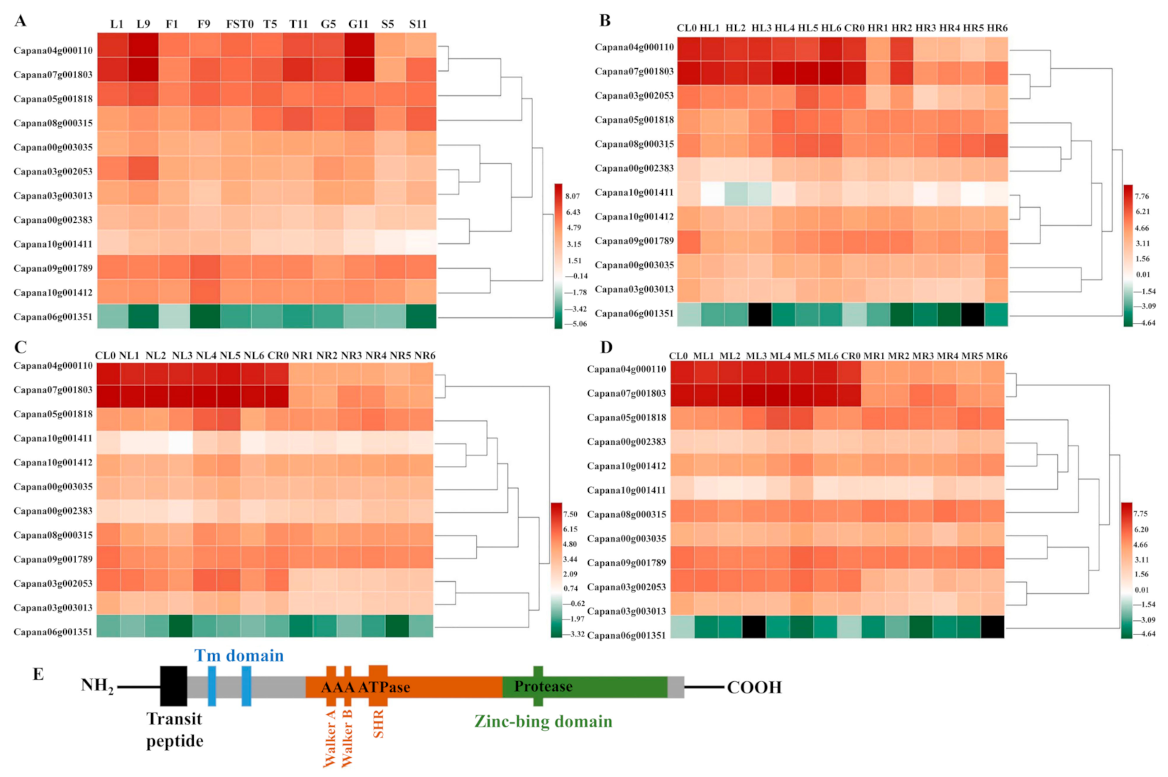
2.1. The Expression Pattern of CaFtsH Genes in Pepper Across Different Tissues during Development and Stress Responses

The transcription data set of the 12 CaFtsHs genes were obtained from the pepper transcriptome database, with transcription data for the CaFtsHs genes at 11 different developmental stages (including six tissues). These data were drawn into a heat map with performing hierarchical clustering (Figure 1A). The heat map profile showed that CaFtsH06, CaFtsH01 and CaFstH05 genes were significantly upregulated. Among them, the CaFtsH06 was expressed in leaves (L1, L9) and flowers (F9). CaFstH05 (S5) was clearly expressed in seeds (S5), CaFtsH01 was expressed at higher levels in fruits (FST0), while the expression of CaFstH04 was highest in seeds (S11). CaFstH09 and CaFstH10 were only expressed at higher levels in flowers (F9), and CaFstH11 also showed higher expression levels in leaves (L9). The expression levels of CaFstH02 and CaFstH08 were not high in all the tested pepper tissues. In addition, the CaFstH12 gene was almost undetectable in all the tested tissues.
The dynamic expression patterns of CaFtsH gene family members under different stress treatments (osmotic, heat, and salt stress) were also observed (Figure 1B–D). The expression level of CaFtsH06 gene in pepper leaves gradually increased after 6 h of continuous heat shock and was quite distinct at 24 h (Figure 1B). However, the expression levels of CaFtsH06 and CaFtsH01 first decreased after heat treatment, slightly increased after 1 h, and again decreased rapidly in the root tissue of pepper plants. After salt stress treatment, the CaFtsH06 gene was mainly accumulated in the leaves and its accumulation at 12 h was highest (Figure 1C). By observing the expression characteristics of CaFtsH homologs under 400 mM mannitol stress (osmotic stress) at different periods (0, 0.5, 1, 3, 6, 12, and 24 h), it was found that the expression of CaFtsH06 in pepper leaves under osmotic stress was most significant at 6 h of treatment (Figure 1D). After osmotic stress, CaFstH01, CaFstH02, CaFstH05, CaFstH06, CaFstH09, and CaFstH10 were upregulated in pepper leaves, while CaFstH02, CaFstH04, CaFstH05, CaFstH07, CaFstH08, CaFstH09, and CaFstH10 were upregulated in the roots. Under the stress of different periods of heat, salt, and osmotic stresses, the CaFstH06 gene had the strongest response in pepper leaves, while the CaFstH12 gene maintained a very low expression level in both pepper leaves and root tissue. The above results indicated that CaFtsH06 may be involved in a variety of environmental stress response processes. Moreover, most of the other genes belonging to the family were not significantly altered in terms of expression across multiple stress conditions (Figure 1). Gene structure and function are closely related, and the homologous LeFtsH6 and AtFtsH6 genes were noted in other reports to have had similar stress responses [30,31]. Therefore, CaFtsH06 was selected as the focus for further research.
FtsH is a typical membrane-bound AAA protease family member39. The structure of FtsH protease in pepper is the same as that of other biological FtsH homologs. It is characterized by a N-terminal region with one or two transmembrane (TM) helix domains required for oligomerization and a central AAA ATPase module. Additionally, the C-terminal domain of the conserved zinc binding site (HEXXH) is required for proteolytic activity [22]. By combining previous studies and the determination of the protein structure from a prediction website, Figure 1E and Figure 3 provides a schematic diagram of FtsH genes, describing the hypothetical structure of FtsH protease in Arabidopsis and pepper.

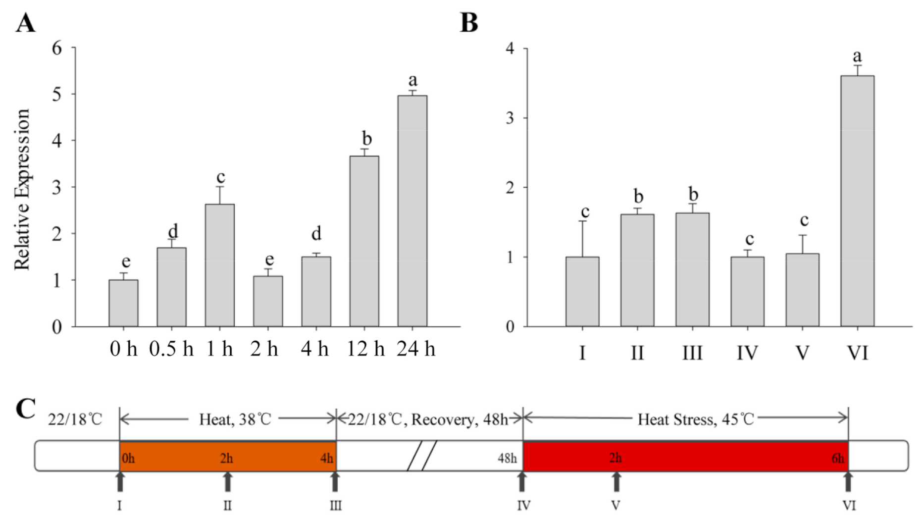
2.2. Expression of CaFtsH06 under Heat Stress in Pepper

All types of exogenous heat stress from the outside acts on plants, and the severity of their damage mainly depends on how fast the plant responds to the stress. The expression patterns of the CaFtsH06 gene in R9 heat-tolerant pepper lines that were exposed to different heat treatments were determined. First, the basic thermotolerance treatment of R9 was analyzed (Figure 2A). At the beginning of the treatment, the expression of CaFtsH06 in pepper increased slowly and then decreased after 2 h of treatment, but after 24 h, the expression of CaFtsH06 increased rapidly to five times the expression level that occurred after 2 h of treatment. The acquired thermotolerance treatment of R9 plants (Figure 2B) between hours 2 and 4 of the heat acclimation was also analyzed. The expression level of CaFtsH06 in leaves was not obvious and tended to be flat. In the recovery phase at 22 °C, the expression level of CaFtsH06 first appeared to decrease, with an expression level that was similar to that level before treatment. However, after the high-temperature treatment at 45 °C, the expression level of CaFtsH06 rose rapidly after treatment and reached its maximum at 6 h, and its expression level was more than 3 times that of the control plant (38 °C, 4 h), as Figure 2C shows, for high-temperature treatment and normal temperature recovery.

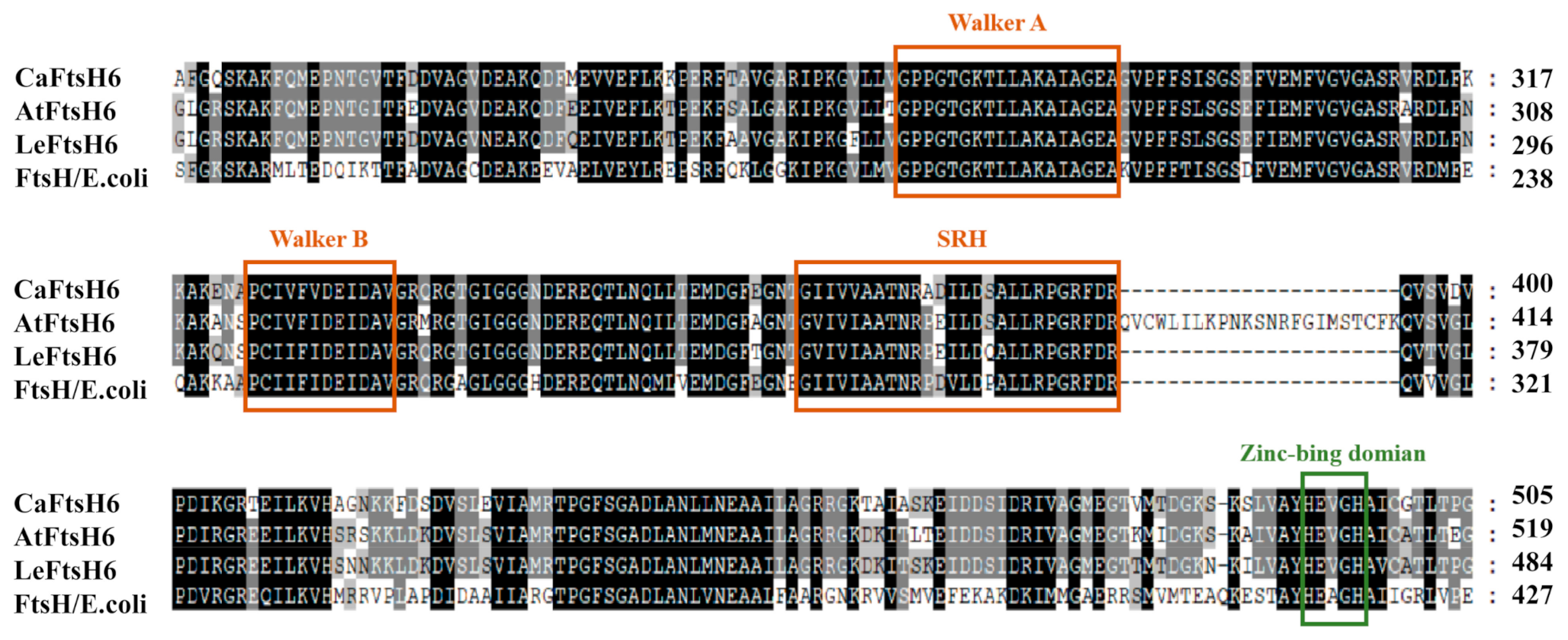
2.3. Structure Analysis of CaFtsH06 Proteins

Based on the close phylogenetic relationship between tomato (Solanum lycopersicum L.) and pepper plants, we determined that the gene should be designated as CaFtsH06 (GenBank accession number XP016580880) based on its homology to tomato metalloproteinase LeFtsH6 (NP001234191). The full-length CaFtsH06 cDNA is 2301 bp, with a 194-bp 5′ untranslated region (5′UTR), 70-bp 3′untranslated region (3′UTR), and 2037-bp ORF. The corresponding protein molecular weight is 77.54 KDa, isoelectric point is 6.29 and instability index is 31.99 (Supplementary Table S1). Multiple alignments of the deduced amino acid sequences of CaFtsH06, AtFtsH6, LeFtsH6, and FtsH/E. coli revealed a consensus region (Figure 3). The central region of the CaFtsH06 contains two putative ATP binding sites (Walker A, Walker-B) and a putative SRH (the second region) for homology. The C-terminus of the CaFtsH06 protein contains a putative zinc-binding motif HEXGH (where each X is a non-conserved amino acid residue).

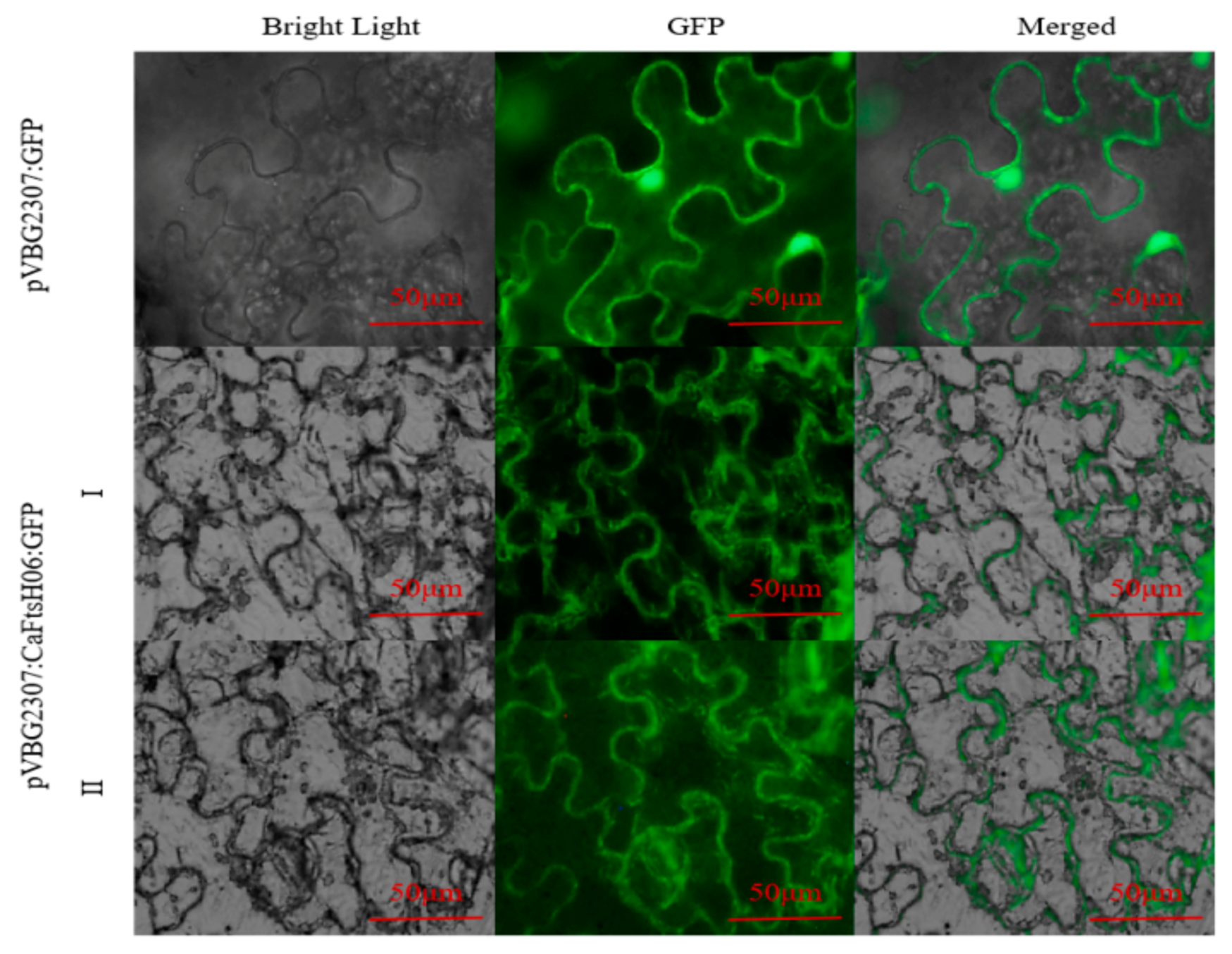
2.4. Subcellular Localization of CaFtsH06 Proteins

The subcellular localization of CaFtsH06 was predicted using the online tool WOLF PSORT (https://wolfpsort.hgc.jp/) (accessed on 10 August 2020), which predicted that CaFtsH06 localized mainly to the mitochondria and cell membrane. To confirm the subcellular localization of CaFtsH06 protein, the pVBG2307:GFP and pVBG2307:CaFtsH06:GFP fusion plasmids were transiently expressed in tobacco leaves. We found that the green fluorescence signal of pVBG2307:CaFtsH06:GFP was detected in the tobacco leaf mitochondria and cell membrane, while the fluorescence of pVBG2307:GFP was distributed throughout the cell, indicating that CaFtsH06 protein in pepper may play a role in the mitochondria and cell membrane (Figure 4).

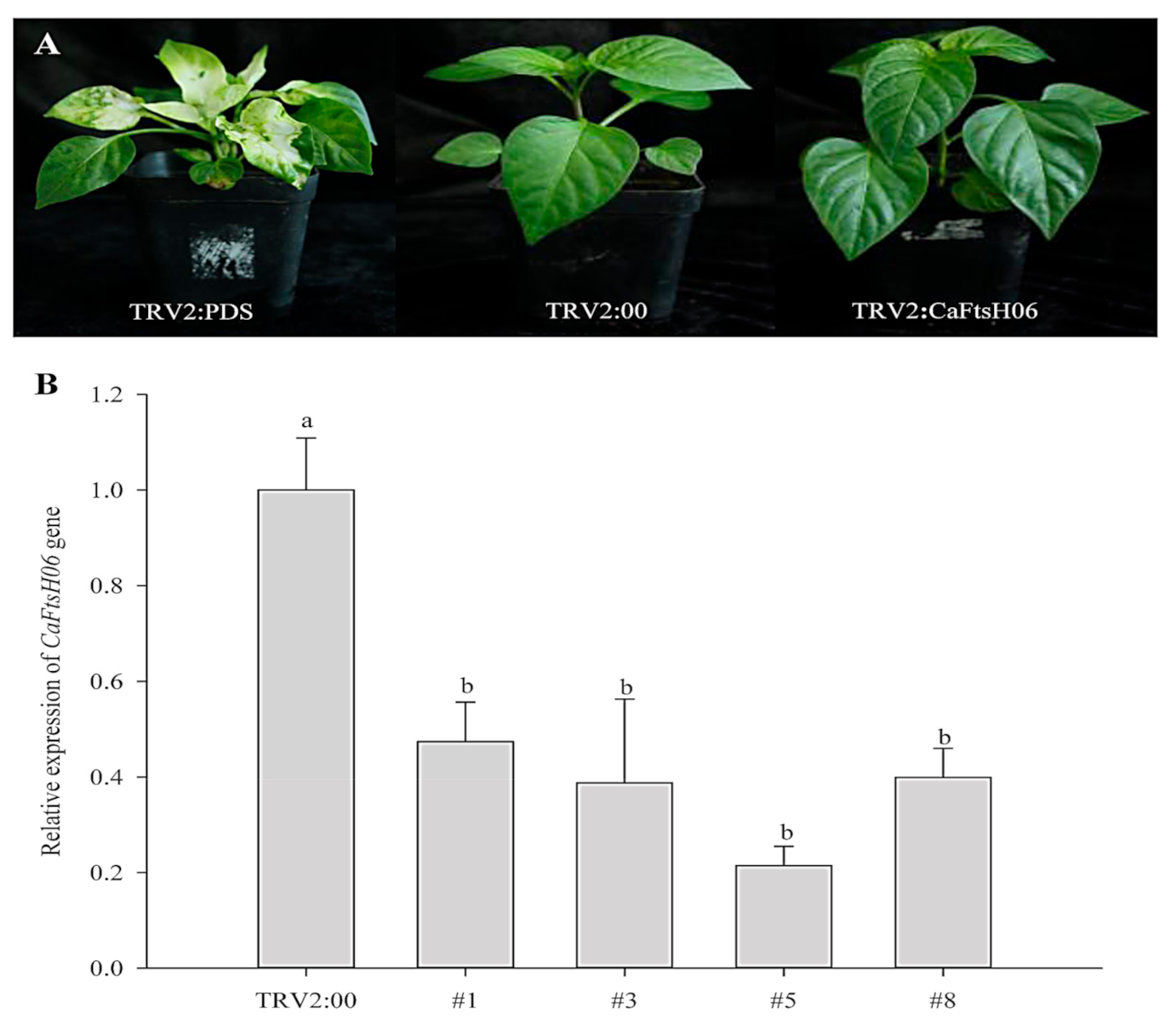
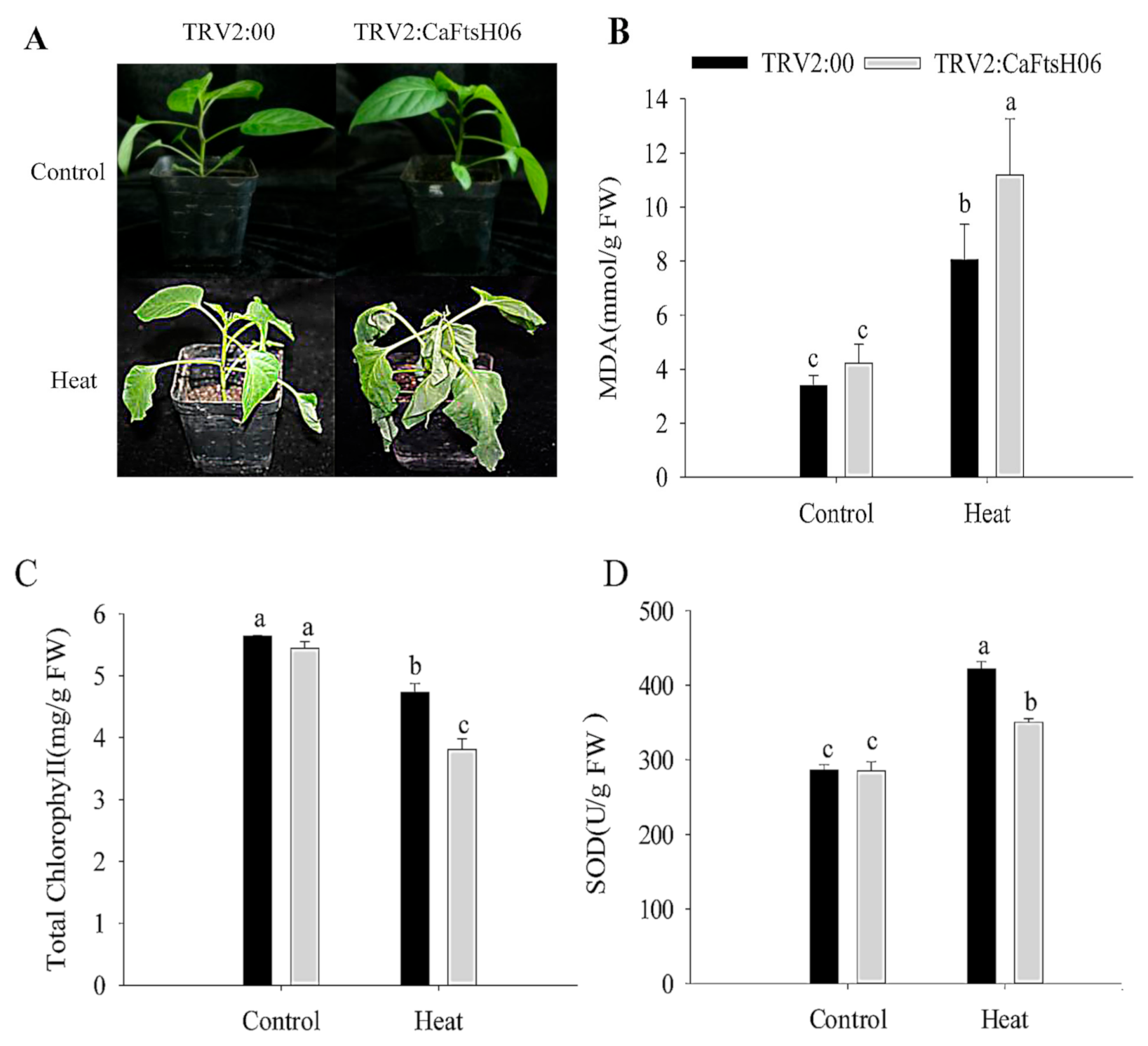
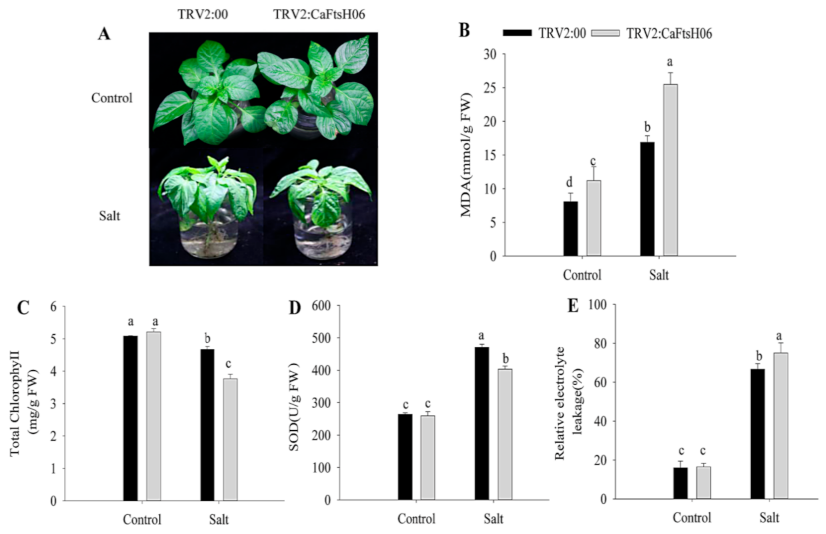
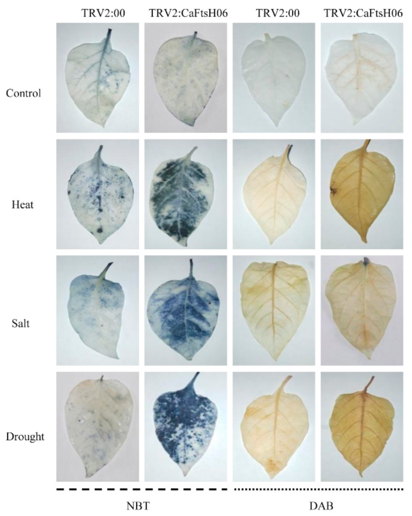
2.5. Knockdown of CaFtsH06 Decreased Abiotic Stress Tolerance in Pepper

The silencing vector TRV2: CaFtsH06 was constructed in the Agrobacterium GV3101 to create CaFtsH06-silenced plants. The leaves of TRV2:CaPDS (positive control) pepper seedlings showed photobleaching symptoms, but no obvious difference in phenotype was observed between TRV2:CaFtsH06 and the negative control TRV2:00 pepper lines under normal growth conditions (Figure 5A). The silencing efficiency of CaFtsH06 in silenced pepper lines exceeded 60% (Figure 5B). Thus, the control plants (TRV2:00) and CaFtsH06-silenced plants (TRV2:CaFtsH06) were used for the subsequent experiment.
The CaFtsH06-silenced and control R9 pepper plants were exposed to 45 °C for 24 h, and significant differences in their phenotypes were observed after high temperature treatment. The leaves of the silenced plants showed severe wilting and drooping, while control plants only exhibited slightly curled edges of leaves (Figure 6A). At the same time, it was determined that the malondialdehyde (MDA) content of the silenced and control plants increased after heat stress, but this increase in the MDA content of the silenced plants was significantly higher than that in control plants (Figure 6B). Similarly, the chlorophyll content of both silenced and control plants decreased after heat shock, but this decrease in the chlorophyll content of the silenced plants was significantly lower than that in control plants (Figure 6C). After the heat stress stimulation, the SOD content of the control and silenced plants was increased, but the increase in the SOD content of the silenced plants were lower than that of the control plants (Figure 6D). The analysis of the above results showed that silencing of the CaFtsH06 gene in pepper reduces its ability to respond to heat shock.
To further explore the function of CaFtsH06 in saline conditions, the CaFtsH06-knockdown and control pepper seedlings were immersed in a 300 mM NaCl solution for 24 h. The leaves of the CaFtsH06-silenced plants showed severe wilting and shrinkage, with the lower leaves having fallen off, while the leaves of the control plants only showed signs of wilting (Figure 7A). At the same time, the MDA content and REL of the two plant types increased significantly, but the increase in the silenced plants was statistically more than that in the non-silenced pepper plants (Figure 7B,E). Additionally, the chlorophyll content of the CaFtsH06-silenced and control plants decreased, but this decrease was significantly lower in the silenced plants than in the control plants (Figure 7C). The SOD content of the control and silenced plants increased after salts stress, but this increase in the SOD content of the control plants was significantly higher than that in the silenced plants (Figure 7D).
After treatment, the MDA content of the CaFtsH06-silenced plants increased significantly, more than that of the control plants (Figure 8B). Plant leaves initiate stress responses to external stimuli and rapidly accumulate proline, but the accumulation of proline in the CaFtsH06-silenced plant leaves was not increased (Figure 8C). Similarly, the chlorophyll content of the CaFtsH06-silenced plants was significantly reduced as compared to the control plants (Figure 8D). Thus, it was preliminarily determined that silencing the CaFtsH06 gene in pepper reduces osmotic tolerance in pepper.
Osmotic stress can lead to the disordering of ion distribution and balance in plants, the deposition of large amounts of destructive ROS substances, and damage to the internal structures and the function of cells. Thus, it was important to explore the influence of CaFtsH06 silencing on the accumulation and removal of ROS in the pepper R9 lines. Accordingly, we used histochemical staining DAB and NBT methods to measure the changes in H2O2 and ·O2, respectively.
As shown in Figure 9, the DAB deposition area of the CaFtsH06-silenced and control plants was higher after stressed conditions as compared to the unstressed plants, but the stained areas of the CaFtsH06-silenced plants were significantly larger than those of the control plants. Similarly, after coercion treatment, the NBT deposition areas of silenced and control peppers were significantly larger than those of unstressed plants, while the stained areas of CaFtsH06-silenced plants were significantly larger than those of control plants. These results indicate that as compared to the leaves of control plants, the leaves of CaFtsH06-silenced plants had higher levels of H2O2 and ·O2. Thus, when the CaFtsH06 gene was silenced in pepper, the ability of the plants to remove harmful substances such as peroxides was also reduced, such that the ability of pepper to resist stress was greatly reduced.

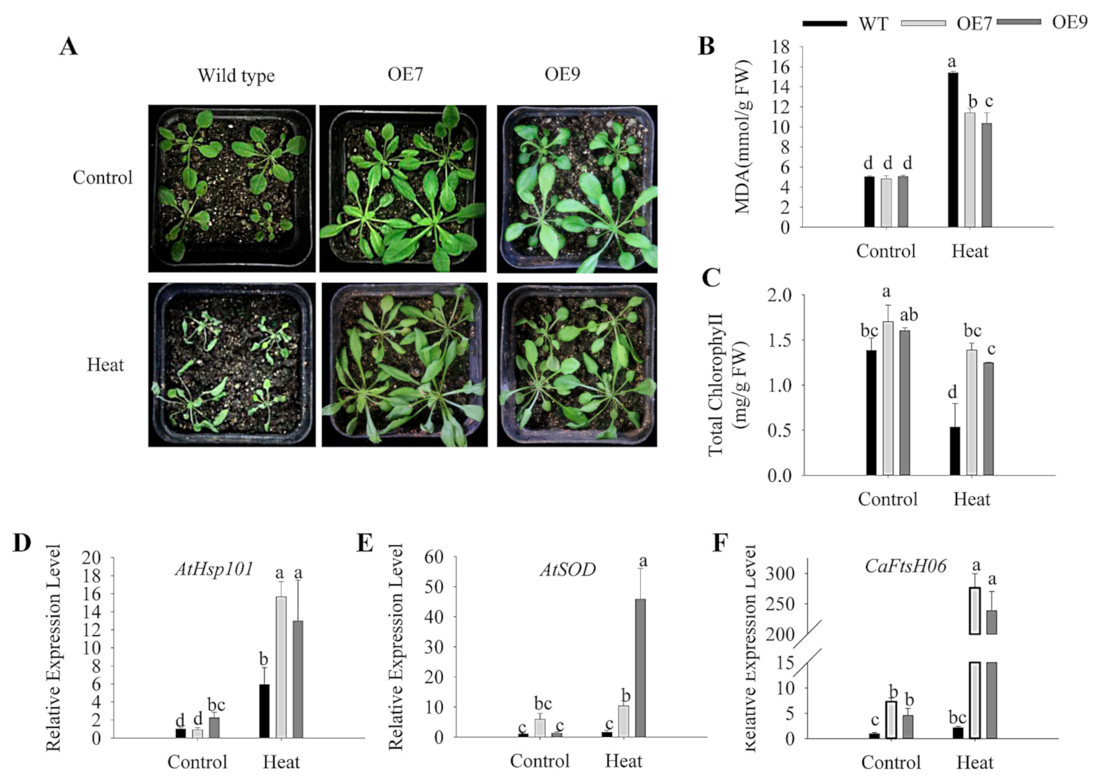
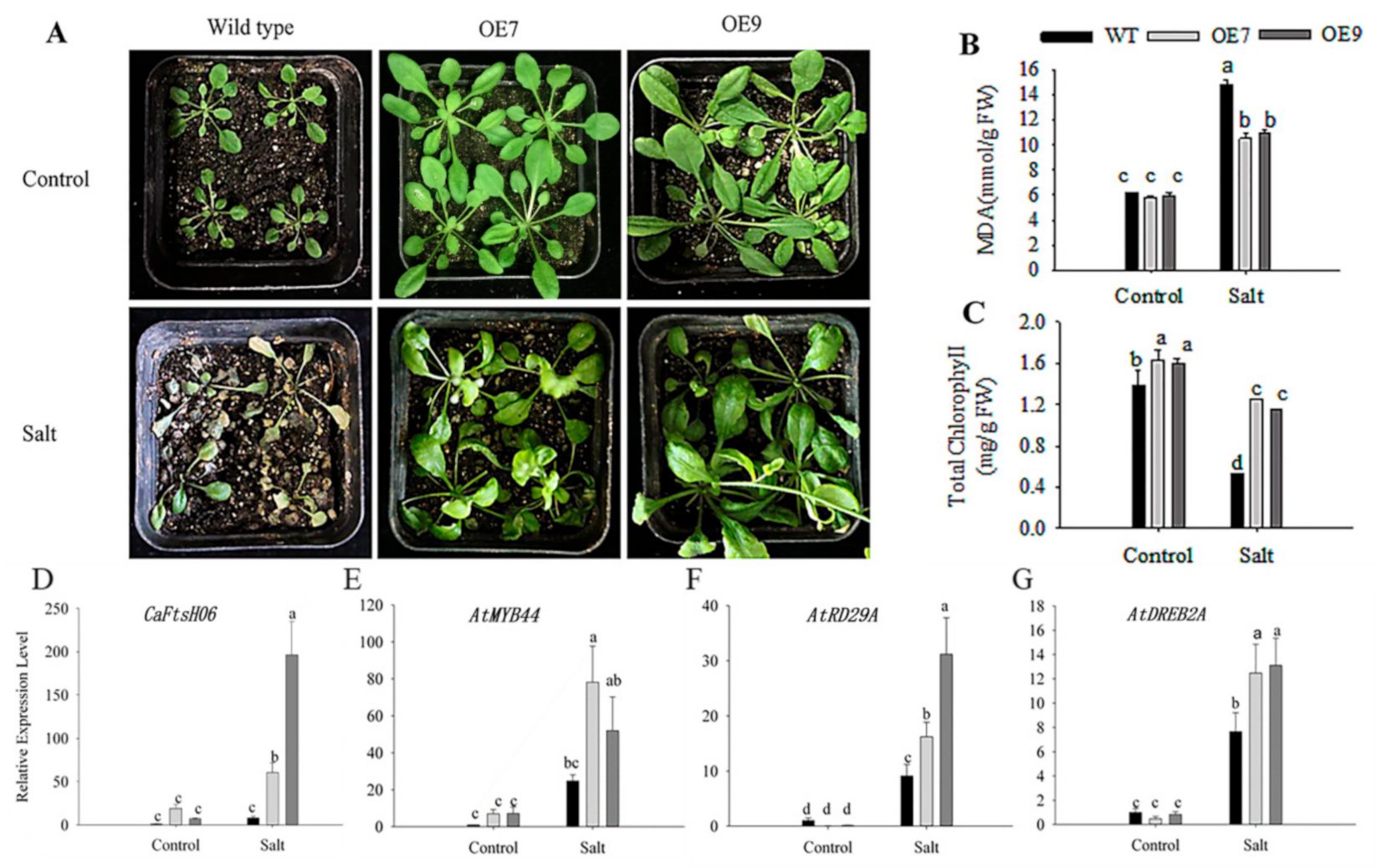
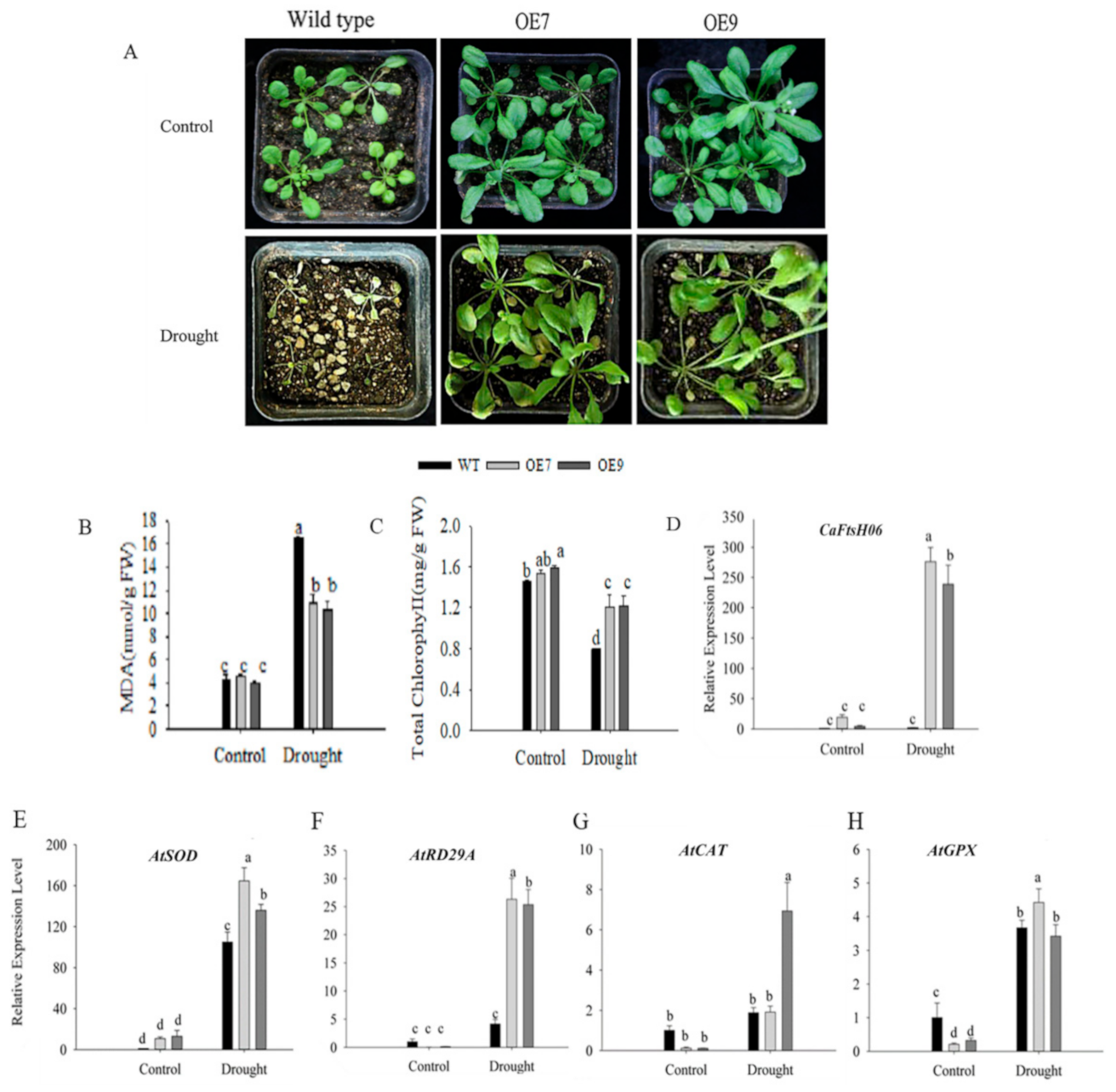
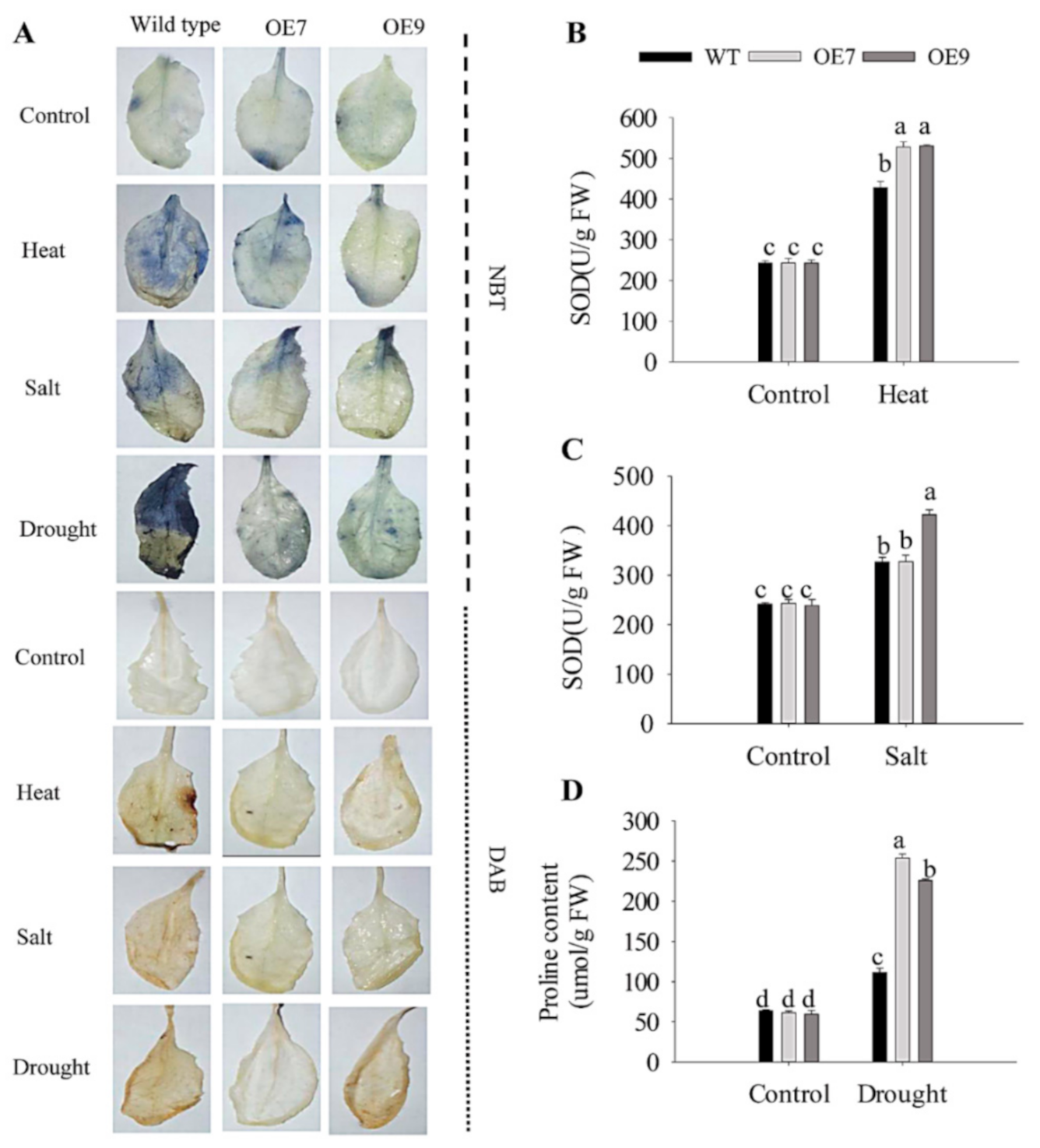
2.6. Ectopic Expression of CaFtsH06 Enhances the Abiotic Stress Tolerance in Arabidopsis

In order to determine whether the CaFtsH06 gene was integrated into Arabidopsis, we first verified Arabidopsis at the DNA level. Based on the performance of this gene in silenced pepper plants under different abiotic stresses and the presence of multiple stress-related cis elements in the promoter region, it was speculated that overexpression of this gene in Arabidopsis can enhance tolerance to multiple abiotic stresses. After PCR detection, we observed that the OE7 and OE9 strains expressed CaFtsH06 genes at a much higher levels than observed in untransformed wild-type (WT) Arabidopsis (Supplementary Figure S3B). Furthermore, we found that the CaFtsH06 gene expression in the two lines of OE7 and OE9 under normal growth conditions was higher and that the leaf area of the transgenic lines was significantly larger than that of WT plants (Figure S4), in addition to their more vigorous phenotype after heat-stress treatment at 45 °C for 16 h. Thus, we selected these two transgenic lines (OE7 and OE9) for the following experiments.
To study the effect of CaFtsH06 on the temperature tolerance of transgenic Arabidopsis, three-week-old WT and transgenic plants with consistent growth were selected and heat treated at 40 °C for 24 h. Phenotypic observation (Figure 10A) revealed that the WT leaves exhibited dryness and wilting, with necrosis on some leaves. In contrast, the CaFtsH06-expressing transgenic line only showed curling and wilting on the leaf edges, and its phenotype was obviously more vigorous than that of WT plants. At the same time, leaves from WT plants and the CaFtsH06-expressing transgenic line were collected before and after treatment for the determination of their physiological indicators related to stress resistance. Analysis of MDA content (Figure 10B) revealed that high-temperature treatment significantly increased the MDA content of OE7, OE9, and WT leaves, though the MDA content of the CaFtsH06-expressing lines were significantly lower than that of WT plants. After heat treatment, the chlorophyll content of the WT leaves decreased significantly (Figure 10C). Additionally, the expression levels of the heat-stress response gene (AtHSP101) and SOD enzyme synthesis gene (AtSOD) in OE7 and OE9 plants were significantly higher than those of WT plants (Figure 10D,E), and at the same time, the expression of CaFtsH06 in OE9 and OE7 was also increased significantly (Figure 10F).
The damage induced by salt stress to plants also involves a series of physiological and biochemical stress responses, which can cause obvious phenotypic damage in severe cases. The WT and transgenic line seedlings at the age of three weeks were watered with a 200 mM NaCl solution once every two days for two weeks. As compared with the control plants, the WT plants showed severe chlorosis and wilting of their leaves after salt stress treatment. However, the transgenic plants showed no obvious difference from the control plants in other characteristics, except for the slight yellowing phenotype in their leaves (Figure 11A). At the same time, the leaves of WT, OE7, and OE9 plants were collected for the determination of their MDA and chlorophyll contents before and after the salt stress treatment. After salt treatment, the MDA content of WT, OE7, and OE9 leaves significantly increased, but the increase in MDA content of WT plants was higher than that of OE7 and OE9 plants (Figure 11B). The chlorophyll content in Arabidopsis leaves was also significantly reduced after the stress treatment, but the chlorophyll content in WT leaves was significantly lower than that of the OE7 and OE9 lines (Figure 11C). To further study the salt tolerance mechanism, qRT-PCR was used to analyze changes in the expression of salt-stress related genes in the WT and OE lines before and after salt treatment. The expression of CaFtsH06 (Figure 11D) and the salt-stress-responsive genes AtMYB44, AtRD29A, and AtDREB2A increased significantly in the OE7 and OE9 lines (Figure 11E–G) as compared to the WT plants. Thus, ectopic expression of the CaFtsH06 gene in Arabidopsis was beneficial to alleviate the salt damage to Arabidopsis plants.
Drought stress, like other stress conditions, also induces a series of morphological, physiological, biochemical, and molecular stress changes in plants. For the drought tolerance assay, the WT and transgenic seedlings (three weeks old) were selected for water-control and drought treatment. After the drought treatment, the leaves of WT seedlings were severely wilted and necrotic, while some of the leaves of the OE7 and OE9 lines appeared dry and yellow (Figure 12A). In addition, the MDA content of WT and transgenic plants increased after drought treatment, but the MDA content of the transgenic plants was significantly lower than that of WT plants (Figure 12B). The chlorophyll content of the WT plants decreased significantly after treatment, though that of the transgenic plants was higher than the WT plants (Figure 12C). Subsequently, we measured the expression levels of the drought-stress response genes and AtRD29A, AtCAT, AtGPX, and the SOD enzyme synthesis gene AtSOD in leaves. The expressions of the CaFtsH06 and the four drought-stress related genes in the transgenic plants were all upregulated after the drought treatment, and each was significantly higher than those of WT plants (Figure 12D,F–H). Collectively, the ectopic expression of the CaFtsH06 gene in Arabidopsis effectively enhanced the resistance of Arabidopsis to drought stress.
In the processes of plant metabolism, redox reactions occur inside and outside of cells. Based on the NBT and DAB staining of the leaves of plants after stress treatment, the ability of transgenic CaFtsH06-expressing Arabidopsis to resist adversity stress and eliminate harmful ROS substances was measured. Compared with the control, the DAB and NBT staining of the WT plants were stronger after the stress treatment, while the accumulation of peroxides in the leaves of the transgenic plants was distinctly lower. For plants not subjected to stress treatment, the stained areas of leaves of the WT plants were significantly higher than those of transgenic plants (Figure 13A). Under heat stress treatment, compared with the wild-type Arabidopsis, the SOD activity of the transgenic lines was higher (Figure 13B). After the same salt treatment, the SOD content of the transgenic Arabidopsis leaves was also higher than that of the wild-type Arabidopsis. The SOD content in the leaves of the WT plants was high (Figure 13C). The proline content in the leaves of the transgenic and WT plants was also measured. The transgenic plants responded quickly to drought stress, with a more obvious increase in the proline content of leaves (Figure 13D). Altogether, the ectopic expression of the CaFtsH06 gene in Arabidopsis improved the ability of plants to resist stress and accelerate the elimination of harmful ROS.

3. Discussion

When plants encounter harsh environments such as extreme temperatures, drought, and high salinity, the resulting oxidative stresses cause disordering of their protein functions to occur. Many proteins lose their functions, owing to denaturation, and then condense together to form aggregates that affect the proteins in the plant or gene functions, which can cause serious damage to all aspects of plants [38,39,40]. FtsH not only participates in repairing damaged proteins, unfolded proteins, and unassembled free subunits, but also can control heat response and lysogen by interacting with some regulatory factors (such as σ32, λCII, LpxC) and other cell activities [11,41,42]. Additionally, FtsH is involved in the EXECUTER1 (EX1)-mediated retrograde signaling network induced by singlet oxygen (1O2) [43]. Recent research by Dogra, et al. [44] claimed that FtsH regulation prevents 1O2 signaling and the expression of related genes in flu mutants. Previous studies have shown that the expression of many FtsH proteases can be induced by abiotic stress [20,21,22,41]. Our results demonstrated that the CaFtsH06 gene responded to abiotic stressors such as extreme temperature, drought, and saline conditions (Figure 1). The CaFtsH06 transcript levels differed and exhibited a complex pattern across different temperature conditions. (Figure 2). Previous reports showed that the FtsH6 gene is involved in thermotolerance in tomatoes [30]. These findings showed that, the CaFtsH06 gene may participate in plant responses to variable environmental stimuli, such as heat, salt, and drought stress.
FtsH localized to various areas [10], such as the protease pairs FtsH3⁄FtsH10 localized to mitochondria [45], FtsH11 localized to both chloroplasts and mitochondria [17], and AtFtsH6 localized to chloroplast, where it may protect the thylakoid membrane of plants under heat stress [31]. In this study, we observed that CaFtsH06 enters the mitochondria and cell membrane, (Figure 4), suggesting that CaFtsH06 protein may play a fundamental role in the mitochondria and cell membrane.
Under heat, drought and salt stress conditions, plant cell structures suffer while physiological, biochemical, and molecular damage may occur, further leading to cell death [1,5]. Additionally, such stress can cause an increase in the MDA content and leaf REL [46]. MDA content is an indicator of the defense signaling of cell membrane lipid peroxidation [47]. In this study, the MDA content of the CaFtsH06-silenced peppers was higher than that of the control (TRV2:00) peppers under drought and salt-stress conditions (Figure 6B, Figure 7B and Figure 8B). This demonstrated that silencing CaFtsH06 increased the damage to the cell membrane in pepper. Furthermore, silencing of CaFtsH06 in pepper reduced the total chlorophyll content as compared to control plants, which may in turn decrease the photosynthetic efficiency (Figure 6C, Figure 7C and Figure 8D). In addition, we found that the CaFtsH06-silenced peppers had higher leaf REL and proline content when exposed to salt and drought stress (Figure 7E and Figure 8C). Under different stress conditions, the excess accumulation of ROS, such as H2O2 and ·O2, likely resulted in membrane damage and damage to other macro molecules, such as proteins, lipids, and the photosynthetic apparatus [48,49]. Therefore, the contents of H2O2 and ·O2 were also indicators of the damage level to plant cells [50]. In the present study, the ROS level in the CaFtsH06-silenced pepper plants were substantially higher than in the control peppers under different environmental conditions (Figure 9). These results demonstrated that this critical gene was involved in abiotic stress tolerance as shown in the silenced plants after the abiotic stress treatments. To further elucidate the biological function of CaFtsH06 in plant response to other environmental stresses, Agrobacterium-mediated ectopic expression CaFtsH06 lines were examined in Arabidopsis. In this study, CaFtsH06-expressing Arabidopsis lines exhibited reduced plant sensitivity to extreme oxidative stress through increased recovery and reduced damage symptoms, MDA content, proline content, and accumulations of H2O2 and ·O2, as well as increased total chlorophyll content and SOD activity compared to the WT plants (Figure 10, Figure 11, Figure 12 and Figure 13). Our results showed that ectopic expression of CaFtsH06 promoted plant stress tolerance. In particular, compared to the WT line, the transgenic Arabidopsis lines exhibited thermostability associated with lower ROS content. Thus, these findings showed that CaFtsH06 positively mediates the plant tolerance to high temperature, excess salt, and drought stress.
Many defense-related genes in plants may be involved in the rapid response to adversity stress and enhancement of plant resistance. For example, AtFtsH2 regulates the transcription of the stress response protein Hsp21, further protecting the photosynthetic system (PS Ⅱ) complex against heat stress [50]. In cotton and tobacco, ectopic expression of the AtHsp101 gene increases heat tolerance in transgenic plants [51], suggesting that it may play a fundamental role in conferring heat stress resistance [52]. In alfalfa, another FtsH homolog is induced by cold stress or high light conditions [22]. The transcription factor AtMYB can regulate the ABA-mediated response of plants to salt and drought stress [53]. The salt-sensitive AtDREB2A gene, which can be induced by NaCl stress, can maintain the ion balance inside across the cell membrane [54]. Defense-related genes such as AtSOD, AtCAT, and the drought stress marker AtRD29A gene were upregulated by high temperatures and salt stress [55,56]. In this study, ectopic expression of the CaFtsH06 gene increased the expression of stress-related genes (AtHSP101, AtMYB44, AtDREB2A, AtCAT, AtGPX, AtSOD, and AtRD29A) under high-temperature, salt, and drought stresses in the transgenic Arabidopsis relative to the WT plants (Figure 10, Figure 11 and Figure 12). Thus, we hypothesize that CaFtsH06 might enhance the stress resistance of pepper plants by regulating the expression of stress-related genes, though further study is required to unravel the associated complex regulatory gene network. Interestingly, our results suggest that CaFtsH06 under the constitutive 35S promoter is upregulated in response to different abiotic stresses, leading the basic tolerance. Furthermore, we will also construct the vector of the gene with its promoter, or using the Arabidopsis stress-induced promoter RD29A, and then after transformation of the recombinant fusion vector into Arabidopsis it would be interesting to further verify whether the gene has acquired tolerance.

4. Materials and Methods

4.1. Plant Materials and Growth Conditions

The R9 thermotolerant pepper line (introduced at the World-Asia Vegetable Research and Development Center, PP0042-51) and Arabidopsis thaliana ecotype Columbia-0 (Col-0) were provided by the College of Horticulture, Northwest A&F University, China. The growth conditions of the test materials were strictly managed in an incubator, with temperatures maintained at 22/18 °C, the light conditions were adjusted to 16 h/8 h (day/night), and a relative humidity of 65% [57]. Temperature was thereafter adjusted according to the experimental requirements detailed below.

4.2. Transcriptome Data Analysis of Pepper

To understand the expression patterns of pepper CaFtsHs family member genes in different tissues during development and various stress response periods, the transcriptomic data from pepper was used for analysis. The Pepper Hub (http://pepperhub.hzau.edu.cn/) (accessed on 15 March 2020) database was used to acquire the transcriptome data for CaFtsHs family members (12) from the Zunla-1 genome [58]. Tissue-specific expression patterns of different tissues at various developmental stages (Supplementary Table S2), including leaves (L1–9), flowers (F1-9), fruits (FST0), pericarp (G1–11), placenta (T3-11), and seeds (S3–11) were analyzed. Expression levels of the CaFtsHs genes in pepper leaves/roots under different stress treatments (Supplementary Table S3), including heat stress (42 °C), salt stress (200 mM NaCl), and osmotic stress (400 mM mannitol), were estimated at 0, 0.5, 1, 3, 6, 12, and 24 h, respectively. Using the HemI software (Available online: http://hemi.biocuckoo.org/down.php) (accessed on 27 June 2021) [59], the transcription data of CaFtsHs family members from pepper were rendered into an expression heat map.

4.3. Sequence Analysis and Identification of Conserved Domains

The open reading frame (ORF) of CaFtsH06 was confirmed using the NCBI ORF Finder. The number of amino acids, the molecular weight, the theoretical isoelectric point (pI), and the protein instability index (>40 considered unstable) were obtained by analyzing amino acid sequences with the EXPASY PROTOPARAM (Available online: http://web.expasy.org/protparam/) (accessed on 27 June 2021) [60]. Conserved prediction domains, motifs, and subcellular localization were inferred using the InterProScan (Available online: http://www.ebi.ac.uk/interpro/) (accessed on 27 June 2021) [61], the PROSITE Scan (Available online: http://www.ebi.ac.uk/InterProScan/) (accessed on 27 June 2021) [62], and WoLF PSORT (Available online: https://wolfpsort.hgc.jp/) (accessed on 27 June 2021) [63], respectively. The ClustalW project (Kyoto University Bioinformatics Center, Uji, Japan) and GenDoc (Available online: http://www.psc.edu/biomed/gendoc/) (accessed on 27 June 2021) [64] were used for sequence alignment.4.4. RNA Extraction and qRT-PCR Analyses.
Total RNA extraction was performed using the method of Guo et al. [65], and reverse transcription was performed according to the manufacturer’s instructions of the TaKaRa reverse transcription kit (TaKaRa, Dalian, China). The samples were analyzed via qRT-PCR using the iQ5.0 Bio-Rad iCycler thermocycler (Bio-Rad, Hercules, CA, USA). All primer pairs used for qRT-PCR were designed using the NCBI Primer-BLAST website (Supplementary Table S4). The reaction mixture used in the PCR analyses was SYBR Green Supermix (Takara, Dalian, China). The ubiquitin ligase protein gene (CaUbi3) was used as a reference gene for pepper, and the AtActin2 was used as a reference gene for Arabidopsis [65]. The relative expression levels of all the genes were calculated using the 2−ΔΔCT method [66].

4.4. Subcellular Localization

A CaFtsH6 ORF without a stop codon was cloned from pepper cDNA. The CaFtsH06 fragment obtained via PCR amplification was then cloned into pVBG2307:GFP vector, and the empty pVBG2307:GFP vector without CaFtsH06 was used as a control. Constructs were confirmed using specific primer pairs (Supplementary Table S5); the constructed vector was transformed with Agrobacterium tumefaciens strain GV3101 to achieve transient expression of the recombinant fusion vector in the leaves of Nicotiana benthamiana.

4.5. Silencing of CaFtsH06 in Pepper

CaFtsH06 expression was knocked down in pepper by using the virus-induced gene silencing (VIGS) technique. In the CDS region of the CaFtsH06 sequence (Supplementary Table S6), fragments with higher specificity were used to design the primers to obtain amplicons that could be used for the construction of the pTRV2:CaFtsH6 vector (Supplementary Table S7). A TRV2:00 empty vector was used as a negative control, and aTRV2:CaPDS (phytane desaturase gene) construct was used as a positive control. The TRV2:CaFtsH06-, TRV2:00-, and TRV2:CaPDS-constructed vectors were transformed by using A. tumefaciens strain GV3101 (Supplementary Figure S1). They were then infiltrated into the leaves of pepper R9 line pepper plants at the two-true-leaf stage. After 40–45 days, when most of the leaves of the positive control pepper plant appeared chlorotic, the TRV2:CaFtsH06- and TRV2:00-injected plants were analyzed via qRT-PCR for analysis of CaFtsH06 expression, and the corresponding silencing efficiency was obtained [57].

4.6. Construction of Transgenic Arabidopsis Line Expressing CaFtsH06

The full-length of the CaFtsH06 open reading frame (ORF) was cloned from pepper cDNA, and a specific primer pair containing the BamHI and KpnI restriction enzyme sites was used (Supplementary Table S8). Subsequently, the CaFtsH06 fragment obtained via PCR amplification was cloned into the pVBG2307 vector (Supplementary Figure S2). The constructed vector was transformed into Arabidopsis using the A. tumefaciens strain GV3101 to realize the transformation of the recombinant fusion vector into Arabidopsis [67]. Transgenic plants were placed in an MS medium with kanamycin (50 mmol/L) to screen for resistance conferred by the vector. To determine the successfully transformed lines, first of all, DNA was extracted from the leaves of the three-week-old seedlings of T3 generation seedlings. The extraction was performed using the method of Huang, et al. [68] and subsequent PCR detection. At the same time, RNA was extracted separately, and the expression level of CaFtsH06 was detected via qRT-PCR, thereby identifying five transgenic lines (OE6, OE7, OE8, OE9, and OE14) (Supplementary Figure S3). Three-week-old seedlings from the transgenic Arabidopsis lines (OE6, OE7, OE8, OE9, and OE14) were treated at 45 °C for 16 h, and the phenotypes were then observed. After comprehensively assessing the expression of CaFtsH06 in transgenic Arabidopsis before and after heat stress as well as assaying the heat-resistant phenotype, two transgenic lines (OE7 and OE9) were selected.

4.7. Plant Abiotic Stress Treatments

The pepper seedlings from line R9 were grown in growth chambers with controlled photoperiod and temperature conditions. The seedlings were raised at 22/18 °C and with a photoperiod of 16 h/8 h (day/night) and a relative humidity of 65%, and samples were collected at the six-to-eight-leaf stage after the treatment with heat, salt, and drought stresses [57,65,69]. The basic thermotolerance treatment involved placing seedlings in a growth chamber set to 45 °C, with collection of plant leaves at 0, 0.5, 1, 2, 4, 12, and 24 h. The acquired thermotolerance treatment involved placing the seedlings in a growth chamber set to 38 °C for thermal acclimation and sampling after 0, 2, and 4 h of acclimatization. After the 4 h thermal acclimation, the seedlings were restored to 22 °C conditions for 48 h. Subsequently, the recovered pepper plants were treated again at a high temperature of 45 °C, and the leaves were sampled and collected at 0, 2, and 6 h, respectively. The time course of the thermal stress treatment is described in Figure 1C. In total, six to eight strains were respectively selected for mixed sampling, with three biological replicates. The samples were immediately frozen in liquid nitrogen and stored in a −80 °C freezer prior to RNA extraction.
To analyze the loss of function of the CaFtsH06 plants under different stresses, the CaFtsH06-silenced pepper plants were generated as mentioned earlier. For heat stress, the seedlings were placed in a growth chamber set to 45 °C for 24 h. After exposure to heat, the collected pepper leaves were used for the determination of MDA, chlorophyll, and SOD content levels, as well as DAB and NBT staining. For salt stress, the roots of the seedlings were washed and soaked in a 300 mM NaCl solution for 24 h. Pepper leaves were collected for determination of MDA, chlorophyll, and SOD content levels, relative electrolytic leakage (REL), and staining of the leaves with DAB and NBT. For osmotic stress, after washing the roots of pTRV2:00 negative control plants and pTRV2:CaFtsH06-silenced plants, they were soaked in a mannitol solution (300 mM, treatment for 36 h). Pepper leaves were collected for determination of MDA, chlorophyll, SOD and proline content levels, as well as and DAB and NBT staining, while RNA was also extracted from the leaves.
To analyze the function of CaFtsH06 in responding to abiotic stress, the three-week-old CaFtsH06-expressing and WT Arabidopsis lines were used. For heat stress, the seedlings were placed in a growth incubator set to 40 °C and incubated for 24 h. For salt stress, the three-week-old seedlings were watered with an NaCl solution (200 mM) every other day, with sampling after 14 d. For drought stress, water was withheld from the three-week-old seedlings, and sampling was conducted seven days later. Plants under normal conditions were used as the control. The leaves of plants before and after the treatment were sampled at random for the determination of the accumulation of MDA, chlorophyll, proline and SOD content levels, DAB and NBT staining, and RNA extraction across three replicates.

4.8. Biochemical Indices

The malondialdehyde content, superoxide dismutase (SOD) activity, and REL determination methods followed those described by Stewart and Bewley [70]. The chlorophyll content was determined following the method of Arkus [71]. Proline content was measured following the method described by Naser [72]. ROS presence was determined through histochemical staining with DAB and NBT following the method of Sekulska-Nalewajko [73].

4.9. Statistical Analysis

The statistical analyses were conducted using the SPSS 19.0 (IBM Corp. Armonk, NY, USA). Analysis of variance (ANOVA) was conducted, with significant differences considered at a p ≤ 0.05 threshold. All the analyses were conducted with three biological replicates.

5. Conclusions

Our findings show that CaFtsH06 responds significantly to heat, salt, and drought stress and encodes a protein that localizes to the mitochondria and cell membrane. The silencing of CaFtsH06 enhanced pepper sensitivity to heat, salt, and drought stress. However, the CaFtsH06-expressing Arabidopsis lines showed decreased sensitivity to heat, salt, and drought stress tolerance. Furthermore, our results provide important information about CaFtsH06, indicating that it may play a crucial role in tolerance of the above-mentioned stresses by inhibiting the accumulation of H2O2 and inducing the transcriptional levels of defense-related genes. Consequently, CaFtsH06 appears to play a pivotal role in plant adaptation to environmental stimuli, and additional work should focus on the adaptive mechanisms underlying the interaction between the CaFtsH family genes and plant development in extreme environmental conditions.

Supplementary Materials

Author Contributions

Conceptualization, J.-J.X. and Z.-H.G.; methodology, J.-J.X., R.-X.Z. and Z.-H.G.; software, R.-X.Z.; validation, J.-J.X., R.-X.Z. and Z.-H.G.; formal analysis, W.-X.G.; investigation, R.-X.Z.; resources, R.-X.Z.; data curation, R.-X.Z. and J.-J.X.; writing—original draft preparation, J.-J.X. and R.-X.Z.; writing—review and editing, J.-J.X., R.-X.Z., A.K., S.u.H. and Z.-H.G.; visualization, A.K.; supervision, J.-J.X., R.-X.Z. and Z.-H.G.; project administration, Z.-H.G.; funding acquisition, Z.-H.G. All authors have read and agreed to the published version of the manuscript.

Funding

This research was supported through funding from the National Key R&D Program of China (no. 2016YFD0101900) and National Natural Science Foundation of China (no. U1603102).

Institutional Review Board Statement

Not applicable.

Informed Consent Statement

Not applicable.

Data Availability Statement

Data is contained within the article.

Conflicts of Interest

The authors declare there are no conflict of interest.

Abbreviations

ROSreactive oxygen species
PDSphytoene desaturase
SODsuperoxide dismutase
NBTnitro-blue tetrazolium
DAB diaminobenzidine
H2O2hydrogen peroxide
·O2superoxide
MDAmalondialdehyde
mg/gmilligram per liter
OEoverexpression
OE#7No. 7 Arabidopsis line with expressing CaFtsH06
OE#9No. 9 Arabidopsis line with expressing CaFtsH06
UBIubiquitin-conjugating protein
ORFopen reading frame
TRVtobacco rattle virus
VIGSvirus-induced gene silencing

References

  1. Zhai, Y.; Wang, H.; Liang, M.; Lu, M. Both silencing-and over-expression of pepper CaATG8c gene compromise plant tolerance to heat and salt stress. Environ. Exp. Bot. 2017, 141, 10–18. [Google Scholar] [CrossRef]
  2. Zhu, J.K. Abiotic stress signaling and responses in plants. Cell 2016, 167, 313–324. [Google Scholar] [CrossRef] [Green Version]
  3. Khan, A.; Li, R.J.; Sun, J.T.; Ma, F.; Zhang, H.X.; Jin, J. Genome-wide analysis of dirigent gene family in pepper (Capsicum annuum L.) and characterization of CaDIR7 in biotic and abiotic stresses. Sci. Rep. 2018, 8, 1–21. [Google Scholar]
  4. Allakhverdiev, S.I.; Kreslavski, V.D.; Klimov, V.V.; Los, D.A.; Carpentier, R.; Mohanty, P. Heat stress: An overview of molecular responses in photosynthesis. Photosynth. Res. 2008, 98, 541–550. [Google Scholar] [CrossRef] [PubMed]
  5. Ahuja, I.; de Vos, R.C.; Bones, A.M.; Hall, R.D. Plant molecular stress responses face climate change. Trends Plant Sci. 2010, 15, 664–674. [Google Scholar] [CrossRef]
  6. Van Breusegem, F.; Dat, J.F. Reactive oxygen species in plant cell death. Plant Physiol. 2006, 141, 384–390. [Google Scholar] [CrossRef] [PubMed] [Green Version]
  7. Cramer, G.R.; Urano, K.; Delrot, S.; Pezzotti, M.; Shinozaki, K. Effects of abiotic stress on plants: A systems biology perspective. BMC Plant Biol. 2011, 11, 163. [Google Scholar] [CrossRef] [Green Version]
  8. Haq, S.U.; Khan, A.; Ali, M.; Khattak, A.M.; Gai, W.X.; Zhang, H.X.; Wei, A.M.; Gong, Z.H. Heat shock proteins: Dynamic biomolecules to counter plant biotic and abiotic stresses. Int. J. Mol. Sci. 2019, 20, 5321. [Google Scholar] [CrossRef] [Green Version]
  9. Gottesman, S.; Wickner, S.; Maurizi, M.R. Protein quality control: Triage by chaperones and proteases. Genes Dev. 1997, 11, 815–823. [Google Scholar] [CrossRef] [Green Version]
  10. Wagner, R.; Aigner, H.; Funk, C. FtsH proteases located in the plant chloroplast. Physiol. Plant 2012, 145, 203–214. [Google Scholar] [CrossRef]
  11. Ito, K.; Akiyama, Y. Cellular functions, mechanism of action, and regulation of FtsH protease. Annu. Rev. Microbiol. 2005, 59, 211–231. [Google Scholar] [CrossRef]
  12. Schumann, W. FtsH—A single-chain charonin? FEMS Microbiol. Rev. 1999, 23, 1–11. [Google Scholar] [CrossRef]
  13. Tülek, A.; Özdemir, F.; Ramadhan, S. Cloning, expression and characterization of membrane bound FtsH protease of Geobacillus kaustophilus. Appl. Biochem. Microbiol. 2020, 56, 678–684. [Google Scholar] [CrossRef]
  14. Suzuki, C.K.; Rep, M.; van Dijl, J.M.; Suda, K.; Grivell, L.A.; Schatz, G. ATP-dependent proteases that also chaperone protein biogenesis. Trends Biochem. Sci. 1997, 22, 118–123. [Google Scholar] [CrossRef]
  15. Janska, H.; Kwasniak, M.; Szczepanowska, J. Protein quality control in organelles—AAA/FtsH story. Biochim. Biophys. Acta BBA Mol. Cell Res. 2013, 1833, 381–387. [Google Scholar] [CrossRef] [Green Version]
  16. Adam, Z.; Adamska, I.; Nakabayashi, K.; Ostersetzer, O.; Haussuhl, K.; Manuell, A.; Zheng, B.; Vallon, O.; Rodermel, S.R.; Shinozaki, K. Chloroplast and mitochondrial proteases in Arabidopsis. A proposed nomenclature. Plant Physiol. 2001, 125, 1912–1918. [Google Scholar] [CrossRef] [Green Version]
  17. Urantowka, A.; Knorpp, C.; Olczak, T.; Kolodziejczak, M.; Janska, H. Plant mitochondria contain at least two i-AAA-like complexes. Plant Mol. Biol. 2005, 59, 239–252. [Google Scholar] [CrossRef] [PubMed]
  18. Herman, C.; Lecat, S.; D’Ari, R.; Bouloc, P. Regulation of the heat-shock response depends on divalent metal ions in an hfIB mutant of Escherichia coli. Mol. Microbiol. 1995, 18, 247–255. [Google Scholar] [CrossRef] [PubMed]
  19. Seo, S.; Okamoto, M.; Iwai, T.; Iwano, M.; Fukui, K.; Isogai, A.; Nakajima, N.; Ohashi, Y. Reduced levels of chloroplast FtsH protein in tobacco mosaic virus-infected tobacco leaves accelerate the hypersensitive reaction. Plant Cell 2000, 12, 917–932. [Google Scholar]
  20. Lindahl, M.; Spetea, C.; Hundal, T.; Oppenheim, A.B.; Adam, Z.; Andersson, B. The thylakoid FtsH protease plays a role in the light-induced turnover of the photosystem II D1 protein. Plant Cell 2000, 12, 419–431. [Google Scholar] [CrossRef] [PubMed] [Green Version]
  21. Sakamoto, W.; Zaltsman, A.; Adam, Z.; Takahashi, Y. Coordinated regulation and complex formation of yellow variegated1 and yellow variegated2, chloroplastic FtsH metalloproteases involved in the repair cycle of photosystem II in Arabidopsis thylakoid membranes. Plant Cell 2003, 15, 2843–2855. [Google Scholar] [CrossRef] [Green Version]
  22. Ivashuta, S.; Imai, R.; Uchiyama, K.; Gau, M.; Shimamoto, Y. Changes in chloroplast FtsH-like gene during cold acclimation in alfalfa (Medicago sativa). J. Plant Physiol. 2002, 159, 85–90. [Google Scholar] [CrossRef]
  23. Żelisko, A.; García-Lorenzo, M.; Jackowski, G.; Jansson, S.; Funk, C. AtFtsH6 is involved in the degradation of the light-harvesting complex II during high-light acclimation and senescence. Proc. Natl. Acad. Sci. USA 2005, 102, 13699–13704. [Google Scholar] [CrossRef] [Green Version]
  24. Yoshioka-Nishimura, M.; Yamamoto, Y. Quality control of photosystem II: The molecular basis for the action of FtsH protease and the dynamics of the thylakoid membranes. J. Photochem. Photobiol. B Biol. 2014, 137, 100–106. [Google Scholar] [CrossRef] [Green Version]
  25. Yoshioka, M.; Uchida, S.; Mori, H.; Komayama, K.; Ohira, S.; Morita, N.; Nakanishi, T.; Yamamoto, Y. Quality control of photosystem II: Cleavage of reaction center D1 protein in spinach thylakoids by FtsH protease under moderate heat stress. J. Biol. Chem. 2006, 281, 21660–21669. [Google Scholar] [CrossRef] [Green Version]
  26. Okuno, T.; Ogura, T. FtsH protease-mediated regulation of various cellular functions. Regul. Proteolysis Microorg. 2013, 53–69. [Google Scholar] [CrossRef]
  27. Deuerling, E.; Paeslack, B.; Schumann, W. The ftsH gene of Bacillus subtilis is transiently induced after osmotic and temperature upshift. J. Bacteriol. 1995, 177, 4105–4112. [Google Scholar] [CrossRef] [PubMed] [Green Version]
  28. Deuerling, E.; Mogk, A.; Richter, C.; Purucker, M.; Schumann, W. The ftsH gene of Bacillus subtilis is involved in major cellular processes such as sporulation, stress adaptation and secretion. Mol. Microbiol. 1997, 23, 921–933. [Google Scholar] [CrossRef] [PubMed] [Green Version]
  29. Bourdineaud, J.P.; Nehmé, B.; Tesse, S.; Lonvaud-Funel, A. The ftsH gene of the wine bacterium Oenococcus oeni is involved in protection against environmental stress. Appl. Environ. Microbiol. 2003, 69, 2512–2520. [Google Scholar] [CrossRef] [PubMed] [Green Version]
  30. Sun, A.Q.; Yi, S.Y.; Yang, J.Y.; Zhao, C.M.; Liu, J. Identification and characterization of a heat-inducible ftsH gene from tomato (Lycopersicon esculentum Mill.). Plant Sci. 2006, 170, 551–562. [Google Scholar] [CrossRef]
  31. Sedaghatmehr, M.; Mueller-Roeber, B.; Balazadeh, S. The plastid metalloprotease FtsH6 and small heat shock protein HSP21 jointly regulate thermomemory in Arabidopsis. Nat. Commun. 2016, 7, 1–14. [Google Scholar] [CrossRef] [PubMed] [Green Version]
  32. Yu, E.; Fan, C.; Yang, Q.; Li, X.; Wan, B.; Dong, Y.; Wang, X.; Zhou, Y. Identification of heat responsive genes in Brassica napus siliques at the seed-filling stage through transcriptional profiling. PLoS ONE 2014, 9, e101914. [Google Scholar] [CrossRef]
  33. Xue, G.P.; Drenth, J.; McIntyre, C.L. TaHsfA6f is a transcriptional activator that regulates a suite of heat stress protection genes in wheat (Triticum aestivum L.) including previously unknown Hsf targets. J. Exp. Bot. 2015, 66, 1025–1039. [Google Scholar] [CrossRef] [PubMed] [Green Version]
  34. Johnson, S.M.; Lim, F.L.; Finkler, A.; Fromm, H.; Slabas, A.R.; Knight, M.R. Transcriptomic analysis of Sorghum bicolor responding to combined heat and drought stress. BMC Genom. 2014, 15, 456. [Google Scholar] [CrossRef] [PubMed] [Green Version]
  35. Kim, S.; Park, M.; Yeom, S.I.; Kim, Y.M.; Lee, J.M.; Lee, H.A.; Seo, E.; Choi, J.; Cheong, K.; Kim, K.-T. Genome sequence of the hot pepper provides insights into the evolution of pungency in Capsicum species. Nat. Genet. 2014, 46, 270–278. [Google Scholar] [CrossRef]
  36. Guo, M.; Liu, J.H.; Ma, X.; Zhai, Y.-F.; Gong, Z.H.; Lu, M.H. Genome-wide analysis of the Hsp70 family genes in pepper (Capsicum annuum L.) and functional identification of CaHsp70-2 involvement in heat stress. Plant Sci. 2016, 252, 246–256. [Google Scholar] [CrossRef]
  37. Zhang, R.X.; Cheng, G.X.; Liu, G.T.; Chen, S.Y.; ul Haq, S.; Khan, A.; Li, Q.H.; Gong, Z.H. Assessing the functional role of color-related CaMYB gene under cold stress using virus-induced gene silencing in the fruit of pepper (Capsicum annuum L.). Sci. Hortic. 2020, 272, 109504. [Google Scholar] [CrossRef]
  38. Wang, H.; Niu, H.; Zhai, Y.; Lu, M. Characterization of BiP genes from pepper (Capsicum annuum L.) and the role of CaBiP1 in response to endoplasmic reticulum and multiple abiotic stresses. Front. Plant Sci. 2017, 8, 1122. [Google Scholar] [CrossRef] [Green Version]
  39. Tucker, P.A.; Sallai, L. The AAA+ superfamily—A myriad of motions. Curr. Opin. Struct. Biol. 2007, 17, 641–652. [Google Scholar] [CrossRef]
  40. Salehi-Lisar, S.Y.; Bakhshayeshan-Agdam, H. Drought stress in plants: Causes, consequences, and tolerance. In Drought Stress Tolerance in Plants; Springer: New York, NY, USA, 2016; Volume 1, pp. 1–16. [Google Scholar]
  41. Kato, Y.; Sakamoto, W. FtsH protease in the thylakoid membrane: Physiological functions and the regulation of protease activity. Front. Plant Sci. 2018, 9, 855. [Google Scholar] [CrossRef] [Green Version]
  42. Bujaldon, S.; Kodama, N.; Rappaport, F.; Subramanyam, R.; de Vitry, C.; Takahashi, Y.; Wollman, F.-A. Functional accumulation of antenna proteins in chlorophyll bless mutants of Chlamydomonas reinhardtii. Mol. Plant 2017, 10, 115–130. [Google Scholar] [CrossRef] [Green Version]
  43. Wang, L.; Kim, C.; Xu, X.; Piskurewicz, U.; Dogra, V.; Singh, S.; Mahler, H.; Apel, K. Singlet oxygen-and EXECUTER1-mediated signaling is initiated in grana margins and depends on the protease FtsH2. Proc. Natl. Acad. Sci. USA 2016, 113, E3792–E3800. [Google Scholar] [CrossRef] [Green Version]
  44. Dogra, V.; Duan, J.; Lee, K.P.; Lv, S.; Liu, R.; Kim, C. FtsH2-dependent proteolysis of EXECUTER1 is essential in mediating singlet oxygen-triggered retrograde signaling in Arabidopsis thaliana. Front. Plant Sci. 2017, 8, 1145. [Google Scholar] [CrossRef] [Green Version]
  45. Janska, H.; Piechota, J.; Kwasniak, M. ATP-dependent proteases in biogenesis and maintenance of plant mitochondria. Biochim. Biophys. Acta BBA Bioenerg. 2010, 1797, 1071–1075. [Google Scholar] [CrossRef] [PubMed] [Green Version]
  46. War, A.R.; Paulraj, M.G.; War, M.Y.; Ignacimuthu, S. Differential defensive response of groundnut germplasms to Helicoverpa armigera (Hubner) (Lepidoptera: Noctuidae). J. Plant Interact. 2012, 7, 45–55. [Google Scholar] [CrossRef]
  47. Dai, H.; Wei, S.; Twardowska, I.; Han, R.; Xu, L. Hyperaccumulating potential of Bidens pilosa L. for Cd and elucidation of its translocation behavior based on cell membrane permeability. Environ. Sci. Pollut. Res. 2017, 24, 23161–23167. [Google Scholar] [CrossRef]
  48. Gill, S.S.; Tuteja, N. Reactive oxygen species and antioxidant machinery in abiotic stress tolerance in crop plants. Plant Physiol. Biochem. 2010, 48, 909–930. [Google Scholar] [CrossRef] [PubMed]
  49. Srivastava, V.K.; Raikwar, S.; Tuteja, R.; Tuteja, N. Ectopic expression of phloem motor protein pea forisome PsSEO-F1 enhances salinity stress tolerance in tobacco. Plant Cell Rep. 2016, 35, 1021–1041. [Google Scholar] [CrossRef]
  50. Li, M.; Ji, L.; Jia, Z.; Yang, X.; Meng, Q.; Guo, S. Constitutive expression of CaHSP22. 5 enhances chilling tolerance in transgenic tobacco by promoting the activity of antioxidative enzymes. Funct. Plant Biol. 2018, 45, 575–585. [Google Scholar] [CrossRef]
  51. Burke, J.J.; Chen, J. Enhancement of reproductive heat tolerance in plants. PLoS ONE 2015, 10, e0122933. [Google Scholar] [CrossRef] [PubMed] [Green Version]
  52. Kumar, R.; Khungar, L.; Shimphrui, R.; Tiwari, L.D.; Tripathi, G.; Sarkar, N.K.; Agarwal, S.K.; Agarwal, M.; Grover, A. AtHsp101 research sets course of action for the genetic improvement of crops against heat stress. J. Plant Biochem. Biotechnol. 2020, 29, 715–732. [Google Scholar] [CrossRef]
  53. Jung, C.; Seo, J.S.; Han, S.W.; Koo, Y.J.; Kim, C.H.; Song, S.I.; Nahm, B.H.; Do Choi, Y.; Cheong, J.J. Overexpression of AtMYB44 enhances stomatal closure to confer abiotic stress tolerance in transgenic Arabidopsis. Plant Physiol. 2008, 146, 623–635. [Google Scholar] [CrossRef] [PubMed] [Green Version]
  54. Pruthvi, V.; Narasimhan, R.; Nataraja, K.N. Simultaneous expression of abiotic stress responsive transcription factors, AtDREB2A, AtHB7 and AtABF3 improves salinity and drought tolerance in peanut (Arachis hypogaea L.). PLoS ONE 2014, 9, e111152. [Google Scholar] [CrossRef] [PubMed]
  55. Rasheed, S.; Bashir, K.; Matsui, A.; Tanaka, M.; Seki, M. Transcriptomic analysis of soil-grown Arabidopsis thaliana roots and shoots in response to a drought stress. Front. Plant Sci. 2016, 7, 180. [Google Scholar] [CrossRef] [PubMed]
  56. Nakashima, K.; Shinwari, Z.K.; Sakuma, Y.; Seki, M.; Miura, S.; Shinozaki, K.; Yamaguchi-Shinozaki, K. Organization and expression of two Arabidopsis DREB2 genes encoding DRE-binding proteins involved in dehydration-and high-salinity-responsive gene expression. Plant Mol. Biol. 2000, 42, 657–665. [Google Scholar] [CrossRef] [PubMed]
  57. Feng, X.H.; Zhang, H.X.; Ali, M.; Gai, W.X.; Cheng, G.X.; Yu, Q.H.; Yang, S.B.; Li, X.X.; Gong, Z.H. A small heat shock protein CaHsp25.9 positively regulates heat, salt, and drought stress tolerance in pepper (Capsicum annuum L.). Plant Physiol. Biochem. 2019, 142, 151–162. [Google Scholar] [CrossRef]
  58. Qin, C.; Yu, C.; Shen, Y.; Fang, X.; Chen, L.; Min, J.; Cheng, J.; Zhao, S.; Xu, M.; Luo, Y.; et al. Whole-genome sequencing of cultivated and wild peppers provides insights into Capsicum domestication and specialization. Proc. Natl. Acad. Sci. USA 2014, 111, 5135–5140. [Google Scholar] [CrossRef] [Green Version]
  59. Deng, W.; Wang, Y.; Liu, Z.; Cheng, H.; Xue, Y. HemI: A toolkit for illustrating heatmaps. PLoS ONE 2014, 9, e111988. [Google Scholar] [CrossRef]
  60. Gasteiger, E.; Hoogland, C.; Gattiker, A.; Duvaud, S.; Wilkins, M.R.; Appel, R.D.; Bairoch, A. Protein identification and analysis tools on the ExPASy server. In The Proteomics Protocols Handbook; Springer Science and Business Media: Berlin/Heidelberg, Germany, 2005; pp. 571–607. [Google Scholar]
  61. Jones, P.; Binns, D.; Chang, H.Y.; Fraser, M.; Li, W.; McAnulla, C.; McWilliam, H.; Maslen, J.; Mitchell, A.; Nuka, G.; et al. InterProScan 5: Genome-scale protein function classification. Bioinformatics 2014, 30, 1236–1240. [Google Scholar] [CrossRef] [Green Version]
  62. de Castro, E.; Sigrist, C.J.; Gattiker, A.; Bulliard, V.; Langendijk-Genevaux, P.S.; Gasteiger, E.; Bairoch, A.; Hulo, N. ScanProsite: Detection of PROSITE signature matches and ProRule-associated functional and structural residues in proteins. Nucleic Acids Res. 2006, 34, W362–W365. [Google Scholar] [CrossRef]
  63. Horton, P.; Park, K.J.; Obayashi, T.; Fujita, N.; Harada, H.; Adams-Collier, C.J.; Nakai, K. WoLF PSORT: Protein localization predictor. Nucleic Acids Res. 2007, 35, W585–W587. [Google Scholar] [CrossRef] [Green Version]
  64. Nicholas, K.B.; Nicholas, H.B., Jr.; Deerfield, D.W. GeneDoc: Analysis and visualization of genetic variation. EMBNEW News 1997, 4, 14. [Google Scholar]
  65. Guo, M.; Zhai, Y.F.; Lu, J.P.; Chai, L.; Chai, W.G.; Gong, Z.H.; Lu, M.H. Characterization of CaHsp70-1, a pepper heat-shock protein gene in response to heat stress and some regulation exogenous substances in Capsicum annuum L. Int. J. Mol. Sci. 2014, 15, 19741–19759. [Google Scholar] [CrossRef] [PubMed] [Green Version]
  66. Livak, K.J.; Schmittgen, T.D. Analysis of relative gene expression data using real-time quantitative PCR and the 2(-Delta Delta C(T)) method. Methods 2001, 25, 402–408. [Google Scholar] [CrossRef]
  67. Labra, M.; Vannini, C.; Grassi, F.; Bracale, M.; Balsemin, M.; Basso, B.; Sala, F. Genomic stability in Arabidopsis thaliana transgenic plants obtained by floral dip. Appl. Genet. 2004, 109, 1512–1518. [Google Scholar] [CrossRef] [Green Version]
  68. Huang, L.J.; Cheng, G.X.; Khan, A.; Wei, A.M.; Yu, Q.-H.; Yang, S.B.; Luo, D.X.; Gong, Z.H. CaHSP16. 4, a small heat shock protein gene in pepper, is involved in heat and drought tolerance. Protoplasma 2019, 256, 39–51. [Google Scholar] [CrossRef] [PubMed]
  69. Wan, H.; Yuan, W.; Ruan, M.; Ye, Q.; Wang, R.; Li, Z.; Zhou, G.; Yao, Z.; Zhao, J.; Liu, S.; et al. Identification of reference genes for reverse transcription quantitative real-time PCR normalization in pepper (Capsicum annuum L.). Biochem. Biophys. Res. Commun. 2011, 416, 24–30. [Google Scholar] [CrossRef]
  70. Stewart, R.R.; Bewley, J.D. Lipid peroxidation associated with accelerated aging of soybean axes. Plant Physiol. 1980, 65, 245–248. [Google Scholar] [CrossRef] [Green Version]
  71. Arkus, K.A.; Cahoon, E.B.; Jez, J.M. Mechanistic analysis of wheat chlorophyllase. Arch. Biochem. Biophys. 2005, 438, 146–155. [Google Scholar] [CrossRef]
  72. Naser, L.; Kourosh, V.; Bahman, K.; Reza, A. Soluble sugars and proline accumulation play a role as effective indices for drought tolerance screening in Persian walnut (Juglans regia L.) during germination. Fruits 2010, 65, 97–112. [Google Scholar] [CrossRef] [Green Version]
  73. Sekulska-Nalewajko, J.; Goclawski, J.; Chojak-Kozniewska, J.; Kuzniak, E. Automated image analysis for quantification of reactive oxygen species in plant leaves. Methods 2016, 109, 114–122. [Google Scholar] [CrossRef] [PubMed]
Figure 1. Heat map of CaFtsHs family gene expression in pepper. (A) Dynamic heat map of the CaFtsHs family genes in different tissues and organs of pepper. L1 and L9 stand for leaves collected at 2 d and 60 d after emergence, respectively; F1 and F9 describe the smallest and largest flower buds, respectively; FST0 indicates the fruit collected at 3 days after flowering (DAF); T5 and T11 stand for placenta collected at 30 and 60 (DAF), respectively; S5 and S11 show seeds collected at 30 and 60 DAF, respectively; G5 and G11 represent peels collected at 30 and 60 DAF, respectively. (B) Expression pattern of CaFtsHs family genes in pepper under heat stress. CL0/CR0 stand for control in leaves/roots, respectively, and HL/HR stand for expression levels of CaFtsHs genes after heat stress (42 °C) in leaves/roots at 0, 0.5, 1, 3, 6, 12, and 24 h t, respectively. (C) Expression of CaFtsHs family genes in pepper under salt stress (200 mM NaCl). CL0/CR0 represent control in leaves/roots, respectively; NL/NR stand for levels of CaFtsHs genes in leaves/roots at 0, 0.5, 1, 3, 6, 12, and 24 h post-treatment, respectively. (D) Profile of CaFtsHs family genes in pepper leaf and root tissues under osmotic stress (400 mM mannitol). CL0/CR0, ML1/MR1, ML2/MR2, ML3/MR3, ML4/MR4, ML5/MR5, and ML6/MR6 show the expression levels of CaFtsHs genes in leaves/roots at 0, 0.5, 1, 3, 6, 12, and 24 h post-treatment, respectively. The date was normalized with log2 transformation. (E) The conserve motifs of CaFtsH are shown in different colors. TM (transmembrane domain), SRH (second region of homology), Zn2+ metalloprotease domain (protease), and the conserved ATPase domains of Walker A and B are marked. Two transmembrane domains (TM domain, blue) and transit peptide (black) regions are shown.
Ijms 22 06953 g001
Figure 2. Analysis of the change in CaFtsH06 expression in pepper plants under heat stress. (A) Detection of background heat tolerance of CaFtsH06 in pepper. (B) Detection of acquired heat tolerance associated with CaFtsH06 in pepper. (C) A time course of high temperature treatment and normal temperature recovery, with the arrows indicating the time points at which pepper leaves were collected (samples I–VI). Different letters denote statistical significance (p < 0.05).
Ijms 22 06953 g002
Figure 3. Analysis of the characteristics of FtsH protein structures. Multiple alignments of the deduced amino acid sequences of CaFtsH06, AtFtsH6, LeFtsH6, and FtsH/E. coli, with the FtsH-specific motif shown by a rectangular box. Part of the protein sequence is drawn with a colored rectangle to match the figure in (Figure 1E).
Ijms 22 06953 g003
Figure 4. Analysis of the characteristics of CaFtsH06 protein’s subcellular localization. The localization of pVBG2307:CaFtsH6:GFP fusion protein in tobacco cells (I-II), with pVBG2307:GFP as a control. Bright light: cells in bright field; GFP: fluorescence of GFP protein under green fluorescence; merged: overlapped image of GFP and bright light. Bar = 50 μm.
Ijms 22 06953 g004
Figure 5. The phenotypes and silencing efficiency of CaFtsH06 in pepper leaves. (A) The phenotypes of TRV2:CaPDS positive control plants, TRV2:00 negative control plants, and pTRV2:CaFtsH06-silenced plants are shown, respectively. (B) Silencing efficiency of CaFtsH06 in pepper. Different letters denote statistical significance (p < 0.05).
Ijms 22 06953 g005
Figure 6. Analysis of heat tolerance of CaFtsH06-silenced pepper. (A) Phenotype identification of pTRV2:00 and pTRV2:CaFtsH06-silenced plants after heat treatment. (B) The malondialdehyde (MDA) content of plants after heat stress. (C) Total chlorophyll content of plants after heat stress. (D) The SOD content of plants after heat stress. Different letters denote statistical significance (p < 0.05).
Ijms 22 06953 g006
Figure 7. Salt stress tolerance analysis in silenced pepper. (A) Phenotypes of pTRV2:00 and pTRV2:CaFtsH06-silenced plants after salt stress. (B) The MDA content of plants after salt stress. (C) Total chlorophyll content of plants after salt stress. (D) The SOD content of plants after salt stress. (E) The relative electrolyte leakage of plants after salt stress. Different letters denote statistical significance (p < 0.05). For the osmotic tolerance analysis of silent and control plants, the roots of the plant samples were cleaned first and then completely immersed in a 300 mM mannitol solution. The silent and control plants were then subjected to 36 h of drought treatment, and the leaves of CaFtsH06-silenced plants were observed to exhibited lost water tension, sagging and shrinking, while the control plants did not change significantly (Figure 8A).
Ijms 22 06953 g007
Figure 8. Analysis of osmotic tolerance of CaFtsH06-silenced pepper. (A) Phenotype identification of pTRV2:00 and pTRV2:CaFtsH6 plants after osmotic stress. (B) The MDA content of plants after osmotic stress. (C) The proline content of plants after osmotic stress. (D) Total chlorophyll content of plants after osmotic stress. Different letters denote statistical significance (p < 0.05).
Ijms 22 06953 g008
Figure 9. Histochemical staining of pepper leaves under heat, salt, and osmotic stresses in the CaFtsH06-silenced and control pepper plants. The NBT and DAB staining in CaFtsH06-silenced plant leaves after heat, salt, and drought stress treatments revealed darker stanning patterns than the control plants, indicating the accumulation of ·O2 and H2O2, respectively.
Ijms 22 06953 g009
Figure 10. Heat stress tolerance analysis of WT and CaFtsH06-expressing Arabidopsis lines following 40 °C treatment for 24 h. (A) The phenotype of WT and CaFtsH06-expressing Arabidopsis lines. (B,C) MDA and total chlorophyll contents of activity of WT and transgenic Arabidopsis. (DF) Relative expression level of WT and CaFtsH06-expressing Arabidopsis lines. Different letters denote statistical significance (p < 0.05).
Ijms 22 06953 g010
Figure 11. Phenotypic observations and physiological and transcriptional analysis after salt stress in the WT and CaFtsH06-expressing Arabidopsis plants watered with a 200 mM NaCl solution for 14 days. (A) Phenotypes of CaFtsH06-OE Arabidopsis and WT plants. (B,C) MDA and chlorophyll contents of the WT and transgenic Arabidopsis plants. (DG) Expression levels of CaFtsH06 and other stress-related genes in transgenic Arabidopsis and WT plants under salt stress. Different letters denote statistical significance (p < 0.05).
Ijms 22 06953 g011
Figure 12. Drought stress tolerance analysis of wild-type (WT) and transgenic CaFtsH06-expressing Arabidopsis seedlings. (A) Phenotypes of WT and CaFtsH06-expressing Arabidopsis seedlings following the withholding of water for seven days. (B,C) MDA content and total chlorophyll content of CaFtsH06-expressing and WT Arabidopsis plants. The drought stress was applied by not watering the seedlings for seven days. (DH) The transcript levels of genes related to drought stress in the transgenic and WT Arabidopsis plants. Different letters denote statistical significance (p < 0.05).
Ijms 22 06953 g012
Figure 13. Study on the scavenging characteristics of reactive oxygen species (ROS) of wild-type and transgenic plants. (A) NBT and DAB staining of WT and transgenic plants after different stress treatments. (B) SOD activity of WT and transgenic lines under heat stress. (C) SOD activity of WT and transgenic plants under salt stress. (D) Proline content of WT and transgenic lines under drought stress. NBT and DAB staining showed accumulation of ·O2 and H2O2, respectively. The error bars show the SD of three biological replicates, and values shown with different lowercase letters were significantly different at a p ≤ 0.05 level of significance. Different letters denote statistical significance (p < 0.05).
Ijms 22 06953 g013
Publisher’s Note: MDPI stays neutral with regard to jurisdictional claims in published maps and institutional affiliations.

