Article

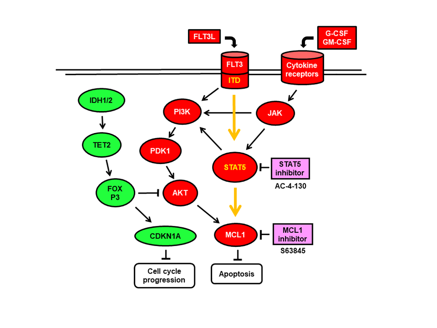
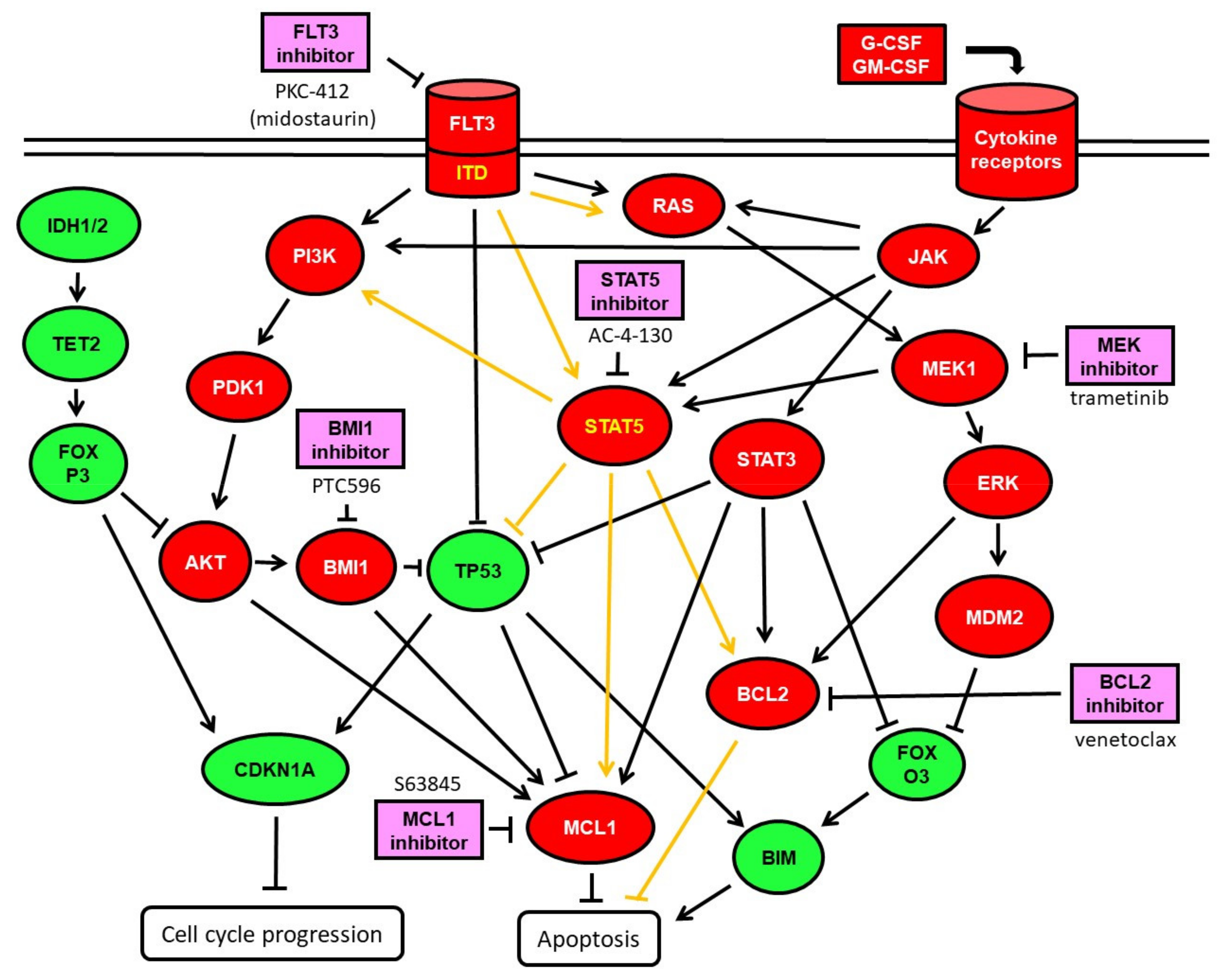
Rationale for a Combination Therapy with the STAT5 Inhibitor AC-4-130 and the MCL1 Inhibitor S63845 in the Treatment of FLT3-Mutated or TET2-Mutated Acute Myeloid Leukemia

1
Department for Biomedical Research, University of Bern, 2008 Bern, Switzerland
2
Department of Hematology, University Hospital Bern, 3010 Bern, Switzerland
3
Department of Medical Oncology, University Hospital Bern, 3010 Bern, Switzerland
*
Authors to whom correspondence should be addressed.
Int. J. Mol. Sci. 2021, 22(15), 8092; https://0-doi-org.brum.beds.ac.uk/10.3390/ijms22158092
Submission received: 28 May 2021 / Revised: 19 July 2021 / Accepted: 26 July 2021 / Published: 28 July 2021
(This article belongs to the Special Issue Hematological Malignancies)

Abstract

:
The FMS-like tyrosine kinase 3 (FLT3) gene is mutated in one-third of patients with de novo acute myeloid leukemia (AML). Mutated FLT3 variants are constitutively active kinases signaling via AKT kinase, MAP kinases, and STAT5. FLT3 inhibitors have been approved for the treatment of FLT3-mutated AML. However, treatment response to FLT3 inhibitors may be short-lived, and resistance may emerge. Compounds targeting STAT5 may enhance and prolong effects of FLT3 inhibitors in this subset of patients with FLT3-mutated AML. Here STAT5-inhibitor AC-4-130, FLT3 inhibitor midostaurin (PKC412), BMI-1 inhibitor PTC596, MEK-inhibitor trametinib, MCL1-inhibitor S63845, and BCL-2 inhibitor venetoclax were assessed as single agents and in combination for their ability to induce apoptosis and cell death in leukemic cells grown in the absence or presence of bone marrow stroma. Synergistic effects on cell viability were detected in both FLT3-mutated and FLT3-wild-type AML cells treated with AC-4-130 in combination with the MCL1 inhibitor S63845. AML patient samples with a strong response to AC-4-130 and S63845 combination treatment were characterized by mutated FLT3 or mutated TET2 genes. Susceptibility of AML cells to AC-4-130, PTC596, trametinib, PKC412, and venetoclax was altered in the presence of HS-5 stroma. Only the MCL1 inhibitor S63845 induced cell death with equal efficacy in the absence or presence of bone marrow stroma. The combination of the STAT5-inhibitor AC-4-130 and the MCL1 inhibitor S63845 may be an effective treatment targeting FLT3-mutated or TET2-mutated AML.

Graphical Abstract

1. Introduction

FMS-like tyrosine kinase 3 (FLT3) is an inducible growth factor receptor signaling via PI3K-PDK1-AKT and via RAS-RAF-MEK-ERK leading to cell growth and proliferation [1,2]. Mutations of the FLT3 gene are detected in around a third of patients with de novo acute myeloid leukemia (AML). Mutated FLT3 variants are constitutively active kinases signaling via AKT and MAP kinases, and as a gain of function via STAT5 [3]. The FLT3 inhibitor midostaurin was approved together with intensive chemotherapy for first-line treatment of FLT3-mutated AML by the FDA in 2017, and was also authorized for use in the EU. However, treatment response to FLT3 inhibitors may be short-lived, and leukemia relapse is the major cause of treatment failure, as resistance may frequently emerge [4]. Moreover, the stromal microenvironment provides an escape route from FLT3 inhibitors through the GAS6-AXL-STAT5 axis [5,6].
STAT5 refers to two highly related genes, STAT5A and STAT5B, which are both located on human chromosome 17 [7]. STAT5 proteins are not only activated by a wide variety of ligands that control proliferation, survival, and cell communication, but their dysregulation also facilitates tumor progression in various human cancers, particularly leukemia and lymphoma [8]. The STAT5 proteins are key downstream transcription factors in FLT3-mutated AML. STAT5 inhibition was reported to be a promising strategy for FLT3-ITD+ AML treatment [5]. In AML cells, STAT5 can be activated by FLT3-ITD, but also by activated cytokine receptors [3,7,9]. Myeloid cytokine receptors can be activated by granulocyte and macrophage colony-stimulating factors (G-CSF, GM-CSF, M-CSF), stem cell factor (SCF), thrombopoetin (THPO), and interleukins. These cytokines are secreted by bone marrow stroma cells to support the growth of normal hematopoietic stem and progenitor cells in the bone marrow. The same cytokines will also support the growth of leukemic stem and progenitor cells in the bone marrow. In normal cord blood cells, STAT5 phosphorylation can be efficiently induced by THPO, IL-3, and GM-CSF. SCF-induced STAT5 phosphorylation is largely restricted to the megakaryocyte–erythroid progenitor (MEP) compartment, while G-CSF, as well as IL-3 and GM-CSF, are most efficient in inducing STAT5 phosphorylation in the myeloid progenitor compartments [10]. Stromal cells can induce multidrug resistance in AML cells via upregulation of the PI3K/Akt signaling pathway, or via upregulation of STAT3 signaling [11,12].
Compounds directly targeting STAT5 canonical functions may inhibit dimerization, DNA binding, or transcriptional activity. The mechanisms of direct STAT5 inhibition include disruption of tyrosine phosphorylation, dimerization, nuclear translocation, and/or DNA binding. Targeting the SH2 domain was, therefore, the main focus for the design and identification of selective inhibitors [13]. AC-4-130 directly binds to the STAT5 SH2 domain and disrupts STAT5 activation, dimerization, nuclear translocation, and STAT5-dependent gene transcription [14]. AC-4-130 impairs the proliferation and clonogenic growth of human AML cell lines and primary FLT3-ITD+ AML patient cells in vitro and in vivo. Activated STAT5 can block apoptosis via induction of BCL-2 and MCL-1 proteins [15,16]. The BCL2 inhibitor venetoclax was approved by the FDA in 2018 in combination with azacitidine, decitabine, or cytarabine for the treatment of newly diagnosed acute myeloid leukemia (AML) in adults who are 75 years or older, or who have comorbidities that preclude use of intensive induction chemotherapy. The MCL1 inhibitor S63845 was effective in combination with the MEK inhibitor trametinib in hematological cells with elevated MCL1- and MEK-protein levels, independent of the mutational status of FLT3 and TP53 [17].
Here, we assessed the STAT5-inhibitor AC-4-130, FLT3 inhibitor midostaurin, BMI-1 inhibitor PTC596, MEK-inhibitor trametinib, MCL1-inhibitor S63845, and BLC2 inhibitor venetoclax as single agents and in combination for their ability to induce apoptosis and cell death in leukemic cells grown in the absence or presence of bone marrow stroma. AML cells represented all major morphologic and molecular subtypes with normal karyotype, including FLT3 mutated or wild-type, NPM1 mutated or wild-type, as well as TP53 mutated or wild-type cells.

2. Results

2.1. Susceptibility of AML Cell Lines Grown in the Absence or Presence of HS-5 Stroma Cells to AC-4-130 and Venetoclax

To determine the sensitivity of AML cells to the STAT5 inhibitor AC-4-130, AML cells were subjected to in vitro cytotoxicity assays. AML cells were treated with the compound for 20 h in dose-escalation experiments before cell-viability assessment. Cell viability was also determined in AML cells grown in the presence of bone marrow stroma cells secreting granulocyte and macrophage colony-stimulating factors (G-CSF, GM-CSF, M-CSF), and other cytokines thereby inducing STAT signaling. The AML cell lines covered the major morphologic and molecular subtypes including, particularly, FLT3 mutated or wild-type, NPM1 mutated or wild-type, as well as TP53 wild-type, mutated, hemizygous, and null cells (Table 1).
The susceptibility to AC-4-130 was elevated in TP53-mutated SKM-1 cells, with IC50 of 0.7 µM; intermediate in TP53 wild-type MOLM-13 and ML-2 cell lines, with IC50 values of 3 µM; reduced in the TP53-mutant MOLM-16 and TP53 wild-type cell line OCI-AML3, with IC50 values of 10 µM; and very low in the TP53 null HL-60 cells (Figure 1A). In the presence of stroma cytokines, the susceptibility to AC-4-130 was enhanced in SKM-1 cells, with IC50 at 0.5 µM; and reduced in MOLM-13 and ML-2, with IC50 at 4.4 µM and 10 µM, respectively (Figure 1B). The sensitivity of AML cell lines to BMI1 inhibitor PTC596, MEK-inhibitor trametinib, and MCL1-inhibitor S63845 was determined in previous studies [17,18]. To determine the sensitivity of the AML cell lines to the BCL2 inhibitor venetoclax, dose-escalation experiments were performed. The susceptibility to venetoclax was elevated in ML-2 and MOLM-13, with IC50 values of 0.3 and 0.8 µM, respectively; intermediate in HL-60; and very low in SKM-1, MOLM-16, and OCI-AML3 cells, with IC50 > 10 µM (Figure 1C). In the presence of stroma cytokines, the susceptibility to venetoclax was much enhanced in SKM-1 cells, with IC50 of 0.4 µM, in OCI-AML3 with IC50 of 1.2 µM, and reduced in ML-2 with IC50 of 4 µM (Figure 1D). In order to define the most effective treatment combinations, we focused on inhibitors expected to elicit synergistic effects in combination with AC-4-130 based on previous studies with BMI1, FLT3, MCL1- and MEK inhibitors [17,18,19,20], as well as the BCL2 inhibitor venetoclax, as indicated in Figure 2.

2.2. AC-4-130 Combination Treatment in AML Cell Lines

Cell viability was determined in AML cell lines treated with increasing dosages of single compounds and in combination treatments using the STAT5 inhibitor AC-4-130 and a variety of targeted therapies, including the BMI-1 inhibitor PTC596, the MCL1 inhibitor S63845, and the MEK inhibitor trametinib (Figure 3). Combination indexes were calculated according to Chou Talalay (Table 2). Some cell lines were additionally treated with AC-4-130 in combination with the FLT3-ITD inhibitor PKC-412 (midostaurin) or the BCL2 inhibitor venetoclax. Synergistic effects were present in TP53 mutated cell line SKM-1 treated with AC-4-130 and PTC596, trametinib, S63845, or venetoclax (Figure 3A, Figure S1). For the FLT3-ITD cell line MOLM-13, response to combination treatments was detected with moderate synergy with AC-4-130 in combination with PKC-412, S63845, or venetoclax, as well as additive effects with AC-4-130 in combination with PTC596, and antagonistic effects in the combination of AC-4-130 with trametinib (Figure 3B, Figure S1). The FLT3 and TP53 wild-type cell line ML-2 response was moderately synergistic to AC-4-130 in combination with S63845, and mildly synergistic in combination with trametinib or venetoclax (Figure S1).

2.3. Changed Susceptibility of AML Cell Lines to Combination Treatment with AC-4-130 in the Presence of Bone Marrow Stroma

To investigate the elevated susceptibility of FLT3 wild-type SKM-1 compared to FLT3-mutated MOLM-13 cells to AC-4-130, cell viability was determined in AML cells grown in the presence of bone marrow stroma cells secreting granulocyte and macrophage colony-stimulating factors (G-CSF, GM-CSF, M-CSF), and other cytokines thereby inducing STAT signaling. The STAT5 inhibitor AC-4-130 was more effective in SKM-1 (Figure 3C, Figure S1), and less effective in MOLM-13 cells grown in the presence of HS-5 stroma (Figure 3D), indicating a moderate cell- and context-specific dependence of STAT5 signaling. The BMI1 inhibitor PTC596 was more effective in SKM-1 and less effective in MOLM-13 cells grown in the presence of HS-5 cells, indicating an elevated cell and context dependence of BMI1 signaling. The BCL2 inhibitor venetoclax was more effective in SKM-1 and less effective in ML-2 cells in the presence of HS-5 stroma (Figure S1), indicating a cell and context dependence of BCL2 function. In contrast, the MCL1 inhibitor S63845 had similar efficacy in SKM-1 and MOLM-13 cells independent of HS-5 presence. The elevated susceptibility of SKM-1 cells to AC-4-130 may be due to dominant signaling of cytokine receptors via STAT5 in this cell line. SKM-1 cells’ response to the BCL2 inhibitor was very low in the absence, but substantial in the presence of HS-5 stroma, indicating BCL2 induction by cytokine receptor signaling in this cell line. In MOLM-13 cells, STAT5 signaling is induced by FLT3-ITD. Here, the presence of stroma cytokines may have activated other STAT proteins and reduced the susceptibility to the STAT5 inhibitor. In contrast to the STAT5 inhibitor AC-4-130 and the BMI1 inhibitor PTC596, whose efficacies were cell- and context-dependent, the MEK1 inhibitor trametinib was generally less effective in the AML cells in the presence of HS-5 stroma (Figure 3). While susceptibility to the MEK inhibitor trametinib was reduced in SKM-1 cells, MOLM-13 cells were resistant to trametinib in the presence of stroma cytokines. MEK1 may activate STAT5 in SKM-1, but not in MOLM-13 cells. For the FLT3 inhibitor midostaurin (PKC412), the susceptibility of MOLM-13 cells was reduced in the presence of HS-5 stroma. Because of the altered susceptibilities, the effects of combination treatments on AML cells grown in the absence of HS-5 stroma were altered in AML cells grown in the presence of HS-5 stroma (Table 2).

2.4. Induction of Apoptosis and Cell Death in AML Cell Lines

The effects of treatment with AC-4-130 and PTC596, S63845, or trametinib alone and in combination on induction of apoptosis, cell-cycle arrest, and cell death were determined in cytometric assays (Figure 4A–F), while effects on the expression of STAT5 target genes CDKN1A and BCL2 were determined by qRT-PCR (Figure 4G,H). Apoptosis and cell death were strongly induced in MOLM-13 cells treated with AC-4-130 and further enhanced in combination with the BMI1 inhibitor PTC596 (Figure 4A,B). Apoptosis and cell death were also induced in MOLM-13 cells with AC-4-130 in combination with the FLT3 inhibitor midostaurin (PKC412) or the BCL2 inhibitor venetoclax (Figure 4C,D). Induction of apoptosis and cell death were less pronounced in SKM-1 cells treated with AC-4-130 in combination with trametinib, S63845, or PTC596 (Figure 4E). Here, the induction of cell-cycle arrest was substantiated as G1 arrest in midostaurin-treated cells and G2 arrest in PTC596-treated cells (Figure 4F), and evident in the induction of CDKN1A gene expression and reduction of BCL2 gene expression in SKM-1 cells treated with AC-4-130 and trametinib (Figure 4G,H).

2.5. AC-4-140 Combination Treatments in Leukemic Cells In Vitro

After initial studies in AML cell lines, the treatment combinations of AC-4-130 with PTC596, trametinib, or S63845 were applied to patient-derived hematological cells. Primary cells of a variety of hematological malignancies were included to determine the specificity of the treatment combinations. A total of 18 AML, 1 MDS, 2 multiple myeloma (MM), 2 B-ALL, and 1 CML, as well as PBMCs of 4 healthy donors (HD), were subjected to single-compound and combination treatments (Table 3). Treatment with the STAT5 inhibitor AC-4-130 (2 µM) on its own had minimal effects on cell viability (Figure S2). Treatment with the BMI inhibitor PTC596 on its own or in combination with AC-4-130 had minimal effects on cell viability (Figure S2A). The cytotoxic effects induced by trametinib (100 nM) treatment were significant in a few AML samples, but not further enhanced in combination with AC-4-130 (Figure S2B). Cytotoxic effects induced by treatment with the MCL1 inhibitor S63845 (100 nM) were significant in 12 AML, the MDS sample and 1 B-ALL, with further enhancement in combination with AC-4-130 (Figure S2C). Treatment with the FLT3 inhibitor midostaurin (PKC412) on its own or in combination with AC-4-130 had minimal effects on cell viability (Figure S2D). Treatment with the BCL2 inhibitor venetoclax on its own had significant effects on cell viability in half of the tested AML samples, with further enhancement in combination with AC-4-130 (Figure S2E).
Compared to the healthy donor cells, the tested patient samples split into two groups with respect to response to combination treatment with AC-4-130 and S63845 (Figure 5). Cell viability was reduced to 75% in healthy donor cells, to 80% in AML cells with normal response (NR), and to 40% in AML cells with strong response (SR). The AML samples with strong response to AC-4-130 and S63845 combination treatment comprised eight FLT3 mutated, five TET2 mutated AML, and one AML with IDH2 and DNMT3A mutations. One of the secondary AMLs susceptible to the combination of AC-4-130 and S63845 carried a TP53 mutation with a variant allele frequency of 5%, while two AMLs less susceptible to this combination treatment carried TP53 mutations with a variant allele frequency of 50% and 92%. Of the two B-ALL samples, one was very susceptible to the combination of AC-4-130 and S63845, while the other one had a normal response. However, to determine whether this treatment combination may be effective in B-ALL, a wider array of ALL samples would have to be tested.

3. Discussion

As treatment response to FLT3 inhibitors may be short-lived, with leukemia relapse as the major cause of treatment failure, compounds targeting STAT5 may enhance and prolong effects of FLT3 inhibitors in this subset of patients with FLT3-mutated AML. To characterize the susceptibility of various AML cell lines to the STAT5 inhibitor AC-4-130, we performed a dose-escalation screening. Susceptibility to AC-4-130 varied in the tested AML cell lines with elevated susceptibility of the TP53-mutated secondary AML cell line SKM-1, intermediate susceptibility of the TP53 wild-type cell lines MOLM-13 and ML-2, as well as reduced susceptibility of the TP53 wild-type cell line OCI-AML3, the TP53 mutated MOLM-16, and the TP53 null cell line HL-60. The elevated susceptibility of the SKM-1 cell line was unexpected, as there is no mutated FLT3 activating STAT5 signaling in these cells, and the presence of mutated tumor suppressor TP53 is often associated with chemo-resistance. In view of the cell-viability studies in AML patient cells, the susceptibility of SKM-1 cells to AC-4-130 may be due to the presence of a TET2 mutation in this cell line. To investigate the elevated susceptibility of FLT3 wild-type SKM-1 compared to FLT3-mutated MOLM-13 cells to AC-4-130, cell viability was determined in AML cells grown in the presence of bone marrow stroma secreting granulocyte and macrophage colony-stimulating factors (G-CSF, GM-CSF, M-CSF), and other cytokines thereby inducing STAT5 signaling. In the presence of bone marrow stroma, AC-4-130 and the BMI1 inhibitor PTC596 were more effective in SKM-1 cells and less effective in MOLM-13 cells, indicating that the elevated susceptibility of SKM-1 cells to AC-4-130 and PTC596 may be due to dominant signaling of cytokine receptors via STAT5 and via PI3K/AKT in this cell line. In addition, co-culture with HS-5 stroma may induce the STAT5 target protein BCL2, as has been suggested for HL-60 cells [25]. In contrast to AC-4-130 and PTC596, the MEK1 inhibitor trametinib and the FLT3 inhibitor PKC412 (midostaurin) were less effective in the AML cells in the presence of HS-5 stroma. HS-5 stroma may induce resistance to FLT3 and MEK inhibitors via upregulation of the PI3K/AKT signaling pathway [11]. Similar effects have been described in c-kit mutant AML cells, where cytokines secreted by bone marrow stromal cells protect c-KIT mutant AML cells from c-KIT inhibitor-induced apoptosis [26]. The differential efficacies of targeted compounds in the context of the peripheral blood and bone marrow environment may be relevant for therapeutic success. Early relapse may arise when treatments are effective at eradicating leukemic cells in the peripheral blood but not in the bone marrow, where leukemic cells are sheltered. Thus, novel biological systems are currently established that enable the investigation of leukemia–stroma cross-talk and verification of novel therapies’ effectiveness under such bone marrow niche-mimicking conditions [27]. In our study, we employed a simple co-culture system with the HS-5 stroma cell line to grow AML cells in a bone-marrow niche environment. We discovered that several targeted compounds had reduced efficacy in AML cells grown on HS-5 stroma, while the MCL1 inhibitor S63845 induced cell death with equal efficacy in the absence or presence of bone marrow stroma, making it an excellent candidate to target leukemic stem cells in the bone marrow.
To determine which compounds may be effective in AML cells treated in combination with a STAT5 inhibitor, we focused on inhibitors expected to elicit synergistic cytotoxic effects in combination treatments based on previous studies with BMI1, FLT3, MCL1, and MEK inhibitors [17,18,19,20]. In the current study, we found synergistic cytotoxic effects in FLT3 wild-type AML cell lines treated with combinations of the STAT5 inhibitor AC-4-130 and the MCL1 inhibitor S63845, and in the FLT3-ITD-positive MOLM-13 cells treated with combinations of the STAT5 inhibitor AC-4-130 and the FLT3 inhibitor midostaurin (PKC412). However, concurrent STAT5 and FLT3 inhibitor treatment was not effective in FLT3-mutated AML patient samples. There were also clearly antagonistic effects observed in MOLM-13 cells treated with a combination of STAT5 inhibitor AC-4-130 and the MEK inhibitor trametinib. Moreover, the effects of concurrent STAT5 and MEK inhibition were rather antagonistic in AML patient samples, independent of FLT3 status. This antagonistic effect in the presence of STAT5 and MEK inhibition may result from induced signaling via PI3K and AKT, thereby preventing induction of apoptosis.
To validate the findings in a translational setting, we applied the STAT5 inhibitor AC-4-130 in combination with BMI1, FLT3, MCL1, MEK, and BCL2 inhibitors to AML patient samples. While treatments with combinations of the STAT5 inhibitor AC-4-130 with the FLT3 inhibitor midostaurin (PKC412), the BMI1 inhibitor PTC596 or the MEK inhibitor trametinib were rather ineffective, the combination of AC-4-130 and the MCL1 inhibitor S63845 led to a significant reduction in cell viability in a larger subset of AML. The susceptible AML patient samples were characterized by the presence of either mutated FLT3 or mutated TET2, and one case of mutated IDH2 and DNMT3A genes. The susceptibility of FLT3-mutated cells to the STAT5 and MCL1 inhibitor may be based on the gain of function of the mutated FLT3 activating STAT5 [3] and its downstream target MCL1. The susceptibility of TET2 mutant cells to the STAT5 inhibitor may be related to a cooperativity of TET2 and STAT5 in the activation of downstream targets [28,29]. The susceptibility of TET2 mutated cells to the MCL1 inhibitor may arise due to AKT-induced MCL1 over-activation in these cells, where AKT may no longer be regulated by mutated TET2 protein [22,23]. Leukemic IDH1 and IDH2 mutations disrupt TET2 function and impair hematopoietic differentiation [30,31]. As TET2 function is dependent on the metabolites produced by the IDH proteins [21,32], susceptibility to STAT5 and MCL1 inhibitors may be similar in AML cells with IDH mutations, as in TET2 mutated cells.
Susceptibility to the combination treatment of AC-4-130 and S63845 was significant in a secondary AML carrying a TP53 mutation with a variant allele frequency (VAF) of 5%. Two AML samples less susceptible to the combination of AC-4-130 and S63845 carried TP53 mutations at frequencies of 50% and 92%. TP53 mutation variant allele frequency may be associated with clinical outcome of patients with myelodysplastic syndrome (MDS) and AML [33,34]. In newly diagnosed AML, the presence of a TP53 mutation with VAF > 40% was independently associated with a significantly higher cumulative incidence of relapse and worse relapse-free and overall survival in patients treated with a cytarabine-based regimen.
It is important to note that there was a considerable effect of the MCL1 inhibitor S63845 in combination with the STAT5 inhibitor AC-4-130 on cell viability of normal PBMCs of healthy donors. This is in contrast to the marginal effect of the MCL1 inhibitor S63845 in combination with the MEK inhibitor trametinib on normal PBMCs previously described in [17], indicating that MCL1 function is of vital importance in normal PBMCs in the context of activated STAT5 signaling, but not in the context of activated MEK signaling. Such a considerable effect on normal monocytes may constitute a limitation in a treatment strategy using a combination of STAT5 and MCL1 inhibitor in a therapeutic context. The side effects of a STAT5 and MCL1 inhibitor combination therapy may be comparable to the side effects of the conventional standard induction therapy, including elevated risk of infection and increased bruising or bleeding. A possible advantage of a STAT5 and MCL1 inhibitor combination compared to the standard induction therapy may be the effective reduction of leukemic stem cells in the bone marrow.
Compared to previous studies, we now, for the first time, included the bone marrow stroma in our preclinical testing of AML cells. This is a new approach that introduces a new level of testing of potentially effective combination therapies. New insights may be gained by restating preclinical testing in AML cells grown in the presence of bone marrow stroma.

4. Materials and Methods

4.1. Patient Samples

Mononuclear cells of AML patients diagnosed and treated at the University Hospital, Bern, Switzerland, between 2005 and 2018 were included in this study. Informed consent from all patients was obtained according to the Declaration of Helsinki, and the studies were approved by decisions of the local ethics committee of Bern, Switzerland. Peripheral blood mononuclear cells (PBMCs) and bone marrow mononuclear cells (BMMCs) were collected at the time of diagnosis before initiation of treatment. The AML cells were analyzed at the central hematology laboratory of the University Hospital Bern according to state-of-the-art techniques [35]. Mutational screening for FLT3, NPM1, TP53, and conventional karyotype analysis of at least 20 metaphases were performed in all samples. In addition, all samples were analyzed by NGS sequencing of the myeloid panel genes.

4.2. Cell Lines and Cell Culture

Human AML cells lines OCI-AML3 (AML-M4, FLT3wt, DNMT3A R882C, NPM1mut, TP53wt), MOLM-13 (AML-M5, t(9;11), FLT3-ITD, TP53wt), MOLM-16 (AML-M0, FLT3wt, TP53mut), ML-2 (AML-M4, t(6;11), FLT3wt, TP53mut), and HL-60 (AML-M2, FLT3wt, TP53 null) were supplied by the Leibniz Institute DSMZ, German Collection of Micro-Organisms and Cell Cultures. AML cells were grown in RPMI 1640 media (SIGMA-ALDRICH, St. Louis, MO, USA) supplemented with 20% fetal bovine serum (FBS, Biochrom GmbH, Berlin, Germany) in a standard cell culture incubator at 37 °C with 5% CO2. The human bone marrow stroma cell line HS-5 was obtained from ATCC (ATCC® CRL-11882™). HS-5 cells were grown in DMEM media (SIGMA-ALDRICH, St. Louis, MO, USA) supplemented with 10% fetal bovine serum (FBS, Biochrom GmbH, Berlin, Germany). The HS-5 cells secreted granulocyte colony-stimulating factor (G-CSF), granulocyte-macrophage-CSF (GM-CSF), macrophage-CSF (M-CSF), Kit ligand (KL), macrophage-inhibitory protein-1 alpha, interleukin-1 alpha (IL-1alpha), IL-1beta, IL-1RA, IL-6, IL-8, IL-11, and leukemia inhibitory factor (LIF). For the co-culture assays, HS-5 cells were plated on six-well plates on day 1. On day 2, Nunc 0.4 uM cell culture inserts (ThermoFisher, Roskilde, Denmark) were placed over the HS-5 feeder layer and AML cells were filled into the cell culture inserts. On day 3, AML cells were collected from the six-well inserts and replated on 96-well plates, before addition of compounds. Cytotoxicity assays were performed on day 4.

4.3. Cytotoxicity Assays

For assays with AML cell lines, cells were plated at a density of 5 × 10⁵/mL and treated with targeted compounds or conventional induction therapy. For assays with patient-derived mononuclear cells, the cells were cultured for 2 h prior to treatment. The STAT5 inhibitor AC-4-130 was purchased at Aobious, Inc. Gloucester, MA, USA. The BMI1 inhibitor PTC596, the FLT3 inhibitor midostaurin (PKC412), the MCL1 inhibitor S63845, and the MEK inhibitor trametinib were purchased at MedChemExpress (Monmouth Junction, NJ, USA). A stock solution of venetoclax was prepared by dissolving a venclexta tablet (Abbvie Inc., North Chicago, IL, USA) in DMSO. Cell viability was determined after 20 h of treatment using the MTT-based cell-proliferation kit I (Roche Diagnostics, Mannheim, Germany). This time point was selected because the cellular responses were effectual for the calculation of combination indexes after 20 h of treatment with two compounds in leukemic cells. For AML cell lines, four independent assays (biological replicates) with four measurements (technical replicates) per dosage were performed. For hematological patient samples, two independent assays with three technical replicates per dosage were performed. Statistical analysis was done on GraphPad Prism (GraphPad Software, San Diego, CA, USA). Data are depicted as XY graphs, box plots or scatter plots with mean values and SD. For the calculation of combination indexes, three dosages of AC-4-130 and two dosages of the other compounds were applied alone and in combination. Combination indexes were calculated on CompuSyn software (version 1.0; ComboSyn, Inc., Paramus, NJ, USA).

4.4. Measurement of mRNA Expression by qPCR

RNA was extracted from AML cells and quantified using qPCR. The RNA extraction kit was supplied by Macherey-Nagel, Düren, Germany. Reverse transcription was done with MMLV-RT (Promega, Madison, WI, USA). Real-time PCR was performed on the QuantStudio 7 Real-Time PCR Instrument using FastStart Universal master mix (Roche Diagnostics, Mannheim, Germany) and gene-specific probes (Cat# 4331182, Thermo Fisher Scientific, Waltham, MA, USA) Hs00355782_m1 (CDKN1A), Hs04986394_s1 (BCL2), and Hs02758991_g1 (GAPDH). Measurements for CDKN1A and BCL2 were normalized with GAPDH values (ddCt relative quantitation). Assays were performed in three or more independent experiments. Data are depicted in column bar graphs plotting mean with SD values.

4.5. Antibodies and Cytometry

Staining for apoptosis was done using annexinV-CF488A (Biotium Inc., Fremont, CA, USA) in annexinV buffer and Hoechst 33,342 (10 μg/mL) for 15 min at 37 °C, followed by several washes. Propidium iodide was added shortly before imaging on a NC-3000 imager (ChemoMetec, Allerod, Denmark). For cell-cycle analysis, cells were incubated in lysis buffer with DAPI (10 μg/mL) for 5 min at 37 °C and analyzed on NC-3000 imager.

Supplementary Materials

Author Contributions

Conceptualization, K.S.; investigation, K.S., C.G. and L.F.; resources, U.B. and T.P.; writing—original draft preparation, K.S.; writing—review and editing, K.S., U.B. and T.P.; visualization, K.S.; supervision, K.S.; funding acquisition, T.P. All authors have read and agreed to the published version of the manuscript.

Funding

This work was supported by a grant from the Swiss National Science Foundation (SNF), #310030_127509 to TP.

Institutional Review Board Statement

The study was conducted according to the guidelines of the Declaration of Helsinki, and approved by decisions of the local ethics committee of Bern, Switzerland, decision number #221/15, last approval 4 November 2020.

Informed Consent Statement

Informed consent was obtained from all subjects involved in the study according to the Declaration of Helsinki. General consent form available.

Data Availability Statement

Data is contained within the article or the Supplementary Materials.

Conflicts of Interest

The authors declare no conflict of interest.

References

  1. Chang, F.; Steelman, L.S.; Lee, J.T.; Shelton, J.G.; Navolanic, P.M.; Blalock, W.L.; Franklin, R.A.; McCubrey, J.A. Signal Transduction Mediated by the Ras/Raf/MEK/ERK Pathway from Cytokine Receptors to Transcription Factors: Potential Targeting for Therapeutic Intervention. Leukemia 2003, 17, 1263–1293. [Google Scholar] [CrossRef]
  2. Takahashi, S. Downstream Molecular Pathways of FLT3 in the Pathogenesis of Acute Myeloid Leukemia: Biology and Therapeutic Implications. J. Hematol. Oncol. 2011, 4, 13. [Google Scholar] [CrossRef] [Green Version]
  3. Spiekermann, K.; Bagrintseva, K.; Schwab, R.; Schmieja, K.; Hiddemann, W. Overexpression and Constitutive Activation of FLT3 Induces STAT5 Activation in Primary Acute Myeloid Leukemia Blast Cells. Clin. Cancer Res. 2003, 9, 2140–2150. [Google Scholar]
  4. Lam, S.S.Y.; Leung, A.Y.H. Overcoming Resistance to FLT3 Inhibitors in the Treatment of FLT3-Mutated AML. Int. J. Mol. Sci. 2020, 21, 1537. [Google Scholar] [CrossRef] [Green Version]
  5. Orlova, A.; Neubauer, H.A.; Moriggl, R. The Stromal Microenvironment Provides an Escape Route from FLT3 Inhibitors through the GAS6-AXL-STAT5 Axis. Haematologica 2019, 104, 1907–1909. [Google Scholar] [CrossRef]
  6. Dumas, P.-Y.; Naudin, C.; Martin-Lannerée, S.; Izac, B.; Casetti, L.; Mansier, O.; Rousseau, B.; Artus, A.; Dufossée, M.; Giese, A.; et al. Hematopoietic Niche Drives FLT3-ITD Acute Myeloid Leukemia Resistance to Quizartinib via STAT5-and Hypoxia-Dependent Upregulation of AXL. Haematologica 2019, 104, 2017–2027. [Google Scholar] [CrossRef] [PubMed] [Green Version]
  7. Jatiani, S.S.; Baker, S.J.; Silverman, L.R.; Reddy, E.P. JAK/STAT Pathways in Cytokine Signaling and Myeloproliferative Disorders. Genes Cancer 2010, 1, 979–993. [Google Scholar] [CrossRef] [Green Version]
  8. Wingelhofer, B.; Neubauer, H.A.; Valent, P.; Han, X.; Constantinescu, S.N.; Gunning, P.T.; Müller, M.; Moriggl, R. Implications of STAT3 and STAT5 Signaling on Gene Regulation and Chromatin Remodeling in Hematopoietic Cancer. Leukemia 2018, 32, 1713–1726. [Google Scholar] [CrossRef] [PubMed] [Green Version]
  9. Schepers, H.; Wierenga, A.T.J.; Vellenga, E.; Schuringa, J.J. STAT5-Mediated Self-Renewal of Normal Hematopoietic and Leukemic Stem Cells. JAK-STAT 2012, 1, 13–25. [Google Scholar] [CrossRef] [Green Version]
  10. Han, L.; Wierenga, A.T.J.; Rozenveld-Geugien, M.; van de Lande, K.; Vellenga, E.; Schuringa, J.J. Single-Cell STAT5 Signal Transduction Profiling in Normal and Leukemic Stem and Progenitor Cell Populations Reveals Highly Distinct Cytokine Responses. PLoS ONE 2009, 4, e7989. [Google Scholar] [CrossRef] [PubMed] [Green Version]
  11. Chen, P.; Jin, Q.; Fu, Q.; You, P.; Jiang, X.; Yuan, Q.; Huang, H. Induction of Multidrug Resistance of Acute Myeloid Leukemia Cells by Cocultured Stromal Cells via Upregulation of the PI3K/Akt Signaling Pathway. Oncol. Res. 2016, 24, 215–223. [Google Scholar] [CrossRef]
  12. Hou, D.; Wang, B.; You, R.; Wang, X.; Liu, J.; Zhan, W.; Chen, P.; Qin, T.; Zhang, X.; Huang, H. Stromal Cells Promote Chemoresistance of Acute Myeloid Leukemia Cells via Activation of the IL-6/STAT3/OXPHOS Axis. Ann. Transl. Med. 2020, 8, 1346. [Google Scholar] [CrossRef] [PubMed]
  13. Brachet-Botineau, M.; Polomski, M.; Neubauer, H.A.; Juen, L.; Hédou, D.; Viaud-Massuard, M.-C.; Prié, G.; Gouilleux, F. Pharmacological Inhibition of Oncogenic STAT3 and STAT5 Signaling in Hematopoietic Cancers. Cancers 2020, 12, 240. [Google Scholar] [CrossRef] [Green Version]
  14. Wingelhofer, B.; Maurer, B.; Heyes, E.C.; Cumaraswamy, A.A.; Berger-Becvar, A.; de Araujo, E.D.; Orlova, A.; Freund, P.; Ruge, F.; Park, J.; et al. Pharmacologic Inhibition of STAT5 in Acute Myeloid Leukemia. Leukemia 2018, 32, 1135–1146. [Google Scholar] [CrossRef] [PubMed] [Green Version]
  15. Debierre-Grockiego, F. Anti-Apoptotic Role of STAT5 in Haematopoietic Cells and in the Pathogenesis of Malignancies. Apoptosis 2004, 9, 717–728. [Google Scholar] [CrossRef]
  16. Yoshimoto, G.; Miyamoto, T.; Jabbarzadeh-Tabrizi, S.; Iino, T.; Rocnik, J.L.; Kikushige, Y.; Mori, Y.; Shima, T.; Iwasaki, H.; Takenaka, K.; et al. FLT3-ITD up-Regulates MCL-1 to Promote Survival of Stem Cells in Acute Myeloid Leukemia via FLT3-ITD-Specific STAT5 Activation. Blood 2009, 114, 5034–5043. [Google Scholar] [CrossRef] [PubMed] [Green Version]
  17. Seipel, K.; Schmitter, K.; Bacher, U.; Pabst, T. Rationale for a Combination Therapy Consisting of MCL1- and MEK-Inhibitors in Acute Myeloid Leukemia. Cancers 2019, 11, 1779. [Google Scholar] [CrossRef] [PubMed] [Green Version]
  18. Seipel, K.; Kopp, B.; Bacher, U.; Pabst, T. BMI1-Inhibitor PTC596 in Combination with MCL1 Inhibitor S63845 or MEK Inhibitor Trametinib in the Treatment of Acute Leukemia. Cancers 2021, 13, 581. [Google Scholar] [CrossRef]
  19. Seipel, K.; Marques, M.A.T.; Sidler, C.; Mueller, B.U.; Pabst, T. MDM2- and FLT3-Inhibitors in the Treatment of FLT3-ITD Acute Myeloid Leukemia, Specificity and Efficacy of NVP-HDM201 and Midostaurin. Haematologica 2018, 103, 1862–1872. [Google Scholar] [CrossRef] [PubMed] [Green Version]
  20. Seipel, K.; Marques, M.A.T.; Sidler, C.; Mueller, B.U.; Pabst, T. The Cellular P53 Inhibitor MDM2 and the Growth Factor Receptor FLT3 as Biomarkers for Treatment Responses to the MDM2-Inhibitor Idasanutlin and the MEK1 Inhibitor Cobimetinib in Acute Myeloid Leukemia. Cancers 2018, 10, 170. [Google Scholar] [CrossRef] [PubMed] [Green Version]
  21. Pasca, S.; Jurj, A.; Zdrenghea, M.; Tomuleasa, C. The Potential Equivalents of TET2 Mutations. Cancers 2021, 13, 1499. [Google Scholar] [CrossRef]
  22. Basu, S.; Hubbard, B.; Shevach, E.M. Foxp3-Mediated Inhibition of Akt Inhibits Glut1 (Glucose Transporter 1) Expression in Human T Regulatory Cells. J. Leukoc. Biol. 2015, 97, 279–283. [Google Scholar] [CrossRef] [PubMed] [Green Version]
  23. Chen, Y.-J.; Huang, C.-H.; Shi, Y.-J.; Lee, Y.-C.; Wang, L.-J.; Chang, L.-S. The Suppressive Effect of Arsenic Trioxide on TET2-FOXP3-Lyn-Akt Axis-Modulated MCL1 Expression Induces Apoptosis in Human Leukemia Cells. Toxicol. Appl. Pharmacol. 2018, 358, 43–55. [Google Scholar] [CrossRef] [PubMed]
  24. Chou, T.-C. Drug Combination Studies and Their Synergy Quantification Using the Chou-Talalay Method. Cancer Res. 2010, 70, 440–446. [Google Scholar] [CrossRef] [PubMed] [Green Version]
  25. Guan, D.; Qing, W.; Ma, C.; Zhang, Z.; Wei, H.; Wu, G. Bone Marrow Stromal-Cell Line HS-5 Affects Apoptosis of Acute Myeloid Leukemia Cells HL-60 through GLI1 Activation. Biomed. Res. 2018, 29, 865–868. [Google Scholar] [CrossRef] [Green Version]
  26. Gordon, P.M.; Dias, S.; Williams, D.A. Cytokines Secreted by Bone Marrow Stromal Cells Protect C-KIT Mutant AML Cells from c-KIT Inhibitor-Induced Apoptosis. Leukemia 2014, 28, 2257–2260. [Google Scholar] [CrossRef] [Green Version]
  27. Podszywalow-Bartnicka, P.; Kominek, A.; Wolczyk, M.; Kolba, M.D.; Swatler, J.; Piwocka, K. Characteristics of Live Parameters of the HS-5 Human Bone Marrow Stromal Cell Line Cocultured with the Leukemia Cells in Hypoxia, for the Studies of Leukemia-Stroma Cross-Talk. Cytom. A 2018, 93, 929–940. [Google Scholar] [CrossRef] [Green Version]
  28. Ma, H.; Gao, W.; Sun, X.; Wang, W. STAT5 and TET2 Cooperate to Regulate FOXP3-TSDR Demethylation in CD4+ T Cells of Patients with Colorectal Cancer. J. Immunol. Res. 2018, 2018, 6985031. [Google Scholar] [CrossRef] [Green Version]
  29. Yang, R.; Qu, C.; Zhou, Y.; Konkel, J.E.; Shi, S.; Liu, Y.; Chen, C.; Liu, S.; Liu, D.; Chen, Y.; et al. Hydrogen Sulfide Promotes Tet1- and Tet2-Mediated Foxp3 Demethylation to Drive Regulatory T Cell Differentiation and Maintain Immune Homeostasis. Immunity 2015, 43, 251–263. [Google Scholar] [CrossRef] [Green Version]
  30. Figueroa, M.E.; Abdel-Wahab, O.; Lu, C.; Ward, P.S.; Patel, J.; Shih, A.; Li, Y.; Bhagwat, N.; Vasanthakumar, A.; Fernandez, H.F.; et al. Leukemic IDH1 and IDH2 Mutations Result in a Hypermethylation Phenotype, Disrupt TET2 Function, and Impair Hematopoietic Differentiation. Cancer Cell 2010, 18, 553–567. [Google Scholar] [CrossRef] [Green Version]
  31. Solary, E.; Bernard, O.A.; Tefferi, A.; Fuks, F.; Vainchenker, W. The Ten-Eleven Translocation-2 (TET2) Gene in Hematopoiesis and Hematopoietic Diseases. Leukemia 2014, 28, 485–496. [Google Scholar] [CrossRef] [PubMed]
  32. Wang, Y.; Xiao, M.; Chen, X.; Chen, L.; Xu, Y.; Lv, L.; Wang, P.; Yang, H.; Ma, S.; Lin, H.; et al. WT1 Recruits TET2 to Regulate Its Target Gene Expression and Suppress Leukemia Cell Proliferation. Mol. Cell 2015, 57, 662–673. [Google Scholar] [CrossRef] [Green Version]
  33. Belickova, M.; Vesela, J.; Jonasova, A.; Pejsova, B.; Votavova, H.; Merkerova, M.D.; Zemanova, Z.; Brezinova, J.; Mikulenkova, D.; Lauermannova, M.; et al. TP53 Mutation Variant Allele Frequency Is a Potential Predictor for Clinical Outcome of Patients with Lower-Risk Myelodysplastic Syndromes. Oncotarget 2016, 7, 36266–36279. [Google Scholar] [CrossRef] [PubMed] [Green Version]
  34. Sallman, D.A.; Komrokji, R.; Vaupel, C.; Cluzeau, T.; Geyer, S.M.; McGraw, K.L.; Al Ali, N.H.; Lancet, J.; McGinniss, M.J.; Nahas, S.; et al. Impact of TP53 Mutation Variant Allele Frequency on Phenotype and Outcomes in Myelodysplastic Syndromes. Leukemia 2016, 30, 666–673. [Google Scholar] [CrossRef]
  35. Shumilov, E.; Flach, J.; Kohlmann, A.; Banz, Y.; Bonadies, N.; Fiedler, M.; Pabst, T.; Bacher, U. Current Status and Trends in the Diagnostics of AML and MDS. Blood Rev. 2018, 32, 508–519. [Google Scholar] [CrossRef] [PubMed]
Figure 1. Dose-response curves of AML cell lines. AML cells were treated with the STAT5 inhibitor AC-4-130 (A,B) or the BCL2 inhibitor venetoclax (C,D) for 20 hours in the absence (A,C) or in the presence (B,D) of HS-5 stroma cells. Cell-viability data are average values of multiple repeat measurements per dosage. The standard deviation was 3–6%.
Ijms 22 08092 g001
Figure 2. Schematic representation of STAT5 signaling pathways in myeloid cells. STAT5 can be activated by FLT3-ITD and by cytokine receptor signaling via Janus tyrosine kinases (JAKs). FLT3-ITD is a constitutively active growth factor receptor signaling via PI3K-AKT [2], via RAS-MEK-ERK [3], and via STAT5 [16], leading to cell growth and proliferation via p53 inhibition and MCL1 induction. Hematopoietic cytokine receptor signaling is largely mediated by JAKs and their downstream transcription factors, the STATs [7]. Mutations in the tumor suppressors IDH1/2 and TET2 may be functionally equivalent [21]. The tumor suppressor FoxP3 may inhibit AKT and its downstream target MCL-1 [22,23]. Oncogenic functions are indicated in red, tumor suppressor functions in green, and chemical inhibitors in pink.
Ijms 22 08092 g002
Figure 3. Susceptibility of SKM-1 and MOLM-13 AML cells to various treatment combinations. Cell viability was determined by AML cells grown in the absence (A,B) and in the presence (C,D) of HS-5 stroma cells. SKM-1 (A,C) and MOLM-13 (B,D) cells were treated for 20 hours with single compounds and in combination with AC-4-130 (AC, yellow) and PTC596 (P, red), S63845 (S, blue), or trametinib (T, pink). Concentrations of inhibitors are nM for PTC596, S63845, and trametinib, and µM for AC-4-130. All values are in reference to mock-treated cells (=100% viability).
Ijms 22 08092 g003
Figure 4. Induction of apoptosis and cell death in AML cells treated with AC-4-130 alone and in combination with targeted compounds. Cytometric analysis of MOLM-13 cells treated for 20 h with AC-4-130 alone and in combination with PTC596 (A,D), PKC412 or venetoclax (B,E) to measure induction of apoptosis using annexinV and PI staining (A,B), and induction of cell-cycle arrest and cell death (subG1 fraction) using DAPI staining (B,E). Cytometric analysis of SKM-1 cells treated for 20 h with AC-4-130 (AC) alone and in combination with trametinib (T), S63845 (S), or PTC596 to measure induction of apoptosis using annexinV and PI staining (C), and induction of cell-cycle arrest and cell death (subG1 fraction) using DAPI staining (F). Relative quantitation of STAT5A target genes CDKN1A and BCL2 in SKM-1 cells treated for 20 hours with AC-4-130 and trametinib (G,H). Concentrations of inhibitors were nM for PKC412 (PKC), PTC596 (P), S63845 (S), trametinib (T), and venetoclax (VC), and uM for AC-4-130 (AC). Cells were grouped into four categories each: low- and high-intensity signal in Annexin staining (Ann lo, Ann hi), and low- and high-intensity signal in PI staining (PI lo, PI hi). DNA content in DAPI staining < 2n, 2n, >2n, 4n (subG1, G0/G1, S, G2).
Ijms 22 08092 g004
Figure 5. Susceptibility of hematological cells in vitro to combination treatment with AC-4-130 and S63845. Cell viability was determined in hematological cells after 20 h treatment with 2 µM AC-4-130 and 100 nM S63845The patient samples were sorted into two groups, one with normal response (NR) and one with strong response (SR). Significance was calculated by Mann-Whitney test. Primary data are presented in Figure S2.
Ijms 22 08092 g005
Table 1. Genetic variants in acute myeloid leukemia (AML) cell lines.
IDFABOriginFLT3TP53Gene VariantsKaryotype
HL-60M2de novowtnullCDKN2A R80X
NRAS Q61L
hypotetraploid
ML-2M4de novowtwtKMT2A-AFDN
KRAS A146T
t(6;11)
MOLM-13M5a, relapseMDSITDwtKMT2A-MLLT3t(9;11)
MOLM-16M0, relapsede novowtV173M
C238S
MLL V1368L
MTOR T571K
hypotetraploid
OCI-AML3M4de novowtwtNRAS Q61L
NPM1 L287fs
DNMT3A R882C
+1, +5, +8
SKM-1M5, refractoryMDSwtR248Q
R248Q
ASXL1 Y591X
KRAS K117N
TET2 C1419fs
del9q12
AML, acute myeloid leukemia; MDS, myelodysplastic syndrome. FAB, French-American-British classification.
Table 2. Combination index values in AML cell lines.
AML
Cell Line
HS-5
Stroma
AC-4-130
+PTC596
AC-4-130
+Trametinib
AC-4-130
+S63845
AC-4-130
+PKC412
AC-4-130
+Venetoclax
SKM-1absent0.6–0.80.6–0.80.7–0.9nd0.4–0.6
present1.0–1.20.8–1.01.1–1.3nd0.5–0.7
MOLM-13absent0.9–1.11.2–1.70.8–1.00.4–0.60.6–0.8
present0.6–0.8nc0.8–1.00.4–0.60.8–0.9
Combination indexes were calculated according to Chou Talalay [24]. Interpretation of combinatorial effects. Strong synergy CI = 0.1–0.3, moderate synergy CI = 0.3–0.7, mild synergy CI = 0.7–0.9, additive CI = 0.9–1.1, antagonism CI > 1.1. nc, not calculable; nd, not determined.
Table 3. Clinical characteristics of hematological patient samples.
IDDiseaseFABAntecedentGene VariantsKaryotypeAC+S *
AML1sAMLM0 MDSEVI1 (overexpressed)−7NR
AML2AMLM0 TP53 (VAF 92%)complexNR
AML3sAML MDS CEPBA, ASXL1, EZH2, RUNX1normalNR
AML4sAML ETASXL1, CALR, KMT2A (amp), TP53 (VAF 50%), 49, +der(11)NR
AML5AMLM5 FLT3-TKD (0.63), NPM1, DNMT3AnormalSR
AML6sAMLM4bicytopeniaNPM1,FLT3-TKD (0.57), TET2, TP53 (VAF 5%), SRSF2+8SR
AML7AMLM5 ASXL1, TET2, KRAS, SH2B3, U2AF1−7SR
AML8AMLM5 FLT3-ITD (0.58), NPM1, DNMT3AnormalSR
AML9sAMLM1MDSNPM1, TET2, DNMT3AnormalSR
AML10AMLM1 FLT3-ITD (0.833), NPM1normalSR
AML11AMLM2 normalcomplex, −7,−9NR
AML12sAML MPNJAK2normalNR
AML13sAMLM4breast cancerNPM1, TET2normalSR
AML14AMLM1 FLT3-ITD (>1.0), NPM1normalSR
AML15AMLM4 FLT3-ITD (0.504), NPM1normalSR
AML16AMLM2 NPM1, IDH2, DNMT3AnormalSR
AML17AMLM1 FLT-3-ITD (0.783), BCOR, TET2, U2AF1del20q11, +8SR
AML18AMLM4 FLT3-TKD (0.487), NRAS, KRAS, KMT2A-MLLT10t(10;11), +8SR
MDS1MDS TET2, ETV6, KRAS, SRSF2, CBLdel7q SR
BA1B-ALL IGH (rearranged)normalNR
BA2B-ALL normalnormalSR
CML1CML BCR-ABL1t(9;22)NR
MM1MM normalNR
MM2MM t(4;14)NR
HDn.a. normalnormalNR
AML, acute myeloid leukemia; sAML, secondary AML; ALL, acute lymphoblastic leukemia; CML, chronic myeloid leukemia; ET, essential thrombocytopenia; HD, healthy donor MDS, myelodysplastic syndrome; MPN, myeloproliferative neoplasm; MM, multiple myeloma. For the FLT3 gene, the mutant allele ratio, and for the TP53 gene, the mutant allele frequency (VAF) are indicated in parentheses. * Response to combination treatment with AC-4-130 and S63845; normal response (NR), strong response (SR).
Publisher’s Note: MDPI stays neutral with regard to jurisdictional claims in published maps and institutional affiliations.

