Review

The 3,4-Quinones of Estrone and Estradiol Are the Initiators of Cancer whereas Resveratrol and N-acetylcysteine Are the Preventers

1
Eppley Institute for Research in Cancer and Allied Diseases, University of Nebraska Medical Center, 986805 Nebraska Medical Center, Omaha, NE 68198-6805, USA
2
Department of Environmental, Agricultural and Occupational Health, University of Nebraska Medical Center, 984388 Nebraska Medical Center, Omaha, NE 68198-4388, USA
*
Author to whom correspondence should be addressed.
Int. J. Mol. Sci. 2021, 22(15), 8238; https://0-doi-org.brum.beds.ac.uk/10.3390/ijms22158238
Submission received: 15 June 2021 / Revised: 14 July 2021 / Accepted: 21 July 2021 / Published: 30 July 2021
(This article belongs to the Special Issue Estrogens in Human Cancer)

Abstract

:
This article reviews evidence suggesting that a common mechanism of initiation leads to the development of many prevalent types of cancer. Endogenous estrogens, in the form of catechol estrogen-3,4-quinones, play a central role in this pathway of cancer initiation. The catechol estrogen-3,4-quinones react with specific purine bases in DNA to form depurinating estrogen-DNA adducts that generate apurinic sites. The apurinic sites can then lead to cancer-causing mutations. The process of cancer initiation has been demonstrated using results from test tube reactions, cultured mammalian cells, and human subjects. Increased amounts of estrogen-DNA adducts are found not only in people with several different types of cancer but also in women at high risk for breast cancer, indicating that the formation of adducts is on the pathway to cancer initiation. Two compounds, resveratrol, and N-acetylcysteine, are particularly good at preventing the formation of estrogen-DNA adducts in humans and are, thus, potential cancer-prevention compounds.

1. Introduction

Estrogens play a role in the development of multiple types of prevalent cancers. This relationship has been observed for more than a century. However, the role of estrogens has only been elucidated in recent years. Estrogen-induced cancers include breast, ovarian and endometrial cancer in women and prostate cancer in men. In postmenopausal women, a two-fold greater risk of breast cancer has been observed in women with higher levels of circulating estrogen than in women with lower levels [1]. Limited data have been compiled addressing this relationship in premenopausal women [1]. Prolonged exposure of women to elevated levels of endogenous estrogens has also been associated with an increased risk for breast cancer [2]. Data also support a relationship between levels of circulating estrogens and risk of ovarian or endometrial cancer in postmenopausal women [1].
The role of estrogens in the etiology of prostate cancer has been recognized for a shorter period of time. In 1995, Bosland et al. hypothesized that prostate cancer is initiated by estrogens and promoted by androgens [3]. More recently, epidemiologic evidence supports the role of estrogens in the etiology of prostate cancer [4]. For example, circulating estrogen levels in African-American men, who have the highest incidence of prostate cancer in the United States, are higher than those in Caucasian men [5]. On the other hand, Japanese men have a low incidence of prostate cancer and relatively low circulating estrogen levels compared to Dutch European men [6]. The ratio of total estradiol (E2) to total testosterone and total estrone (E1) to androstenedione was also found to be higher in Blacks than in other groups [7]. The enzyme aromatase catalyzes the conversion of androgens into estrogens. Aromatase activity has been demonstrated in the prostate, enabling the synthesis of estrogens from androgens in situ in the gland [8]. Interestingly, aromatase knock-out mice cannot produce E2 in the prostate, and they develop benign prostate hypertrophy rather than prostate cancer [9]. All of this information demonstrates the critical role of estrogens in the etiology of prostate cancer.
The oncogenic role of estrogens has been investigated in terms of hormonal, nonhormonal, and immunological responses. Chemical carcinogenesis, however, is the basis for the initiation of cancer. The chemicals that cause most human cancers are estrogens, and all of us, both men and women, form endogenous estrogens.
There is a common mechanism leading to the initiation of many prevalent types of cancer. Cancer can be initiated by increased formation of reactive estrogen metabolites called catechol estrogen-3,4-quinones. If estrogen metabolism becomes unbalanced and significant amounts of these quinones arise, depurinating estrogen-DNA adducts are primarily formed, leading to cancer-causing mutations [10,11,12,13,14].
A legitimate question can be raised about the relative importance of depurinating estrogen-DNA adducts compared with stable adducts. The depurinating adducts are quickly detached from the strand of DNA, leaving behind apurinic sites, whereas stable adducts remain in DNA unless removed by repair enzymes [13,14]. The essential role of depurinating DNA adducts, and the apurinic sites they generate during the initiation of cancer, was first demonstrated with adducts of three potent carcinogenic polycyclic aromatic hydrocarbons (PAH), namely: benzo[a]pyrene; 7,12-dimethylbenz[a]anthracene, and dibenzo[a,l]pyrene [13]. For all three PAH, the site of mutations in the H-ras oncogene in mouse skin tumors is dependent on the site of depurinating PAH-DNA adducts [15]. If the depurinating adducts are predominantly formed at adenines, the oncogenic H-ras mutations are at adenines. Similarly, the formation of depurinating adducts at guanines correlates with H-ras mutations at guanines in the tumors [15]. Similar results have been observed with depurinating estrogen-DNA adducts in both mouse skin [16] and rat mammary glands [17]. By contrast, there is no correlation between stable DNA adducts with oncogenic mutations. Thus, depurinating estrogen-DNA adducts play a critical role in the etiology of various prevalent types of cancer.

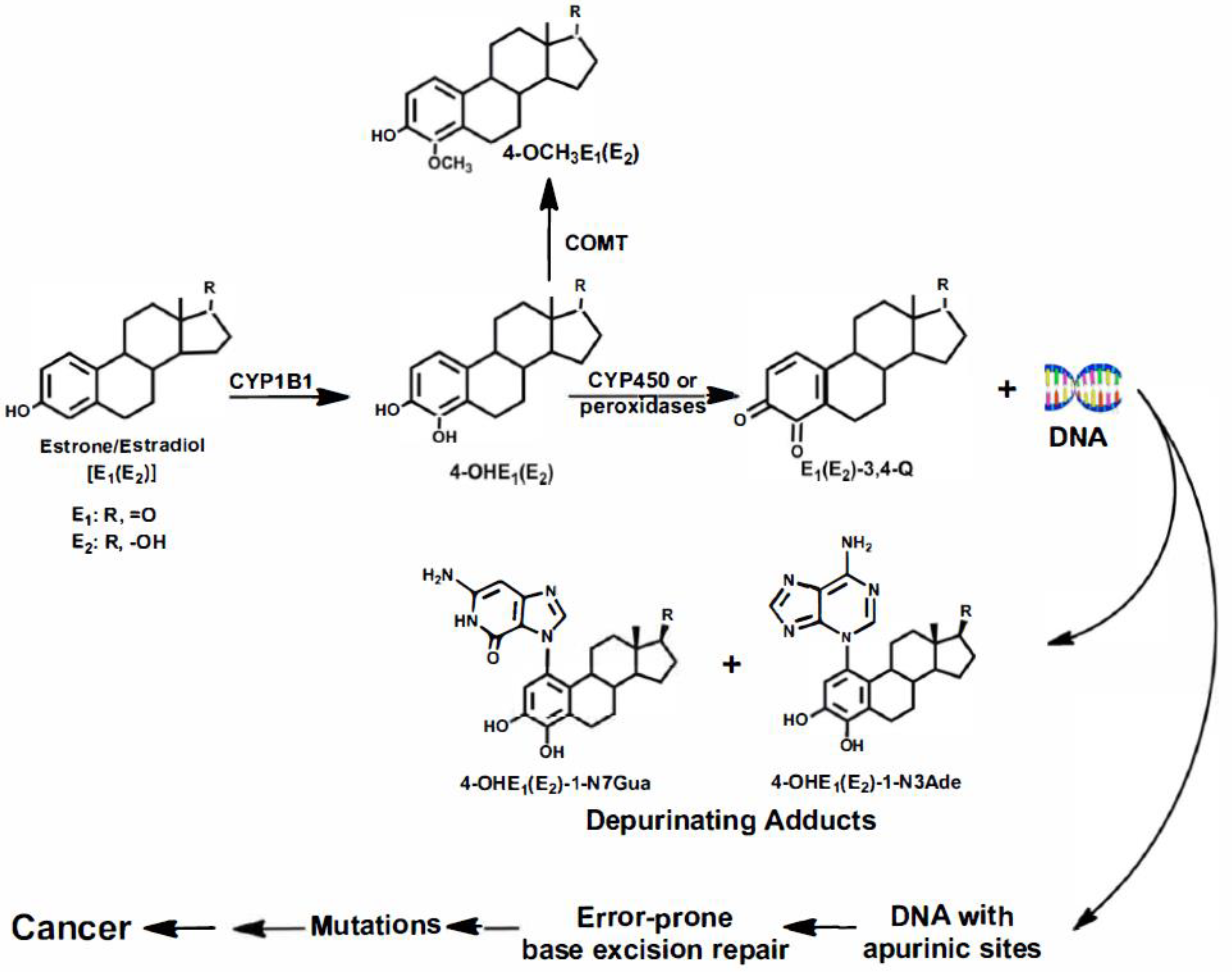
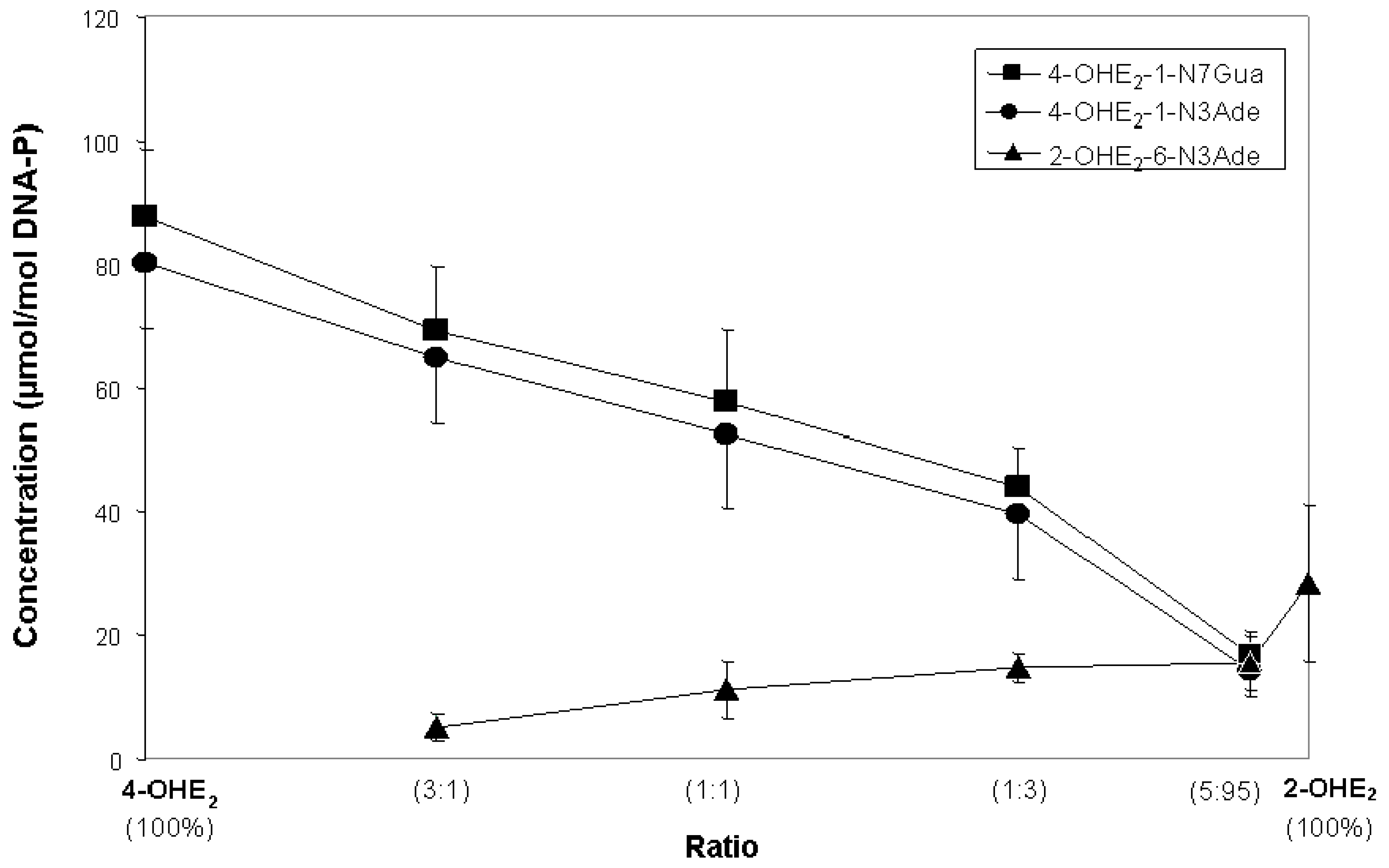
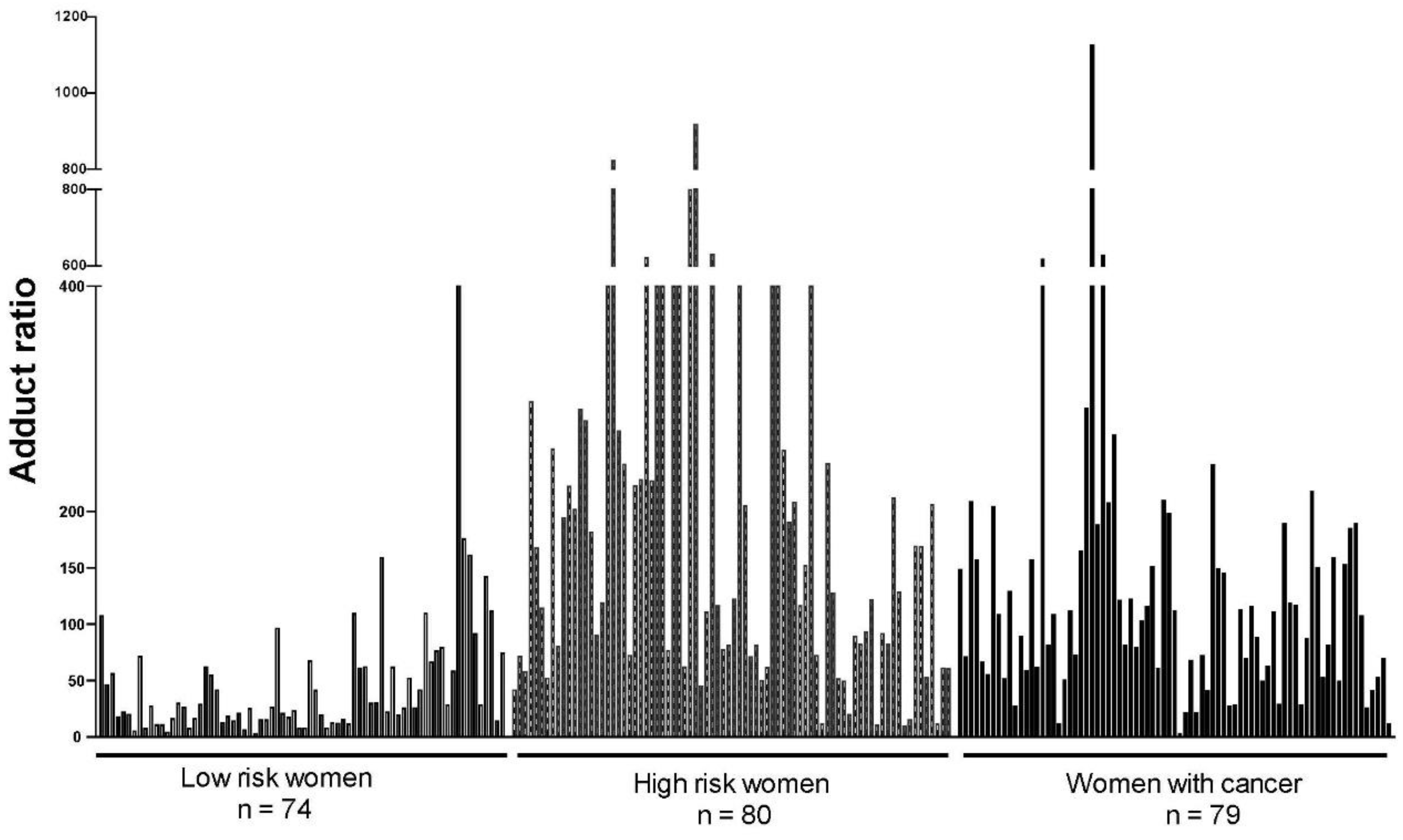
2. Initiation of Cancer

The pathway by which estrogens can cause cancer is shown in Figure 1. In this pathway, the estrogens estrone (E1) and estradiol (E2) are oxidized by cytochrome P450 (CYP)1B1 to the 4-catechol estrogens, 4-OHE1(E2) [18], which are further oxidized to the E1(E2)-3,4-quinones [E1(E2)-3,4-Q] by cytochrome P450 or peroxidases. Estrogen metabolism can be balanced or unbalanced in terms of the levels of catechol estrogen quinones formed. Normally, estrogen metabolism is balanced and low levels of E1(E2)-3,4-Q are formed, but when estrogen metabolism becomes unbalanced, higher levels of 4-OHE1(E2) and E1(E2)-3,4-Q are formed [11,12,13,14]; this has been demonstrated in the kidneys of Syrian golden hamsters [11], prostates of Noble rats [3,19], mammary glands of estrogen receptor knock-out (ERKO)/Wnt-1 mice [20], and breasts of women [12].
The E1(E2)-3,4-Q overwhelmingly react with DNA at specific guanines and adenines to form the estrogen-DNA adducts 4-OHE1(E2)-1-N7Gua and 4-OHE1(E2)-1-N3Ade (Figure 1), which are released from DNA by depurination, leaving apurinic sites in the DNA [16,17]. Repair of the apurinic sites can lead to errors, resulting in mutations; this has been demonstrated in both mice and rats [16,17]. These genetically altered cells then undergo a series of changes resulting in the growth of malignant cells into detectable cancer. The development of estrogen-initiated cells into malignant tumors does not require the involvement of the estrogen receptor (ER), although the ER certainly plays a role in the promotion of cancer cells; the development of mammary tumors in 100% of female ERKO/Wnt-1 mice demonstrated this. These mice have no functional ER-α, and they have the Wnt-1 transgene, which induces mammary tumors in all female mice [21,22]. Initially, it was thought that knocking out ER-α would demonstrate that the estrogen receptor is necessary for the development of mammary tumors, but the tumors developed in the absence of the receptor, disproving a critical role of the receptor in the development of mammary tumors [21,22]. However, these tumors are dependent on E2, and tumor development can be controlled by the dose of E2 given to ovariectomized females [23,24]. To further prove that ER-α does not play an essential role in the development of mammary tumors, it was found that tumors arise even in female ERKO/Wnt-1 mice treated with an antiestrogen known to block the receptor [25]. Nonetheless, mammary tumors develop in ERKO/Wnt-1 mice more slowly than in mice that have ER-α [22,23,24,25], suggesting a role of ER-α in tumor promotion.
The immortalized human breast epithelial MCF-10F cell line does not contain functional ER-α. Nonetheless, these cells can also be transformed by E2 into malignant cells that induce tumors when implanted into immunodeficient mice [26,27,28,29]. The transformation of MCF-10F cells into malignant cells can be increased or decreased by manipulating the levels and/or activity of estrogen-metabolizing enzymes such as CYP1B1 and catechol-O-methyltransferase (COMT) [30,31,32,33]. With more CYP1B1 activity, more E1(E2)-3,4-Q, higher levels of adducts, and more cell transformation occur. By contrast, with more COMT activity, less E1(E2)-3,4-Q, lower levels of adducts, and less cell transformation occur. MCF-10F cells are transformed by estrogens in the presence of antiestrogens, such as tamoxifen or ICI-182,780, demonstrating that the estrogen receptor does not play a role in the transformation [34]. These effects contribute to understanding how estrogens initiate cancer by metabolism into E1(E2)-3,4-Q, which react with DNA to generate specific mutations.
E1(E2)-3,4-Q are highly electrophilic compounds, seeking reaction with nucleophilic sites in the adenine (Ade) and guanine (Gua) bases in DNA [10,35,36]. The reaction of the electrophilic 3,4-keto groups with the N-7 of Gua and the N-3 of Ade occurs specifically at the nucleophilic C-1 position of the estrogen, resulting in the reduction of the vicinal keto groups to hydroxyl groups [13,14]; this causes the E1(E2)-3,4-Q to be far more reactive with DNA than the E1(E2)-2,3-Q, leading to many more depurinating DNA adducts formed with 4-OHE1(E2) than with 2-OHE1(E2). For example, in an in vitro reaction with DNA catalyzed by lactoperoxidase, a mixture of 95% 2-OHE2 and 5% 4-OHE2 is needed to form approximately similar amounts of the depurinating 2-OHE2 adducts as 4-OHE2 adducts (Figure 2) [37]. Similar results were obtained when mixtures of horseradish peroxidase-activated 2-OHE2 and 4-OHE2, or the quinones themselves, reacted with DNA [37]. In all of these reactions, the level of stable adducts formed was no more than approximately 1% of the total adducts.
It is not surprising, therefore, that the 4-OHE1(E2) are far more potent as carcinogens than the 2-OHE1(E2) [38,39,40]. The 4-OHE1(E2) induce kidney tumors in male Syrian golden hamsters, but the 2-OHE1(E2) do not [38,39]. Neonatal exposure of CD-1 mice to 4-OHE2 led to uterine adenocarcinomas, but 2-OHE2 had only borderline ability to induce the tumors [40]. These differences in carcinogenic potency cannot be attributed to redox cycling, which forms hydroxyl radicals, because 2-OHE2 and 4-OHE2 have identical redox potentials [41,42]. The formation of reactive oxygen species by oxidative stress has been shown to increase estrogen carcinogenesis [43]; this is considered by some to induce cancer-causing mutations, as evidenced by 8-oxo-dG [2]. However, this is not consistent with the greater potency of 4-OHE1(E2) compared with 2-OHE1(E2) [38,39,40]. Instead, the greater potency arises because E1(E2)-3,4-Q form depurinating DNA adducts more efficiently than E1(E2)-2,3-Q (Figure 2) [37].
Depurinating estrogen-DNA adducts can be detected in humans [13,14]. These adducts migrate out of cells and tissues, travel through the bloodstream, and are excreted in urine. By using ultraperformance liquid chromatography/tandem mass spectrometry (UPLC-MS/MS), the adducts 4-OHE1(E2)-1-N7Gua, 4-OHE1(E2)-1-N3Ade, and 2-OHE1(E2)-6-N3Ade can be detected in human urine and serum, although the level of the 2-OHE1(E2) adducts is generally much lower than the level of the 4-OHE1(E2) adducts [44,45,46,47,48,49,50,51]. However, the level of the adducts itself is not predictive of cancer risk since the level of estrogens in humans continuously varies. Instead, the molar ratio of depurinating estrogen-DNA adducts to estrogen metabolites and estrogen conjugates defines the balance of estrogen metabolism in a person and thus predicts that person’s risk for developing cancer [13,14,45]. This ratio is defined as follows:
ratio = ( 4 - OHE 1 ( E 2 ) - 1 - N 3 Ade + 4 - OHE 1 ( E 2 ) - 1 - N 7 Gua 4 - catechol   estrogens + 4 - catechal   estrogen   conjugates + 2 - OHE 1 ( E 2 ) - 6 - N 3 Ade 2 - catechol   estrogens + 2 - catechal   estrogen   conjugates ) ×   1000
The ratio of depurinating estrogen-DNA adducts to estrogen metabolites and conjugates has been analyzed extensively in women diagnosed with breast cancer compared with women at normal or high risk of developing the disease (Figure 3) [44,45,46]. This ratio has no units because they cancel out between the numerator and denominator. The ratio in women at normal risk of developing breast cancer was significantly lower than the ratio in women at high risk or diagnosed with breast cancer (p < 0.001) in three different studies [44,45,46]. Similar estrogen-DNA adduct ratios are found in postmenopausal women and premenopausal women [46], establishing that the balance of estrogen metabolism does not depend on the level of circulating estrogens in a person. Instead, the balance of estrogen metabolism depends on a person’s enzymological makeup and can be affected by environmental factors that alter the balance of enzyme activities.
In the initial study, estrogen-DNA adduct ratios in urine samples from 46 normal-risk women, 12 high-risk women, and 17 women with breast cancer were compared, and the average ratio of the normal-risk women was significantly lower than the average ratios of the high-risk and women with breast cancer (both p < 0.001) [44]. In the second study, groups of 40 women at normal risk, high risk, or diagnosed with breast cancer were compared; again, the average ratio of the normal-risk women was significantly lower than those of the other two groups (both p < 0.001) [45]. In the third study, estrogen-DNA adduct ratios in serum from 74 women at normal risk, 80 women at high risk, and 79 women diagnosed with breast cancer were compared; the average ratio in the normal-risk women was again significantly lower than those in the high-risk and breast cancer groups (both p < 0.0001, Figure 3) [46]. By contrast, the groups of women at high risk or diagnosed with breast cancer have similarly high average ratios (Figure 3) [44,45,46]. The critical observation from these studies is that women at high risk of developing breast cancer have a high ratio of adducts to metabolites and conjugates. This finding indicates that the formation of depurinating estrogen-DNA adducts begins before breast cancer develops and suggests that the formation of these adducts is a critical event in the initiation of cancer.
The three studies of women described above were conducted primarily with Caucasian women. An additional study was conducted with African-American women, but in this study, the women were divided into only two groups: those diagnosed with breast cancer and those who had not been diagnosed with breast cancer [47]. Once again, a significant difference was observed in the estrogen-DNA adduct ratios in the two groups of women. The women not diagnosed with breast cancer had an average ratio of 38.5, whereas those diagnosed with breast cancer had an average ratio of 92.4 (p < 0.001) [47]. These results support the role of estrogen-DNA adducts in the initiation of breast cancer.
The ratio of depurinating estrogen-DNA adducts to estrogen metabolites and conjugates are also found to be significantly higher in women with ovarian (p < 0.0001) or thyroid (p < 0.0001) cancer [48,49]. The study of women with and without ovarian cancer included urine samples for analysis of estrogen metabolites, conjugates and depurinating DNA adducts, as well as saliva samples for purification of DNA and analysis of single nucleotide polymorphisms (SNPs) in several genes for the enzymes involved in estrogen metabolism, CYP1B1 and COMT [49]. The selected SNPs resulted in a more active CYP1B1, which increased the formation of E1(E2)-3,4-Q, and a less active COMT, which decreased methylation of 4-OHE1(E2), thereby increasing the amount of E1(E2)-3,4-Q formed. Both of these SNPs thus resulted in more E1(E2)-3,4-Q available to react with DNA. When women had one or two copies of the SNP for a more active CYP1B1 plus two copies of the SNP for a less active COMT, they were three times more likely to have ovarian cancer, and had approximately twice the ratio of estrogen-DNA adducts to estrogen metabolites and conjugates as women without the SNPs. When the women had two copies of both the CYP1B1 and COMT SNPs, they were six times more likely to have the disease and had even higher estrogen-DNA adduct ratios [49]. Similar to the high ratios of estrogen-DNA adducts to estrogen metabolites and conjugates in women at high risk for breast cancer, these results suggest that the formation of estrogen-DNA adducts is a precursor to the development of cancer.
As we know, estrogens are present in both men and women. Therefore, estrogen metabolites, estrogen conjugates, and estrogen-DNA adducts can be analyzed in men and women, and the ratio of adducts to metabolites and conjugates can be determined. In fact, the first study of depurinating estrogen-DNA adducts in humans was conducted in men with and without prostate cancer [52]. As noted previously, Bosland et al. hypothesized that prostate cancer is initiated by estrogens and promoted by androgens [3]. In agreement with this hypothesis, it was found that men with prostate cancer have significantly higher ratios of adducts to metabolites and conjugates (p < 0.001) than healthy men not diagnosed with cancer [50]. The same observation was made in men with and without non-Hodgkin lymphoma (p < 0.0007) [51].
In summary, these results in both men and women strongly suggest that the formation of depurinating estrogen-DNA adducts is a critical event in the initiation of cancer.

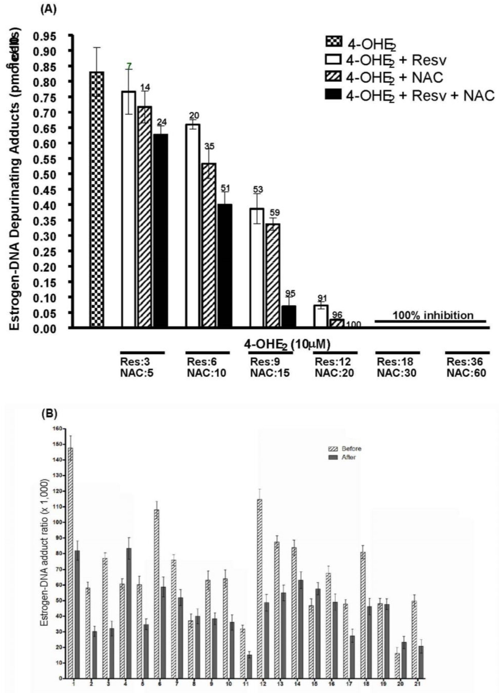
3. Prevention of Cancer by Inhibition of Estrogen-DNA Adduct Formation

Estrogens need to be metabolized to the reactive catechol estrogen-3,4-quinones to be carcinogenic. Therefore, it should be possible to prevent cancer by inhibiting this metabolic process and/or blocking reaction of the quinones with DNA. Two compounds are particularly good at blocking the metabolism of estrogens to quinones and/or scavenging the reactive quinones, they are: resveratrol and N-acetylcysteine (NAC) [13,14,30,31,32,53]. Cysteine is equally effective but it cannot be used because of its toxicity in humans. Resveratrol is a natural product found in grape skins, red wine, and peanuts, and it has various biological actions. Resveratrol is found to have several therapeutic effects; it is known to be anti-inflammatory, antioxidant, antihyperlipidemic, and anticarcinogenic, possessing immune-modulating, cardioprotective, hepatoprotective, and neuroprotective properties [54]. Among these effects are specific abilities to reduce catechol estrogen semiquinones back to catechol estrogens [30,53] and induce the estrogen-protective enzyme quinone reductase [30,31,32,53]. Resveratrol also modulates CYP1B1, thereby reducing its activity and thus the formation of 4-OHE1(E2). NAC has somewhat different properties. It, too, can reduce estrogen semiquinones back to catechol estrogens, but its primary effect is to react with quinones to form conjugates [30,31,32,53], thus preventing the formation of estrogen-DNA adducts.
Both resveratrol and NAC have been shown to inhibit the formation of depurinating estrogen-DNA adducts in cultured mammalian cells [30,31,32]. Resveratrol was found to inhibit the malignant transformation of the human MCF-10F breast epithelial cell line [31]. NAC was found to inhibit the malignant transformation of both MCF-10F and immortalized mouse mammary cells [33,55]. The two compounds work together additively to reduce the formation of depurinating estrogen-DNA adducts in MCF-10F cells treated with 4-OHE2 (Figure 4A) [30]. These results lay the foundation for investigating the ability of resveratrol and NAC to reduce estrogen-DNA adduct formation in humans as an approach to cancer prevention.
In order to consider resveratrol and/or NAC as potential cancer-preventing compounds, it is necessary to demonstrate their ability to reduce the formation of estrogen-DNA adducts in humans. Therefore, a study was conducted to test the two compounds’ ability to affect adduct formation in normal healthy women. A total of 21 women provided a urine sample before and after a 3-month treatment regimen that included taking resveratrol and NAC daily for 3 months. The urine samples were analyzed for estrogen metabolites, conjugates, and depurinating DNA adducts. In these women, the ratio of estrogen-DNA adducts to estrogen metabolites and conjugates was reduced by a statistically significant amount (p = 0.03, Figure 4B) during the three-month study period [56]. These results suggest that ingestion of NAC and resveratrol may constitute a safe, low-cost, effective measure to prevent breast cancer and other types of cancer.

4. Conclusions

Estrogens initiate cancer through metabolism to catechol estrogen-3,4-quinones. The quinones react with DNA to primarily form the depurinating DNA adducts 4-OHE1(E2)-1-N7Gua and 4-OHE1(E2)-1-N3Ade. The resulting apurinic sites in specific locations in DNA generate mutations that can initiate cancer; this has been shown in laboratory animals and human beings. Two compounds, resveratrol and NAC, are potential cancer-preventive compounds that can reduce the formation of estrogen-DNA adducts in people, thereby blocking the initiation of estrogen-induced cancer.
The two major mechanisms of estrogen-induced cancer are ER-dependent and ER-independent mechanisms. A variety of evidence has been cited to support an ER-dependent mechanism. Working through ER-α, estrogens increase the rate of cell proliferation, which decreases opportunities for cells to repair mutations; in turn, this may increase the risk for malignant transformation [57]. Additionally, the ability of anti-estrogens tamoxifen and raloxifene to prevent breast cancer in women also supports an ER-dependent mechanism [58,59,60].
Other evidence points to an ER-independent mechanism. The development of estrogen-dependent mammary tumors in ERKO/Wnt-1 mice, even in the presence of an anti-estrogen, constitutes some of the strongest evidence for an ER-independent mechanism of cancer initiation by estrogens. The observation of high ratios of depurinating estrogen-DNA adducts to estrogen metabolites and conjugates in women at high risk for breast cancer, as well as women with breast cancer [45,46,47], is consistent with an ER-independent mechanism of initiation. In addition, the presence of SNPs in CYP1B1 and COMT that increase both the formation of depurinating estrogen-DNA adducts and the likelihood of ovarian cancer (six-fold) [50] supports an ER-independent mechanism of cancer initiation by estrogens.
Of the estimated number of people in the United States who died of cancer in 2020 (606,520) [61], 140,000 were smokers. Cancer would have been preventable for approximately 466,000 people by ingestion of resveratrol and NAC; only the smokers would have died because they produce different carcinogens while smoking. If people cease their smoking habits, cancer may become a rare disease for human beings.

Funding

No specific grant from funding agencies in the public, commercial or not-for-profit sectors was received for the preparation of this article.

Institutional Review Board Statement

The studies in humans were conducted according to the guidelines of the Declaration of Helsinki, and approved by the Institutional Review Board for the University of Nebraska Medical Center (Protocols #579-13, 601-14 and 450-16, approved in 2013, 2014 and 2016).

Informed Consent Statement

Informed consent was obtained from all subjects involved in the studies.

Data Availability Statement

To obtain the data used in the results reported in this article contact Eleanor Rogan.

Conflicts of Interest

The authors declare no potential conflict of interest.

References

  1. Brown, S.B.; Hankinson, S.E. Endogenous estrogens and the risk of breast, endometrial, and ovarian cancers. Steroids 2015, 99, 8–10. [Google Scholar] [CrossRef]
  2. Yager, J.D. Mechanisms of estrogen carcinogenesis: The role of E2/E1-quinone metabolites suggests new approaches to preventive intervention—A review. Steroids 2015, 99, 56–60. [Google Scholar] [CrossRef] [Green Version]
  3. Bosland, M.C.; Ford, H.; Horton, L. Induction at high incidence of ductal prostate adenocarcinomas in NBL/Cr and Sprague-Dawley Hsd:SD rats treated with a combination of testosterone and estradiol-17β or diethylstilbestrol. Carcinogenesis 1995, 16, 1311–1317. [Google Scholar] [CrossRef]
  4. Ho, S.-M.; Lee, M.-T.; Lam, H.-M.; Keung, Y.-K. Estrogens and prostate cancer: Etiology, mediators, prevention, and management. Endocrinol. Metab. Clin. N. Am. 2011, 40, 591–614. [Google Scholar] [CrossRef] [Green Version]
  5. Rohrmann, S.; Nelson, W.G.; Rifari, N.; Brown, T.R.; Dobs, A.; Kanarek, N.; Yager, J.D.; Platz, E.A. Serum estrogen, but not testosterone, levels differ between black and white men in a nationally representative sample of Americans. J. Clin. Endocrinol. Metab. 2007, 92, 2519–2525. [Google Scholar] [CrossRef]
  6. Ross, R.; Bernstein, L.; Judd, H.; Hanisch, R.; Pike, M.; Henderson, B. Serum testosterone levels in healthy young black and white men. J. Natl. Cancer Inst. 1986, 76, 45–58. [Google Scholar]
  7. Orwoll, E.S.; Nielson, C.M.; Labrie, F.; Barrett-Connor, E.; Cauley, J.A.; Cummings, S.R.; Ensrud, K.; Karlsson, M.; Lau, E.; Leung, P.C.; et al. Evidence of geographical and racial variation in serum sex steroid levels in older men. J. Clin. Endocrinol. Metab. 2010, 95, E151–E160. [Google Scholar] [CrossRef]
  8. Krieg, M.; Nass, R.; Runn, S. Effect of aging on endogenous level of 5 alpha-dihydrotestosterone, testosterone, estradiol, and estrone in epithelium and stroma of normal and hyperplastic human prostate. J. Clin. Endocrinol. Metab. 1993, 77, 375–381. [Google Scholar] [PubMed] [Green Version]
  9. McPherson, S.J.; Wang, H.; Jones, M.E.; Pedersen, J.; Iismaa, T.O.; Wreford, N.; Simpson, E.R.; Risbridger, G.P. Elevated androgens and prolactin in aromatase-deficient mice cause enlargement, but not malignancy, of the prostate gland. Endocrinology 2001, 142, 2458–2467. [Google Scholar] [CrossRef] [PubMed]
  10. Cavalieri, E.L.; Stack, D.E.; Devanesan, P.D.; Todorovic, R.; Dwivedy, I.; Higginbotham, S.; Johansson, S.L.; Patil, K.D.; Gross, M.L.; Gooden, J.K.; et al. Molecular origin of cancer: Catechol estrogen-3,4-quinones as endogenous tumor initiators. Proc. Natl. Acad. Sci. USA 1997, 94, 10937–10942. [Google Scholar] [CrossRef] [Green Version]
  11. Cavalieri, E.L.; Kumar, S.; Todorovic, R.; Higginbotham, S.; Badawi, A.F.; Rogan, E.G. Imbalance of estrogen homeostasis in kidney and liver of hamsters treated with estradiol: Implications for estrogen-induced initiation of renal tumors. Chem. Res. Toxicol. 2001, 14, 1041–1050. [Google Scholar] [CrossRef] [PubMed]
  12. Rogan, E.G.; Badawi, A.F.; Devanesan, P.D.; Meza, J.L.; Edney, J.A.; West, W.W.; Higginbotham, S.M.; Cavalieri, E.L. Relative imbalances in estrogen metabolism and conjugation in breast tissue of women with carcinoma: Potential biomarkers of susceptibility to cancer. Carcinogenesis 2003, 24, 697–702. [Google Scholar] [CrossRef] [Green Version]
  13. Cavalieri, E.; Rogan, E. The molecular etiology and prevention of estrogen-initiated cancers. Mol. Asp. Med. 2014, 36, 1–55. [Google Scholar] [CrossRef] [Green Version]
  14. Cavalieri, E.L.; Rogan, E.G.; Zahid, M. Critical depurinating DNA adducts: Estrogen adducts in the etiology and prevention of cancer and dopamine adducts in the etiology and prevention of Parkinson’s disease. Int. J. Cancer 2017, 141, 1078–1090. [Google Scholar] [CrossRef] [Green Version]
  15. Chakravarti, D.; Pelling, J.C.; Cavalieri, E.L.; Rogan, E.G. Relating aromatic hydrocarbon-induced DNA adducts and c-Harvey-ras mutations in mouse skin papillomas: The role of apurinic sites. Proc. Natl. Acad. Sci. USA 1995, 92, 10422–10426. [Google Scholar] [CrossRef] [PubMed] [Green Version]
  16. Chakravarti, D.; Mailander, P.; Li, K.M.; Higginbotham, S.; Zhang, H.L.; Gross, M.L.; Cavalieri, E.; Rogan, E. Evidence that a burst of DNA depurination in SENCAR mouse skin induces error-prone repair and forms mutations in the H-ras gene. Oncogene 2001, 20, 7945–7953. [Google Scholar] [CrossRef] [Green Version]
  17. Mailander, P.C.; Meza, J.L.; Higginbotham, S.; Chakravarti, D. Induction of A.T to G.C mutations by erroneous repair of depurinated DNA following estrogen treatment of the mammary gland of ACI rats. J. Steroid. Biochem. Mol. Biol. 2006, 101, 204–215. [Google Scholar] [CrossRef]
  18. Zhu, B.T.; Conney, A.H. Functional role of estrogen metabolism in target cells: Review and perspectives. Carcinogenesis 1998, 9, 1–27. [Google Scholar] [CrossRef] [PubMed] [Green Version]
  19. Cavalieri, E.L.; Devanesan, P.; Bosland, M.C.; Badawi, A.F.; Rogan, E.G. Catechol estrogen metabolites and conjugates in different regions of the prostate of Noble rats treated with 4-hydroxyestradiol: Implications for estrogen-induced initiation of prostate cancer. Carcinogenesis 2002, 23, 329–333. [Google Scholar] [CrossRef] [Green Version]
  20. Devanesan, P.; Santen, R.J.; Bocchinfuso, W.P.; Korach, K.S.; Rogan, E.G.; Cavalieri, E.L. Catechol estrogen metabolites and conjugates in mammary tumors and hyperplastic tissue from estrogen receptor alpha knock out (ERKO)/Wnt 1 mice; implications for initiation of mammary tumors. Carcinogenesis 2001, 22, 1573–1576. [Google Scholar] [CrossRef] [Green Version]
  21. Bocchinfuso, W.P.; Korach, K.S. Mammary gland development and tumorigenesis in estrogen receptor knockout mice. J. Mammary Gland Biol. Neoplasia 1997, 2, 323–334. [Google Scholar] [CrossRef]
  22. Bocchinfuso, W.P.; Hively, W.P.; Couse, J.F.; Varmus, H.E.; Korach, K.S. A mouse mammary tumor virus-Wnt-1 transgene induces mammary gland hyperplasia and tumorigenesis in mice lacking estrogen receptor-α. Cancer Res. 1999, 59, 1869–1876. [Google Scholar] [PubMed]
  23. Santen, R.J.; Yue, W.; Bocchinfuso, W.; Korach, K.; Wang, J.P.; Rogan, E.G.; Yang, L.; Cavalieri, E.; Russo, J.; Devanesan, P.; et al. Estradiol-induced carcinogenesis via formation of genotoxic metabolites. In Advances in Endocrine Therapy of Breast Cancer; Ingle, J.N., Dowsett, M., Eds.; Marcel Dekker: New York, NY, USA, 2003; pp. 163–177. [Google Scholar]
  24. Yue, W.; Santen, R.J.; Wang, J.P.; Li, Y.; Verderame, M.F.; Bocchinfuso, W.P.; Korach, K.S.; Todorovic, R.; Rogan, E.G.; Cavalieri, E.L. Genotoxic metabolites of estradiol in breast: Potential mechanism of estradiol induced carcinogenesis. J. Steroid Biochem. Mol. Biol. 2003, 86, 477–486. [Google Scholar] [CrossRef]
  25. Santen, R.; Cavalieri, E.; Rogan, E.; Russo, J.; Guttenplan, J.; Ingle, J.; Yue, W. Estrogen mediation of breast tumor formation involves estrogen receptor-dependent, as well as independent, genotoxic effects. Ann. N. Y. Acad. Sci. 2009, 1155, 132–140. [Google Scholar] [CrossRef] [PubMed]
  26. Russo, J.; Lareef, M.H.; Tahin, Q.; Hu, Y.F.; Slater, C.; Ao, X.; Russo, I.H. 17-Beta-estradiol is carcinogenic in human breast epithelial cells. J. Steroid Biochem. Mol. Biol. 2002, 80, 149–162. [Google Scholar] [CrossRef]
  27. Russo, J.; Hasan Lareef, M.; Balogh, G.; Guo, S.; Russo, I.H. Estrogen and its metabolites are carcinogenic agents in human breast epithelial cells. J. Steroid Biochem. Mol. Biol. 2003, 87, 1–25. [Google Scholar] [CrossRef]
  28. Russo, J.; Russo, I.H. Genotoxicity of steroidal estrogens. Trends Endocrinol. Metab. 2004, 15, 211–215. [Google Scholar] [CrossRef]
  29. Russo, J.; Fernandez, S.V.; Russo, P.A.; Fernbaugh, R.; Sheriff, F.S.; Lareef, H.M.; Garber, J.; Russo, I.H. 17β-Estradiol induces transformations and tumorigenesis in human breast epithelial cells. FASEB J. 2006, 20, 1622–1634. [Google Scholar] [CrossRef] [PubMed]
  30. Zahid, M.; Saeed, M.; Beseler, C.; Rogan, E.G.; Cavalieri, E.L. Resveratrol and N-acetylcysteine block the cancer-initiating step in MCF-10F cells. Free Radic. Biol. Med. 2011, 50, 78–85. [Google Scholar] [CrossRef] [Green Version]
  31. Lu, F.; Zahid, M.; Wang, C.; Saeed, M.; Cavalieri, E.L.; Rogan, E.G. Resveratrol prevents estrogen-DNA adduct formation and neoplastic transformation in MCF-10F cells. Cancer Prev. Res. 2008, 1, 135–145. [Google Scholar] [CrossRef] [Green Version]
  32. Zahid, M.; Gaikwad, N.W.; Ali, M.F.; Lu, F.; Saeed, M.; Yang, L.; Rogan, E.G.; Cavalieri, E.L. Prevention of estrogen-DNA adducts in MCF-10F cells by resveratrol. Free Radic. Biol. Med. 2008, 45, 136–145. [Google Scholar] [CrossRef] [PubMed] [Green Version]
  33. Zahid, M.; Saeed, M.; Ali, M.F.; Rogan, E.G.; Cavalieri, E.L. N-Acetylcysteine blocks formation of cancer-initiating estrogen-DNA adducts in cells. Free Radic. Biol. Med. 2010, 49, 392–400. [Google Scholar] [CrossRef] [Green Version]
  34. Lareef, M.H.; Garber, J.; Russo, P.A.; Russo, I.H.; Heulings, R.; Russo, J. The estrogen antagonist ICI-182-780 does not inhibit the transformation phenotypes induced by 17-beta-estradiol and 4-OH estradiol in human breast epithelial cells. Int. J. Oncol. 2005, 26, 423–429. [Google Scholar] [PubMed]
  35. Stack, D.E.; Byun, J.; Gross, M.L.; Rogan, E.G.; Cavalieri, E.L. Molecular characteristics of catechol estrogen quinones in reactions with deoxyribonucleosides. Chem. Res. Toxicol. 1996, 9, 851–859. [Google Scholar] [CrossRef]
  36. Li, K.M.; Todorovic, R.; Devanesan, P.; Higginbotham, S.; Kofeler, H.; Ramanathan, R.; Gross, M.L.; Rogan, E.G.; Cavalieri, E.L. Metabolism and DNA binding studies of 4-hydroxyestradiol and estradiol-3,4-quinone in vitro and in female ACI rat mammary gland in vivo. Carcinogenesis 2004, 25, 289–297. [Google Scholar] [CrossRef] [Green Version]
  37. Zahid, M.; Kohli, E.; Saeed, M.; Rogan, E.; Cavalieri, E. The greater reactivity of estradiol-3,4-quinone versus estradiol-2,3-quinone with DNA in the formation of depurinating adducts. Implications for tumor-initiating activity. Chem. Res. Toxicol. 2006, 19, 164–172. [Google Scholar] [CrossRef]
  38. Liehr, J.G.; Fang, W.F.; Sirbasku, D.A.; Ari-Ulubelen, A. Carcinogenicity of catechol estrogens in Syrian hamsters. J. Steroid Biochem. 1986, 24, 353–356. [Google Scholar] [CrossRef]
  39. Li, J.J.; Li, S.A. Estrogen carcinogenesis in Syrian hamster tissues: Role of metabolism. Fed. Proc. 1987, 46, 1858–1863. [Google Scholar]
  40. Newbold, R.R.; Liehr, J.G. Induction of uterine adenocarcinoma in CD-1 mice by catechol estrogens. Cancer Res. 2000, 60, 235–237. [Google Scholar]
  41. Cavalieri, E. Minisymposium on endogenous carcinogens: The catechol estrogen pathway. An introduction. Polycycl. Aromat. Compd. 1994, 6, 223–228. [Google Scholar] [CrossRef]
  42. Mobley, J.A.; Bhat, A.S.; Brueggemeier, R.W. Measurement of oxidative DNA damage by catechol estrogens and analogues in vitro. Chem. Res. Toxicol. 1999, 12, 270–277. [Google Scholar] [CrossRef]
  43. Singh, B.; Mense, S.M.; Remotti, F.; Liu, X.; Bhat, H.K. Antioxidant butylated hydroxyanisole inhibits estrogen-induced breast carcinogenesis in female ACI rats. J. Biochem. Mol. Toxicol. 2009, 23, 302–311. [Google Scholar] [CrossRef] [Green Version]
  44. Gaikwad, N.W.; Yang, L.; Muti, P.; Meza, J.L.; Pruthi, S.; Ingle, J.N.; Rogan, E.G.; Cavalieri, E.L. The molecular etiology of breast cancer: Evidence from biomarkers of risk. Int. J. Cancer 2008, 122, 1949–1957. [Google Scholar] [CrossRef] [PubMed] [Green Version]
  45. Gaikwad, N.W.; Yang, L.; Pruthi, S.; Ingle, J.N.; Sandhu, N.; Rogan, E.; Cavalieri, E.L. Urine biomarkers of risk in the molecular etiology of breast cancer. Breast Cancer Basic Clin. Res. 2009, 3, BCBCR-S2112. [Google Scholar] [CrossRef] [PubMed]
  46. Pruthi, S.; Yang, L.; Sandhu, N.P.; Ingle, J.N.; Beseler, C.L.; Suman, V.J.; Cavalieri, E.L.; Rogan, E.G. Evaluation of serum estrogen-DNA adducts as potential biomarkers of breast cancer risk. J. Steroid Biochem. Mol. Biol. 2012, 132, 73–79. [Google Scholar] [CrossRef] [PubMed] [Green Version]
  47. Zahid, M.; Mondal, B.; LeVan, T.D.; Rogan, E.G. Estrogen metabolism in African-American women with and without breast cancer: A pilot study. Chem. Res. Toxicol. 2018, 32, 190–194. [Google Scholar] [CrossRef]
  48. Zahid, M.; Goldner, W.; Beseler, C.L.; Rogan, E.G.; Cavalieri, E.L. Unbalanced estrogen metabolism in thyroid cancer. Int. J. Cancer 2013, 133, 2642–2649. [Google Scholar] [CrossRef] [Green Version]
  49. Zahid, M.; Beseler, C.L.; Hall, J.B.; LeVan, T.; Cavalieri, E.L.; Rogan, E.G. Unbalanced estrogen metabolism in ovarian cancer. Int. J. Cancer 2014, 134, 2414–2423. [Google Scholar] [CrossRef] [Green Version]
  50. Yang, L.; Gaikwad, N.; Meza, J.; Cavalieri, E.; Muti, P.; Trock, B.; Rogan, E. Novel biomarkers for risk of prostate cancer: Results from a case-control study. Prostate 2009, 69, 41–48. [Google Scholar] [CrossRef] [PubMed] [Green Version]
  51. Gaikwad, N.W.; Yang, L.; Weisenburger, D.D.; Vose, J.; Beseler, C.; Rogan, E.G.; Cavalieri, E. Urinary biomarkers suggest that estrogen-DNA adducts may play a role in the aetiology of non-Hodgkin lymphoma. Biomarker 2009, 14, 502–512. [Google Scholar] [CrossRef] [Green Version]
  52. Markushin, Y.; Gaikwad, N.; Zhang, H.; Rogan, E.; Cavalieri, E.; Trock, B.; Pavlovich, C.; Jankowiak, R. Potential biomarker for early risk assessment of prostate cancer. Prostate 2006, 66, 1565–1571. [Google Scholar] [CrossRef]
  53. Zahid, M.; Gaikwad, N.; Rogan, E.G.; Cavalieri, E.L. Inhibition of depurinating estrogen-DNA adduct formation by natural compounds. Chem. Res. Toxicol. 2007, 20, 1947–1953. [Google Scholar] [CrossRef]
  54. Shaito, A.; Posadino, A.M.; Younes, N.; Hasan, H.; Halabi, S.; Alhababi, D.; Mohannadi, A.N.; Abdel-Rahman, W.M.; Eid, A.H.; Nasrallah, G.K.; et al. Potential adverse effects of resveratrol: A literature review. Int. J. Mol. Sci. 2020, 21, 2084. [Google Scholar] [CrossRef] [PubMed] [Green Version]
  55. Venugopal, D.; Zahid, M.; Mailander, P.C.; Meza, J.L.; Rogan, E.G.; Cavalieri, E.L.; Chakravarti, D. Reduction of estrogen-induced transformation of mouse mammary epithelial cells by N-acetylcysteine. J. Steroid Biochem. Mol. Biol. 2008, 109, 22–30. [Google Scholar] [CrossRef] [PubMed] [Green Version]
  56. Muran, P.; Muran, S.; Beseler, C.L.; Cavalieri, E.L.; Rogan, E.G.; Zahid, M. Breast health and reducing breast cancer risk, a functional medicine approach. J. Altern. Complement. Med. 2015, 21, 321–326. [Google Scholar] [CrossRef] [PubMed]
  57. Preston-Martin, S.; Pike, M.C.; Ross, R.K.; Jones, P.A.; Henderson, B.E. Increased cell division as a cause of human cancer. Cancer Res. 1990, 50, 7415–7421. [Google Scholar]
  58. Cuzick, J. International-breast-cancer-intervention-study. A brief review of the international breast cancer intervention study (IBIS), the other current breast cancer prevention trials, and proposals for future trials. Ann. N. Y. Acad. Sci. 2001, 949, 123–133. [Google Scholar] [CrossRef]
  59. Cummings, S.R.; Eckert, S.; Krueger, K.A.; Grady, D.; Powles, T.G.; Cauley, J.A.; Norton, L.; Nickelsen, T.; Bjarnason, N.H.; Morrow, M.; et al. The effect of raloxifene on risk of breast cancer in postmenopausal women: Results from the MORE randomized trial. Multiple outcomes of raloxifene evaluation. JAMA 1999, 281, 2189–2197. [Google Scholar] [CrossRef] [Green Version]
  60. Martino, S.; Cauley, J.A.; Barrett-Connor, E.; Powles, T.J.; Mershon, J.; Disch, D.; Secrest, R.J.; Cummings, S.R. For the CORE Investigators. Continuing outcomes relevant to Evista: Breast cancer incidence in postmenopausal osteoporotic women in a randomized trial of raloxifene. J. Natl. Cancer Inst. 2004, 96, 1751–1761. [Google Scholar] [CrossRef] [Green Version]
  61. American Cancer Society. Cancer Facts and Figures; American Cancer Society: Atlanta, GA, USA, 2020; p. 4. [Google Scholar]
Figure 1. Pathway of cancer initiation by estrogens.
Ijms 22 08238 g001
Figure 2. Depurinating adducts formed after 10 h (to allow complete depurination of 4-OHE2-1-N7Gua) by mixtures of lactoperoxidase-activated 4-OHE2 and 2-OHE2 reacted with DNA at different ratios. The level of stable adducts formed in the mixtures was less than 1% of the total adducts [37].
Ijms 22 08238 g002
Figure 3. Ratios of depurinating estrogen-DNA adducts to estrogen metabolites and conjugates in serum of women at low risk, women at high risk and women diagnosed with breast cancer (p < 0.0001) [46].
Ijms 22 08238 g003
Figure 4. Inhibition of estrogen-DNA adduct formation by N-acetylcysteine and resveratrol in (A) MCF-10F cells treated with 4-OHE2 [30]; and (B) urine from healthy women before and after 3 months of daily ingestion of the two compounds [56].
Ijms 22 08238 g004
Publisher’s Note: MDPI stays neutral with regard to jurisdictional claims in published maps and institutional affiliations.

