Review

Receptor-Loaded Virion Endangers GPCR Signaling: Mechanistic Exploration of SARS-CoV-2 Infections and Pharmacological Implications

Laboratory for GPCR Biology, Department of Pharmacology and Chemical Biology, University of Pittsburgh, Pittsburgh, PA 15261, USA
*
Author to whom correspondence should be addressed.
Int. J. Mol. Sci. 2021, 22(20), 10963; https://0-doi-org.brum.beds.ac.uk/10.3390/ijms222010963
Submission received: 30 August 2021 / Revised: 7 October 2021 / Accepted: 9 October 2021 / Published: 11 October 2021
(This article belongs to the Special Issue Role of Signaling Pathways in the Viral Life Cycle 2.0)

Abstract

:
SARS-CoV-2 exploits the respiratory tract epithelium including lungs as the primary entry point and reaches other organs through hematogenous expansion, consequently causing multiorgan injury. Viral E protein interacts with cell junction-associated proteins PALS1 or ZO-1 to gain massive penetration by disrupting the inter-epithelial barrier. Conversely, receptor-mediated viral invasion ensures limited but targeted infections in multiple organs. The ACE2 receptor represents the major virion loading site by virtue of its wide tissue distribution as demonstrated in highly susceptible lung, intestine, and kidney. In brain, NRP1 mediates viral endocytosis in a similar manner to ACE2. Prominently, PDZ interaction involves the entire viral loading process either outside or inside the host cells, whereas E, ACE2, and NRP1 provide the PDZ binding motif required for interacting with PDZ domain-containing proteins PALS1, ZO-1, and NHERF1, respectively. Hijacking NHERF1 and β-arrestin by virion loading may impair specific sensory GPCR signalosome assembling and cause disordered cellular responses such as loss of smell and taste. PDZ interaction enhances SARS-CoV-2 invasion by supporting viral receptor membrane residence, implying that the disruption of these interactions could diminish SARS-CoV-2 infections and be another therapeutic strategy against COVID-19 along with antibody therapy. GPCR-targeted drugs are likely to alleviate pathogenic symptoms-associated with SARS-CoV-2 infection.

1. Introduction

With over four million confirmed deaths as of August 2021 [1], the COVID-19 pandemic caused by SARS-CoV-2 poses a serious global health crisis. The causative virus has a linear, positive-sense, single-stranded RNA genome sharing highly similar gene sequences with SARS-CoV that caused the 2003 SARS outbreaks [2]. The clinical presentation of SARS-CoV-2 patients ranges from asymptomatic or mild to severe pneumonia-like symptoms and mortality [3].
The SARS-CoV-2 genome encodes 16 non-structural proteins (NSPs) such as 3CL protease NSP5, several accessory proteins, for example, ORF3a [4] and four structural proteins including spike (S), envelope (E), membrane (M), and nucleoprotein (N) shared by all coronaviruses [5]. As in the case of SARS-CoV [6], ACE2 has been identified as the receptor for entry into respiratory epithelial cells in SARS-CoV-2 infections [7]. During viral infection, the trimeric S glycoprotein is cleaved into S1 and S2 subunits, and the resulting S1 containing the receptor binding domain (RBD) is responsible for binding to ACE2 and S2 for membrane fusion [8]. Extensive vaccination strategies have been developed to introduce neutralizing antibodies mostly targeting the S protein to prevent ACE2-mediated viral invasion [9]. Rapidly developed vaccines, particularly mRNA-based COVID-19 vaccine inducing S protein-specific neutralizing antibodies, have achieved substantial success in countering the spread of SARS-CoV-2 [10]. However, in addition to vaccines, alternative therapeutics as in the case of Tamiflu (oseltamivir carboxylate) for treating influenza [11] or Molnupiravir (MK-4482, EIDD-2801) for COVID-19 [12], need to be developed to contain or mitigate the pandemic, especially considering the urgent concerns of the immune escape caused by emerging and widespread SARS-CoV-2 variants [13,14]. Herein, we discuss the functional role of the PDZ interaction in mediating SARS-CoV-2 invasion, the potentially impaired GPCR signaling and the related pharmacological implications. Comprehensive characterization of SARS-CoV-2 biology, cell signaling, and pathophysiology will provide significant clues for developing novel therapeutics to treat SARS-CoV-2 infection in humans.

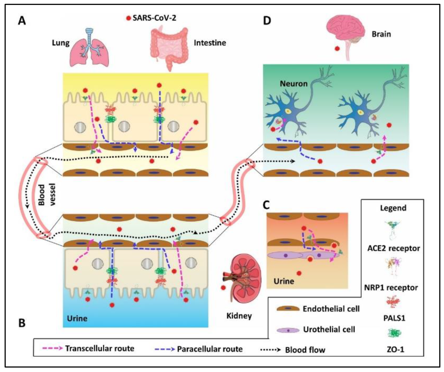
2. PDZ Interaction Enhances SARS-CoV-2 Invasion by Paracellular Leaky Route and Receptor-Mediated Endocytosis

Accumulating evidence indicates that SARS-CoV-2 penetrates host cells through distinct transcellular and paracellular pathways (Figure 1). In the transcellular route, viral S protein binds ACE2 [7] to gain cell entry in lung [15], intestine (Figure 1A) [16], and kidney (Figure 1B,C) [17]. In the brain, NRP1 has been identified as the entry receptor to facilitate the virion internalization (Figure 1D) [18,19]. Alternatively, paracellular leak caused by epithelial barrier disorganization also facilitates SARS-CoV-2 penetration in the lung [20,21], intestine [22], and possibly in the kidney [23] and brain [24]. Notably, internalized virions and those from leaky penetration end up in the circulation by a mixed and relayed delivery combining receptor loading and paracellular leak. For instance, ACE2 mediates viral entry into the host cells and the progeny virions are generated that can be released and pass through the inter-endothelial space to blood vessels or receive ACE2 reloading and bud from the endothelial cells to the same destination (Figure 1A,B). Circulating virions subsequently reach other organs by following a hematogenous route and cause multiple organ failure as reported recently [25].
Remarkably, several proteins critical for viral invasion through either paracellular or transcellular route possess a canonical, carboxy-terminal PDZ-recognition sequence. Named for the structural domain shared by the postsynaptic density protein (PSD95), Drosophila disc large tumor suppressor (DlgA), and ZO-1, PDZ domains typically bind a target motif of 3-to-4 residues in length at the carboxy-tail of host proteins. These short linear motifs with certain consensus sequences are presented in three different classes: Type I, S/T-X-Φ; Type II, Φ-X-Φ; and Type III, D/E-X-Φ, where X represents any amino acid and Φ is a hydrophobic amino acid residue [28], as shown in Table 1. On the extracellular side, SARS-CoV-2 E protein containing a Type II PDZ binding motif DLLV that can interact with the PDZ domain-containing proteins PALS1 [20] or ZO-1 [23]. Utilization of cell junction-associated proteins PALS1 or ZO-1 by forming PDZ interactions disrupts the intercellular barrier and as a consequence, increases paracellular permeability facilitating viral penetration into the interstitial space and providing a mechanism through which it can gain entry to the circulation [20,23,29]. SARS-CoV-2 E protein is a short integral membrane of 75 residues embedded in the viral envelope (Figure 2A), raising the question of how it interacts with PALS1 or ZO-1, especially considering that the long stalk of viral S protein presents a physical obstacle for E reaching into the hydrophobic pocket of PDZ domains. Furthermore, the orientation of E remains unknown. More experimental evidence is needed to functionally define the role of E in mediating viral invasion compared to the well-characterized S protein. For this to end, it is required to obtain the SARS-CoV-2 spike and envelope doubly-pseudotyped virus, where E remains intact, and S has defective mutations causing its inability to interact with the ACE2 receptor.
On the intracellular side, viral receptors ACE2 and NRP1 harbor C-terminal Type I PDZ ligands, -QTSF, and -YSEA, respectively. We recently found that the cytoplasmic PDZ domain-containing protein NHERF1 interacts with ACE2, which further aids in assembling the ACE2, B0AT1, NHERF1, and β-arrestin super-complex that attached to SARS-CoV-2 virions by ACE2-mediated recognition of the viral S protein. The super-complex enhances SARS-CoV-2 invasion, and the related scenario may be common in lung, intestine, and kidney, all of which express NHERF1 [35] and even recur in NRP1-mediated viral entry. Herein, the SARS-CoV-2 virion acts as a ligand that specifically binds to ACE2 or NRP1 receptors and mediates viral transcytosis. Receptor-plus-ligand-based viral entry often requires virion ligand binding to a specific receptor such as the phosphate housekeeping transporter PiT1 for leukemia virus [36], sodium/bile acid cotransporter for hepatitis B/D virus [37], or G protein-coupled chemokine coreceptors CCR5 and CXCR4 for human immunodeficiency virus (HIV-1) [38]. In the case of SARS-CoV-2, both identified receptors are single-pass membrane proteins that require the coreceptor B0AT1 for ACE2 [8,15] and likely VEGF-A for NRP1 [39] to maintain stable membrane residence that better supports virion loading. Additionally, SARS-CoV-2 N and 3a protein remain intracellular during viral replication and also have PDZ-binding motifs [40], -STQA, and -SVPL, respectively (Table 1). The host PDZ proteins interacting with these two proteins are not characterized and their roles remain to be ascertained. Given N protein contains a specific viral RNA packing signal [41], the unidentified cellular PDZ protein for N may serve as a docking site, scaffolding N protein-mediated genome assembly into the complete progeny virions.

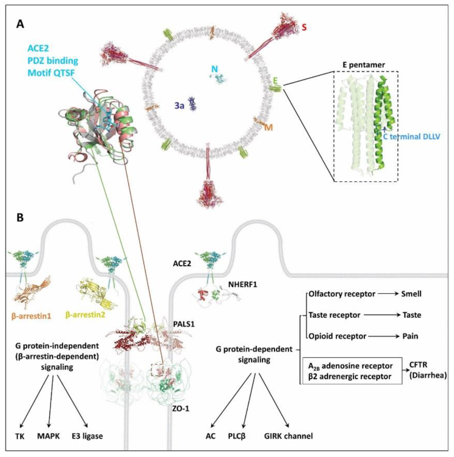
3. Possible Damaged GPCR Signaling by SARS-CoV-2 Invasion

Upon infection, the innate immune response is triggered as the initial line of defense [42], which further prompts activation of the adaptive immune responses to provide long-lasting resistance against SARS-CoV-2 [43,44]. Except for the pathological immune responses, SARS-CoV-2 infection in various target cells impairs some normal cellular physiological functions by conscripting intercellular or intracellular signaling and causing illness. Typical symptoms of infected patients often include fever or chills, cough, shortness of breath or difficult breathing, headache, fatigue, muscle or body aches, diarrhea, and loss of smell and taste, as described previously [45].
As above-mentioned, the PDZ interaction between viral E protein and host PALS1 [46,47] or ZO-1 [23] could demolish cell tight junctions and, consequently, interfere with intercellular signaling and communication. Accordingly, the neighboring cells will lose the direct exchange of metabolites and signaling molecules such as extracellular messengers and hormones required for ensuing intracellular signaling transduction, as in the case of the functional definition of ATP and serotonin in paracrine signaling [48].
In addition to interrupted intercellular signaling, the noted symptoms are also relevant to dysregulation of physiological activities by specific GPCRs such as for pain, loss of smell and taste, and diarrhea. Viruses could directly utilize and impair GPCR-related intracellular signaling, which has been reported in the case of human JC polyomavirus loaded by the serotonergic receptor 5HT2AR [49] and HIV-1 by G protein-coupled chemokine coreceptors CCR5 and CXCR4 [38]. Instead, SARS-CoV-2 employs ACE2 or NRP1 rather than GPCRs to gain cell entry. As stated in our recent work [35], SARS-CoV-2 may hijack components such as NHERF1 and β-arrestin from various GPCR assemblies that, in turn, indirectly damage specific GPCR signaling (Figure 2B). Furthermore, β-arrestin can activate G protein-independent signaling and regulate multiple cellular signaling pathways including MAPK signaling that supports cell growth and survival [50]. Indeed, affected p38 MAPK signaling has been reported upon SARS-CoV infection [51], where its genomic sequence is highly similar to SARS-CoV-2. In the case of SARS-CoV-2, the infected cells are desensitized but appear normal in the presence of agonist or antagonist due to the lack of sufficient virion-occupied component proteins like NHERF1 and β-arrestin to assemble the GPCR signalosome, further protecting the targeted cells from being attacked by the human immune system. Similar strategies have been exploited by herpesviruses that encode their own GPCR to hijack host cell GPCR signaling networks for survival [52]. For pain responses, various well-established drug targets such as G protein-coupled opioid receptors [53] are evident candidates for investigation (Table 1). In terms of smell and taste, the olfactory receptor (OR) and taste receptor (TASR) belonging to the GPCR superfamily are the responsible sensory receptors [54]. These two receptors, widely expressed in SARS-CoV-2-susceptible lung, intestine, kidney, and brain, merit special attention. Regarding diarrhea, CFTR is of particular interest considering its role in mediating transepithelial fluid secretion primarily in the lung and intestine. CFTR is not a GPCR protein but a member of the ABC (ATP-binding cassette) transporter superfamily, often functionally coupled with and tightly controlled by certain GPCRs like the β2AR [55]. Notably, these GPCRs or non-GPCRs potentially affected by SARS-CoV-2 infection all possess C-terminal PDZ-recognition sequences, particularly NHERF1-favored Type I motif (Table 1). Hijacking NHERF1 and β-arrestin employed by these receptors damages host cellular signaling and contribute to viral pathogenesis. Indeed, a recent report suggests that upon SARS-CoV-2 infection, GPCR signaling mediated by A2BAR and β2AR deleteriously modulates the activity of CFTR that in turn initiates pathophysiological lung edema [56].
Collectively, viral infection directly or indirectly impairs GPCR signaling as discussed above. On the other hand, reverse regulation of GPCR signaling driven by virus invasion could seemingly reduce viral infection as in the case of the influenza virus [57], suggesting these two events are highly correlated if, not in fact, coupled.
Table 2. PDB structures considered in this work.
Protein aUniProtLengthPDB IDReferences
3aP0DTC32756XDCRCSB [58]
EP0DTC475QHD43418ZhangLab [59]
MP0DTC5222QHD43419ZhangLab
NP0DTC9419QHD43423ZhangLab
SP0DTC21273QHD43416ZhangLab
ACE2Q9BYF1805AF-Q9BYF1-F1-model_v1AlphaFold [60]
NRP1O14786923AF-O14786-F1-model_v1AlphaFold
PALS1Q8N3R9675AF-Q8N3R9-F1-model_v1AlphaFold
ZO-1Q071571748AF-Q07157-F1-model_v1AlphaFold
NHERF1O14745358AF-O14745-F1-model_v1AlphaFold
β-arrestin1P49407418AF-P49407-F1-model_v1AlphaFold
β-arrestin2P32121409AF-P32121-F1-model_v1AlphaFold
a All full-length protein structures are from experimental or computational models as referenced. Cartoon presentations of these protein structures were prepared with PyMOL [61].

4. Implications in Specific Drug Development

The mechanistic exploration of SARS-CoV-2 infection holds the opportunity to discover new antiviral therapeutics. Terminating the destructive effect on lung and intestine cell tight junctions can be accomplished by preventing the interaction of PDZ formation with viral E protein and host extracellular PALS1 or ZO-1. Similarly, PDZ interactions mediated by intracellular ACE2/NHERF1 may be inhibited dramatically to reduce viral penetration. Indeed, drugs specifically targeting NHERF1 PDZ domains have been reported [62,63] and merit testing in the case of COVID-19. The fact that a G protein-coupled α2AR agonist inhibits influenza virus replication [57] underscores the potential for viruses to damage GPCR signaling. This mechanism could be harnessed to employ a broad catalog of existing approved drugs and to develop new agents against SARS-CoV-2 infection. Considering the potential side effects [64], regulating selective GPCR signaling may be particularly pertinent to alleviate or eliminate COVID-19 symptoms in affected patients.

5. Concluding Remarks

Enhancing immunity by vaccination is the priority tactic stemming SARS-CoV-2 infection. Novel supplementary strategies are required for treating infected patients. For instance, disrupting extracellular or intracellular PDZ interaction may reduce viral insinuation in the targeted cells. This is of particular importance considering the potential for loading massive amounts of viruses by the paracellular route, where increased penetration across the intestine and lung may cause multi-organ infection by the vascular delivery of SARS-CoV-2. Decreased cellular virion penetration by inhibiting intracellular ACE2/NHERF1 has the potential to minimize the damage to specific target organs. Furthermore, PDZ drugs hold the promise of being more effective when combined with antibody therapy.
Additionally, the regulation of certain GPCR signaling is also a worthy strategy to explore. Very few publications involving GPCR signaling induced by viral infection are currently available. The biggest challenge is to find an appropriate approach to evaluate GPCR signaling in specific infected model cells, particularly in the presence of authentic viruses. Notably, SARS-CoV-2-stressed GPCR signaling is a novel but ignored field, where investigating the relationship between viral infection and cell signaling is warranted. The related research may provide further optional or adjuvant medications for COVID-19 including repurposed GPCR-targeted drugs currently available and enable preparation for emerging variants of SARS-CoV-2.

Author Contributions

Conceptualization, Q.Z. and P.A.F.; Writing—original draft preparation, Q.Z.; Writing—review and editing, Q.Z. and P.A.F.; Visualization, Q.Z.; Funding acquisition, P.A.F. All authors have read and agreed to the published version of the manuscript.

Funding

This work was supported by the National Institutes of Health Grant R01 DK105811.

Data Availability Statement

The data that support the findings of this study are available from the corresponding author upon reasonable request.

Acknowledgments

The authors would like to acknowledge Alex R. Zhang for the technical support.

Conflicts of Interest

The authors declare no conflict of interest.

Abbreviations

GPCR: G protein-coupled receptor; SARS-CoV-2: severe acute respiratory syndrome coronavirus 2; COVID-19: coronavirus disease 2019; WHO: World Health Organization; E: envelope; S: spike; N: nucleocapsid; M: membrane; PALS: protein associated with Lin-7; PDZ: PSD-95-Disc large-ZO-1; ZO: zona occludens; ACE2: angiotensin converting enzyme 2; NRP1: neuropilin 1; PiT-1: phosphate inorganic transporter 1; CCR5: CC chemokine receptor 5; CXCR4: CXC chemokine receptor 4; HIV: human immunodeficiency virus; B0AT1: sodium-dependent neutral amino acid transporter; VEGF: vascular endothelial f0actor; 5HT2AR: 5-hydroxy-tryptamine (serotonin) 2A receptor; NHERF1: sodium-hydrogen exchanger regulatory factor 1; MAPK: mitogen-activated protein kinase; OR: olfactory receptor; TASR: taste receptor; CFTR: cystic fibrosis transmembrane conductance regulator; ABC: ATP-binding cassette; β2AR: β2 adrenergic receptor; A2BAR: adenosine A2B receptor; α2AR: α2 adrenergic receptor; TK: tyrosine kinase; AC: adenylyl cyclase; PLC: phospholipase C; GIRK: G protein-gated inwardly rectifying potassium channel.

References

  1. WHO Coronavirus Disease (COVID-19) Dashboard. Available online: https://covid19.who.int (accessed on 30 August 2021).
  2. Abdelrahman, Z.; Li, M.; Wang, X. Comparative Review of SARS-CoV-2, SARS-CoV, MERS-CoV, and Influenza A Respiratory Viruses. Front. Immunol. 2020, 11, 552909. [Google Scholar] [CrossRef]
  3. Ejaz, H.; Alsrhani, A.; Zafar, A.; Javed, H.; Junaid, K.; Abdalla, A.E.; Abosalif, K.O.A.; Ahmed, Z.; Younas, S. COVID-19 and comorbidities: Deleterious impact on infected patients. J. Infect. Public Health 2020, 13, 1833–1839. [Google Scholar] [CrossRef] [PubMed]
  4. Arya, R.; Kumari, S.; Pandey, B.; Mistry, H.; Bihani, S.C.; Das, A.; Prashar, V.; Gupta, G.D.; Panicker, L.; Kumar, M. Structural insights into SARS-CoV-2 proteins. J. Mol. Biol. 2021, 433, 166725. [Google Scholar] [CrossRef] [PubMed]
  5. Yin, C. Genotyping coronavirus SARS-CoV-2: Methods and implications. Genomics 2020, 112, 3588–3596. [Google Scholar] [CrossRef] [PubMed]
  6. Li, W.; Moore, M.J.; Vasilieva, N.; Sui, J.; Wong, S.K.; Berne, M.A.; Somasundaran, M.; Sullivan, J.L.; Luzuriaga, K.; Greenough, T.C.; et al. Angiotensin-converting enzyme 2 is a functional receptor for the SARS coronavirus. Nature 2003, 426, 450–454. [Google Scholar] [CrossRef] [PubMed] [Green Version]
  7. Hoffmann, M.; Kleine-Weber, H.; Schroeder, S.; Kruger, N.; Herrler, T.; Erichsen, S.; Schiergens, T.S.; Herrler, G.; Wu, N.H.; Nitsche, A.; et al. SARS-CoV-2 Cell Entry Depends on ACE2 and TMPRSS2 and Is Blocked by a Clinically Proven Protease Inhibitor. Cell 2020, 181, 271–280.e8. [Google Scholar] [CrossRef]
  8. Yan, R.; Zhang, Y.; Li, Y.; Xia, L.; Guo, Y.; Zhou, Q. Structural basis for the recognition of SARS-CoV-2 by full-length human ACE2. Science 2020, 367, 1444–1448. [Google Scholar] [CrossRef] [Green Version]
  9. Kyriakidis, N.C.; Lopez-Cortes, A.; Gonzalez, E.V.; Grimaldos, A.B.; Prado, E.O. SARS-CoV-2 vaccines strategies: A comprehensive review of phase 3 candidates. NPJ Vaccines 2021, 6, 28. [Google Scholar] [CrossRef]
  10. Baden, L.R.; El Sahly, H.M.; Essink, B.; Kotloff, K.; Frey, S.; Novak, R.; Diemert, D.; Spector, S.A.; Rouphael, N.; Creech, C.B.; et al. Efficacy and Safety of the mRNA-1273 SARS-CoV-2 Vaccine. N. Engl. J. Med. 2021, 384, 403–416. [Google Scholar] [CrossRef]
  11. McClellan, K.; Perry, C.M. Oseltamivir: A review of its use in influenza. Drugs 2001, 61, 263–283. [Google Scholar] [CrossRef]
  12. Malone, B.; Campbell, E.A. Molnupiravir: Coding for catastrophe. Nat. Struct. Mol. Biol. 2021, 28, 706–708. [Google Scholar] [CrossRef]
  13. Garcia-Beltran, W.F.; Lam, E.C.; St Denis, K.; Nitido, A.D.; Garcia, Z.H.; Hauser, B.M.; Feldman, J.; Pavlovic, M.N.; Gregory, D.J.; Poznansky, M.C.; et al. Multiple SARS-CoV-2 variants escape neutralization by vaccine-induced humoral immunity. Cell 2021, 184, 2372–2383. [Google Scholar] [CrossRef]
  14. Harvey, W.T.; Carabelli, A.M.; Jackson, B.; Gupta, R.K.; Thomson, E.C.; Harrison, E.M.; Ludden, C.; Reeve, R.; Rambaut, A.; Consortium, C.-G.U.; et al. SARS-CoV-2 variants, spike mutations and immune escape. Nat. Rev. Microbiol. 2021, 19, 409–424. [Google Scholar] [CrossRef] [PubMed]
  15. Salahudeen, A.A.; Choi, S.S.; Rustagi, A.; Zhu, J.; van Unen, V.; de la, O.S.; Flynn, R.A.; Margalef-Catala, M.; Santos, A.J.M.; Ju, J.; et al. Progenitor identification and SARS-CoV-2 infection in human distal lung organoids. Nature 2020, 588, 670–675. [Google Scholar] [CrossRef]
  16. Zang, R.; Gomez Castro, M.F.; McCune, B.T.; Zeng, Q.; Rothlauf, P.W.; Sonnek, N.M.; Liu, Z.; Brulois, K.F.; Wang, X.; Greenberg, H.B.; et al. TMPRSS2 and TMPRSS4 promote SARS-CoV-2 infection of human small intestinal enterocytes. Sci. Immunol. 2020, 5. [Google Scholar] [CrossRef]
  17. Batlle, D.; Soler, M.J.; Sparks, M.A.; Hiremath, S.; South, A.M.; Welling, P.A.; Swaminathan, S.; on behalf of the COVID-19 and ACE2 in Cardiovascular, Lung, and Kidney Working Group. Acute Kidney Injury in COVID-19: Emerging Evidence of a Distinct Pathophysiology. J. Am. Soc. Nephrol. 2020, 31, 1380–1383. [Google Scholar] [CrossRef] [PubMed]
  18. Cantuti-Castelvetri, L.; Ojha, R.; Pedro, L.D.; Djannatian, M.; Franz, J.; Kuivanen, S.; van der Meer, F.; Kallio, K.; Kaya, T.; Anastasina, M.; et al. Neuropilin-1 facilitates SARS-CoV-2 cell entry and infectivity. Science 2020, 370, 856–860. [Google Scholar] [CrossRef] [PubMed]
  19. Daly, J.L.; Simonetti, B.; Klein, K.; Chen, K.E.; Williamson, M.K.; Anton-Plagaro, C.; Shoemark, D.K.; Simon-Gracia, L.; Bauer, M.; Hollandi, R.; et al. Neuropilin-1 is a host factor for SARS-CoV-2 infection. Science 2020, 370, 861–865. [Google Scholar] [CrossRef] [PubMed]
  20. De Maio, F.; Lo Cascio, E.; Babini, G.; Sali, M.; Della Longa, S.; Tilocca, B.; Roncada, P.; Arcovito, A.; Sanguinetti, M.; Scambia, G.; et al. Improved binding of SARS-CoV-2 Envelope protein to tight junction-associated PALS1 could play a key role in COVID-19 pathogenesis. Microbes Infect. 2020, 22, 592–597. [Google Scholar] [CrossRef] [PubMed]
  21. Robinot, R.; Hubert, M.; de Melo, G.D.; Lazarini, F.; Bruel, T.; Smith, N.; Levallois, S.; Larrous, F.; Fernandes, J.; Gellenoncourt, S.; et al. SARS-CoV-2 infection induces the dedifferentiation of multiciliated cells and impairs mucociliary clearance. Nat. Commun. 2021, 12, 4354. [Google Scholar] [CrossRef] [PubMed]
  22. Lamers, M.M.; Beumer, J.; van der Vaart, J.; Knoops, K.; Puschhof, J.; Breugem, T.I.; Ravelli, R.B.G.; Paul van Schayck, J.; Mykytyn, A.Z.; Duimel, H.Q.; et al. SARS-CoV-2 productively infects human gut enterocytes. Science 2020, 369, 50–54. [Google Scholar] [CrossRef]
  23. Shepley-McTaggart, A.; Sagum, C.A.; Oliva, I.; Rybakovsky, E.; DiGuilio, K.; Liang, J.; Bedford, M.T.; Cassel, J.; Sudol, M.; Mullin, J.M.; et al. SARS-CoV-2 Envelope (E) protein interacts with PDZ-domain-2 of host tight junction protein ZO1. PLoS ONE 2021, 16, e0251955. [Google Scholar] [CrossRef]
  24. Llorens, S.; Nava, E.; Munoz-Lopez, M.; Sanchez-Larsen, A.; Segura, T. Neurological Symptoms of COVID-19: The Zonulin Hypothesis. Front. Immunol. 2021, 12, 665300. [Google Scholar] [CrossRef]
  25. Liu, J.; Li, Y.; Liu, Q.; Yao, Q.; Wang, X.; Zhang, H.; Chen, R.; Ren, L.; Min, J.; Deng, F.; et al. SARS-CoV-2 cell tropism and multiorgan infection. Cell Discov. 2021, 7, 17. [Google Scholar] [CrossRef] [PubMed]
  26. Peng, L.; Liu, J.; Xu, W.; Luo, Q.; Chen, D.; Lei, Z.; Huang, Z.; Li, X.; Deng, K.; Lin, B.; et al. SARS-CoV-2 can be detected in urine, blood, anal swabs, and oropharyngeal swabs specimens. J. Med. Virol. 2020, 92, 1676–1680. [Google Scholar] [CrossRef]
  27. Mumm, J.N.; Osterman, A.; Ruzicka, M.; Stihl, C.; Vilsmaier, T.; Munker, D.; Khatamzas, E.; Giessen-Jung, C.; Stief, C.; Staehler, M.; et al. Urinary Frequency as a Possibly Overlooked Symptom in COVID-19 Patients: Does SARS-CoV-2 Cause Viral Cystitis? Eur. Urol. 2020, 78, 624–628. [Google Scholar] [CrossRef]
  28. Romero, G.; von Zastrow, M.; Friedman, P.A. Role of PDZ proteins in regulating trafficking, signaling, and function of GPCRs: Means, motif, and opportunity. Adv. Pharmacol. 2011, 62, 279–314. [Google Scholar] [PubMed] [Green Version]
  29. Linfield, D.T.; Raduka, A.; Aghapour, M.; Rezaee, F. Airway tight junctions as targets of viral infections. Tissue Barriers 2021, 9, 1883965. [Google Scholar] [CrossRef] [PubMed]
  30. Mandala, V.S.; McKay, M.J.; Shcherbakov, A.A.; Dregni, A.J.; Kolocouris, A.; Hong, M. Structure and drug binding of the SARS-CoV-2 envelope protein transmembrane domain in lipid bilayers. Nat. Struct. Mol. Biol. 2020, 27, 1202–1208. [Google Scholar] [CrossRef] [PubMed]
  31. Pera, T.; Tompkins, E.; Katz, M.; Wang, B.; Deshpande, D.A.; Weinman, E.J.; Penn, R.B. Specificity of NHERF1 regulation of GPCR signaling and function in human airway smooth muscle. FASEB J. 2019, 33, 9008–9016. [Google Scholar] [CrossRef] [PubMed]
  32. Ardura, J.A.; Friedman, P.A. Regulation of G protein-coupled receptor function by Na+/H+ exchange regulatory factors. Pharmacol. Rev. 2011, 63, 882–900. [Google Scholar] [CrossRef] [Green Version]
  33. Hilger, D.; Masureel, M.; Kobilka, B.K. Structure and dynamics of GPCR signaling complexes. Nat. Struct. Mol. Biol. 2018, 25, 4–12. [Google Scholar] [CrossRef]
  34. Luttrell, L.M.; Gesty-Palmer, D. Beyond desensitization: Physiological relevance of arrestin-dependent signaling. Pharmacol. Rev. 2010, 62, 305–330. [Google Scholar] [CrossRef] [Green Version]
  35. Zhang, Q.; Gefter, J.; Sneddon, W.B.; Mamonova, T.; Friedman, P.A. ACE2 interaction with cytoplasmic PDZ protein enhances SARS-CoV-2 invasion. iScience 2021, 24, 102770. [Google Scholar] [CrossRef]
  36. Farrell, K.B.; Russ, J.L.; Murthy, R.K.; Eiden, M.V. Reassessing the role of region A in Pit1-mediated viral entry. J. Virol. 2002, 76, 7683–7693. [Google Scholar] [CrossRef] [Green Version]
  37. Yan, H.; Peng, B.; Liu, Y.; Xu, G.; He, W.; Ren, B.; Jing, Z.; Sui, J.; Li, W. Viral entry of hepatitis B and D viruses and bile salts transportation share common molecular determinants on sodium taurocholate cotransporting polypeptide. J. Virol. 2014, 88, 3273–3284. [Google Scholar] [CrossRef] [Green Version]
  38. Bleul, C.C.; Wu, L.; Hoxie, J.A.; Springer, T.A.; Mackay, C.R. The HIV coreceptors CXCR4 and CCR5 are differentially expressed and regulated on human T lymphocytes. Proc. Natl Acad. Sci. USA 1997, 94, 1925–1930. [Google Scholar] [CrossRef] [PubMed] [Green Version]
  39. Parker, M.W.; Xu, P.; Li, X.; Vander Kooi, C.W. Structural basis for selective vascular endothelial growth factor-A (VEGF-A) binding to neuropilin-1. J. Biol. Chem. 2012, 287, 11082–11089. [Google Scholar] [CrossRef] [PubMed] [Green Version]
  40. Caillet-Saguy, C.; Durbesson, F.; Rezelj, V.V.; Gogl, G.; Tran, Q.D.; Twizere, J.C.; Vignuzzi, M.; Vincentelli, R.; Wolff, N. Host PDZ-containing proteins targeted by SARS-CoV-2. FEBS J. 2021, 288, 5148–5162. [Google Scholar] [CrossRef] [PubMed]
  41. Narayanan, K.; Chen, C.J.; Maeda, J.; Makino, S. Nucleocapsid-independent specific viral RNA packaging via viral envelope protein and viral RNA signal. J. Virol. 2003, 77, 2922–2927. [Google Scholar] [CrossRef] [PubMed] [Green Version]
  42. Cheemarla, N.R.; Watkins, T.A.; Mihaylova, V.T.; Wang, B.; Zhao, D.; Wang, G.; Landry, M.L.; Foxman, E.F. Dynamic innate immune response determines susceptibility to SARS-CoV-2 infection and early replication kinetics. J. Exp. Med. 2021, 218, e20210583. [Google Scholar] [CrossRef]
  43. Azkur, A.K.; Akdis, M.; Azkur, D.; Sokolowska, M.; van de Veen, W.; Bruggen, M.C.; O’Mahony, L.; Gao, Y.; Nadeau, K.; Akdis, C.A. Immune response to SARS-CoV-2 and mechanisms of immunopathological changes in COVID-19. Allergy 2020, 75, 1564–1581. [Google Scholar] [CrossRef]
  44. Wheatley, A.K.; Juno, J.A.; Wang, J.J.; Selva, K.J.; Reynaldi, A.; Tan, H.X.; Lee, W.S.; Wragg, K.M.; Kelly, H.G.; Esterbauer, R.; et al. Evolution of immune responses to SARS-CoV-2 in mild-moderate COVID-19. Nat. Commun. 2021, 12, 1162. [Google Scholar] [CrossRef]
  45. Zheng, J. SARS-CoV-2: An Emerging Coronavirus that Causes a Global Threat. Int. J. Biol. Sci. 2020, 16, 1678–1685. [Google Scholar] [CrossRef] [Green Version]
  46. Toto, A.; Ma, S.; Malagrino, F.; Visconti, L.; Pagano, L.; Stromgaard, K.; Gianni, S. Comparing the binding properties of peptides mimicking the Envelope protein of SARS-CoV and SARS-CoV-2 to the PDZ domain of the tight junction-associated PALS1 protein. Protein Sci. 2020, 29, 2038–2042. [Google Scholar] [CrossRef]
  47. Javorsky, A.; Humbert, P.O.; Kvansakul, M. Structural basis of coronavirus E protein interactions with human PALS1 PDZ domain. Commun. Biol. 2021, 4, 724. [Google Scholar] [CrossRef] [PubMed]
  48. Huang, Y.A.; Dando, R.; Roper, S.D. Autocrine and paracrine roles for ATP and serotonin in mouse taste buds. J. Neurosci. 2009, 29, 13909–13918. [Google Scholar] [CrossRef] [PubMed]
  49. Elphick, G.F.; Querbes, W.; Jordan, J.A.; Gee, G.V.; Eash, S.; Manley, K.; Dugan, A.; Stanifer, M.; Bhatnagar, A.; Kroeze, W.K.; et al. The human polyomavirus, JCV, uses serotonin receptors to infect cells. Science 2004, 306, 1380–1383. [Google Scholar] [CrossRef] [Green Version]
  50. Ahn, S.; Shenoy, S.K.; Luttrell, L.M.; Lefkowitz, R.J. SnapShot: Beta-Arrestin Functions. Cell 2020, 182, 1362. [Google Scholar] [CrossRef] [PubMed]
  51. Jimenez-Guardeno, J.M.; Nieto-Torres, J.L.; DeDiego, M.L.; Regla-Nava, J.A.; Fernandez-Delgado, R.; Castano-Rodriguez, C.; Enjuanes, L. The PDZ-binding motif of severe acute respiratory syndrome coronavirus envelope protein is a determinant of viral pathogenesis. PLoS Pathog. 2014, 10, e1004320. [Google Scholar] [CrossRef] [Green Version]
  52. Vischer, H.F.; Siderius, M.; Leurs, R.; Smit, M.J. Herpesvirus-encoded GPCRs: Neglected players in inflammatory and proliferative diseases? Nat. Rev. Drug Discov. 2014, 13, 123–139. [Google Scholar] [CrossRef] [PubMed]
  53. Geppetti, P.; Veldhuis, N.A.; Lieu, T.; Bunnett, N.W. G Protein-Coupled Receptors: Dynamic Machines for Signaling Pain and Itch. Neuron 2015, 88, 635–649. [Google Scholar] [CrossRef] [PubMed] [Green Version]
  54. Lee, S.J.; Depoortere, I.; Hatt, H. Therapeutic potential of ectopic olfactory and taste receptors. Nat. Rev. Drug Discov. 2019, 18, 116–138. [Google Scholar] [CrossRef] [PubMed]
  55. Vijftigschild, L.A.; Berkers, G.; Dekkers, J.F.; Zomer-van Ommen, D.D.; Matthes, E.; Kruisselbrink, E.; Vonk, A.; Hensen, C.E.; Heida-Michel, S.; Geerdink, M.; et al. beta2-Adrenergic receptor agonists activate CFTR in intestinal organoids and subjects with cystic fibrosis. Eur. Respir. J. 2016, 48, 768–779. [Google Scholar] [CrossRef] [Green Version]
  56. Abdel Hameid, R.; Cormet-Boyaka, E.; Kuebler, W.M.; Uddin, M.; Berdiev, B.K. SARS-CoV-2 may hijack GPCR signaling pathways to dysregulate lung ion and fluid transport. Am. J. Physiol. Lung Cell Mol. Physiol. 2021, 320, L430–L435. [Google Scholar] [CrossRef]
  57. Matsui, K.; Ozawa, M.; Kiso, M.; Yamashita, M.; Maekawa, T.; Kubota, M.; Sugano, S.; Kawaoka, Y. Stimulation of alpha2-adrenergic receptors impairs influenza virus infection. Sci. Rep. 2018, 8, 4631. [Google Scholar] [CrossRef] [Green Version]
  58. Kern, D.M.; Sorum, B.; Mali, S.S.; Hoel, C.M.; Sridharan, S.; Remis, J.P.; Toso, D.B.; Kotecha, A.; Bautista, D.M.; Brohawn, S.G. Cryo-EM structure of SARS-CoV-2 ORF3a in lipid nanodiscs. Nat. Struct. Mol. Biol. 2021, 28, 573–582. [Google Scholar] [CrossRef] [PubMed]
  59. Zheng, W.; Zhang, C.; Li, Y.; Pearce, R.; Bell, E.W.; Zhang, Y. Folding non-homologous proteins by coupling deep-learning contact maps with I-TASSER assembly simulations. Cell Rep. Methods 2021, 1, 100014. [Google Scholar] [CrossRef]
  60. Jumper, J.; Evans, R.; Pritzel, A.; Green, T.; Figurnov, M.; Ronneberger, O.; Tunyasuvunakool, K.; Bates, R.; Zidek, A.; Potapenko, A.; et al. Highly accurate protein structure prediction with AlphaFold. Nature 2021, 596, 583–589. [Google Scholar] [CrossRef]
  61. Schrödinger, L.; DeLano, W. PyMOL. 2020. Available online: http://www.pymol.org/pymol (accessed on 30 August 2021).
  62. Coluccia, A.; La Regina, G.; Naccarato, V.; Nalli, M.; Orlando, V.; Biagioni, S.; De Angelis, M.L.; Baiocchi, M.; Gautier, C.; Gianni, S.; et al. Drug Design and Synthesis of First in Class PDZ1 Targeting NHERF1 Inhibitors as Anticancer Agents. ACS Med. Chem. Lett. 2019, 10, 499–503. [Google Scholar] [CrossRef] [Green Version]
  63. Mayasundari, A.; Ferreira, A.M.; He, L.; Mahindroo, N.; Bashford, D.; Fujii, N. Rational design of the first small-molecule antagonists of NHERF1/EBP50 PDZ domains. Bioorg. Med. Chem. Lett. 2008, 18, 942–945. [Google Scholar] [CrossRef]
  64. Tummino, T.A.; Rezelj, V.V.; Fischer, B.; Fischer, A.; O’Meara, M.J.; Monel, B.; Vallet, T.; White, K.M.; Zhang, Z.; Alon, A.; et al. Drug-induced phospholipidosis confounds drug repurposing for SARS-CoV-2. Science 2021, 373, 541–547. [Google Scholar] [CrossRef]
Figure 1. Invasive pathway of SARS-CoV-2 in the lung, intestine, kidney, and brain. (A) In the lung and intestine, SARS-CoV-2 may cross the epithelium barrier by ACE2 receptor-mediated endocytosis or paracellular leaky pathway. The virion can then enter the bloodstream by the paracellular leak route or ACE2-mediated transcytosis in vascular endothelial cells. (B) In the kidney, SARS-CoV-2 detected in urine [26] may cause acute kidney injury (AKI) [17] by ACE2-mediated renal reabsorption or paracellular leak. (C) SARS-CoV-2 in the urine may come from urothelial cells that receive the released virions from vascular endothelial cells [27] through ACE2-mediated transport. (D) In the brain, SARS-CoV-2 from the bloodstream can pass through the intercellular space or be internalized into vascular endothelial cells by ACE2-mediated endocytosis. The virions across the vascular blood–brain barrier subsequently infect brain neurons by NRP1-mediated transcytosis.
Ijms 22 10963 g001
Figure 2. SARS-CoV-2 infection endangers GPCR signaling. (A) Intracellular or extracellular PDZ interaction enhances SARS-CoV-2 cell entry. SARS-CoV-2 contains viral proteins S, E, M, N, and ORF3a (Top middle panel). The virion ligand binds the ACE2 receptor and is internalized with the help of NHERF1 and β-arrestin. The virion can also cross the cellular barrier through leaky paracellular pathways. NHERF1 (gray), PALS1 (lime), and ZO-1 (salmon) have a similar β-sandwich-like PDZ domain containing six β-strands flanked by two α-helices (top left panel), where the characteristic hydrophobic pocket can accommodate the PDZ binding motif such as -QTSF (top left panel, cyan sticks) from ACE2 or -DLLV in E (top right panel, light blue sticks). The pentameric structure of full-length E is built using the available envelope protein model determined by solid-state NMR (PDB code: 7K3G) [30]. PDB identification codes (ID) for all protein structures used in this figure are listed in Table 2. (B) Possible damaged GPCR signaling by SARS-CoV-2 infection. SARS-CoV-2 hijacks the component proteins of the GPCR signaling machinery including NHERF1 and β-arrestins. Insufficient NHERF1 for some specific GPCRs may cause dysfunctional NHERF1-related G protein-dependent signaling [31,32] that includes the activity of AC, PLCβ, or GIRK channel [33] (lower right panel). Non-selective preference for β-arrestins hijacked by SARS-CoV-2 cell entry may cause uncontrolled β-arrestin-dependent signaling (lower left panel) that affects the activity of TK, MAPK, and E3 ligases [34], which consequently modifies downstream signaling.
Ijms 22 10963 g002
Table 1. GPCRs and non-GPCRs containing various PDZ binding motifs.
Protein aUniProtC-Tail SequenceType ProteinUniProtC-Tail SequenceType
Smell
GPCR
OR5F1O95221SSFLIGPCRA2BARP29275GVGLII
OR5M10Q6IEU7KIAVIIβ2ARP07550DSLLI
OR10A7 Q8NGE5 LDVFIIINon-GPCRCFTRP13569DTRLI
Taste
GPCR
TAS2R20 P59543 QSTPIACE2Q9BYF1QTSFI
TAS2R39 P59534 EWTLIINRP1O14786YSEAI
TAS2R42 Q7RTR8 ALPLIINP0DTC9STQAI
Pain
GPCR
MOR-1 P35372 APLP II EP0DTC4DLLVII
MOR C7E9I8 ETAP I ORF3aP0DTC3SVPLII
a Selected GPCRs related to smell, taste, and pain, proteins harboring PDZ-binding ligand regulating CFTR, associated with secretary diarrhea, or related to SARS-CoV-2. GPCR types are shaded with different colors.
Publisher’s Note: MDPI stays neutral with regard to jurisdictional claims in published maps and institutional affiliations.

