
A Comparison Study on the Magneto-Responsive Properties and Swelling Behaviors of a Polyacrylamide-Based Hydrogel Incorporating with Magnetic Particles


Abstract
:1. Introduction
2. Results
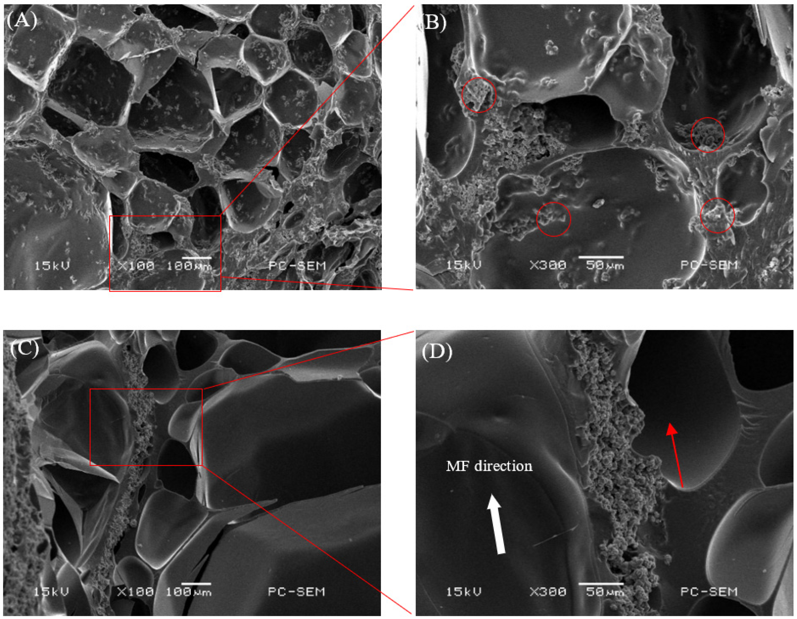
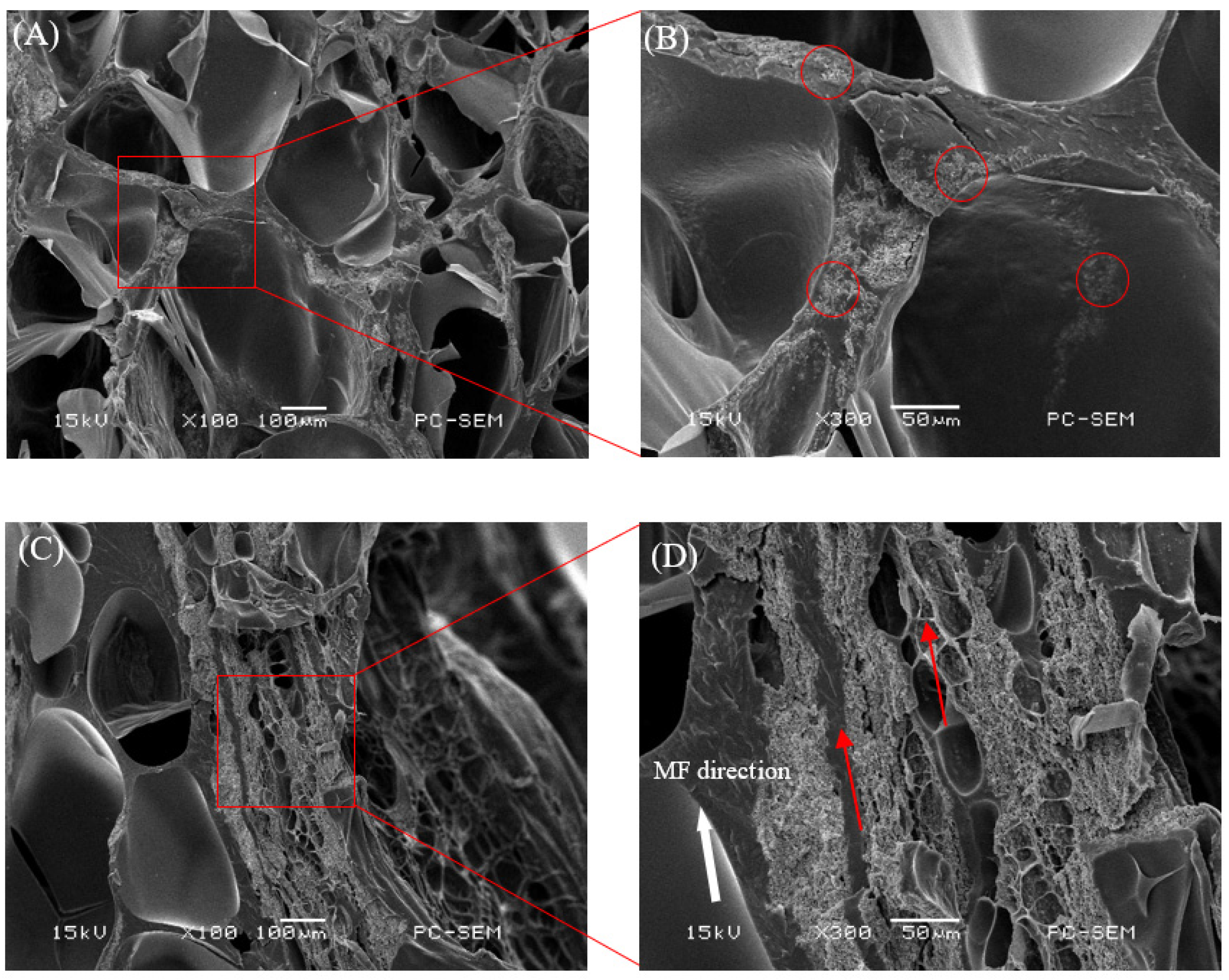
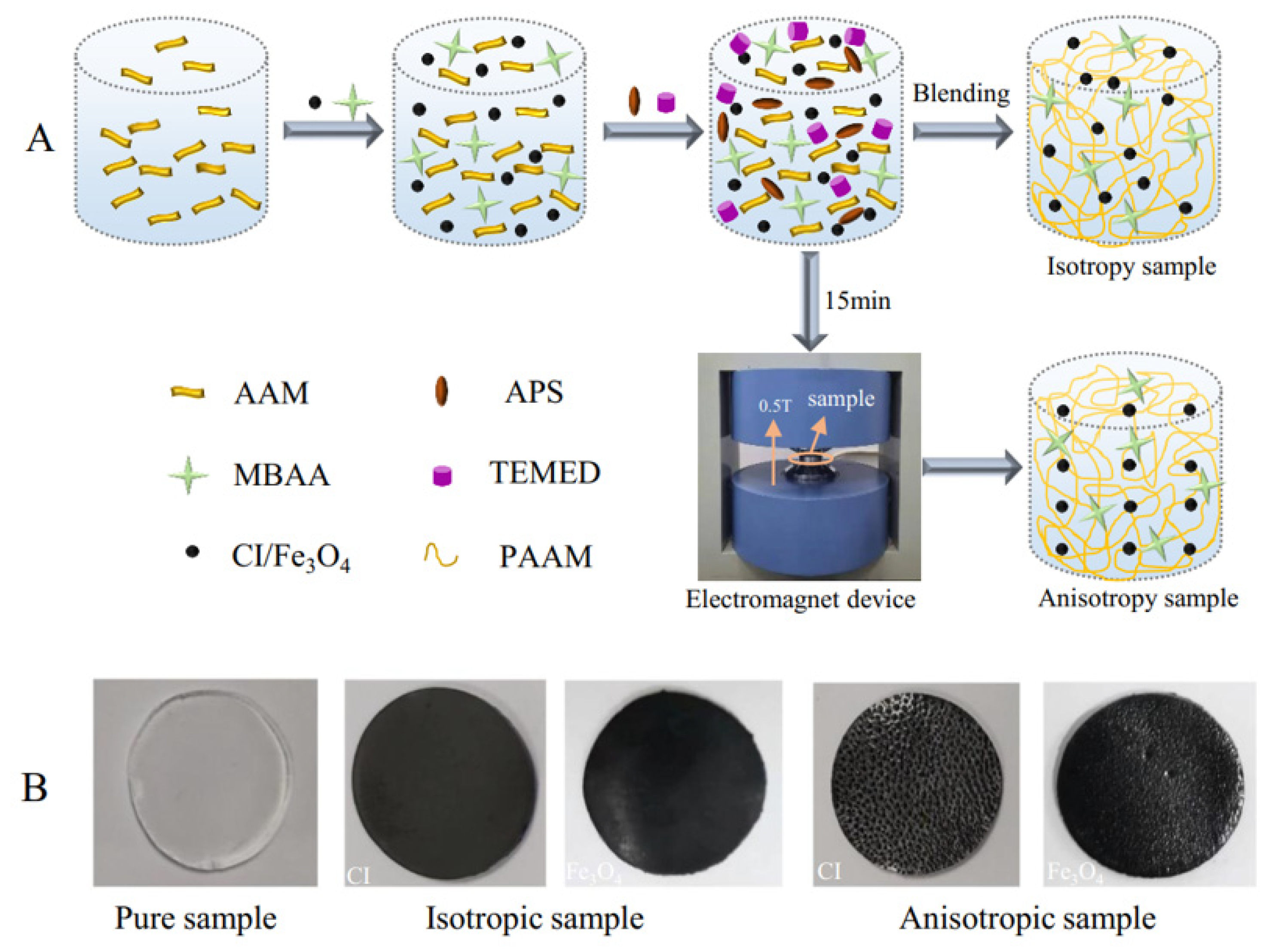
2.1. Morphology
2.2. The Rheological Properties of Magnetic Hydrogels with Fe3O4
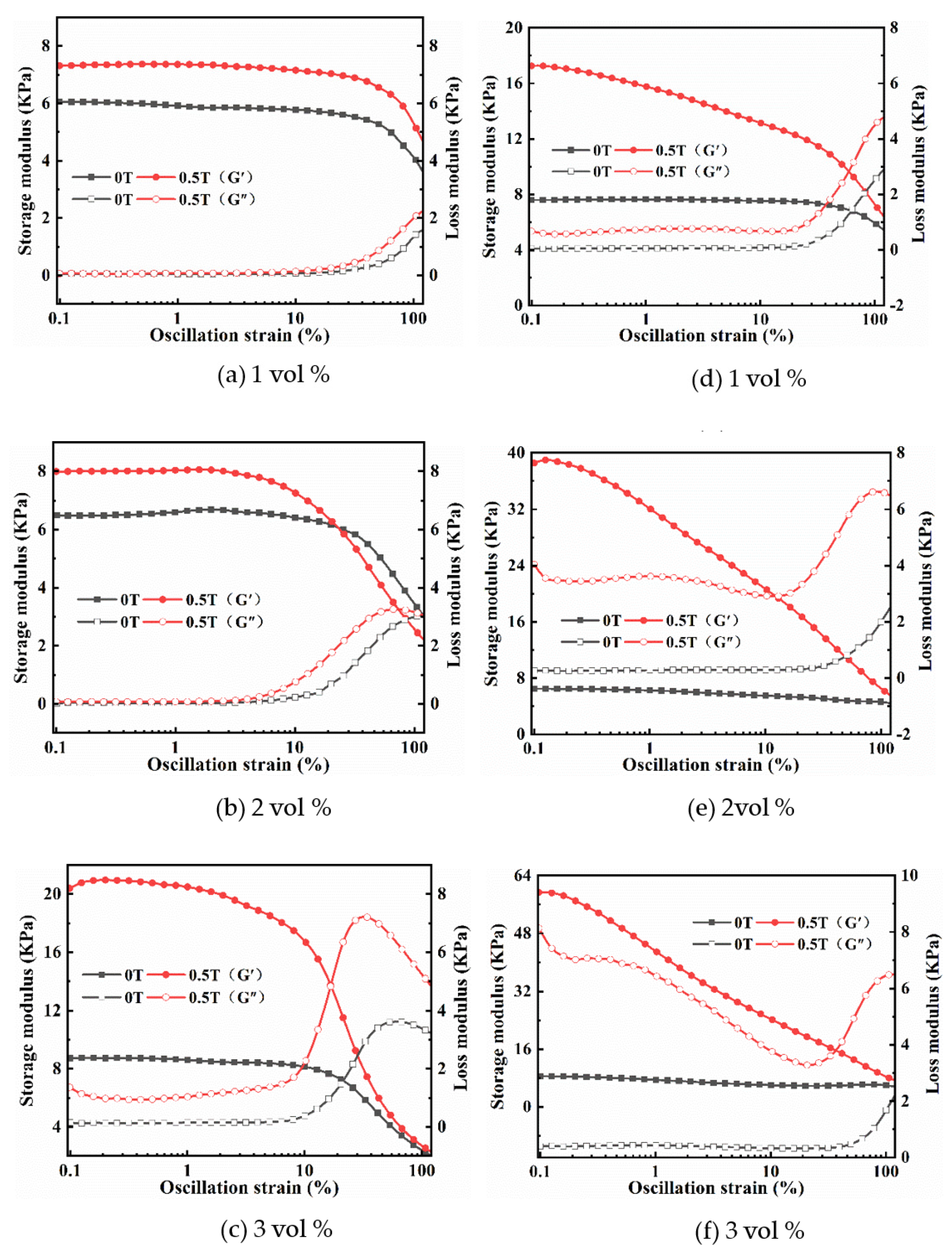
2.2.1. Strain Amplitude Sweep
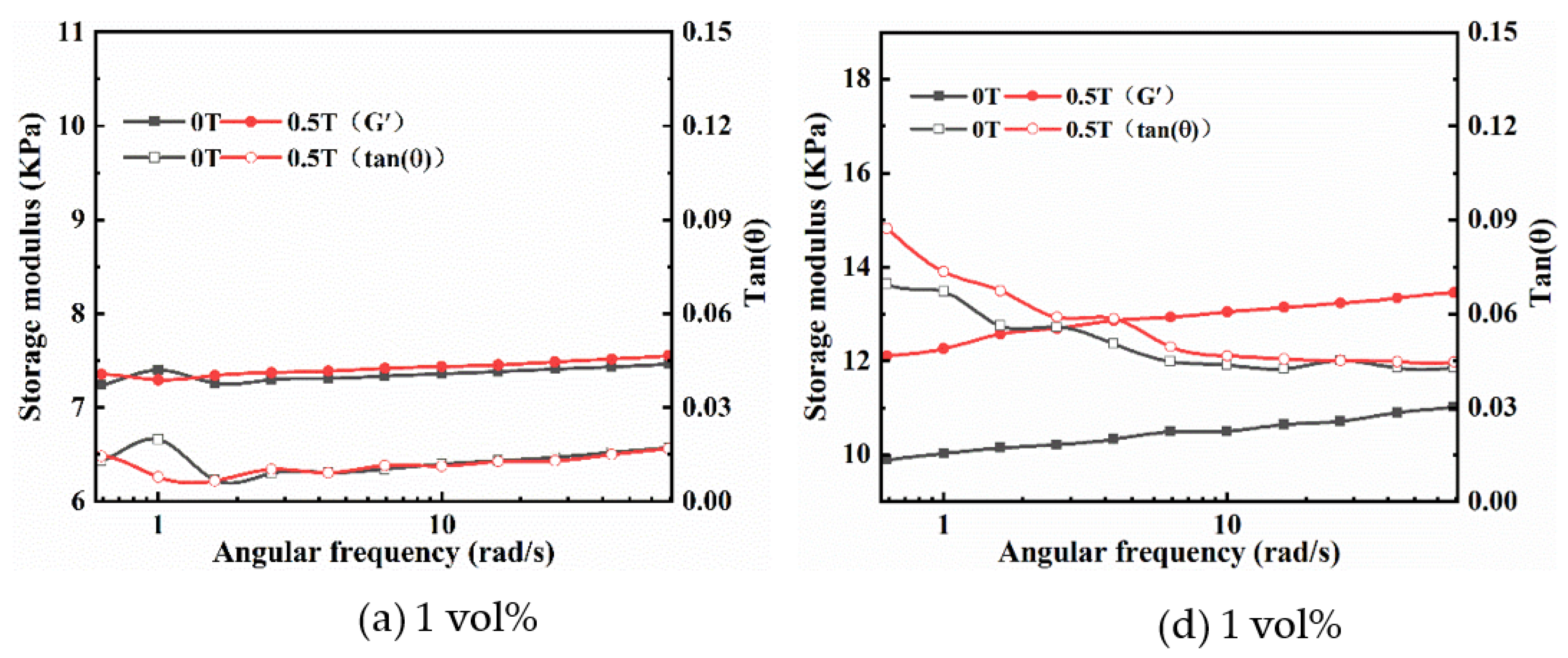
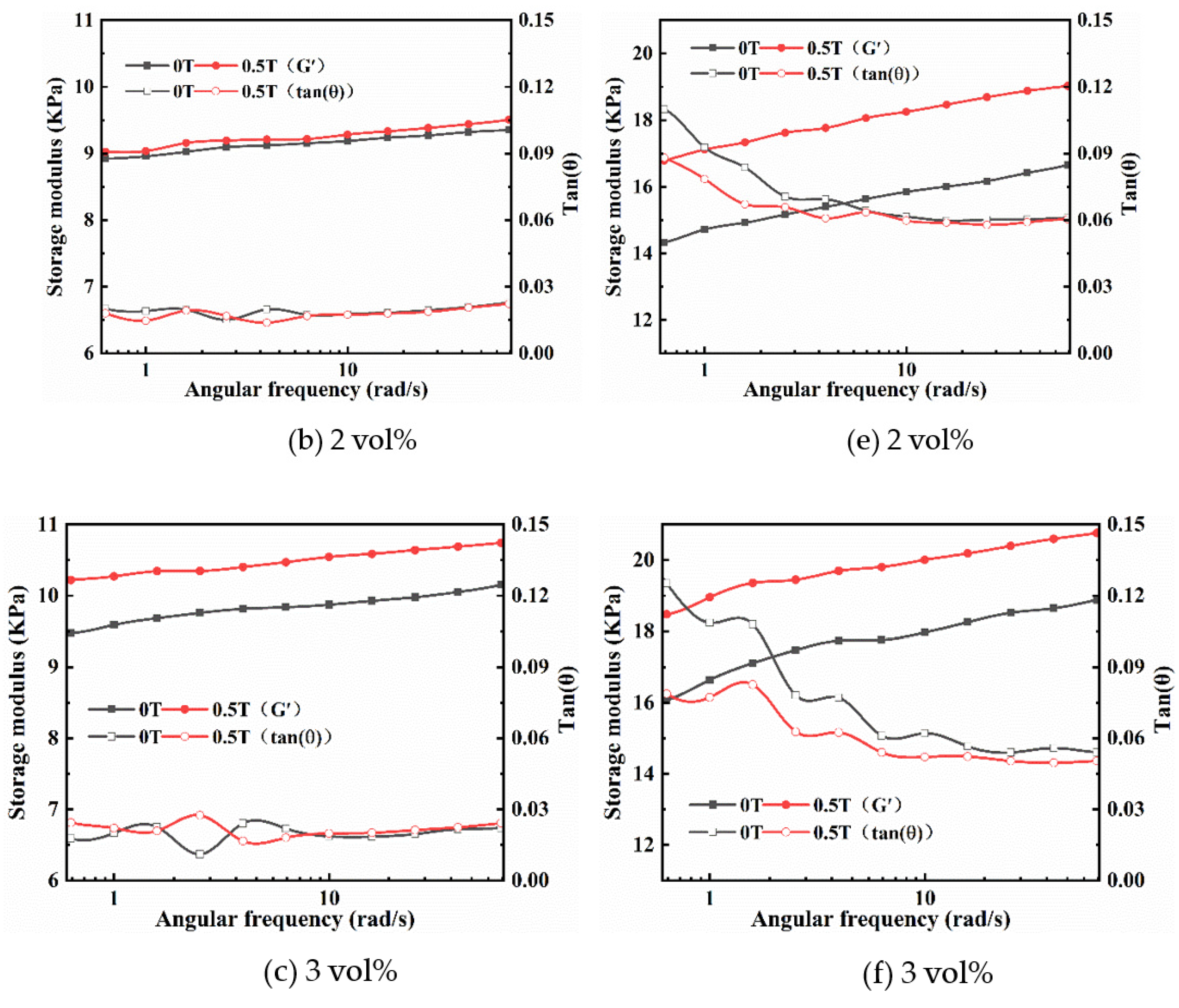
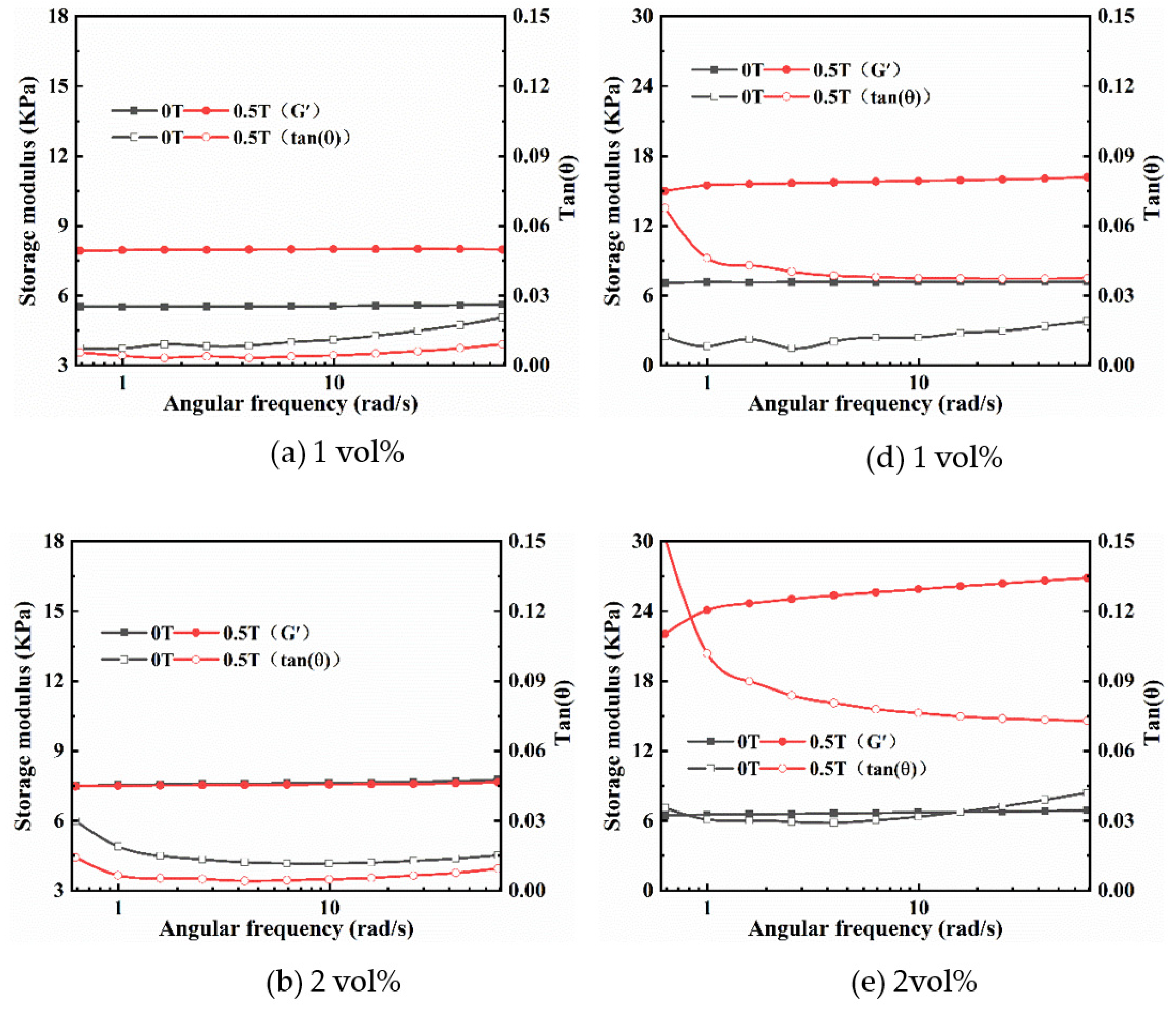
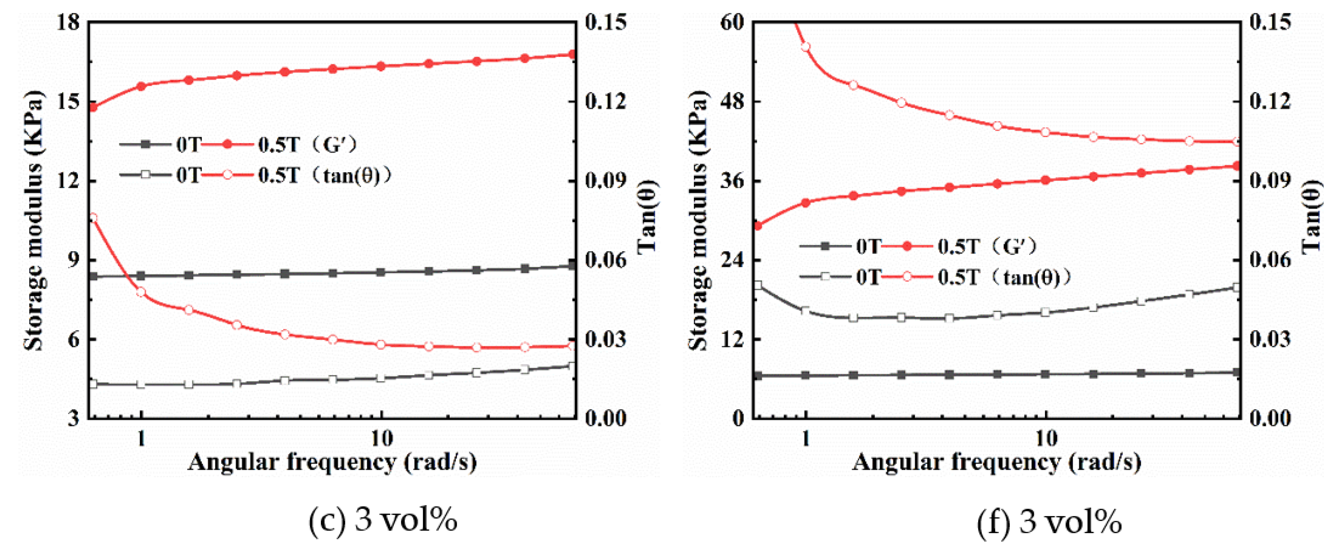
2.2.2. Frequency Sweep
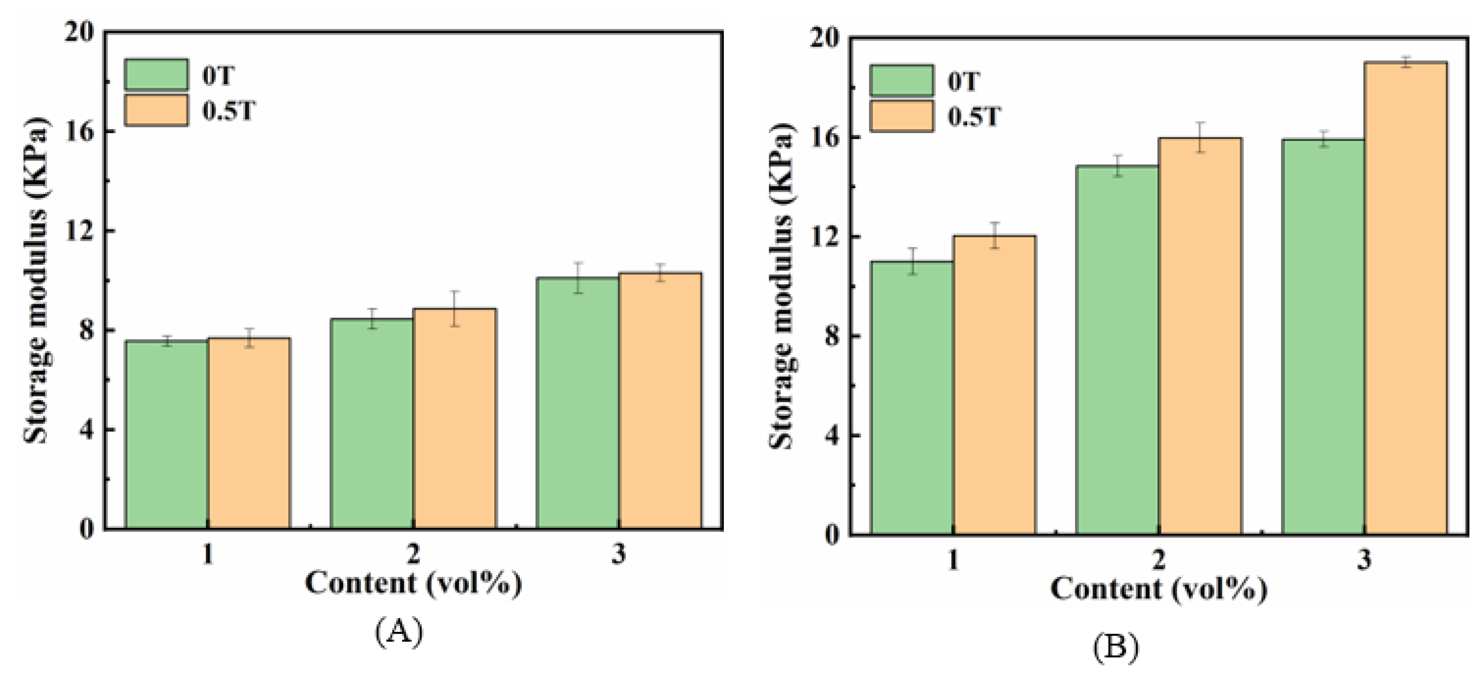
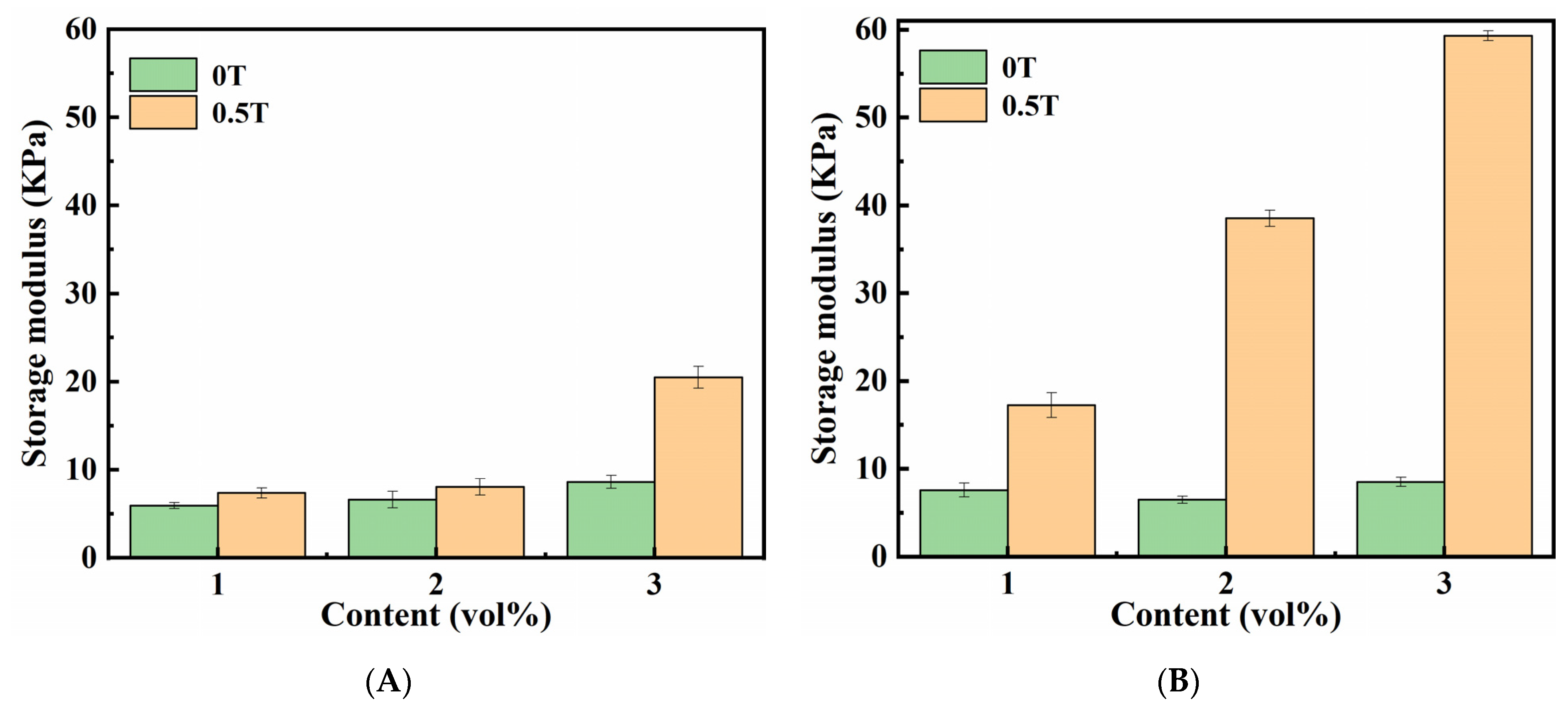
2.2.3. MR Effect
2.3. The Rheological Properties of Magnetic Hydrogels with CI
2.3.1. Strain Amplitude Sweep
2.3.2. Frequency Sweep
2.3.3. MR Effect
2.4. Swelling Behavior
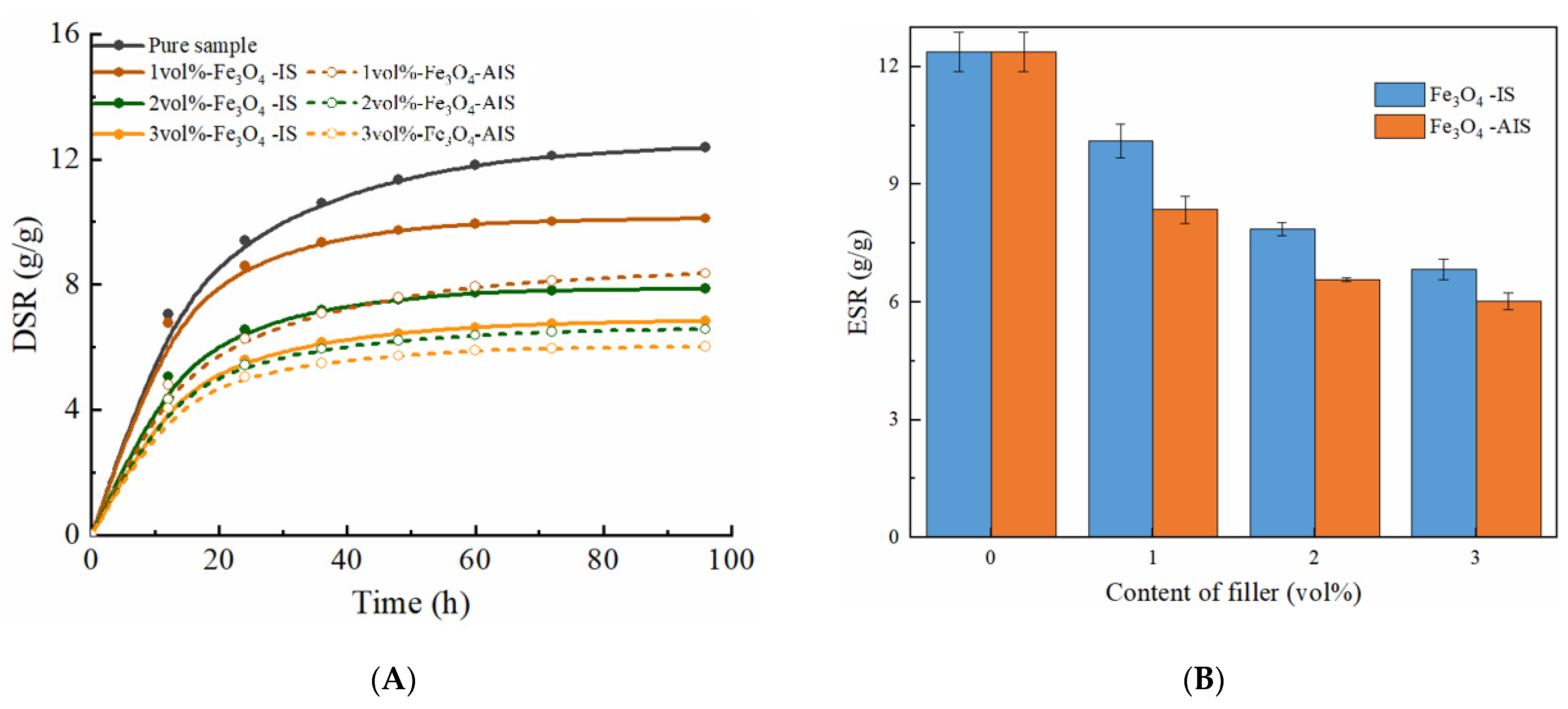
2.4.1. Swelling Behavior of Magnetic Hydrogels with Fe3O4
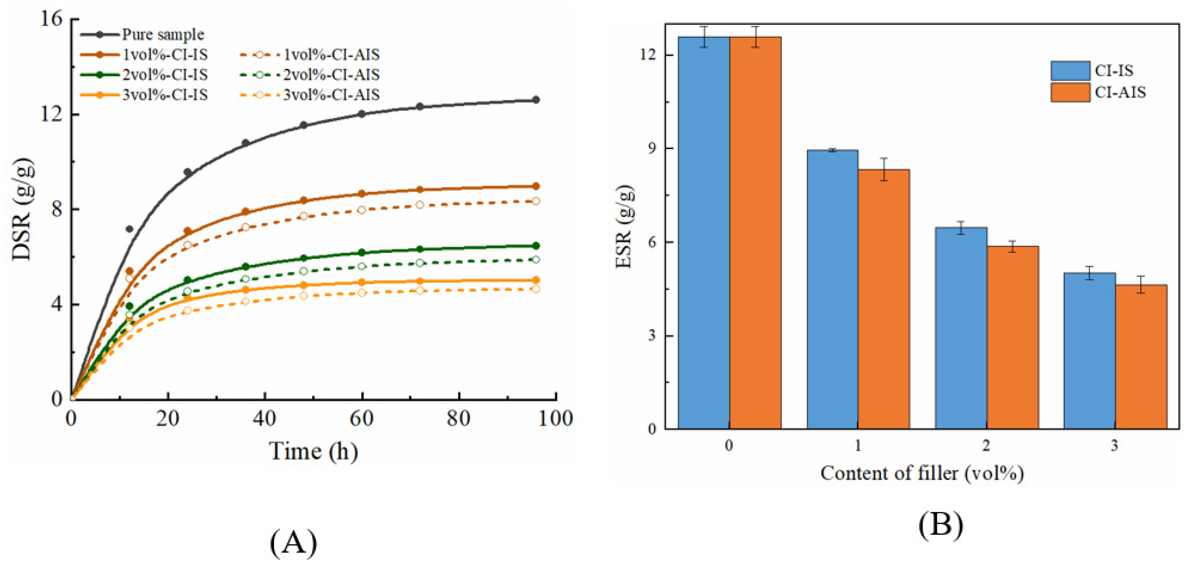
2.4.2. Swelling Behavior of Magnetic Hydrogels with CI
3. Materials and Methods
3.1. Material Preparations
3.1.1. Raw Materials
3.1.2. Surface Pre-Treatment of CI
3.1.3. Preparation of Magnetic Hydrogels
3.2. Characterizations
3.2.1. Morphology
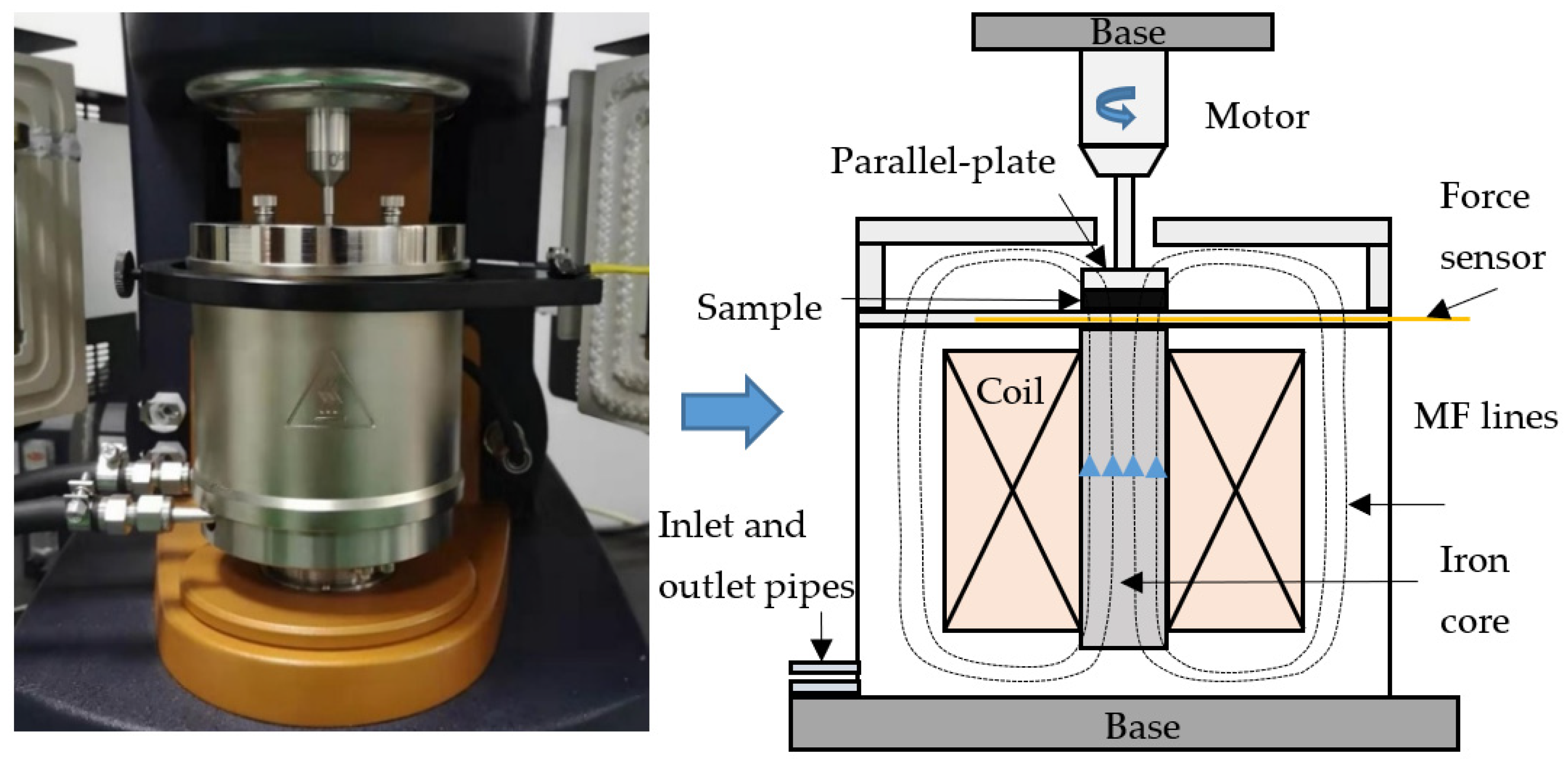
3.2.2. Rheological Properties
3.2.3. Swelling Studies
4. Conclusions
Author Contributions
Funding
Institutional Review Board Statement
Informed Consent Statement
Data Availability Statement
Conflicts of Interest
References
- Oyen, M.L. Mechanical characterisation of hydrogel materials. Int. Mater. Rev. 2013, 59, 44–59. [Google Scholar] [CrossRef]
- Bahram, M.; Mohseni, N.; Moghtader, M. An Introduction to Hydrogels and Some Recent Applications. In Emerging Concepts in Analysis and Applications of Hydrogels; IntechOpen: London, UK, 2016. [Google Scholar]
- Zhang, T.; Yuk, H.; Lin, S.; Parada, G.A.; Zhao, X. Tough and tunable adhesion of hydrogels: Experiments and models. Acta Mech. Sin. 2017, 33, 543–554. [Google Scholar] [CrossRef]
- Memic, A.; Alhadrami, H.A.; Hussain, M.A.; Aldhahri, M.; Al Nowaiser, F.; Al-Hazmi, F.; Oklu, R.; Khademhosseini, A. Hydrogels 2.0: Improved properties with nanomaterial composites for biomedical applications. Biomed. Mater. 2015, 11, 014104. [Google Scholar] [CrossRef]
- Tang, N.; Peng, Z.; Guo, R.; An, M.; Chen, X.; Li, X.; Yang, N.; Zang, J. Thermal Transport in Soft PAAm Hydrogels. Polymers 2017, 9, 688. [Google Scholar] [CrossRef] [Green Version]
- Rao, P.; Sun, T.L.; Chen, L.; Takahashi, R.; Shinohara, G.; Guo, H.; King, D.R.; Kurokawa, T.; Gong, J.P. Tough Hydrogels with Fast, Strong, and Reversible Underwater Adhesion Based on a Multiscale Design. Adv. Mater. 2018, 30, e1801884. [Google Scholar] [CrossRef] [PubMed] [Green Version]
- Serrano-Aroca, Á. Enhancement of Hydrogels’ Properties for Biomedical Applications: Latest Achievements. In Hydrogels; IntechOpen: London, UK, 2018. [Google Scholar]
- Davaran, S.; Alimirzalu, S.; Nejati-Koshki, K.; Nasrabadi, H.T.; Akbarzadeh, A.; Khandaghi, A.A.; Abbasian, M.; Alimohammadi, S. Physicochemical Characteristics of Fe3O4Magnetic Nanocomposites Based on Poly(N-isopropylacrylamide) for Anti-cancer Drug Delivery. Asian Pac. J. Cancer Prev. 2014, 15, 49–54. [Google Scholar] [CrossRef] [PubMed] [Green Version]
- Reddy, N.N.; Varaprasad, K.; Ravindra, S.; Reddy, G.V.S.; Reddy, K.M.S.; Mohan Reddy, K.M.; Raju, K.M. Evaluation of blood compatibility and drug release studies of gelatin based magnetic hydrogel nanocomposites. Colloids Surf. A Physicochem. Eng. Asp. 2011, 385, 20–27. [Google Scholar] [CrossRef]
- Kurdtabar, M.; Bardajee, G.R. Drug release and swelling behavior of magnetic iron oxide nanocomposite hydrogels based on poly(acrylic acid) grafted onto sodium alginate. Polym. Bull. 2020, 77, 3001–3015. [Google Scholar] [CrossRef]
- Zhang, J.X.; Huang, Q.T.; Du, J.Z. Recent advances in magnetic hydrogels. Polym. Int. 2016, 65, 1365–1372. [Google Scholar] [CrossRef]
- Huang, C.; Li, Y.; Duan, L.; Wang, L.; Ren, X.; Gao, G. Enhancing the self-recovery and mechanical property of hydrogels by macromolecular microspheres with thermal and redox initiation systems. RSC Adv. 2017, 7, 16015–16021. [Google Scholar] [CrossRef] [Green Version]
- Ilg, P. Stimuli-responsive hydrogels cross-linked by magnetic nanoparticles. Soft Matter 2013, 9, 3465–3468. [Google Scholar] [CrossRef] [Green Version]
- Baqiya, M.A.; Taufiq, A.; Sunaryono, A.; Munaji, M.; Sari, D.P.; Dwihapsari, Y.; Darminto, J. Development of PVA/Fe3O4 as Smart Magnetic Hydrogels for Biomedical Applications. In Hydrogels; IntechOpen: London, UK, 2018. [Google Scholar]
- Abrougui, M.M.; Lopez-Lopez, M.T.; Duran, J.D.G. Mechanical properties of magnetic gels containing rod-like composite particles. Philos. Trans. A Math. Phys. Eng. Sci. 2019, 377, 20180218. [Google Scholar] [CrossRef] [Green Version]
- Lee, Y.W.; Ceylan, H.; Yasa, I.C.; Kilic, U.; Sitti, M. 3D-Printed Multi-Stimuli-Responsive Mobile Micromachines. ACS Appl. Mater. Interfaces 2021, 13, 12759–12766. [Google Scholar] [CrossRef] [PubMed]
- Goudu, S.R.; Yasa, I.C.; Hu, X.; Ceylan, H.; Hu, W.; Sitti, M. Biodegradable Untethered Magnetic Hydrogel Milli-Grippers. Adv. Funct. Mater. 2020, 30, 9. [Google Scholar] [CrossRef]
- Li, Y.; Huang, G.; Zhang, X.; Li, B.; Chen, Y.; Lu, T.; Lu, T.J.; Xu, F. Magnetic Hydrogels and Their Potential Biomedical Applications. Adv. Funct. Mater. 2013, 23, 660–672. [Google Scholar] [CrossRef]
- Liu, Z.; Liu, J.; Cui, X.; Wang, X.; Zhang, L.; Tang, P. Recent Advances on Magnetic Sensitive Hydrogels in Tissue Engineering. Front. Chem. 2020, 8, 124. [Google Scholar] [CrossRef] [PubMed]
- Lei, J.; Zhou, Z.; Liu, Z. Side Chains and the Insufficient Lubrication of Water in Polyacrylamide Hydrogel—A New Insight. Polymers 2019, 11, 1845. [Google Scholar] [CrossRef] [Green Version]
- Tanasa, E.; Zaharia, C.; Radu, I.C.; Surdu, V.A.; Vasile, B.S.; Damian, C.M.; Andronescu, E. Novel Nanocomposites Based on Functionalized Magnetic Nanoparticles and Polyacrylamide: Preparation and Complex Characterization. Nanomaterials 2019, 9, 1384. [Google Scholar] [CrossRef] [Green Version]
- Selzer, L.; Odenbach, S. Effects of carbonyl iron particles on the rheological behavior of nanocomposite hydrogels. J. Magn. Magn. Mater. 2020, 501, 166394. [Google Scholar] [CrossRef]
- Kasgoz, H.; Durmus, A.; Kasgoz, A.; Aydin, I. Structurally Enhanced Hydrogel Nanocomposites with Improved Swelling and Mechanical Properties. J. Macromol. Sci. Part A 2012, 49, 92–99. [Google Scholar] [CrossRef]
- Jang, J.; Lee, J.; Seol, Y.-J.; Jeong, Y.H.; Cho, D.-W. Improving mechanical properties of alginate hydrogel by reinforcement with ethanol treated polycaprolactone nanofibers. Compos. Part B Eng. 2013, 45, 1216–1221. [Google Scholar] [CrossRef]
- Crippa, F.; Moore, T.L.; Mortato, M.; Geers, C.; Haeni, L.; Hirt, A.M.; Rothen-Rutishauser, B.; Petri-Fink, A. Dynamic and biocompatible thermo-responsive magnetic hydrogels that respond to an alternating magnetic field. J. Magn. Magn. Mater. 2017, 427, 212–219. [Google Scholar] [CrossRef] [Green Version]
- Gila-Vilchez, C.; Manas-Torres, M.C.; Contreras-Montoya, R.; Alaminos, M.; Duran, J.D.G.; de Cienfuegos, L.A.; Lopez-Lopez, M.T. Anisotropic magnetic hydrogels: Design, structure and mechanical properties. Philos. Trans. A Math. Phys. Eng. Sci. 2019, 377, 20180217. [Google Scholar] [CrossRef] [Green Version]
- Meharthaj, H.; Sivakumar, S.M.; Arockiarajan, A. Significance of particle size on the improved performance of magnetorheological gels. J. Magn. Magn. Mater. 2019, 490, 165483. [Google Scholar] [CrossRef]
- Hu, X.; Nian, G.; Liang, X.; Wu, L.; Yin, T.; Lu, H.; Qu, S.; Yang, W. Adhesive Tough Magnetic Hydrogels with High Fe3O4 Content. ACS Appl. Mater. Interfaces 2019, 11, 10292–10300. [Google Scholar] [CrossRef]
- Bonini, M.; Lenz, S.; Falletta, E.; Ridi, F.; Carretti, E.; Fratini, E.; Wiedenmann, A.; Baglioni, P. Acrylamide-Based Magnetic Nanosponges: A New Smart Nanocomposite Material. Langmuir 2008, 24, 12644–12650. [Google Scholar] [CrossRef]
- Konwar, A.; Gogoi, A.; Chowdhury, D. Magnetic alginate–Fe3O4 hydrogel fiber capable of ciprofloxacin hydrochloride adsorption/separation in aqueous solution. RSC Adv. 2015, 5, 81573–81582. [Google Scholar] [CrossRef]
- Lee, J.H.; Han, W.J.; Jang, H.S.; Choi, H.J. Highly Tough, Biocompatible, and Magneto-Responsive Fe3O4/Laponite/PDMAAm Nanocomposite Hydrogels. Sci. Rep. 2019, 9, 15024. [Google Scholar] [CrossRef] [Green Version]
- Pang, L.; Dong, X.; Niu, C.; Qi, M. Dynamic viscoelasticity and magnetorheological property of magnetic hydrogels. J. Magn. Magn. Mater. 2020, 498, 166140. [Google Scholar] [CrossRef]
- Wu, J.K.; Gong, X.L.; Fan, Y.C.; Xia, H.S. Physically crosslinked poly(vinyl alcohol) hydrogels with magnetic field controlled modulus. Soft Matter 2011, 7, 6205–6212. [Google Scholar] [CrossRef]
- Mitsumata, T.; Honda, A.; Kanazawa, H.; Kawai, M. Magnetically Tunable Elasticity for Magnetic Hydrogels Consisting of Carrageenan and Carbonyl Iron Particles. J. Phys. Chem. B 2012, 116, 12341–12348. [Google Scholar] [CrossRef] [PubMed]
- Ikeda, J.; Takahashi, D.; Watanabe, M.; Kawai, M.; Mitsumata, T. Particle Size in Secondary Particle and Magnetic Response for Carrageenan Magnetic Hydrogels. Gels 2019, 5, 39. [Google Scholar] [CrossRef] [PubMed] [Green Version]
- Cvek, M.; Zahoranova, A.; Mrlik, M.; Sramkova, P.; Minarik, A.; Sedlacik, M. Poly(2-oxazoline)-based magnetic hydrogels: Synthesis, performance and cytotoxicity. Colloids Surf. B Biointerfaces 2020, 190, 110912. [Google Scholar] [CrossRef] [PubMed]
- Bonhome-Espinosa, A.B.; Campos, F.; Rodriguez, I.A.; Carriel, V.; Marins, J.A.; Zubarev, A.; Duran, J.D.G.; Lopez-Lopez, M.T. Effect of particle concentration on the microstructural and macromechanical properties of biocompatible magnetic hydrogels. Soft Matter 2017, 13, 2928–2941. [Google Scholar] [CrossRef]
- Bin, L.; Xu, C.; Dong, S.; Wang, X. Alignment of magnetic particles in hydrogel matrix: A novel anisotropic magnetic hydrogels for soft robotics. J. Intell. Mater. Syst. Struct. 2020, 32, 1432–1440. [Google Scholar] [CrossRef]
- Hapipi, N.M.; Mazlan, S.A.; Ubaidillah, U.; Homma, K.; Aziz, S.A.A.; Nordin, N.A.; Bahiuddin, I.; Nazmi, N. The Rheological Studies on Poly(vinyl) Alcohol-Based Hydrogel Magnetorheological Plastomer. Polymers 2020, 12, 2332. [Google Scholar] [CrossRef]
- Hong, K.P.; Song, K.H.; Cho, M.W.; Kwon, S.H.; Choi, H.J. Magnetorheological properties and polishing characteristics of silica-coated carbonyl iron magnetorheological fluid. J. Intel. Mat. Syst. Str. 2017, 29, 137–146. [Google Scholar] [CrossRef] [Green Version]
- Lin, F.C.; Zheng, J.J.; Guo, W.H.; Zhu, Z.T.; Wang, Z.; Dong, B.Y.; Lin, C.S.; Huang, B.; Lu, B.L. Smart cellulose-derived magnetic hydrogel with rapid swelling and deswelling properties for remotely controlled drug release. Cellulose 2019, 26, 6861–6877. [Google Scholar] [CrossRef]


| Functions | Raw Materials | Producers |
|---|---|---|
| Hydrogel matrix | AAM | Sinopharm Chemical Reagent Co., Ltd., Shanghai, China |
| MBAA | Sinopharm Chemical Reagent Co., Ltd., Shanghai, China | |
| APS | Sinopharm Chemical Reagent Co., Ltd., Shanghai, China | |
| TEMED | Sinopharm Chemical Reagent Co., Ltd., Shanghai, China | |
| Magnetic particles | CI with an average diameter ranges from 1 to 3 μm | Jiangsu Tianyi Ultra-fine metal powder Co., Ltd., Huaiyin, China |
| Fe3O4 with an average diameter ranges from 1 to 10 nm | Sinopharm Chemical Reagent Co., Ltd., Shanghai, China | |
| Surface pre-treatment of CI | MAA (99%) | Sigma-Aldrich, St. Louis, MO, USA |
| VTMOS (98.0%) | Sigma-Aldrich, St. Louis, MO, USA | |
| Ethanol (99.8%) | Sinopharm Chemical Reagent Co., Ltd., Shanghai, China |
| S. No. | CI (vol%) | Fe3O4 (vol%) | CI (g) | Fe3O4 (g) | AAM (mL) | MBAA (mL) | APS (mL) | TEMED (µL) |
|---|---|---|---|---|---|---|---|---|
| Isotropic samples | ||||||||
| 1 | 1 | 0 | 1.78 | 0 | 20 | 1.6 | 0.8 | 20 |
| 2 | 2 | 0 | 3.60 | 0 | 20 | 1.6 | 0.8 | 20 |
| 3 | 3 | 0 | 5.45 | 0 | 20 | 1.6 | 0.8 | 20 |
| 4 | 0 | 1 | 0 | 1.17 | 20 | 1.6 | 0.8 | 20 |
| 5 | 0 | 2 | 0 | 2.37 | 20 | 1.6 | 0.8 | 20 |
| 6 | 0 | 3 | 0 | 3.59 | 20 | 1.6 | 0.8 | 20 |
| Anisotropic samples | ||||||||
| 7 | 1 | 0 | 1.78 | 0 | 20 | 1.6 | 0.8 | 20 |
| 8 | 2 | 0 | 3.60 | 0 | 20 | 1.6 | 0.8 | 20 |
| 9 | 3 | 0 | 5.45 | 0 | 20 | 1.6 | 0.8 | 20 |
| 10 | 0 | 1 | 0 | 1.17 | 20 | 1.6 | 0.8 | 20 |
| 11 | 0 | 2 | 0 | 2.37 | 20 | 1.6 | 0.8 | 20 |
| 12 | 0 | 3 | 0 | 3.59 | 20 | 1.6 | 0.8 | 20 |
Publisher’s Note: MDPI stays neutral with regard to jurisdictional claims in published maps and institutional affiliations. |
© 2021 by the authors. Licensee MDPI, Basel, Switzerland. This article is an open access article distributed under the terms and conditions of the Creative Commons Attribution (CC BY) license (https://creativecommons.org/licenses/by/4.0/).
Share and Cite
Xu, C.; Li, B.; Wang, X. A Comparison Study on the Magneto-Responsive Properties and Swelling Behaviors of a Polyacrylamide-Based Hydrogel Incorporating with Magnetic Particles. Int. J. Mol. Sci. 2021, 22, 12342. https://0-doi-org.brum.beds.ac.uk/10.3390/ijms222212342
Xu C, Li B, Wang X. A Comparison Study on the Magneto-Responsive Properties and Swelling Behaviors of a Polyacrylamide-Based Hydrogel Incorporating with Magnetic Particles. International Journal of Molecular Sciences. 2021; 22(22):12342. https://0-doi-org.brum.beds.ac.uk/10.3390/ijms222212342
Chicago/Turabian StyleXu, Chanchan, Bin Li, and Xiaojie Wang. 2021. "A Comparison Study on the Magneto-Responsive Properties and Swelling Behaviors of a Polyacrylamide-Based Hydrogel Incorporating with Magnetic Particles" International Journal of Molecular Sciences 22, no. 22: 12342. https://0-doi-org.brum.beds.ac.uk/10.3390/ijms222212342