
Adult-Onset Still’s Disease: Novel Biomarkers of Specific Subsets, Disease Activity, and Relapsing Forms


Abstract
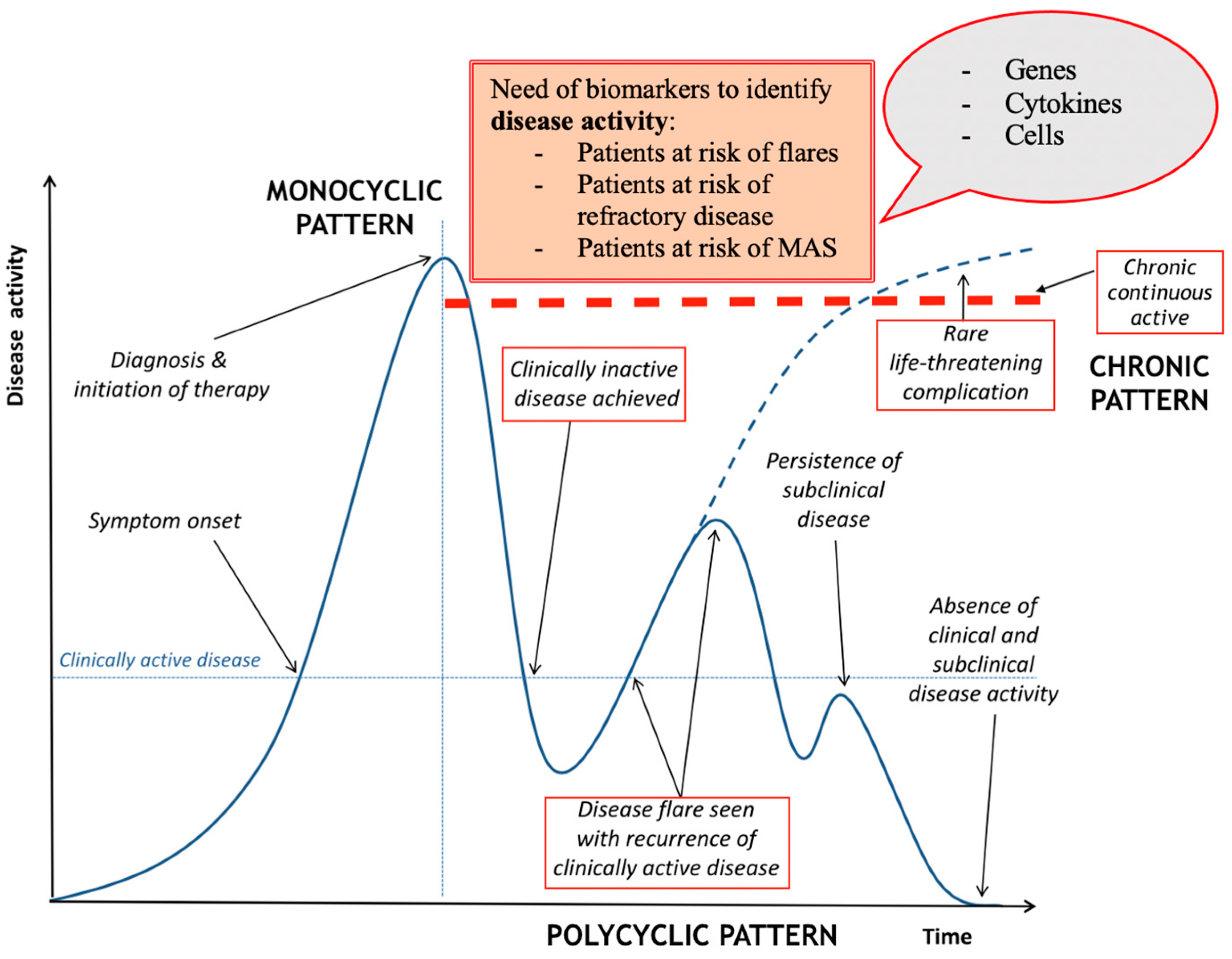
:1. Introduction
2. Gene Polymorphisms
3. The Role of Ferritin
4. Cellular Signaling Imbalance
5. Innate System Activation
6. Cytokine Storm
7. Clinical Hallmarks
8. Biomarkers for Differential Diagnosis
9. Biomarkers of Organs’ Damage
10. Ongoing Trials
11. Conclusions
Author Contributions
Funding
Institutional Review Board Statement
Informed Consent Statement
Data Availability Statement
Conflicts of Interest
References
- Bagnari, V.; Colina, M.; Ciancio, G.; Govoni, M.; Trotta, F. Adult-onset Still’s disease. Rheumatol. Int. 2010, 30, 855–862. [Google Scholar] [CrossRef] [Green Version]
- Bywaters, E.G. Still’s disease in the adult. Ann. Rheum. Dis. 1971, 30, 121–133. [Google Scholar] [CrossRef] [PubMed] [Green Version]
- Efthimiou, P.; Kontzias, A.; Hur, P.; Rodha, K.; Ramakrishna, G.S.; Nakasato, P. Adult-onset Still’s disease in focus: Clinical manifestations, diagnosis, treatment, and unmet needs in the era of targeted therapies. Semin. Arthritis Rheum. 2021, 51, 858–874. [Google Scholar] [CrossRef]
- Kastner, D.L.; Aksentijevich, I.; Goldbach-Mansky, R. Autoinflammatory disease reloaded: A clinical perspective. Cell 2010, 140, 784–790. [Google Scholar] [CrossRef] [Green Version]
- McGonagle, D.; Aziz, A.; Dickie, L.J.; McDermott, M.F. An integrated classification of pediatric inflammatory diseases, based on the concepts of autoinflammation and the immunological disease continuum. Pediatr. Res. 2009, 65 Pt 2, 38r–45r. [Google Scholar] [CrossRef]
- Jamilloux, Y.; Gerfaud-Valentin, M.; Martinon, F.; Belot, A.; Henry, T.; Seve, P. Pathogenesis of adult-onset Still’s disease: New insights from the juvenile counterpart. Immunol. Res. 2015, 61, 53–62. [Google Scholar] [CrossRef]
- Feist, E.; Mitrovic, S.; Fautrel, B. Mechanisms, biomarkers and targets for adult-onset Still’s disease. Nat. Rev. Rheumatol. 2018, 14, 603–618. [Google Scholar] [CrossRef]
- Gerfaud-Valentin, M.; Jamilloux, Y.; Iwaz, J.; Seve, P. Adult-onset Still’s disease. Autoimmun. Rev. 2014, 13, 708–722. [Google Scholar] [CrossRef] [PubMed] [Green Version]
- Yamaguchi, M.; Ohta, A.; Tsunematsu, T.; Kasukawa, R.; Mizushima, Y.; Kashiwagi, H.; Kashiwazaki, S.; Tanimoto, K.; Matsumoto, Y.; Ota, T.; et al. Preliminary criteria for classification of adult Still’s disease. J. Rheumatol. 1992, 19, 424–430. [Google Scholar] [PubMed]
- Fautrel, B. Adult-onset Still disease. Best Pract. Res. Clin. Rheumatol. 2008, 22, 773–792. [Google Scholar] [CrossRef] [PubMed]
- Perdan-Pirkmajer, K.; Praprotnik, S.; Tomsic, M. A case of refractory adult-onset Still’s disease successfully controlled with tocilizumab and a review of the literature. Clin. Rheumatol. 2010, 29, 1465–1467. [Google Scholar] [CrossRef]
- Lee, W.S.; Yoo, S.H.; Ko, R.E.; Yoo, W.H. Adalimumab in the treatment of refractory adult-onset Still’s disease. Int. J. Rheum. Dis. 2017, 20, 1798–1799. [Google Scholar] [CrossRef]
- Lahiri, M.; Teng, G.G. A case of refractory adult-onset Still’s disease treated with anakinra. Int. J. Rheum. Dis. 2010, 13, e36–e41. [Google Scholar] [CrossRef]
- Aikawa, N.E.; Ribeiro, A.C.; Saad, C.G.; Pereira, R.M.; Levy, M.; Silva, C.A.; Bonfa, E.; de Carvalho, J.F. Is anti-TNF switching in refractory Still’s disease safe and effective? Clin. Rheumatol. 2011, 30, 1129–1134. [Google Scholar] [CrossRef] [PubMed]
- Kalyoncu, U.; Solmaz, D.; Emmungil, H.; Yazici, A.; Kasifoglu, T.; Kimyon, G.; Balkarli, A.; Bes, C.; Ozmen, M.; Alibaz-Oner, F.; et al. Response rate of initial conventional treatments, disease course, and related factors of patients with adult-onset Still’s disease: Data from a large multicenter cohort. J. Autoimmun. 2016, 69, 59–63. [Google Scholar] [CrossRef] [PubMed]
- Cush, J.J.; Medsger, T.A., Jr.; Christy, W.C.; Herbert, D.C.; Cooperstein, L.A. Adult-onset Still’s disease. Clinical course and outcome. Arthritis Rheum. 1987, 30, 186–194. [Google Scholar] [CrossRef] [PubMed]
- Yin, R.; Wang, G.; Yang, X.; Zhang, L.; Wang, S.; Li, T.; Liu, S. Identification of prognostic factors and construction of a nomogram for patients with relapse/refractory adult-onset Still’s disease. Clin. Rheumatol. 2021, 40, 3951–3960. [Google Scholar] [CrossRef]
- Colina, M.; Zucchini, W.; Ciancio, G.; Orzincolo, C.; Trotta, F.; Govoni, M. The evolution of adult-onset Still disease: An observational and comparative study in a cohort of 76 Italian patients. Semin. Arthritis Rheum. 2011, 41, 279–285. [Google Scholar] [CrossRef] [PubMed]
- Gasparyan, A.Y.; Ayvazyan, L.; Blackmore, H.; Kitas, G.D. Writing a narrative biomedical review: Considerations for authors, peer reviewers, and editors. Rheumatol. Int. 2011, 31, 1409–1417. [Google Scholar] [CrossRef]
- Hsieh, C.W.; Chang, C.Y.; Chen, Y.M.; Chen, H.H.; Hung, W.T.; Gung, N.R.; Wey, S.J.; Chen, D.Y. Impaired autophagic flux and its related inflammation in patients with adult-onset Still’s disease. Oncotarget 2018, 9, 110–121. [Google Scholar] [CrossRef] [Green Version]
- Hung, W.T.; Hung, S.I.; Chen, Y.M.; Hsieh, C.W.; Chen, H.H.; Tang, K.T.; Chen, D.Y.; Lan, T.H. The Association of ATG16L1 Variations with Clinical Phenotypes of Adult-Onset Still’s Disease. Genes 2021, 12, 904. [Google Scholar] [CrossRef]
- Romanov, J.; Walczak, M.; Ibiricu, I.; Schüchner, S.; Ogris, E.; Kraft, C.; Martens, S. Mechanism and functions of membrane binding by the Atg5-Atg12/Atg16 complex during autophagosome formation. EMBO J. 2012, 31, 4304–4317. [Google Scholar] [CrossRef] [PubMed]
- Hampe, J.; Franke, A.; Rosenstiel, P.; Till, A.; Teuber, M.; Huse, K.; Albrecht, M.; Mayr, G.; De La Vega, F.M.; Briggs, J.; et al. A genome-wide association scan of nonsynonymous SNPs identifies a susceptibility variant for Crohn disease in ATG16L1. Nat. Genet. 2007, 39, 207–211. [Google Scholar] [CrossRef] [PubMed]
- Rioux, J.D.; Xavier, R.J.; Taylor, K.D.; Silverberg, M.S.; Goyette, P.; Huett, A.; Green, T.; Kuballa, P.; Barmada, M.M.; Datta, L.W.; et al. Genome-wide association study identifies new susceptibility loci for Crohn disease and implicates autophagy in disease pathogenesis. Nat. Genet. 2007, 39, 596–604. [Google Scholar] [CrossRef]
- Mangan, M.S.J.; Olhava, E.J.; Roush, W.R.; Seidel, H.M.; Glick, G.D.; Latz, E. Targeting the NLRP3 inflammasome in inflammatory diseases. Nat. Rev. Drug. Discov. 2018, 17, 688. [Google Scholar] [CrossRef] [PubMed] [Green Version]
- Hung, W.T.; Chen, Y.M.; Hung, S.I.; Chen, H.H.; Gung, N.R.; Hsieh, C.W.; Tang, K.T.; Chen, D.Y. CARD8 SNP rs11672725 Identified as a Potential Genetic Variant for Adult-Onset Still’s Disease. Life 2021, 11, 382. [Google Scholar] [CrossRef]
- Kirino, Y.; Kawaguchi, Y.; Tada, Y.; Tsukamoto, H.; Ota, T.; Iwamoto, M.; Takahashi, H.; Nagasawa, K.; Takei, S.; Horiuchi, T.; et al. Beneficial use of serum ferritin and heme oxygenase-1 as biomarkers in adult-onset Still’s disease: A multicenter retrospective study. Mod. Rheumatol. 2018, 28, 858–864. [Google Scholar] [CrossRef]
- Kim, J.W.; Jung, J.Y.; Suh, C.H.; Kim, H.A. Systemic immune-inflammation index combined with ferritin can serve as a reliable assessment score for adult-onset Still’s disease. Clin. Rheumatol. 2021, 40, 661–668. [Google Scholar] [CrossRef] [PubMed]
- Shimojima, Y.; Kishida, D.; Ueno, K.I.; Ushiyama, S.; Ichikawa, T.; Sekijima, Y. Characteristics of Circulating Natural Killer Cells and Their Interferon-γ Production in Active Adult-onset Still Disease. J. Rheumatol. 2019, 46, 1268–1276. [Google Scholar] [CrossRef]
- Jia, J.; Wang, M.; Ma, Y.; Teng, J.; Shi, H.; Liu, H.; Sun, Y.; Su, Y.; Meng, J.; Chi, H.; et al. Circulating Neutrophil Extracellular Traps Signature for Identifying Organ Involvement and Response to Glucocorticoid in Adult-Onset Still’s Disease: A Machine Learning Study. Front. Immunol. 2020, 11, 563335. [Google Scholar] [CrossRef] [PubMed]
- Curtale, G.; Citarella, F.; Carissimi, C.; Goldoni, M.; Carucci, N.; Fulci, V.; Franceschini, D.; Meloni, F.; Barnaba, V.; Macino, G. An emerging player in the adaptive immune response: microRNA-146a is a modulator of IL-2 expression and activation-induced cell death in T lymphocytes. Blood 2010, 115, 265–273. [Google Scholar] [CrossRef]
- Liao, T.L.; Chen, Y.M.; Hsieh, C.W.; Chen, H.H.; Lee, H.C.; Hung, W.T.; Tang, K.T.; Chen, D.Y. Upregulation of circulating microRNA-134 in adult-onset Still’s disease and its use as potential biomarker. Sci. Rep. 2017, 7, 4214. [Google Scholar] [CrossRef] [PubMed] [Green Version]
- Liao, T.L.; Chen, Y.M.; Tang, K.T.; Chen, P.K.; Liu, H.J.; Chen, D.Y. MicroRNA-223 inhibits neutrophil extracellular traps formation through regulating calcium influx and small extracellular vesicles transmission. Sci. Rep. 2021, 11, 15676. [Google Scholar] [CrossRef]
- Winkle, M.; El-Daly, S.M.; Fabbri, M.; Calin, G.A. Noncoding RNA therapeutics—Challenges and potential solutions. Nat. Rev. Drug. Discov. 2021, 20, 629–651. [Google Scholar] [CrossRef]
- Ciancio, G.; Ferracin, M.; Saccenti, E.; Bagnari, V.; Farina, I.; Furini, F.; Galuppi, E.; Zagatti, B.; Trotta, F.; Negrini, M.; et al. Characterisation of peripheral blood mononuclear cell microRNA in early onset psoriatic arthritis. Clin. Exp. Rheumatol. 2017, 35, 113–121. [Google Scholar]
- Ciancio, G.; Colina, M.; Zagatti, B.; Seri, M.; Bagnari, V.; Negrini, M.; Trotta, F.; Govoni, M. MicroRNA Expression Profile in Peripheral Blood Cells of Psoriatic Arthritis. Clin. Drug Investig. Suppl. Suppl. Auckl. 2013, 33, S99–S103. [Google Scholar]
- Ciancio, G.; Negrini, M.; Govoni, M. The Role of Micro-RNAs in Rheumatic Diseases: An Update. Innov. Rheumatol. 2013, 51, 51–69. [Google Scholar]
- Li, C.X.; Chen, J.; Lv, S.K.; Li, J.H.; Li, L.L.; Hu, X. Whole-Transcriptome RNA Sequencing Reveals Significant Differentially Expressed mRNAs, miRNAs, and lncRNAs and Related Regulating Biological Pathways in the Peripheral Blood of COVID-19 Patients. Mediat. Inflamm. 2021, 2021, 6635925. [Google Scholar] [CrossRef]
- Yang, C.A.; Chen, P.K.; Lan, J.L.; Chang, C.K.; Chang, J.G.; Chang, S.H.; Lin, C.C.; Chen, D.Y. Expression signature of inflammation-associated long non-coding RNAs in adult-onset Still’s disease. Clin. Exp. Rheumatol. 2021, 39 (Suppl. 132), 67–74. [Google Scholar] [PubMed]
- Jung, J.Y.; Kim, J.W.; Suh, C.H.; Kim, H.A. Roles of Interactions Between Toll-Like Receptors and Their Endogenous Ligands in the Pathogenesis of Systemic Juvenile Idiopathic Arthritis and Adult-Onset Still’s Disease. Front. Immunol. 2020, 11, 583513. [Google Scholar] [CrossRef] [PubMed]
- Jung, J.Y.; Suh, C.H.; Kim, H.A. The role of damage-associated molecular pattern for pathogenesis and biomarkers in adult-onset Still’s disease. Expert. Rev. Mol. Diagn. 2019, 19, 459–468. [Google Scholar] [CrossRef]
- Fujita, Y.; Yago, T.; Asano, T.; Matsumoto, H.; Matsuoka, N.; Temmoku, J.; Sato, S.; Yashiro-Furuya, M.; Suzuki, E.; Watanabe, H.; et al. Clinical relevance for circulating cold-inducible RNA-binding protein (CIRP) in patients with adult-onset Still’s disease. PLoS ONE 2021, 16, e0255493. [Google Scholar] [CrossRef] [PubMed]
- Fujita, Y.; Asano, T.; Matsumoto, H.; Matsuoka, N.; Temmoku, J.; Sato, S.; Furuya, M.Y.; Suzuki, E.; Watanabe, H.; Koga, T.; et al. Elevated serum levels of checkpoint molecules in patients with adult Still’s disease. Arthritis Res. Ther. 2020, 22, 174. [Google Scholar] [CrossRef] [PubMed]
- Kim, H.A.; An, J.M.; Nam, J.Y.; Jeon, J.Y.; Suh, C.H. Serum S100A8/A9, but not follistatin-like protein 1 and interleukin 18, may be a useful biomarker of disease activity in adult-onset Still’s disease. J. Rheumatol. 2012, 39, 1399–1406. [Google Scholar] [CrossRef]
- Thornton, S.; Tan, R.; Sproles, A.; Do, T.; Schick, J.; Grom, A.A.; DeLay, M.; Schulert, G.S. A Multiparameter Flow Cytometry Analysis Panel to Assess CD163 mRNA and Protein in Monocyte and Macrophage Populations in Hyperinflammatory Diseases. J. Immunol. 2019, 202, 1635–1643. [Google Scholar] [CrossRef] [PubMed]
- Guo, Q.; Zha, X.; Li, C.; Jia, Y.; Zhu, L.; Guo, J.; Su, Y. Serum calprotectin--a promising diagnostic marker for adult-onset Still’s disease. Clin. Rheumatol. 2016, 35, 73–79. [Google Scholar] [CrossRef] [Green Version]
- Wang, Z.; Chi, H.; Sun, Y.; Teng, J.; Feng, T.; Liu, H.; Cheng, X.; Ye, J.; Shi, H.; Hu, Q.; et al. Serum sTREM-1 in adult-onset Still’s disease: A novel biomarker of disease activity and a potential predictor of the chronic course. Rheumatology 2020, 59, 3293–3302. [Google Scholar] [CrossRef]
- Wu, J.; Li, J.; Salcedo, R.; Mivechi, N.F.; Trinchieri, G.; Horuzsko, A. The proinflammatory myeloid cell receptor TREM-1 controls Kupffer cell activation and development of hepatocellular carcinoma. Cancer Res. 2012, 72, 3977–3986. [Google Scholar] [CrossRef] [Green Version]
- Gibot, S.; Cravoisy, A.; Levy, B.; Bene, M.C.; Faure, G.; Bollaert, P.E. Soluble triggering receptor expressed on myeloid cells and the diagnosis of pneumonia. N. Engl. J. Med. 2004, 350, 451–458. [Google Scholar] [CrossRef] [PubMed]
- Kasama, T.; Furuya, H.; Yanai, R.; Ohtsuka, K.; Takahashi, R.; Yajima, N.; Miwa, Y.; Kobayashi, K. Correlation of serum CX3CL1 level with disease activity in adult-onset Still’s disease and significant involvement in hemophagocytic syndrome. Clin. Rheumatol. 2012, 31, 853–860. [Google Scholar] [CrossRef]
- Han, J.H.; Suh, C.H.; Jung, J.Y.; Nam, J.Y.; Kwon, J.E.; Yim, H.; Kim, H.A. Association of CXCL10 and CXCL13 levels with disease activity and cutaneous manifestation in active adult-onset Still’s disease. Arthritis Res. Ther. 2015, 17, 260. [Google Scholar] [CrossRef] [Green Version]
- Han, J.H.; Suh, C.H.; Jung, J.Y.; Ahn, M.H.; Han, M.H.; Kwon, J.E.; Yim, H.; Kim, H.A. Elevated circulating levels of the interferon-γ-induced chemokines are associated with disease activity and cutaneous manifestations in adult-onset Still’s disease. Sci. Rep. 2017, 7, 46652. [Google Scholar] [CrossRef] [PubMed] [Green Version]
- Kim, H.A.; Kim, Y.H.; Jeon, Y.K.; Yang, W.I.; Kwon, J.E.; Han, J.H. Histopathology and expression of the chemokines CXCL10, CXCL13, and CXCR3 and the endogenous TLR-4 ligand S100A8/A9 in lymph nodes of patients with adult-onset Still’s disease. Sci. Rep. 2019, 9, 7517. [Google Scholar] [CrossRef] [PubMed] [Green Version]
- Han, J.H.; Ahn, M.H.; Jung, J.Y.; Suh, C.H.; Kwon, J.E.; Yim, H.; Kim, H.A. The levels of CXCL12 and its receptor, CXCR4, as a biomarker of disease activity and cutaneous manifestation in adult-onset Still’s disease. Clin. Exp. Rheumatol. 2019, 37 (Suppl. 121), 67–73. [Google Scholar] [PubMed]
- Colafrancesco, S.; Priori, R.; Alessandri, C.; Perricone, C.; Pendolino, M.; Picarelli, G.; Valesini, G. IL-18 Serum Level in Adult Onset Still’s Disease: A Marker of Disease Activity. Int. J. Inflamm. 2012, 2012, 156890. [Google Scholar] [CrossRef] [Green Version]
- Bae, C.B.; Suh, C.H.; An, J.M.; Jung, J.Y.; Jeon, J.Y.; Nam, J.Y.; Kim, H.A. Serum S100A12 may be a useful biomarker of disease activity in adult-onset Still’s disease. J. Rheumatol. 2014, 41, 2403–2408. [Google Scholar] [CrossRef]
- Kudela, H.; Drynda, S.; Lux, A.; Horneff, G.; Kekow, J. Comparative study of Interleukin-18 (IL-18) serum levels in adult onset Still’s disease (AOSD) and systemic onset juvenile idiopathic arthritis (sJIA) and its use as a biomarker for diagnosis and evaluation of disease activity. BMC Rheumatol. 2019, 3, 4. [Google Scholar] [CrossRef]
- Han, J.H.; Suh, C.H.; Jung, J.Y.; Ahn, M.H.; Kwon, J.E.; Yim, H.; Kim, H.A. Serum Levels of Interleukin 33 and Soluble ST2 Are Associated with the Extent of Disease Activity and Cutaneous Manifestations in Patients with Active Adult-onset Still’s Disease. J. Rheumatol. 2017, 44, 740–747. [Google Scholar] [CrossRef]
- Ruscitti, P.; Barile, A.; Berardicurti, O.; Iafrate, S.; Di Benedetto, P.; Vitale, A.; Caso, F.; Costa, L.; Bruno, F.; Ursini, F.; et al. The joint involvement in adult onset Still’s disease is characterised by a peculiar magnetic resonance imaging and a specific transcriptomic profile. Sci. Rep. 2021, 11, 12455. [Google Scholar] [CrossRef] [PubMed]
- Takakuwa, Y.; Hanaoka, H.; Kiyokawa, T.; Iida, H.; Ishimori, K.; Uekusa, T.; Yamada, H.; Kawahata, K. Adult-onset Still’s disease-associated interstitial lung disease represents severe phenotype of the disease with higher rate of haemophagocytic syndrome and relapse. Clin. Exp. Rheumatol. 2019, 37 (Suppl. 121), 23–27. [Google Scholar]
- Mitrovic, S.; Fautrel, B. New Markers for Adult-Onset Still’s Disease. Jt. Bone Spine 2018, 85, 285–293. [Google Scholar] [CrossRef]
- Ruscitti, P.; Cipriani, P.; Masedu, F.; Iacono, D.; Ciccia, F.; Liakouli, V.; Guggino, G.; Carubbi, F.; Berardicurti, O.; Di Benedetto, P.; et al. Adult-onset Still’s disease: Evaluation of prognostic tools and validation of the systemic score by analysis of 100 cases from three centers. BMC Med. 2016, 14, 194. [Google Scholar] [CrossRef] [Green Version]
- Kim, H.A.; Sung, J.M.; Suh, C.H. Therapeutic responses and prognosis in adult-onset Still’s disease. Rheumatol. Int. 2012, 32, 1291–1298. [Google Scholar] [CrossRef]
- Giacomelli, R.; Ruscitti, P.; Shoenfeld, Y. A comprehensive review on adult onset Still’s disease. J. Autoimmun. 2018, 93, 24–36. [Google Scholar] [CrossRef] [PubMed]
- Kong, X.D.; Xu, D.; Zhang, W.; Zhao, Y.; Zeng, X.; Zhang, F. Clinical features and prognosis in adult-onset Still’s disease: A study of 104 cases. Clin. Rheumatol. 2010, 29, 1015–1019. [Google Scholar] [CrossRef] [PubMed]
- Chen, Y.; Zhong, H.; Zhao, Y.; Luo, X.; Gao, W. Role of platelet biomarkers in inflammatory response. Biomark. Res. 2020, 8, 28. [Google Scholar] [CrossRef]
- Kapoor, S.R.; Filer, A.; Fitzpatrick, M.A.; Fisher, B.A.; Taylor, P.C.; Buckley, C.D.; McInnes, I.B.; Raza, K.; Young, S.P. Metabolic profiling predicts response to anti-tumor necrosis factor alpha therapy in patients with rheumatoid arthritis. Arthritis Rheum. 2013, 65, 1448–1456. [Google Scholar] [CrossRef]
- Chen, D.Y.; Chen, Y.M.; Chien, H.J.; Lin, C.C.; Hsieh, C.W.; Chen, H.H.; Hung, W.T.; Lai, C.C. Metabolic Disturbances in Adult-Onset Still’s Disease Evaluated Using Liquid Chromatography/Mass Spectrometry-Based Metabolomic Analysis. PLoS ONE 2016, 11, e0168147. [Google Scholar] [CrossRef]
- Sun, Y.; Wang, F.; Zhou, Z.; Teng, J.; Su, Y.; Chi, H.; Wang, Z.; Hu, Q.; Jia, J.; Liu, T.; et al. Urinary Proteomics Identifying Novel Biomarkers for the Diagnosis of Adult-Onset Still’s Disease. Front. Immunol. 2020, 11, 2112. [Google Scholar] [CrossRef]
- Chen, P.K.; Lan, J.L.; Huang, P.H.; Hsu, J.L.; Chang, C.K.; Tien, N.; Lin, H.J.; Chen, D.Y. Interleukin-18 Is a Potential Biomarker to Discriminate Active Adult-Onset Still’s Disease From COVID-19. Front. Immunol. 2021, 12, 719544. [Google Scholar] [CrossRef] [PubMed]
- Rodrigues, T.S.; de Sá, K.S.G.; Ishimoto, A.Y.; Becerra, A.; Oliveira, S.; Almeida, L.; Gonçalves, A.V.; Perucello, D.B.; Andrade, W.A.; Castro, R.; et al. Inflammasomes are activated in response to SARS-CoV-2 infection and are associated with COVID-19 severity in patients. J. Exp. Med. 2021, 218, e20201707. [Google Scholar] [CrossRef]
- Tian, R.; Chen, X.; Yang, C.; Teng, J.; Qu, H.; Liu, H.L. Serum Heparin-Binding Protein as a Potential Biomarker to Distinguish Adult-Onset Still’s Disease From Sepsis. Front. Immunol. 2021, 12, 654811. [Google Scholar] [CrossRef]
- Zhou, Y.; Liu, Z.; Huang, J.; Li, G.; Li, F.; Cheng, Y.; Xie, X.; Zhang, J. Usefulness of the heparin-binding protein level to diagnose sepsis and septic shock according to Sepsis-3 compared with procalcitonin and C reactive protein: A prospective cohort study in China. BMJ Open 2019, 9, e026527. [Google Scholar] [CrossRef] [PubMed] [Green Version]
- Di Benedetto, P.; Cipriani, P.; Iacono, D.; Pantano, I.; Caso, F.; Emmi, G.; Grembiale, R.D.; Cantatore, F.P.; Atzeni, F.; Perosa, F.; et al. Ferritin and C-reactive protein are predictive biomarkers of mortality and macrophage activation syndrome in adult onset Still’s disease. Analysis of the multicentre Gruppo Italiano di Ricerca in Reumatologia Clinica e Sperimentale (GIRRCS) cohort. PLoS ONE 2020, 15, e0235326. [Google Scholar] [CrossRef]
- Jia, J.; Yang, L.; Cao, Z.; Wang, M.; Ma, Y.; Ma, X.; Liu, Q.; Teng, J.; Shi, H.; Liu, H.; et al. Neutrophil-derived lipocalin-2 in adult-onset Still’s disease: A novel biomarker of disease activity and liver damage. Rheumatology 2021, 60, 304–315. [Google Scholar] [CrossRef]
- Liu, X.; Guo, R.; Meng, X.; Fang, J.; Lu, L. The Role of RIPK1/3 in Adult Onset Still’s Disease Patients With Liver Damage: A Preliminary Study. Front. Immunol. 2020, 11, 560744. [Google Scholar] [CrossRef] [PubMed]
- Junge, G.; Mason, J.; Feist, E. Adult onset Still’s disease-The evidence that anti-interleukin-1 treatment is effective and well-tolerated (a comprehensive literature review). Semin. Arthritis Rheum. 2017, 47, 295–302. [Google Scholar] [CrossRef]
- Kedor, C.; Listing, J.; Zernicke, J.; Weiss, A.; Behrens, F.; Blank, N.; Henes, J.C.; Kekow, J.; Rubbert-Roth, A.; Schulze-Koops, H.; et al. Canakinumab for Treatment of Adult-Onset Still’s Disease to Achieve Reduction of Arthritic Manifestation (CONSIDER): Phase II, randomised, double-blind, placebo-controlled, multicentre, investigator-initiated trial. Ann. Rheum. Dis. 2020, 79, 1090–1097. [Google Scholar] [CrossRef] [PubMed]
- Kaneko, Y.; Kameda, H.; Ikeda, K.; Ishii, T.; Murakami, K.; Takamatsu, H.; Tanaka, Y.; Abe, T.; Takeuchi, T. Tocilizumab in patients with adult-onset still’s disease refractory to glucocorticoid treatment: A randomised, double-blind, placebo-controlled phase III trial. Ann. Rheum. Dis. 2018, 77, 1720–1729. [Google Scholar] [CrossRef] [PubMed] [Green Version]
- Pouchot, J.; Arlet, J.B. Biological treatment in adult-onset Still’s disease. Best Pract. Res. Clin. Rheumatol. 2012, 26, 477–487. [Google Scholar] [CrossRef] [PubMed]
- Hu, Q.; Wang, M.; Jia, J.; Teng, J.; Chi, H.; Liu, T.; Liu, H.L.; Cheng, X.; Ye, J.; Su, Y.; et al. Tofacitinib in refractory adult-onset Still’s disease: 14 cases from a single centre in China. Ann. Rheum. Dis. 2020, 79, 842–844. [Google Scholar] [CrossRef] [PubMed] [Green Version]
- Gabay, C.; Fautrel, B.; Rech, J.; Spertini, F.; Feist, E.; Kotter, I.; Hachulla, E.; Morel, J.; Schaeverbeke, T.; Hamidou, M.A.; et al. Open-label, multicentre, dose-escalating phase II clinical trial on the safety and efficacy of tadekinig alfa (IL-18BP) in adult-onset Still’s disease. Ann. Rheum. Dis. 2018, 77, 840–847. [Google Scholar] [CrossRef] [PubMed]
- Yoo, D.H. Biologics for the treatment of adult-onset still’s disease. Expert. Opin. Biol. Ther. 2019, 19, 1173–1190. [Google Scholar] [CrossRef] [PubMed]


| Relapsing/Refractory Biomarkers | Conflicting Results |
|---|---|
| Age [17] | Erythrocyte sedimentation rate (ESR) [17,63], platelet (PLT) count [17,65,66] |
| Prolonged disease duration [17] | Skin rash, arthritis, sore throat, lymphadenopathy, hepatosplenomegaly [21] |
| Weight loss, fever, monocyclic disease course [8,17,64] | Leukocytosis [21] |
| DAS28 values [18] | Disease activity score based on Pouchot’s score [21] |
| α-1-acid glycoprotein 1 (LRG1), orosomucoid 1 (ORM1) and ORM2 levels [69] | Levels of ferritin [27,28,65], IL-1, IL-6, IL-17A, IL-18 and TNF-α [21] |
Publisher’s Note: MDPI stays neutral with regard to jurisdictional claims in published maps and institutional affiliations. |
© 2021 by the authors. Licensee MDPI, Basel, Switzerland. This article is an open access article distributed under the terms and conditions of the Creative Commons Attribution (CC BY) license (https://creativecommons.org/licenses/by/4.0/).
Share and Cite
Maranini, B.; Ciancio, G.; Govoni, M. Adult-Onset Still’s Disease: Novel Biomarkers of Specific Subsets, Disease Activity, and Relapsing Forms. Int. J. Mol. Sci. 2021, 22, 13320. https://0-doi-org.brum.beds.ac.uk/10.3390/ijms222413320
Maranini B, Ciancio G, Govoni M. Adult-Onset Still’s Disease: Novel Biomarkers of Specific Subsets, Disease Activity, and Relapsing Forms. International Journal of Molecular Sciences. 2021; 22(24):13320. https://0-doi-org.brum.beds.ac.uk/10.3390/ijms222413320
Chicago/Turabian StyleMaranini, Beatrice, Giovanni Ciancio, and Marcello Govoni. 2021. "Adult-Onset Still’s Disease: Novel Biomarkers of Specific Subsets, Disease Activity, and Relapsing Forms" International Journal of Molecular Sciences 22, no. 24: 13320. https://0-doi-org.brum.beds.ac.uk/10.3390/ijms222413320