Article

Na+/H+ Exchangers Involve in Regulating the pH-Sensitive Ion Channels in Mouse Sperm

1
Institute of Life Science and School of Life Science, Nanchang University, Nanchang 330031, Jiangxi, China
2
Institute of Reproductive Medicine, School of Medicine, Nantong University, Nantong 226019, Jiangsu, China
*
Author to whom correspondence should be addressed.
These authors contributed equally to this work.
Int. J. Mol. Sci. 2021, 22(4), 1612; https://0-doi-org.brum.beds.ac.uk/10.3390/ijms22041612
Submission received: 14 January 2021 / Revised: 2 February 2021 / Accepted: 3 February 2021 / Published: 5 February 2021

Abstract

:
Sperm-specific K+ ion channel (KSper) and Ca2+ ion channel (CatSper), whose elimination causes male infertility in mice, determine the membrane potential and Ca2+ influx, respectively. KSper and CatSper can be activated by cytosolic alkalization, which occurs during sperm going through the alkaline environment of the female reproductive tract. However, which intracellular pH (pHi) regulator functionally couples to the activation of KSper/CatSper remains obscure. Although Na+/H+ exchangers (NHEs) have been implicated to mediate pHi in sperm, there is a lack of direct evidence confirming the functional coupling between NHEs and KSper/CatSper. Here, 5-(N,N-dimethyl)-amiloride (DMA), an NHEs inhibitor that firstly proved not to affect KSper/CatSper directly, was chosen to examine NHEs function on KSper/CatSper in mouse sperm. The results of patch clamping recordings showed that, when extracellular pH was at the physiological level of 7.4, DMA application caused KSper inhibition and the depolarization of membrane potential when pipette solutions were not pH-buffered. In contrast, these effects were minimized when pipette solutions were pH-buffered, indicating that they solely resulted from pHi acidification caused by NHEs inhibition. Similarly, DMA treatment reduced CatSper current and intracellular Ca2+, effects also dependent on the buffer capacity of pH in pipette solutions. The impairment of sperm motility was also observed after DMA incubation. These results manifested that NHEs activity is coupled to the activation of KSper/CatSper under physiological conditions.

1. Introduction

Ion channels play critical roles in the regulation of cellular physiological activity. In mammalian sperm, it has been found that sperm-specific K+ ion channel (KSper) and Ca2+ ion channel (CatSper), as two kinds of vital ion channels, were responsible for the modulation of membrane potential and Ca2+ influx, respectively [1,2,3,4,5,6,7,8]. Benefiting the established knockout models, the elimination of KSper or CatSper channels in mice caused male infertility, which resulted from the severe impairments of sperm motility and functions [4,9,10,11,12,13,14,15]. More importantly, the patients who exhibited the abnormal regulation of membrane potential primarily mediated by KSper or the mutation of CatSper-related gene also might suffer from the infertile syndrome [16,17,18]. As the indispensability of KSper and CatSper channels in the fertilizing capacity of sperm, fully illuminating the physiological regulation of these two ion channels is, therefore, worth carrying out.
With the success of the sperm patch-clamp technique, it has been well known that pHi was the dominant inducer for the functional activation of KSper and CatSper channels in mouse sperm [1,5,9,10,15,19]. During the fertilization process, the elevation of pHi could hyperpolarize KSper-mediated membrane potential and promote CatSper-mediated Ca2+ influx [1,10,19,20]. For the KSper channel, Slo3, as the main subunit which accounted for KSper conductance, was found to be regulated by pH in an allosteric manner [21,22]. Deletion of Slo3 in mouse sperm also resulted in the failure of alkalization-induced hyperpolarization [10]. For the CatSper channel, early reports have inferred that the property of pH-sensitivity in the CatSper channel was presumably due to the highly enriched histidine in CatSper1 protein [4]. However, a recent study found that EF-hand calcium-binding domain-containing protein 9 (EFCAB9), which was associated with CatSperζ, was of significance for the pH-sensitive activation of CatSper. Alkalized pHi enhanced the Ca2+ sensitivity of EFCAB9 and diminished the interaction of EFCAB9 and CatSperζ, both of which triggered the activation of CatSper [15]. Thus, the acquisition of cytoplasmic alkalization is perceived as a pivotal factor that facilitates the opening probability of KSper and CatSper.
Several ion channels and transporters have been considered as the underlying pHi regulators of sperm [23]. Among them, Na+/H+ exchangers (NHEs), as the main membrane H+ transporters, were suggested to mediate the proton efflux via responding to the extracellular ascending pH in the female reproductive tract [23,24]. However, whether NHEs activity was functionally coupled to the activation of KSper and CatSper was not yet elucidated. In this study, to define the function of NHEs on the physiological activation of KSper and CatSper in epididymal sperm of mice, we utilized 5-(N,N-dimethyl)-amiloride (also named DMA), a potent inhibitor of sperm NHEs which does not affect KSper and CatSper channel directly. We demonstrated by patch-clamp recordings, Ca2+ fluorimetry, and motility measurements that the treatment of DMA remarkably attenuated the potentiation of KSper and CatSper channels through impairing the regulation of pHi, and thereby, reduced intracellular Ca2+ concentration and motility of sperm. This study may shed insight into the coordinated interaction between key components during sperm function regulation.

2. Results

2.1. DMA Impaired the Activation of KSper Channel via Inhibiting NHEs Function

Although DMA was reported as the potent inhibitor for NHEs (IC50 = 6.9 μM) [25], whether the physiological activity of NHEs in mouse sperm was also impaired by the application of DMA needs to be verified. By employing the pH indicator BCECF, it was found that 20 μM DMA acidified the cytoplasm of mouse sperm (Figure S1), which indicated the involvement of DMA-sensitive NHEs in the regulation of pHi. Ammonium chloride (NH4Cl), a definite stimulus of intracellular alkalization for sperm, was employed as a positive control (Figure S1a). Therefore, DMA was utilized to evaluate the effect of NHEs activity on the pH-dependent KSper and CatSper channels in mouse sperm.
The coupling between NHEs and KSper was examined first. To exclude the possibility that DMA might affect KSper directly, the effect of DMA on KSper currents was tested under pH-buffered pipette solution, and it showed that DMA had no effect on KSper currents (Figure S2), indicating that DMA is suitable to elucidate the coupling of NHEs activity on the activation of KSper channel. The pipette pH was set to 7.0, a level at which both NHEs and KSper should be activated [1,26]. The results showed that DMA only brought about a slight reduction on KSper currents in the pipette solution of pH 7.0 containing the pH buffer (Figure 1a,c). In contrast, when pH buffer in the pipette solution was removed to allow the change of pH in the sperm cytoplasm, DMA inhibited the KSper currents significantly (Figure 1b,c). Since the only difference between Figure 1a and 1b was whether the pipette pH was buffered, the inhibition of KSper currents recorded in Figure 1b should only result from the intracellular acidification caused by the inhibition of DMA on NHEs, thus supporting the coupling between NHEs and KSper.

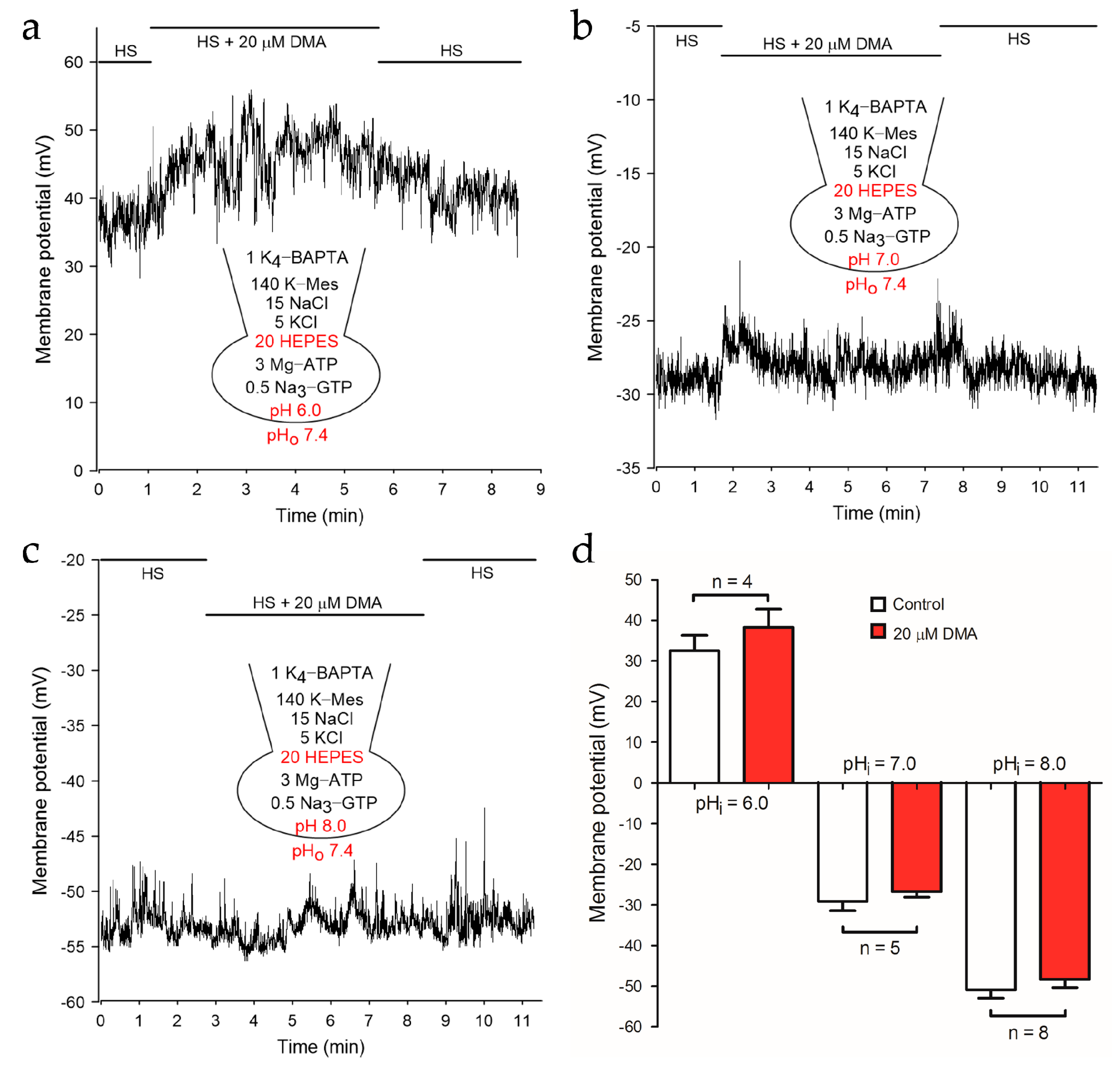
2.2. NHEs Inhibition Resulted in the Depolarization of Membrane Potential

The hyperpolarization of membrane potential during the capacitation process is necessary for the fertilizing capacity of sperm [10,16,19,27,28]. Given that KSper dominates the maintenance of membrane potential in mouse sperm [1], the transfer of DMA inhibition on NHEs to the reduction of KSper activity should be reflected as the change of sperm membrane potential. Since DMA may inhibit other channels/transporters such as the epithelial Na+ channel [25,29], the effect of DMA on the membrane potential with a pH-buffered pipette solution was examined first. Current clamp recordings showed that the membrane potential of mouse sperm had no significant alteration after adding 20 μM DMA at various pHi (Figure 2), suggesting that pH-independent hyperpolarization of membrane potential caused by DMA can be ignored.
Subsequently, we removed the pH buffer in the pipette solutions and explored whether NHEs inhibition caused by DMA could depolarize sperm membrane potential. After adding 20 μM DMA, the membrane potential of mouse sperm depolarized significantly at pHi 6.0 (−1.00 ± 2.58 mV to 20.31 ± 4.32 mV; p < 0.001), 7.0 (−29.09 ± 2.33 mV to −19.54 ± 3.08 mV; p < 0.05) or 8.0 (−40.47 ± 1.56 mV to −28.60 ± 1.78 mV; p < 0.001), respectively (Figure 3). Of note, the depolarized level at pHi 7.0 or 8.0 was less dramatic than that at pHi 6.0, possibly due to the compromised activity of NHEs at pHi 7.0 and 8.0. Consistently, DMA could partially reduce the hyperpolarization of membrane potential caused by 10 mM NH4Cl, which is usually used to alkalize cells at low pHi (Figure S3). Thus, it was concluded that NHEs inhibition remarkably suppressed the activation of the pH sensitive KSper channel and depolarized the membrane potential in mouse sperm.

2.3. Alkalization-Activated CatSper Channel and NHEs Were Functional Coupled

Since CatSper is also pH-dependent, we further investigated whether the inhibition of DMA on NHEs could affect the activity of CatSper. Similar to the strategy of the KSper study, the effect of DMA on CatSper currents was compared by using pipette solutions with or without a pH buffer. When the pipette solution was pH-buffered, the inward current of the CatSper channel was not impaired in the presence of DMA (Figure 4a,c). In contrast, DMA dramatically diminished the CatSper current when the pH buffer was eliminated from the pipette solution (Figure 4b,c). Statistical analysis showed that the inward CatSper currents at −80 mV decreased from −567.1 pA ± 65.15 pA to −234.6 ± 44.29 pA (Figure 4c). It was worth noting that CatSper current at high positive voltage decreased after adding DMA in Figure 4a. This decrease remained even as 50 mM of HEPES was added in the pipette solution to buffer the pHi (Figure S4), implying a disturbance of HEPES on the outward currents through CatSper. Nevertheless, this unphysiological effect should not affect the idea that the activation of the CatSper channel was also attenuated after inhibiting NHEs by DMA.

2.4. DMA Treatment Reduced Intracellular Ca2+ Concentration and Impaired Sperm Motility

Internal Ca2+ concentration regulates almost all of the physiological activities of sperm [30]. Considering that CatSper is the unique approach for Ca2+ influx and the hyperpolarization of membrane potential modulated by KSper provided a driving force of Ca2+, we speculated that the homeostasis of cytoplasmic Ca2+ would be disturbed by DMA. Indeed, the treatment of DMA pronouncedly down-regulated Ca2+ signals (Figure 5a,b). Furthermore, the results showed that the total motility and progressive motility of mouse sperm incubated with DMA exhibited a severe reduction (Figure 5c,d). Surprisingly, the velocity parameters, including those reflecting sperm hyperactivation which is dependent on CatSper, were not affected by DMA (Figure S5). Although the underlying mechanism to explain this apparently inconsistent information needs further investigation, the results generally support that the activity of NHEs is functionally coupled to the activation of CatSper under physiological conditions.

3. Discussion

The pH-regulated activation of KSper and CatSper is important for almost all functional aspects of sperm, such as capacitation, hyperactivation, acrosome reaction, and sperm-egg fusion [23]. By utilizing patch clamping recordings in single sperm together with an NHEs inhibitor DMA which was proved not to affect KSper and CatSper directly, this study clearly showed that the inhibition of NHEs decreased the activities of both KSper and CatSper, supporting the idea that NHEs activity is functionally coupled to the activation of KSper and CatSper channels. In another word, under physiological conditions, NHEs-mediated cytoplasmic alkalization should potentiate the pH-sensitive KSper and CatSper, and as a consequence, regulate sperm functions.
As the main transporters for eliminating the cytosolic protons, six isoforms of NHEs: NHE1 [31,32,33], NHE5 [34], NHE8 [35], sNHE [36,37], NHA1 [38], and NHA2 [39] had been reported to be expressed in sperm. However, which NHE is the determinant modulator for the activation of KSper and CatSper in murine sperm remained ambiguous. Considering that NHE1 and NHE2 were the primary targets for DMA [40], we adopted another selective NHE1 antagonist cariporide to verify NHE1 contribution in the activation of CatSper [32,40]. However, 10 μM cariporide failed to diminish the CatSper current in the pipette solution without a pH buffer (Figure S6a,b). Previous reports manifested that NHE1 was expressed in the midpiece of sperm [34], while this location could not detect any appreciable currents of KSper or CatSper [1,5]. Therefore, we speculated that NHE1 activity did not account for the activation of pH-sensitive ion channels.
sNHE and NHAs expressed in the principal piece of sperm were also candidates for mediating the activation of pH-dependent ion channels [36,39]. Given that NHAs were insensitive to amiloride [41], we supposed that KSper and CatSper were functionally coupled to sNHE. Unfortunately, there is no sNHE specific blocker or commercially available antibodies of sNHE which were capable of inhibiting CatSper currents (Figure S6c,f). Thus, we could not clarify the contribution of sNHE on the activation of the CatSper channel. Nevertheless, several studies implied that sNHE worked hand-in-hand with the KSper channel owing to the occurrence of the voltage sensor in sNHE and the pH sensitivity in KSper [9,20], supporting a functional interaction between sNHE and KSper. In terms of the CatSper channel, however, there was no measurable difference of CatSper currents recorded from WT and sNHE−/− sperm [12]. In that study, the pipette solution contained 10 mM of HEPES, which would minimize the pH regulation effects of sNHE on CatSper [5,12]. Comparison of KSper and CatSper currents recorded with pH-unbuffered pipette solution from WT and sNHE−/− sperm should clarify the contribution of sNHE on the activation of KSper and CatSper channels.
This study revealed a functional coupling between NHEs and pH-dependent KSper and CatSper channels in mouse sperm. Is a similar mechanism shared in human sperm? Human KSper exhibits less pH sensitivity than mouse KSper [2,3]. Besides, the regulatory mechanism of pHi may be different between human and mouse sperm [42]. Although it has been reported that NHE1 was expressed in human spermatozoa, a broad-spectrum NHE inhibitor EIPA could not block the initiation of acrosome reaction induced by progesterone [32]. Moreover, the voltage-gated proton channel Hv1 has been proposed as the major regulator for acid extrusion and physiological functions of human sperm [43]. Therefore, whether the NHEs-mediated cytoplasmic alkalization contributes to the potentiation of KSper and CatSper activities in human sperm needs further investigation.

4. Materials and Methods

4.1. Reagents

Fluo 4-AM and BCECF-AM were purchased from Molecular Probes (Thermo Fisher, Waltham, MA, USA). Bovine serum albumin (BSA) was purchased from Sangon Biotech (Shanghai, China). The anti-sNHE antibodies were purchased from Abcam (Cambridge, MA, USA) and Invitrogen (Thermo Fisher, Waltham, MA, USA). Other chemicals were obtained from Sigma-Aldrich (St Louis, MO, USA).

4.2. Animal Treatment and Sperm Preparation

The C57BL/6 mice (25–35 g; 12–20 weeks) for experiments were received from the Animal Center of Nanchang University. All animal procedures were performed in accord with the recommendations of the Animal Center of Nanchang University guidelines and approved by the Animal Research Ethics Committee of Nanchang University (SYXK2015-0002, January 2015, Nanchang, China). Mice were treated humanely and housed at the temperature of 20–25 °C.
Sperm were obtained from the isolated epididymides of male mice. For measurements of intracellular pH, Ca2+, and motility, the caudal epididymis was gently perforated to collect mature sperm. For patch-clamp recordings, the corpus epididymis was snipped into several parts to collect sperm containing the robust cytoplasmic droplet required for gigaohm seal. Although sperm in corpus epididymis are less mature than sperm in caudal epididymis [44], it was suggested that there are no significant differences in the properties of ion channels between the corpus and caudal epididymis of sperm [45]. Afterward, Minced mouse epididymis was rinsed in HS solution (135 mM NaCl, 5 mM KCl, 1 mM MgSO4, 2 mM CaCl2, 20 mM Hepes, 5 mM Glucose, 10 mM Lactic acid, and 1 mM Na-pyruvate at pH 7.4 with NaOH). Sperm was shaken slowly for about 20 min to swim out at 37 °C. Then, the supernatant was transferred into a 1.5 ml centrifuge tube for experiments.

4.3. Electrophysiology

Whole-cell recordings were conducted on non-capacitated sperm as reported [19]. Briefly, mouse sperm in corpus epididymis were isolated into HS solution. Gigaohm seals (>3 GΩ) between patch pipette (15–25 MΩ) and sperm cytosolic droplet were gained. Transitions into the whole-cell mode were made by applying 5 ms, 350–500 mV voltage pulses, and combined with light suction. For voltage-clamp recordings of KSper, the extracellular K+ symmetrical solution contained 160 mM KOH, 10 mM Hepes, 150 mM methanesulfonic acid (Mes), 2 mM Ca(OH)2, adjusted to pH 7.4 with KOH. A strong pH-buffered pipette solution contained 155 mM KOH, 5 mM KCl, 10 mM 1,2-bis (2-aminoph enoxy) ethane-N,N,N′,N′-tetraacetic acid (BAPTA), 20 mM Hepes, and 115 mM Mes adjusted to pH 8.0 with KOH. The pipette solution without pH buffer or with pH buffer contained: 140 mM KOH, 15 mM NaCl, 5 mM KCl, 1 mM BAPTA, 3 mM MgATP, 0.5 mM Na3GTP, 0 or 20 mM HEPES, adjusted to pH 7.0 with KOH. For current-clamp recordings of KSper, HS was used for primary extracellular solution for the current clamp. The pipette solution without pH buffer or with pH buffer contained 144 mM KOH, 5 mM KCl, 10 mM NaCl, 3 mM MgATP, 0.5 mM Na3GTP, 1 mM BAPTA, 140 mM Mes, and 0 or 20 mM Hepes with pH 6.0, 7.0, and 8.0 adjusted with Mes or KOH. For whole-cell recordings of mouse monovalent CatSper currents, the pipette solution without pH buffer or with pH buffer was filled with: 135 mM Cs-Mes, 5 mM CsCl, 0 or 20 mM HEPES, 10 mM EGTA adjusted to pH 7.2 with CsOH. The sodium-based divalent solution (DVF) comprised 150 mM sodium gluconate, 20 mM HEPES and 5 mM Na3HEDTA adjusted to pH 7.4 with NaOH. The extracellular solutions were applied directly via a local perfusion system. All currents and membrane potentials were recorded by utilizing Axon 200B amplifier (Molecular Devices, San Jose, CA, USA). Signals were filtered at 1 kHz and sampled at 10 kHz. All experiments were at room temperature (~22–25 °C). Data were analyzed with Clampfit 10.4 (Molecular Devices, San Jose, CA, USA) and Grapher 8 (Golden Software Inc., Golden, CO, USA).

4.4. Determination of Sperm Intracellular pH and Ca2+

Sperm intracellular pH and Ca2+ were examined by fluorescence recordings as previously reported [8]. Sperm obtained from the caudal epididymis were loaded with 0.5 μM pH-sensitive dye BCECF-AM or 5 μM Ca2+ indicator Fluo-4 AM in the presence of F-127 (0.1% v/v) for 30 min at 37 °C. Then, sperm were washed once at 300 g for 5 min and resuspended in HS solution. For the recordings, these sperm were loaded in the multimode plate reader (PerkinElmer, Waltham, MA, USA) at 30 °C. When the recordings were stable, HS or 20 μM DMA was added. 10 mM NH4Cl was a positive control. The relative changes of intracellular pH and Ca2+ were showed by (ΔF)/F0 × 100%. ΔF indicates the fluorescence changes before and after the injection of solutions. F0 represents the stable value of basal fluorescence.

4.5. Assessment of Sperm Motility

Mouse sperm for the detection of motility were collected from the caudal epididymis. HS solution containing 25 mM NaHCO3 and 5 mg/mL BSA was prepared for measuring sperm motility. Two sides of caudal epididymides in one mouse were divided into the control group and the experimental group (20 μM DMA), respectively. Caudal epididymis was gently perforated to allow sperm to swim-out for 15 min at 37 °C. Then, sperm samples were injected into 80 μm deep chambers (Hamilton-Throne, Beverly, MA, USA). Sperm motility was analyzed using a computer-assisted sperm analysis (CASA) system (Hamilton-Throne, Beverly, MA, USA) on a Zeiss microscope with a 10 × objective (Oberkochen, German) every 30 min. More than 200 motile sperm were examined. The parameters including the percentage of motile and progressive motile of sperm, average path velocity (VAP), curvilinear velocity (VCL), straight-line velocity (VSL), amplitude of lateral head displacement (ALH), beat cross frequency (BCF) and linearity (LIN) were calculated.

4.6. Statistical Analysis

Data were expressed as mean ± SEM. Statistical significance in the results of sperm total motility and progressive motility were determined by employing an unpaired t-test. Other results were assessed by a paired t-test (for two group comparisons) and a one-way ANOVA (for multi-group comparisons). A statistically significant difference was set at p < 0.05. All statistical analyses were performed in GraphPad Prism, version 5.0 software (San Diego, CA, USA).

Supplementary Materials

The following are available online at https://0-www-mdpi-com.brum.beds.ac.uk/1422-0067/22/4/1612/s1, Figure S1: pHi in mouse sperm was reduced by DMA application, Figure S2: DMA had no impact on KSper channel, Figure S3: The alkalization-induced hyperpolarization was reversibly weakened by DMA application in a pH 6.0 pipette solution without pH buffer, Figure S4: DMA decreased the outward current of CatSper in the pipette solution with a strong buffer of pH, Figure S5: Sperm motility parameters were not affected by DMA incubation, Figure S6: Cariporide and commercially sNHE antibodies failed to inhibit CatSper current.

Author Contributions

Conceptualization, H.K., M.L. and X.-H.Z.; Methodology, H.K., M.L., W.Z., R.-Z.H., N.Z. and C.C.; Software, H.K. and M.L.; Validation, H.K. and M.L.; Formal Analysis, H.K. and M.L.; Investigation, H.K., M.L. and X.-H.Z.; Resources, X.-H.Z.; Data Curation, H.K., M.L. and X.-H.Z.; Writing—Original Draft Preparation, H.K. and M.L.; Writing—Review & Editing, H.K. and X.-H.Z.; Visualization, H.K. and X.-H.Z.; Supervision, X.-H.Z.; Project Administration, X.-H.Z.; Funding Acquisition, X.-H.Z. All authors have read and agreed to the published version of the manuscript.

Funding

This research was funded by the National Natural Science Foundation of China to X.-H.Z. (81871201 and 31673204).

Institutional Review Board Statement

The study was conducted according to the guidelines of the Declaration of Helsinki, and approved by the Animal Research Ethics Committee of Nanchang University (SYXK2015-0002, January, 2015, Nanchang, China).

Informed Consent Statement

Not applicable.

Data Availability Statement

Data is contained within the supplementary material.

Conflicts of Interest

The authors declare no conflict of interest. The funders had no role in the design of the study; in the collection, analyses, or interpretation of data; in the writing of the manuscript, or in the decision to publish the results.

Abbreviations

ALHAmplitude of lateral head displacement
BAPTA1,2-bis(2-aminophenoxy)ethane-N,N,N’,N’-tetraacetic acid
BCECFBCECF-AM
BCFBeat cross frequency
BSABovine serum albumin
CatSperCa2+ channel of sperm
DMA5-(N, N-dimethyl)-amiloride
EFCAB9EF-hand calcium-binding domain-containing protein 9
EGTAEthylene glycol tetraacetic acid
EIPA5-(N-ethyl-N-isopropyl)-amiloride
HEPES4-(2-hydroxyethyl)-1-piperazineethanesulfonic acid
HPHolding potential
HSHigh saline solution
Hv1Voltage-gated proton channel
KSperPotassium channel of sperm
LINLinearity
MesMethanesulfonic acid
Na+DVFSodium-based divalent free solution
NHANa+/H+ antiporter
NHEsNa+/H+ exchangers
NH4ClAmmonium chloride
pHiIntracellular pH
SEMStandard error of the mean
sNHESperm-specific NHE
VAPAverage path velocity
VCLCurvilinear velocity
VSLStraight-line velocity

References

  1. Navarro, B.; Kirichok, Y.; Clapham, D.E. KSper, a pH-sensitive K+ current that controls sperm membrane potential. Proc. Natl. Acad. Sci. USA 2007, 104, 7688–7692. [Google Scholar] [CrossRef] [Green Version]
  2. Brenker, C.; Zhou, Y.; Müller, A.; Echeverry, F.A.; Trötschel, C.; Poetsch, A.; Xia, X.M.; Bönigk, W.; Lingle, C.J.; Kaupp, U.B.; et al. The Ca2+-activated K+ current of human sperm is mediated by Slo3. Elife 2014, 3, e01438. [Google Scholar] [CrossRef]
  3. Mannowetz, N.; Naidoo, N.M.; Choo, S.A.; Smith, J.F.; Lishko, P.V. Slo1 is the principal potassium channel of human spermatozoa. Elife 2013, 2, e01009. [Google Scholar] [CrossRef] [PubMed]
  4. Ren, D.; Navarro, B.; Perez, G.; Jackson, A.C.; Hsu, S.; Shi, Q.; Tilly, J.L.; Clapham, D.E. A sperm ion channel required for sperm motility and male fertility. Nature 2001, 413, 603–609. [Google Scholar] [CrossRef]
  5. Kirichok, Y.; Navarro, B.; Clapham, D.E. Whole-cell patch-clamp measurements of spermatozoa reveal an alkaline-activated Ca2+ channel. Nature 2006, 439, 737–740. [Google Scholar] [CrossRef] [PubMed]
  6. Xia, J.; Reigada, D.; Mitchell, C.H.; Ren, D. CATSPER channel-mediated Ca2+ entry into mouse sperm triggers a tail-to-head propagation. Biol. Reprod. 2007, 77, 551–559. [Google Scholar] [CrossRef] [PubMed] [Green Version]
  7. Lishko, P.V.; Botchkina, I.L.; Kirichok, Y. Progesterone activates the principal Ca2+ channel of human sperm. Nature 2011, 471, 387–391. [Google Scholar] [CrossRef]
  8. Strünker, T.; Goodwin, N.; Brenker, C.; Kashikar, N.D.; Weyand, I.; Seifert, R.; Kaupp, U.B. The CatSper channel mediates progesterone-induced Ca2+ influx in human sperm. Nature 2011, 471, 382–386. [Google Scholar] [CrossRef]
  9. Santi, C.M.; Martinez-Lopez, P.; de la Vega-Beltran, J.L.; Butler, A.; Alisio, A.; Darszon, A.; Salkoff, L. The SLO3 sperm-specific potassium channel plays a vital role in male fertility. FEBS Lett. 2010, 584, 1041–1046. [Google Scholar] [CrossRef] [Green Version]
  10. Zeng, X.H.; Yang, C.; Kim, S.T.; Lingle, C.J.; Xia, X.M. Deletion of the Slo3 gene abolishes alkalization-activated K+ current in mouse spermatozoa. Proc. Natl. Acad. Sci. USA 2011, 108, 5879–5884. [Google Scholar] [CrossRef] [Green Version]
  11. Quill, T.A.; Sugden, S.A.; Rossi, K.L.; Doolittle, L.K.; Hammer, R.E.; Garbers, D.L. Hyperactivated sperm motility driven by CatSper2 is required for fertilization. Proc. Natl. Acad. Sci. USA 2003, 100, 14869–14874. [Google Scholar] [CrossRef] [Green Version]
  12. Qi, H.; Moran, M.M.; Navarro, B.; Chong, J.A.; Krapivinsky, G.; Krapivinsky, L.; Kirichok, Y.; Ramsey, I.S.; Quill, T.A.; Clapham, D.E. All four CatSper ion channel proteins are required for male fertility and sperm cell hyperactivated motility. Proc. Natl. Acad. Sci. USA 2007, 104, 1219–1223. [Google Scholar] [CrossRef] [Green Version]
  13. Chung, J.J.; Navarro, B.; Krapivinsky, G.; Krapivinsky, L.; Clapham, D.E. A novel gene required for male fertility and functional CATSPER channel formation in spermatozoa. Nat. Commun. 2011, 2, 153. [Google Scholar] [CrossRef] [PubMed]
  14. Chung, J.J.; Miki, K.; Kim, D.; Shim, S.H.; Shi, H.F.; Hwang, J.Y.; Cai, X.; Iseri, Y.; Zhuang, X.; Clapham, D.E. CatSperζ regulates the structural continuity of sperm Ca(2+) signaling domains and is required for normal fertility. Elife 2017, 6, 23082. [Google Scholar] [CrossRef] [PubMed]
  15. Hwang, J.Y.; Mannowetz, N.; Zhang, Y.; Everley, R.A.; Gygi, S.P.; Bewersdorf, J.; Lishko, P.V.; Chung, J.J. Dual Sensing of Physiologic pH and Calcium by EFCAB9 Regulates Sperm Motility. Cell 2019, 177, 1480–1494. [Google Scholar] [CrossRef] [PubMed]
  16. Brown, S.G.; Publicover, S.J.; Mansell, S.A.; Lishko, P.V.; Williams, H.L.; Ramalingam, M.; Wilson, S.M.; Barratt, C.L.; Sutton, K.A.; Da Silva, S.M. Depolarization of sperm membrane potential is a common feature of men with subfertility and is associated with low fertilization rate at IVF. Hum. Reprod. 2016, 31, 1147–1157. [Google Scholar] [CrossRef] [PubMed] [Green Version]
  17. Smith, J.F.; Syritsyna, O.; Fellous, M.; Serres, C.; Mannowetz, N.; Kirichok, Y.; Lishko, P.V. Disruption of the principal, progesterone-activated sperm Ca2+ channel in a CatSper2-deficient infertile patient. Proc. Natl. Acad. Sci. USA 2013, 110, 6823–6828. [Google Scholar] [CrossRef] [Green Version]
  18. Luo, T.; Chen, H.Y.; Zou, Q.X.; Wang, T.; Cheng, Y.M.; Wang, H.F.; Wang, F.; Jin, Z.L.; Chen, Y.; Weng, S.Q.; et al. A novel copy number variation in CATSPER2 causes idiopathic male infertility with normal semen parameters. Hum. Reprod. 2019, 34, 414–423. [Google Scholar] [CrossRef]
  19. Zeng, X.H.; Yang, C.; Xia, X.M.; Liu, M.; Lingle, C.J. SLO3 auxiliary subunit LRRC52 controls gating of sperm KSPER currents and is critical for normal fertility. Proc. Natl. Acad. Sci. USA 2015, 112, 2599–2604. [Google Scholar] [CrossRef] [PubMed] [Green Version]
  20. Chávez, J.C.; Ferreira, J.J.; Butler, A.; De La Vega Beltrán, J.L.; Treviño, C.L.; Darszon, A.; Salkoff, L.; Santi, C.M. SLO3 K+ channels control calcium entry through CATSPER channels in sperm. J. Biol. Chem. 2014, 289, 32266–32275. [Google Scholar] [CrossRef] [Green Version]
  21. Schreiber, M.; Wei, A.; Yuan, A.; Gaut, J.; Saito, M.; Salkoff, L. Slo3, a novel pH-sensitive K+ channel from mammalian spermatocytes. J. Biol. Chem. 1998, 273, 3509–3516. [Google Scholar] [CrossRef] [PubMed] [Green Version]
  22. Zhang, X.; Zeng, X.; Lingle, C.J. Slo3 K+ channels: Voltage and pH dependence of macroscopic currents. J. Gen. Physiol. 2006, 128, 317–336. [Google Scholar] [CrossRef] [PubMed] [Green Version]
  23. Nishigaki, T.; José, O.; González-Cota, A.L.; Romero, F.; Treviño, C.L.; Darszon, A. Intracellular pH in sperm physiology. Biochem. Biophys. Res. Commun. 2014, 450, 1149–1158. [Google Scholar] [CrossRef] [PubMed] [Green Version]
  24. Lardy, H.A.; Pounden, W.D.; Phillips, P.H. H ion concentration of various fluids of the genital tract of the cow. Proc. Soc. Exp. Biol. Med. 1940, 44, 517. [Google Scholar] [CrossRef]
  25. Kleyman, T.R.; Cragoe, E.J., Jr. Amiloride and its analogs as tools in the study of ion transport. J. Membr. Biol. 1988, 105, 1–21. [Google Scholar] [CrossRef]
  26. Aronson, P.S.; Nee, J.; Suhm, M.A. Modifier role of internal H+ in activating the Na+-H+ exchanger in renal microvillus membrane vesicles. Nature 1982, 299, 161–163. [Google Scholar] [CrossRef] [PubMed]
  27. Baro Graf, C.; Ritagliati, C.; Torres-Monserrat, V.; Stival, C.; Carizza, C.; Buffone, M.G.; Krapf, D. Membrane Potential Assessment by Fluorimetry as a Predictor Tool of Human Sperm Fertilizing Capacity. Front. Cell Dev. Biol. 2019, 7, 383. [Google Scholar] [CrossRef]
  28. Molina, L.C.P.; Gunderson, S.; Riley, J.; Lybaert, P.; Borrego-Alvarez, A.; Jungheim, E.S.; Santi, C.M. Membrane Potential Determined by Flow Cytometry Predicts Fertilizing Ability of Human Sperm. Front. Cell Dev. Biol. 2019, 7, 387. [Google Scholar] [CrossRef] [Green Version]
  29. Hernández-González, E.O.; Sosnik, J.; Edwards, J.; Acevedo, J.J.; Mendoza-Lujambio, I.; López-González, I.; Demarco, I.; Wertheimer, E.; Darszon, A.; Visconti, P.E. Sodium and epithelial sodium channels participate in the regulation of the capacitation-associated hyperpolarization in mouse sperm. J. Biol. Chem. 2006, 281, 5623–5633. [Google Scholar] [CrossRef] [PubMed] [Green Version]
  30. Publicover, S.; Harper, C.V.; Barratt, C. [Ca2+]i signalling in sperm--making the most of what you’ve got. Nat. Cell Biol. 2007, 9, 235–242. [Google Scholar] [CrossRef] [PubMed]
  31. Martins, A.D.; Bernardino, R.L.; Neuhaus-Oliveira, A.; Sousa, M.; Sá, R.; Alves, M.G.; Oliveira, P.F. Physiology of na+/h+ exchangers in the male reproductive tract: Relevance for male fertility. Biol. Reprod. 2014, 91, 11. [Google Scholar] [CrossRef]
  32. Garcia, M.A.; Meizel, S. Regulation of intracellular pH in capacitated human spermatozoa by a Na+/H+ exchanger. Mol. Reprod. Dev. 1999, 52, 189–195. [Google Scholar] [CrossRef]
  33. Muzzachi, S.; Guerra, L.; Martino, N.A.; Favia, M.; Punzi, G.; Silvestre, F.; Guaricci, A.C.; Roscino, M.T.; Pierri, C.L.; Dell’Aquila, M.E.; et al. Effect of cariporide on ram sperm pH regulation and motility: Possible role of NHE1. Reproduction 2018, 155, 433–445. [Google Scholar] [CrossRef] [PubMed]
  34. Woo, A.L.; James, P.F.; Lingrel, J.B. Roles of the Na,K-ATPase alpha4 isoform and the Na+/H+ exchanger in sperm motility. Mol. Reprod. Dev. 2002, 62, 348–356. [Google Scholar] [CrossRef]
  35. Oberheide, K.; Puchkov, D.; Jentsch, T.J. Loss of the Na(+)/H(+) exchanger NHE8 causes male infertility in mice by disrupting acrosome formation. J. Biol. Chem. 2017, 292, 10845–10854. [Google Scholar] [CrossRef] [PubMed] [Green Version]
  36. Wang, D.; King, S.M.; Quill, T.A.; Doolittle, L.K.; Garbers, D.L. A new sperm-specific Na+/H+ exchanger required for sperm motility and fertility. Nat. Cell Biol. 2003, 5, 1117–1122. [Google Scholar] [CrossRef] [PubMed]
  37. Wang, D.; Hu, J.; Bobulescu, I.A.; Quill, T.A.; McLeroy, P.; Moe, O.W.; Garbers, D.L. A sperm-specific Na+/H+ exchanger (sNHE) is critical for expression and in vivo bicarbonate regulation of the soluble adenylyl cyclase (sAC). Proc. Natl. Acad. Sci. USA 2007, 104, 9325–9330. [Google Scholar] [CrossRef] [PubMed] [Green Version]
  38. Liu, T.; Huang, J.C.; Zuo, W.L.; Lu, C.L.; Chen, M.; Zhang, X.S.; Li, Y.C.; Cai, H.; Zhou, W.L.; Hu, Z.Y.; et al. A novel testis-specific Na+/H+ exchanger is involved in sperm motility and fertility. Front. Biosci. 2010, 2, 566–581. [Google Scholar] [CrossRef] [Green Version]
  39. Chen, S.R.; Chen, M.; Deng, S.L.; Hao, X.X.; Wang, X.X.; Liu, Y.X. Sodium-hydrogen exchanger NHA1 and NHA2 control sperm motility and male fertility. Cell Death Dis. 2016, 7, e2152. [Google Scholar] [CrossRef] [PubMed] [Green Version]
  40. Masereel, B.; Pochet, L.; Laeckmann, D. An overview of inhibitors of Na(+)/H(+) exchanger. Eur. J. Med. Chem. 2003, 38, 547–554. [Google Scholar] [CrossRef]
  41. Xiang, M.; Feng, M.; Muend, S.; Rao, R. A human Na+/H+ antiporter sharing evolutionary origins with bacterial NhaA may be a candidate gene for essential hypertension. Proc. Natl. Acad. Sci. USA 2007, 104, 18677–18681. [Google Scholar] [CrossRef] [PubMed] [Green Version]
  42. Miller, M.R.; Mansell, S.A.; Meyers, S.A.; Lishko, P.V. Flagellar ion channels of sperm: Similarities and differences between species. Cell Calcium. 2015, 58, 105–113. [Google Scholar] [CrossRef] [Green Version]
  43. Lishko, P.V.; Botchkina, I.L.; Fedorenko, A.; Kirichok, Y. Acid extrusion from human spermatozoa is mediated by flagellar voltage-gated proton channel. Cell 2010, 140, 327–337. [Google Scholar] [CrossRef] [PubMed] [Green Version]
  44. Yanagimachi, R. Fertility of mammalian spermatozoa: Its development and relativity. Zygote 1994, 2, 371–372. [Google Scholar] [CrossRef] [PubMed]
  45. Lishko, P.V.; Clapham, D.E.; Navarro, B.; Kirichok, Y. Sperm patch-clamp. Methods Enzym. 2013, 525, 59–83. [Google Scholar] [CrossRef] [Green Version]
Figure 1. 5-(N,N-dimethyl)-amiloride (DMA) prominently reduced K+ ion channel (KSper) currents after the removal of pH buffer in the pipette solution. Representative patch-clamp recordings of KSper currents in the absence or presence of 20 μM DMA elicited by 1 s voltage ramp from −100 mV to + 100 mV with a 20 mM 4-(2-hydroxyethyl)-1-piperazineethanesulfonic acid (HEPES) (a) or 0 mM HEPES (b) pipette solution at pHi 7.0. The high saline solution (HS) containing 135 mM Na+ and 2 mM Ca2+ was employed to maintain Na+/H+ exchangers (NHEs) activity and block the CatSper current, respectively. (c) Statistical analysis of the mean normalized KSper currents at + 80 mV recorded in the pipette solution containing 20 mM HEPES (n = 5) or 0 mM HEPES (n = 5). Data are expressed as mean ± standard error of the mean (SEM). ** p < 0.01 (paired t-test).
Ijms 22 01612 g001
Figure 2. DMA failed to depolarize the membrane potential in a pH buffered pipette solution. Representative traces of membrane potentials recorded by the current clamp at pHi 6.0 (a), pHi 7.0 (b), pHi 8.0 (c) was shown in the absence or presence of 20 μM DMA. HS solution containing 135 mM Na+ was employed to maintain NHEs activity. (d) Mean amplitudes of membrane potential before and after the treatment of DMA at pHi 6.0 (n = 4), pHi 7.0 (n = 5) and pHi 8.0 (n = 8). Data are expressed as mean ± SEM.
Ijms 22 01612 g002
Figure 3. The membrane potential of mouse spermatozoa was depolarized by DMA in the pipette solution with no pH buffer. Sperm membrane potential was reversibly depolarized by 20 μM DMA at pHi 6.0 (a), pHi 7.0 (b), pHi 8.0 (c). Representatives of three current-clamp recordings are shown respectively. HS solution containing 135 mM Na+ was employed to maintain NHEs activity. (d) Mean amplitudes of membrane potential before and after the treatment of DMA at pHi 6.0 (n = 9), pHi 7.0 (n = 5) and pHi 8.0 (n = 16). Data are expressed as mean ± SEM. * p < 0.05, *** p < 0.001 (paired t-test).
Ijms 22 01612 g003
Figure 4. DMA prominently inhibited the Ca2+ ion channel (CatSper) currents via the acidification of pHi. Representative patch-clamp recordings of CatSper currents in the absence or presence of 20 μM DMA elicited by 1 s voltage ramp from −100 mV to +100 mV with a 20 mM HEPES (a) or 0 mM HEPES (b) pipette solution at pHi 7.2. The sodium-based divalent free solution (Na+ DVF) was employed to record the monovalent CatSper current and maintain NHEs activity. (c) Statistical analysis of the mean CatSper currents at −80 mV recorded in the pipette solution containing 20 mM HEPES (n = 6) or 0 mM HEPES (n = 5). Data are expressed as mean ± SEM. ** p < 0.01 (paired t-test).
Ijms 22 01612 g004
Figure 5. The effect of NHEs inhibition by DMA on intracellular Ca2+ and motility of mouse sperm were evaluated. (a) The fluorescence trace of intracellular Ca2+ signals was shown. Arrow indicates the time point of HS solution or DMA injection. (b) Mean fluorescence intensities of Ca2+ signals in the absence and presence of DMA. n = 5 (*** p < 0.001, paired t-test). The effect of DMA on the total motility (c) or the progressive motility (d) of mouse sperm at 0, 30, 60, 90, and 120 min was shown. Data are expressed as mean ± SEM. n = 6 (** p < 0.01; *** p < 0.001, unpaired t-test).
Ijms 22 01612 g005
Publisher’s Note: MDPI stays neutral with regard to jurisdictional claims in published maps and institutional affiliations.

