Article

Synthesis and Self-Assembly Properties of Bola-Amphiphilic Glycosylated Lipopeptide-Type Supramolecular Hydrogels Showing Colour Changes Along with Gel–Sol Transition

1
Graduate School of Integrated Arts and Sciences, Kochi University, 2-5-1, Akebono-cho, Kochi 780-8520, Japan
2
School of Environmental Science and Engineering, Kochi University of Technology, Kami, Kochi 782-8502, Japan
3
Research Center for Molecular Design, Kochi University of Technology, Kami, Kochi 782-8502, Japan
4
Faculty of Science, Kochi University, 2-5-1, Akebono-cho, Kochi 780-8520, Japan
5
Department of Chemistry and Biomolecular Science, Faculty of Engineering, Gifu University, 1-1 Yanagido, Gifu 501-1193, Japan
6
United Graduate School of Drug Discovery and Medical Information Sciences, Gifu University, 1-1 Yanagido, Gifu 501-1193, Japan
7
Interdisciplinary Science Unit, Multidisciplinary Sciences Cluster, Research and Education Faculty, Kochi University, 2-5-1, Akebono-cho, Kochi 780-8520, Japan
8
Faculty of Science and Technology, Kochi University, 2-5-1, Akebono-cho, Kochi 780-8520, Japan
*
Author to whom correspondence should be addressed.
Int. J. Mol. Sci. 2021, 22(4), 1860; https://0-doi-org.brum.beds.ac.uk/10.3390/ijms22041860
Submission received: 19 January 2021 / Revised: 1 February 2021 / Accepted: 9 February 2021 / Published: 13 February 2021
(This article belongs to the Special Issue Self-Assembly Mechanism and Connection of Peptides and Proteins)

Abstract

:
Supramolecular hydrogels formed by self-assembly of low-molecular-weight amphiphiles (hydrogelators) have attracted significant attention, as smart and soft materials. However, most of the observed stimuli-responsive behaviour of these supramolecular hydrogels are limited to gel–sol transitions. In this study, we present bola-amphiphilic glycosylated lipopeptide-type supramolecular hydrogelators that exhibit reversible thermochromism along with a gel–sol transition. The bola-amphiphiles have mono-, di-, tri- or tetra-phenylalanine (F) as a short peptide moiety. We investigate and discuss the effects of the number of F residues on the gelation ability and the morphology of the self-assembled nanostructures.

1. Introduction

Supramolecular hydrogels constructed through the self-assembly of low molecular-weight amphiphiles (hydrogelator) via weak non-covalent interactions, such as hydrogen bonding, hydrophobic interactions, π-π stacking, electrostatic interactions have attracted significant attention, as smart and soft materials [1,2,3,4,5,6,7,8,9,10,11,12,13]. Many supramolecular hydrogels capable of rapidly responding to external stimuli, such as chemical additives [14,15,16,17,18,19,20,21] and biological molecules [22,23,24,25,26,27,28,29,30,31,32,33], have been reported. However, most of the observed stimuli-responsive behaviour of these supramolecular hydrogels is limited to gel–sol transitions. Supramolecular hydrogels that exhibit a colour change (i.e., chromism) in response to the desired external stimuli are useful for developing practical sensing materials [34,35,36,37,38,39,40].
We developed supramolecular hydrogels that exhibit thermochromism along with a gel–sol transition in which the hydrogel state was yellow and the solution state was orange (Figure 1) [36,40]. The hydrogelators have an N-alkyl-2-anilino-3-chloromaleimide (AAC) moiety as a chromophore, capable of acting as a probe to readout the self-assembly state. Considering its solution state counterpart, the blue-shifted absorption band in the hydrogel state indicates that the AAC moiety in the hydrogelators was stacked in an H-type aggregation mode [41,42]. Despite the usefulness of this system as a colorimetric assay, the molecular design of hydrogelators was limited to the glycolipid-type bola-amphiphiles with hydrophilic moiety (saccharide or carboxy group) at each end of the hydrophobic core, including the AAC moiety and the hydrocarbon chain.
In this paper, we report the design, synthesis, and self-assembly properties of glycosylated lipopeptide-type bola-amphiphiles to diversify the molecular design of our colour-change hydrogelation system. The lipopeptides are a class of molecules with one or more lipid chains attached to a short peptide. Recently, self-assembled peptide-based nanomaterials have attracted significant attention, as functional materials [43,44,45,46,47,48,49]. Short peptide-based amphiphiles have been studied as hydrogelators due to their ability to assemble into a large range of novel nanostructures and their rational design for various applications, such as molecular sensors, tissue engineering, and drug-delivery systems [50,51,52,53,54,55,56]. The molecular configurations and intramolecular interactions of short peptides can be controlled by the amino acid sequence. Aromatic amino acids, such as phenylalanine (F), tyrosine (Y), and tryptophan (W) are a popular type of building block with aromatic π-π interactions and hydrophobic interaction. Hydrogelators containing diphenylalanine (F2) peptide, which is the core motif of Alzheimer’s β-amyloid peptide or more extended aromatic sequences have been investigated [57,58,59]. F2 peptide can self-assemble into a nanostructure that is obtained by combining hydrogen bonding and π-π stacking interactions. Despite the significant attention on F2-based hydrogels, a few examples of triphenylalanine (F3)- or tetraphenylalanine (F4)-based hydrogels and self-assembled nanostructures have been investigated [60,61,62,63,64,65]. Here, we evaluate the effects of the number of F residues on the gelation ability and the morphology of the self-assembled nanostructures.

2. Results and Discussion

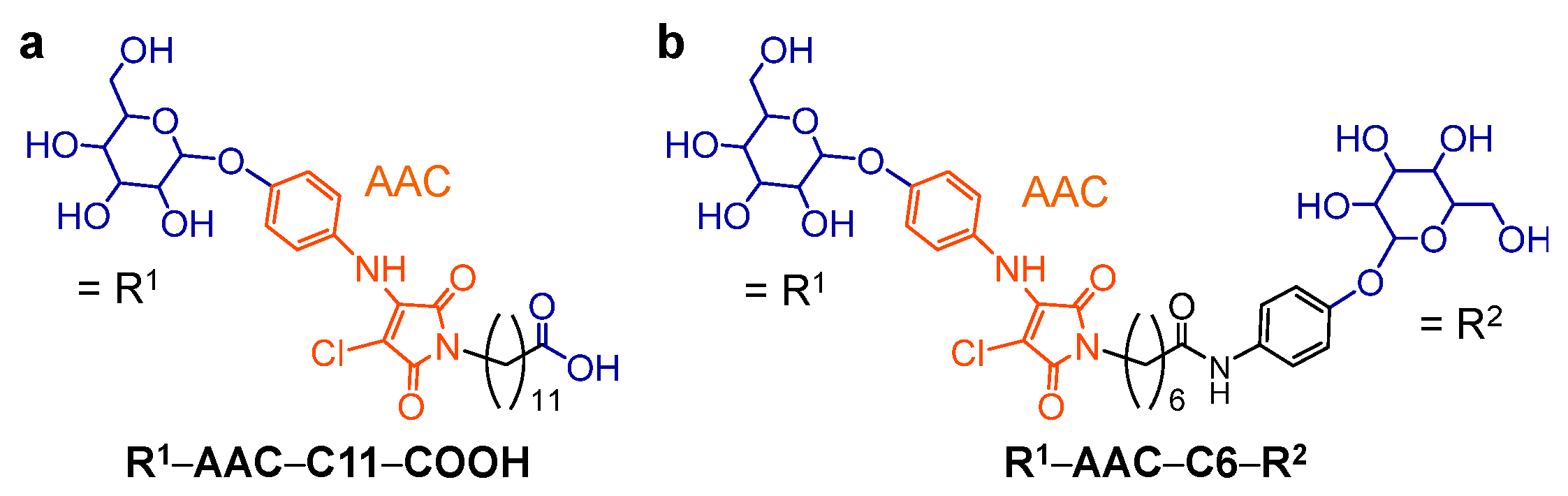
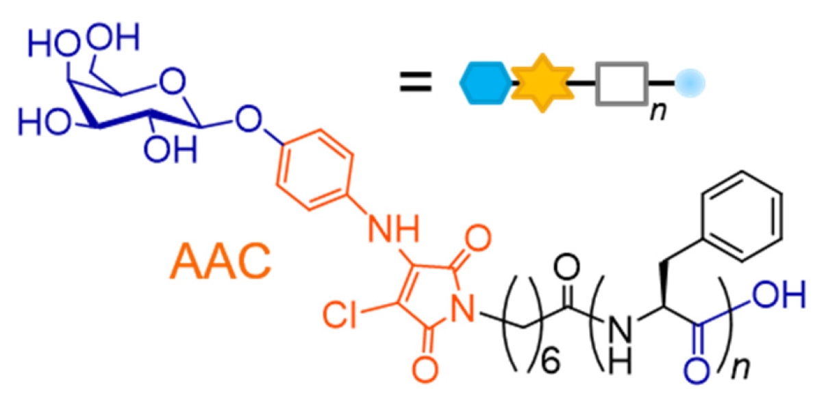
2.1. Molecular Design of Glycosylated Lipopeptide-type Bola-amphiphiles

We designed and synthesized the glycosylated lipopeptide-type bola-amphiphiles with the general formula β-D-galactose (βGal)–AAC–C6–Fn (n = 1–4) (Figure 2, Scheme 1). The saccharide structure and hydrocarbon chain length are essential factors for the self-assembly property of glycolipid-type amphiphiles [36,40,66,67,68,69,70,71,72]. We fixed the saccharide structure as βGal and hydrocarbon chain length as the C6 spacer based on the previous study [40]. Thus, we evaluated the effect of the number of F residues on the self-assembly properties of the prepared bola-amphiphiles.

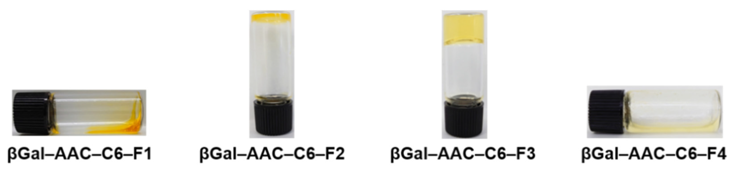
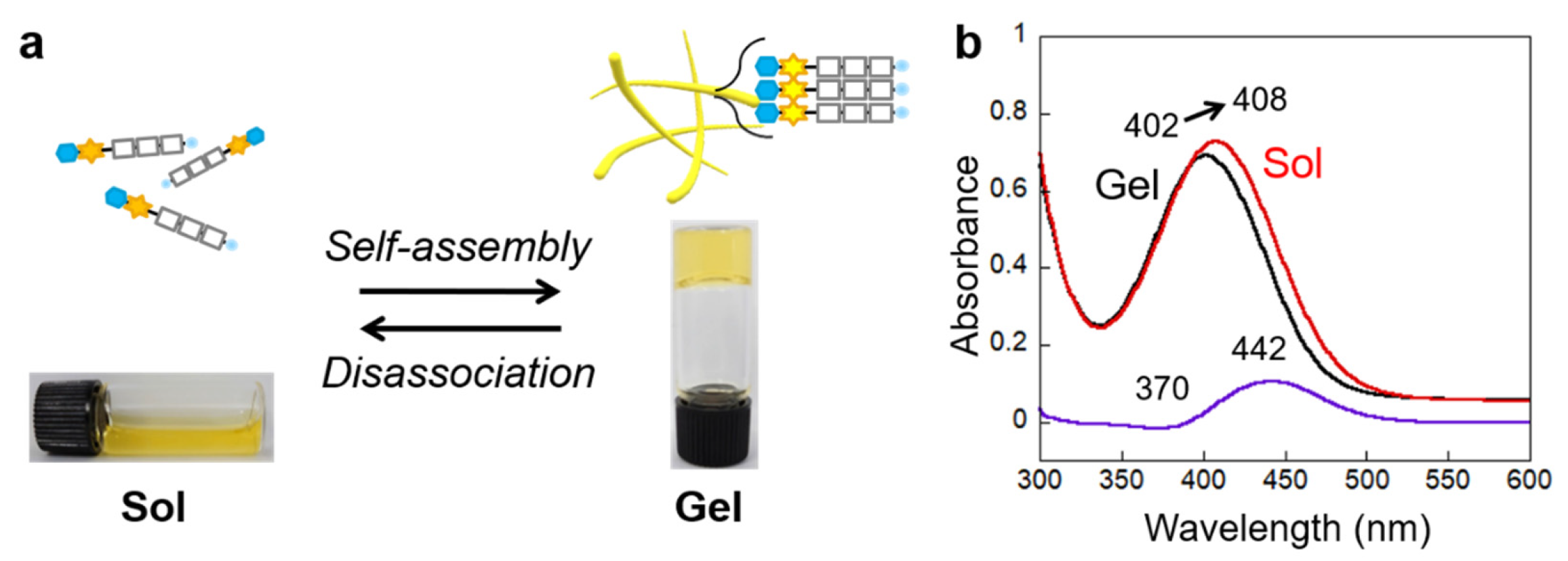
2.2. Self-Assembly Properties of Glycosylated Lipopeptide-Type Bola-Amphiphiles

The gelation ability of the glycosylated lipopeptide-type bola-amphiphiles in 200 mM HEPES–NaOH buffer (pH 8.0) was screened using the tube-inversion method (Table 1, Figure 3). βGal–AAC–C6–F2 formed unstable partial hydrogel, and βGal–AAC–C6–F3 formed stable transparent hydrogel at their critical gelation concentrations (CGCs). However, even at sufficiently high concentration (about 10 wt%), βGal–AAC–C6–F1 remained in solution state (Figure 3a). This behaviour may be attributed to the higher water solubility compared to the other compounds. Furthermore, the sample of βGal–AAC–C6–F4, which showed the lowest solubility, was dispersed at 0.35 wt%. These results indicate that differences in the combination of intermolecular hydrogen bonding between amide groups and side-chain π-π stacking interactions, which were caused by the difference in the number of F residues, have a significant effect on the self-assembly properties. It was revealed that βGal–AAC–C6–F3 has the optimal assembly properties for gel formation. We obtained that the compound’s maximum absorption wavelength (λmax) in 200 mM HEPES–NaOH buffer (pH 8.0) shifted to shorter wavelength as the number of F increased (Table 1, Figure S1, ESI† for absorption spectra), suggesting the larger intermolecular interaction of the molecule. The difference in the chemical structure is only the number of F unit, however, the abrupt change in the phase behaviour was observed. It is presumed that the differences in the aromatic ππ interactions derived from the side chain phenyl groups of F and the hydrogen bonding mode derived from the difference in the amide bonds greatly contribute to the self-assembly ability of the molecules.
The above gelation process was thermally reversible, as shown in Figure 4a for βGal–AAC–C6–F3 (Tgel: 78 °C at 0.19 wt% (CGC)), and a chromic change upon the sol–gel transition was observed similarly to the existing system [39]. The UV–Vis absorption spectral analysis confirmed the colour change. The absorption band arising from the AAC moiety was bathochromically shifted from the absorption maximum being 402 nm for the hydrogel state at 25 °C to 408 nm for the solution state at 85 °C (Figure 4b and Figure S2, ESI† for temperature-dependent absorption spectral change). The differential spectrum showed an increase in absorbance at 442 nm and a decrease of 370 nm. Considering its solution state counterpart, the blue-shifted absorption band in the hydrogel state (Δλmax = 6 nm) indicates that the AAC moieties in the hydrogel are stacked in an H-type aggregation mode. However, the visual colour change between the gel state and the solution state of βGal–AAC–C6–F3 sample was scarce (Figure 4a) compared to previously reported glycolipid-type hydrogels [35,39]. The cause of this phenomenon is unknown, and we intend to investigate the cause in the future.

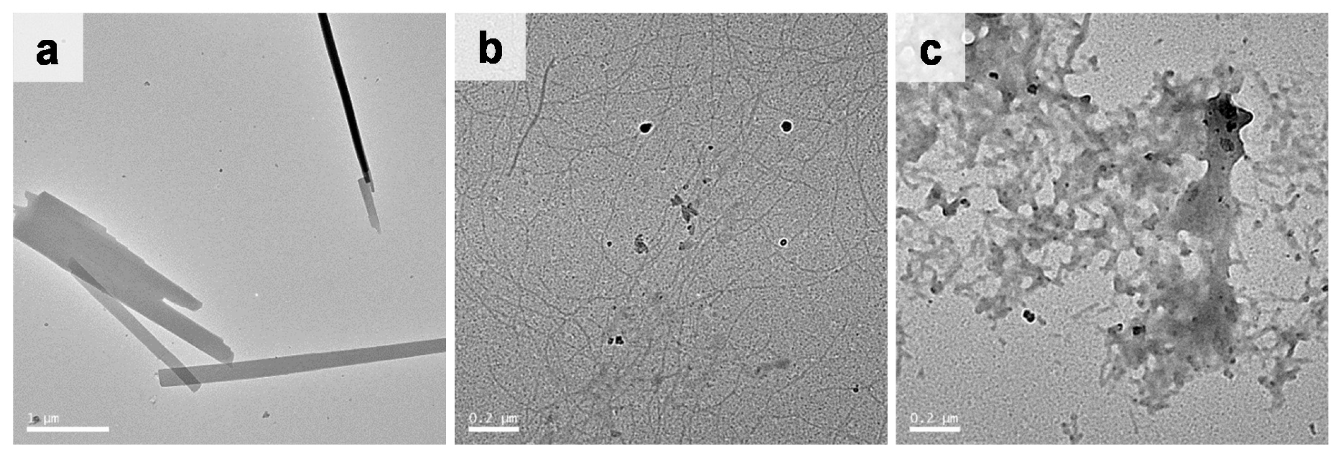
2.3. Morphology of the Self-Assembled Glycosylated Lipopeptide-Type Bola-Amphiphiles

Insight into the morphology of the self-assembled structures was obtained by transmission electron microscopy (TEM) on the βGal–AAC–C6–F2 and βGal–AAC–C6–F3 hydrogels, and βGal–AAC–C6–F4 dispersion. TEM images showed that βGal–AAC–C6–F2 self-assembled into nanoribbons with an averaged width of several tens of nm and up to ca. 100 nm and length of several micrometres (Figure 5a). On the other hand, βGal–AAC–C6–F3 self-assembled into longer and thinner one-dimensional (1D) nanofibers with an averaged width of ca. 25 nm, thereby facilitating the formation of a highly entangled three-dimensional (3D) network (Figure 5b). These conventional TEM images are not cryo-TEM and thus there is the potential influence of the sample preparation process (especially during the drying process) on the observed morphologies. Nevertheless, we speculate that the longer and thinner nanofibers of βGal–AAC–C6–F3, instead of the straight nanoribbons of βGal–AAC–C6–F2, should be responsible for the stable hydrogel formation of βGal–AAC–C6–F3 even at a lower concentration. The detail in the molecular assembly mode and the difference is under investigation. In contrast, the self-assembled structures of βGal–AAC–C6–F4 dispersion (non-hydrogel) were non-networked aggregates of shorter nanofibers (Figure 5c).

3. Materials and Methods

3.1. Generals

Chemical reagents were purchased from Tokyo Chemical Industry Co., Ltd., FUJIFILM Wako Pure Chemical Corporation, Watanabe Chemical Industries, Ltd. and Bachem Holding AG., and used without further purification. Thin-layer chromatography (TLC) was performed on TLC silica gel 60F254 (Merck). Column chromatography was performed on silica gel 60N (Kanto Chemical Co., Inc., spherical neutral, 63 to 210 µm). 1H and 13C NMR spectra were recorded on a JEOL ECA500 spectrometer in CDCl3, CD3OD or dimethyl sulfoxide-d6 (DMSO-d6) with tetramethylsilane (TMS) or residual non-deuterated solvents as the internal references. Multiplicities are abbreviated as follows: s = singlet, d = doublet, t = triplet, q = quartet, m = multiplet, dd = double doublet, and br = broad. LRMS (ESI-MS) analysis was conducted using a Bruker amaZon SL mass spectrometer. HRMS (ESI-FT-ICR-MS) analyses were conducted using a Bruker Solarix spectrometer. The absorption spectra were measured using a Jasco V-650 spectrometer equipped with an ETCS-761 temperature controller. FT-IR spectra were recorded on a JEOL FT/IR-4100 spectrometer using KBr pellets in the range of 4000 to 400 cm−1.

3.2. Gelation Test

The compounds (typically, 2.0 mg) were suspended in 200 mM HEPES–NaOH buffer (pH 8.0) in a mighty vial. The suspensions were heated to form homogeneous solutions. Then, the hot solution was cooled to room temperature (around 23 °C) and incubated for 10 min. Gelation was confirmed by the gravitational flux with inversion of the vial. When no fluid ran down the wall of the test tube upon inversion of the vial, we judged it to be gel. Upon confirming the gel formation, the buffer was added to the samples, heated to dissolution and cooled to room temperature. This process was repeated until the gel formation could no longer be observed. The CGC was considered as the last concentration at which a stable gel phase could be observed.

3.3. Measurement of the Gel–Sol Transition Temperature (Tgel)

The gel–sol phase transition behaviour of the βGal–AAC–C6–F3 hydrogel (0.19 wt% (CGC) in 200 mM HEPES–NaOH buffer (pH 8.0)) was determined using the vial inversion method. The inverted gel in the vial was placed in an oil bath, which was heated from 25 °C to 85 °C, at a rate of 1 °C/step. The vial was immersed at each temperature for 1 min to equilibrate. The temperature, at which the sample completely dissolved, was defined as the Tgel value of the gel.

3.4. Measurements of the Absorption Spectra of the Compounds

An aqueous suspension of the compounds in 200 mM HEPES–NaOH buffer (pH 8.0) was heated to form a homogeneous solution. The hot solution was transferred into a quartz cell (path length: 0.1 mm (assembled quartz cell, GL Sciences Inc., cat. no. AB10-UV-0.1 with cell adaptor, GL Sciences Inc., cat. no. CAS-10-1) for βGal–AAC–C6–F1 and βGal–AAC–C6–F2, or 1 mm for βGal–AAC–C6–F3 and βGal–AAC–C6–F4) and stored at room temperature for 10 min. The absorption spectra were measured at room temperature. Conditions: [βGal–AAC–C6–F1] = 2.4 wt%, [βGal–AAC–C6–F2] = 2.4 wt% (CGC), [βGal–AAC–C6–F3] = 0.19 wt% (CGC), and [βGal–AAC–C6–F4] = 0.35 wt%, 200 mM HEPES–NaOH buffer (pH 8.0).

3.5. Measurements of the Temperature-Dependent Absorption Spectral Changes of βGal–AAC–C6–F3 Hydrogel

An aqueous suspension of βGal–AAC–C6–F3 (0.19 wt% (CGC) in 200 mM HEPES–NaOH buffer (pH 8.0)) was heated to form a homogeneous solution. The hot solution (400 mL) was transferred into a quartz cell (path length: 1 mm) and stored at room temperature for 10 min to complete the gelation. The absorption spectra were measured upon heating from 25 to 85 °C.

3.6. TEM Observation

Sample (ca. 10 µL) was dropped on a copper TEM grid covered by an elastic carbon-support film (20 to 25 nm) with a filter paper underneath. Then, the excess solution was blotted with the filter paper immediately. The TEM images were acquired using a JEOL JEM-2100F (accelerating voltage: 200 kV) equipped with a CCD camera. Conditions: [βGal–AAC–C6–F2] = 2.4 wt% (CGC), [βGal–AAC–C6–F3] = 0.19 wt% (CGC), and [βGal–AAC–C6–F4] = 0.35 wt%, 200 mM HEPES–NaOH buffer (pH 8.0).

3.7. Synthesis

Compound 1 was synthesized according to previously reported methods [39]. H-FF-OH was purchased from Bachem Holding AG. H-FFF-OH and H-FFFF-OH were synthesized by standard liquid phase synthesis using N-α-(9-Fluorenylmethoxycarbonyl)-L-phenylalanine (Fmoc-F-OH) and L-phenylalanine t-butyl ester hydrochloride (H-F-OBut•HCl) [73]. All the amino acids and Fmoc protected peptides were conjugated via active ester method by using 1-[1-(cyano-2-ethoxy-2-oxoethylideneaminooxy)-dimethylamino-morpholino]-uronium hexafluorophosphate (COMU) in the presence of N,N-diisopropylethylamine (DIEA) in dry N,N-dimethylformamide (DMF) under an N2 atmosphere. Fmoc deprotection was done with 20% piperidine in DMF. t-Butyl ester deprotection was done with trifluoroacetic acid:H2O = 95:5 (v/v).

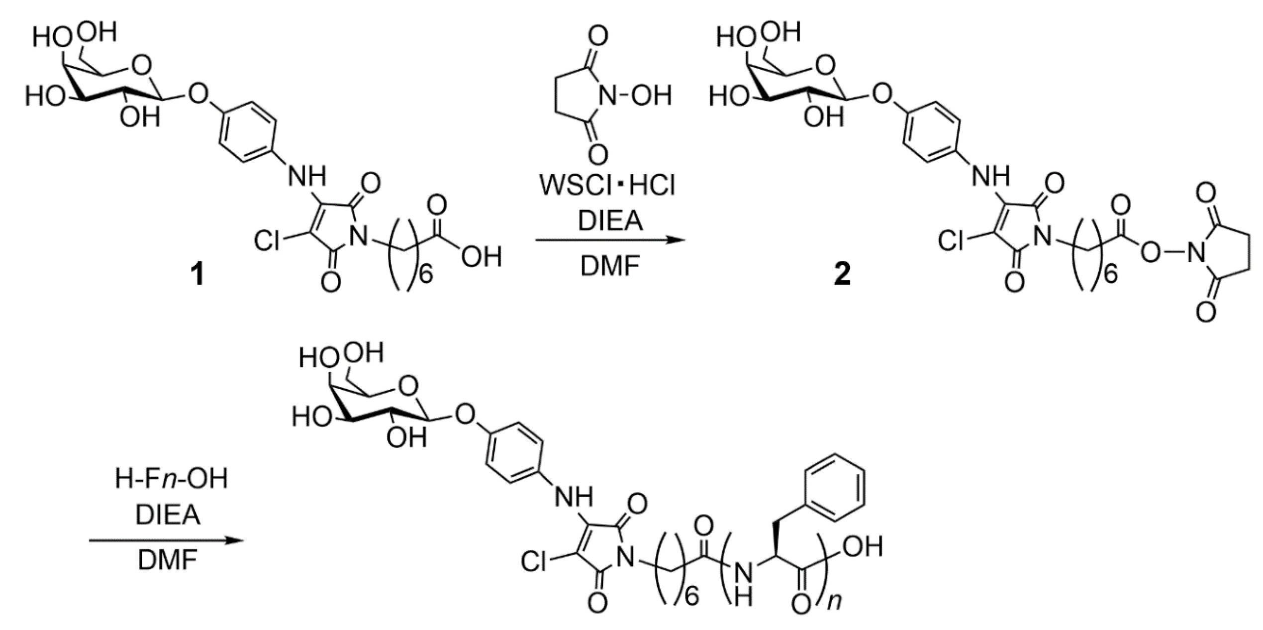
3.7.1. Synthesis of Compound 2

After cooling in an ice bath, N-hydroxysuccinimide (52 mg, 0.45 mmol, 1.5 eq.) and water soluble carbodiimide hydrochloride (WSCI•HCl, 116 mg, 0.60 mmol, 2.0 eq.) were added to a solution of 1 (160 mg, 0.30 mmol, 1.0 eq.) in dry DMF (6 mL), and the mixture was stirred at room temperature overnight under an N2 atmosphere. The solvent was then evaporated, and residue was purified by column chromatography (SiO2, CH2Cl2:MeOH = 6:1 (v/v)). Then, the residue was further purified by reprecipitation with diethyl ether (Et2O) for two times. The resulting product was dried under vacuum to give compound 2 (113 mg, 59%) as a yellow powder. 1H NMR (500 MHz, CD3OD): δ (ppm) = 1.31–1.37 (m, 3H), 1.43–1.49 (m, 3H), 1.58–1.64 (m, 3H), 1.69–1.75 (m, 3H), 2.62 (q, J = 7.1 Hz, 3H), 2.80 (q, J = 6.6 Hz, 4H), 3.51 (t, 2H), 3.57 (dd, J1 = 3.4 Hz, J2 = 9.7 Hz, 1H), 3.67 (q, J = 5.7 Hz, 1H), 3.72–3.80 (m, 3H), 3.89 (d, J = 3.5 Hz, 1H), 4.89 (d, J = 7.4 Hz, 1H), and 7.09–7.13 (m, 4H). 13C NMR (125 MHz, CD3OD): δ (ppm) = 25.51, 26.48, 27.20, 29.15, 29.35, 31.45, 39.07, 62.41, 70.19, 72.22, 74,.75, 76.88, 92.05, 103.07, 117.64, 126.70, 132.29, 139.69, 157.19, 167.07, 169.76, 170.26, 171.90. LRMS (ESI-TOF, positive mode): Calcd. for [M(C27H32ClN3O12)+Na]+: m/z = 648.2; Found: 648.2.

3.7.2. Synthesis of βGal–AAC–C6–F1

H-F-OH (5.8 mg, 0.035 mmol, 1.1 eq.) and DIEA (6.4 µL, 0.038 mmol, 1.2 eq.) were added to a solution of 2 (20 mg, 0.032 mmol, 1.0 eq.) in dry DMF (2 mL), and the mixture was stirred at room temperature overnight under an N2 atmosphere. The solvent was then evaporated, and residue was purified by column chromatography (SiO2, CH2Cl2:MeOH = 2:1 (v/v)). Then, the residue was further purified by reprecipitation with Et2O for two times. The resulting product was dried under vacuum to give βGal–AAC–C6–F1 (17 mg, 78%) as a yellow powder. 1H NMR (500 MHz, CD3OD): δ (ppm) = 1.16–1.28 (m, 4H), 1.43–1.55 (m, 4H), 2.09–2.14 (m, 2H), 2.88–3.00 (m, 2H), 3.47 (t, J1 = 7.2 Hz, 2H), 3.57 (dd, J1 = 2.9 Hz, J2 = 9.8 Hz, 1H), 3.66–3.70 (m, 1H), 3.71–3.80 (m, 3H), 3.88 (d, J = 3.5 Hz, 1H), 4.52 (q, J = 4.7 Hz, 1H), 4.58 (s, 1H), 7.09–7.15 (m, 3H), 7.18–7.20 (m, 3H), and 7.33–7.24 (m, 3H). 13C NMR (125 MHz, DMSO-d6): δ (ppm) = 25.17, 26.05, 28.05, 32.77, 33.05, 35.20, 36.98, 37.84, 48.70, 53.71, 60.48, 68.21, 70.39, 73.57, 90.00, 101.26, 115.89, 125.61, 126.33, 128.14, 129.30, 138.16, 147.53, 151.05, 155.31, 165.55, 167.55, 172.12, 173.84. HRMS (ESI-FT-ICR, positive mode): Calcd. for [M(C32H38ClN3O11)+H]+: m/z = 676.2268; Found: 676.2258. FT-IR (KBr pellet): v = 3422.1, 3029.6, 2937.1, 2861.8, 2359.5, 2344.1, 1771.3, 1712.5, 1654.6, 1607.4, 1508.1, 1442.5, 1409.7, 1231.3, 1151.3, 1074.2, 834.1, 743.4, 703.9 cm–1.

3.7.3. Synthesis of βGal–AAC–C6–F2

H-FF-OH (11 mg, 0.035 mmol, 1.1 eq.) and DIEA (6.4 µL, 0.038 mmol, 1.2 eq.) were added to a solution of 2 (20 mg, 0.032 mmol, 1.0 eq.) in dry DMF (4 mL), and the mixture was stirred at room temperature overnight under an N2 atmosphere. The solvent was then evaporated, and residue was purified by column chromatography (SiO2, CH2Cl2:MeOH = 3:1 to 2:1 to 1:1 to 0:1 (v/v)). Then, the residue was further purified by reprecipitation with Et2O for two times. The resulting product was dried under vacuum to give βGal–AAC–C6–F2 (22 mg, 84%) as a yellow powder. 1H NMR (500 MHz, CD3OD): δ (ppm) = 1.08 (q, J = 8.0 Hz, 2H), 1.16 (q, J = 7.1 Hz, 2H), 1.34–1.38 (m, 2H), 1.48 (q, J = 7.4 Hz, 2H), 2.06 (t, J = 6.9 Hz, 1H), 2.72 (dd, J1 = 5.3 Hz, J2 = 13.5 Hz, 2H), 3.01 (dd, J1 = 3.3 Hz, J2 = 14.0 Hz, 1H), 3.13–3.17 (m, 2H), 3.43–3.48 (m, 2H), 3.57 (dd, J1 = 3.2 Hz, J2 = 10.0 Hz, 1H), 3.67–3.69 (m, 1H), 3.72–3.80 (m, 3H), 3.89 (d, J = 3.4 Hz, 1H), 4.44 (t, J = 5.8 Hz, 1H), 4.57–4.60 (m, 3H), 7.09–7.14 (m, 5H), and 7.15–7.27 (m, 9H). 13C NMR (125 MHz, DMSO-d6): δ (ppm) = 25.09, 25.24, 25.98, 27.97, 29.53, 33.05, 35.14, 36.92, 37.35, 48.30, 54.25, 84. 63, 93.18, 98.30, 101.26, 115.69, 125.51, 126.13, 127.94, 129.14, 129.39, 138.20, 141.81, 150.62, 151.05, 157.12, 167.39, 169.91, 170.97, 171.35, 172.82. HRMS (ESI-FT-ICR, positive mode): Calcd. for [M(C41H47ClN4O12)+H]+: m/z = 823.2952; Found: 823.2958. FT-IR (KBr pellet): v = 3398.0, 3060.5, 3027.7, 2933.2, 2859.9, 2360.4, 2342.1, 1770.3, 1712.5, 1654.6, 1603.5, 1508.1, 1442.5, 1409.7, 1231.3, 1150.3, 1077.1, 832.1, 744.4, 701.0 cm–1.

3.7.4. Synthesis of βGal–AAC–C6–F3

H-FFF-OH (16 mg, 0.035 mmol, 1.1 eq.) and DIEA (6.4 µL, 0.038 mmol, 1.2 eq.) were added to a solution of 2 (20 mg, 0.032 mmol, 1.0 eq.) in dry DMF (3 mL), and the mixture was stirred at 50 °C overnight under an N2 atmosphere. The solvent was then evaporated, and residue was purified by column chromatography (SiO2, CH2Cl2:MeOH = 2:1 to 1:1 to 0:1 (v/v)). Then, the residue was further purified by reprecipitation with Et2O for two times. The resulting product was dried under vacuum to give βGal–AAC–C6–F3 (18 mg, 58%) as a yellow powder. 1H NMR (500 MHz, CD3OD): δ (ppm) = 1.05–1.10 (m, 2H), 1.16 (q, J = 7.2 Hz, 2H), 1.32–1.39 (m, 2H), 1.48 (q, J = 7.1 Hz, 2H), 2.01 (t, J = 7.7 Hz, 2H), 2.79–2.85 (m, 1H), 2.99–3.05 (m, 2H), 3.12–3.16 (m, 2H), 3.18–3.21 (m, 1H), 3.43–3.49 (m, 3H), 3.57 (dd, J1 = 3.7 Hz, J2 = 10.0 Hz, 1H), 3.68 (t, J = 5.7 Hz, 1H), 3.72–3.80 (m, 3H), 3.89 (d, J = 4.0 Hz, 1H), 4.44–4.47 (m, 1H), 4.56–4.61 (m, 3H), 7.11–7.13 (m, 5H), and 7.15–7.27 (m, 14H). HRMS (ESI-FT-ICR, positive mode): Calcd. for [M(C50H56ClN5O13)+H]+: m/z = 970.3636; Found: 970.3606. FT-IR (KBr pellet): v = 3403.7, 3083.6, 3062.4, 3027.7, 2933.2, 2859.0, 2360.4, 2342.1, 1772.3, 1708.6, 1653.7, 1539.9, 1523.5, 1508.1, 1455.0, 1440.6, 1409.7, 1303.6, 1230.4, 1151.3, 1078.0, 817.7, 744.4, 699.1, 669.2 cm–1.

3.7.5. Synthesis of βGal–AAC–C6–F4

H-FFFF-OH (13 mg, 0.018 mmol, 1.1 eq.) and DIEA (4.0 µL, 0.024 mmol, 1.5 eq.) were added to a solution of 2 (10 mg, 0.016 mmol, 1.0 eq.) in dry DMF (5 mL), and the mixture was stirred at 50 °C overnight under an N2 atmosphere. The solvent was then evaporated, and residue was purified by column chromatography (SiO2, CH2Cl2:MeOH = 2:1 to 1:1 to 0:1 (v/v)). Then, the residue was further purified by reprecipitation with Et2O for two times. The resulting product was dried under vacuum to give compound βGal–AAC–C6–F4 (12 mg, 66%) as a yellow powder. 1H NMR (500 MHz, DMSO-d6): δ (ppm) = 0.81–0.86 (m, 1H), 0.96–1.01 (m, 1H), 1.07 (q, J = 7.6 Hz, 2H), 1.20–1.27 (m, 4H), 1.39 (q, J = 6.0 Hz, 1H), 1.76 (s, 1H), 1.89 (q, J = 6.9 Hz, 1H), 2.47–2.51 (m, 2H), 2.55 (m, J = 6.6 Hz, 2H), 2.59 (m, J = 13.7 Hz, 1H), 2.69–2.76 (m, 2H), 2.81–2.85 (m,1H), 2.72 (dd, J1 = 5.2 Hz, J2 = 13.2 Hz, 2H), 3.01–3.07 (m, 4H), 3.46 (t, J = 4.9 Hz, 1H), 3.50–3.54 (m, 3H), 3.66 (t, J = 3.4 Hz, 1H), 3.91 (d, J = 6.9 Hz, 1H), 4.34–4.39 (m, 2H), 4.43–4.48 (m, 2H), 4.69–4.71 (m, 1H), 4.76 (d, J = 8.0 Hz, 1H), 4.89 (d, J = 6.3 Hz, 1H), 5.18 (d, J = 5.1 Hz, 1H), 6.94–7.51 (m, 20H). 7.51 (d, J = 5.2. Hz, 1H), 7.81 (d, J = 6.9 Hz, 1H), 7.91 (d, J = 8.5 Hz, 1H), 8.36 (d, J = 6.9 Hz, 1H). HRMS (ESI-FT-ICR, positive mode): Calcd. for [M(C59H65ClN6O14)+H]+: m/z = 1117.4320; Found: 1117.4341. FT-IR (KBr pellet): v = 3410.5, 3282.3, 3087.5, 3066.3, 3029.6, 2931.3, 2859.0, 2360.4, 2342.1, 1771.3, 1713.4, 1639.2, 1540.9, 1524.5, 1454.1, 1441.5, 1409.7, 1229.4, 1152.3, 1077.1, 832.1, 744.4, 699.1 cm–1.

4. Conclusions

In summary, we have successfully developed glycosylated lipopeptide-type supramolecular hydrogelators exhibiting small but perceptible reversible thermochromism along with the gel–sol transition. The gelation ability and the morphology of the self-assembled nanostructures depend on the number of F residues. βGal–AAC–C6–F2 formed unstable partial hydrogel (CGC = 2.4 wt%), and βGal–AAC–C6–F3 formed stable transparent hydrogel (CGC = 0.19 wt%). On the other hand, βGal–AAC–C6–F1 and βGal–AAC–C6–F4 did not form a hydrogel. The morphology of the self-assembled nanostructures was affected by the number of F residues and in the present molecular scaffold (i.e., βGal–AAC–C6) with βGal as the saccharide structure and C6 alkyl chain as the spacer, F3 peptide was optimal for hydrogel formation. Further research into potential bio-applications, such as the development of sensing materials for peptidase, is in progress.

Supplementary Materials

Supplementary Materials can be found at https://0-www-mdpi-com.brum.beds.ac.uk/1422-0067/22/4/1860/s1.

Author Contributions

Conceptualization: R.O. and N.T.; synthesis and structural analysis: N.T., A.I. (Azumi Ishigamori) and R.O.; data curation (self-assembly properties): N.T.; UV-Vis measurements; N.T. and A.I. (Akitaka Ito); TEM experiments: M.I. (Masato Ikeda); supervision: M.I. (Masato Ikeda) and M.I. (Masayuki Izumi); writing—original draft: R.O. and N.T.; writing—review and editing: R.O., N.T., M.I. (Masato Ikeda), A.I. (Akitaka Ito) and M.I. (Masayuki Izumi). All authors have read and agreed to the published version of the manuscript.

Funding

This work was partially supported by the Kaneko-Narita Grant-in-Aid for Encouragement of Young Scientists (Protein Research Foundation), the Grant for Collaborative Research Promoting Diversity (Kochi University), and the Grant-in-Aid for Early-Career Scientists (JSPS KAKENHI Grant Number JP18K14003, JP20K15416).

Data Availability Statement

The data that support the findings of this study are available from the corresponding author upon reasonable request.

Conflicts of Interest

The authors declare no conflict of interest. The funders had no role in the design of the study; in the collection, analyses, or interpretation of data; in the writing of the manuscript, or in the decision to publish the results.

References

  1. Estroff, L.A.; Hamilton, A.D. Water gelation by small organic molecules. Chem. Rev. 2004, 104, 1201. [Google Scholar] [CrossRef] [PubMed]
  2. Shimizu, T.; Masuda, M.; Minamikawa, H. Supramolecular nanotube architectures based on amphiphilic molecules. Chem. Rev. 2005, 105, 1401. [Google Scholar] [CrossRef]
  3. De Loos, M.; Feringa, B.L.; van Esch, J.H. Design and application of self-assembled low molecular weight hydrogels. Eur. J. Org. Chem. 2005, 3615. [Google Scholar] [CrossRef]
  4. Ikeda, M.; Ochi, R.; Hamachi, I. Supramolecular hydrogel-based protein and chemosensor array. Lab Chip 2010, 10, 3325. [Google Scholar] [CrossRef]
  5. Dawn, A.; Shiraki, T.; Haraguchi, S.; Tamaru, S.; Shinkai, S. What kind of “soft materials” can we design from molecular gels? Chem. Asian J. 2011, 6, 266. [Google Scholar] [CrossRef]
  6. Raeburn, J.; Cardoso, A.Z.; Adams, D.J. The importance of the self-assembly process to control mechanical properties of low molecular weight hydrogels. Chem. Soc. Rev. 2013, 42, 5143. [Google Scholar] [CrossRef] [PubMed]
  7. Weiss, R.G. The past, present, and future of molecular gels. What is the status of the field, and where is it going? J. Am. Chem. Soc. 2014, 136, 7519. [Google Scholar] [CrossRef]
  8. Du, X.; Zhou, J.; Shi, J.; Xu, B. Supramolecular hydrogelators and hydrogels: From soft matter to molecular biomaterials. Chem. Rev. 2015, 115, 13165. [Google Scholar] [CrossRef]
  9. Okesola, B.O.; Smith, D.K. Applying low-molecular weight supramolecular gelators in an environmental setting—Self-assembled gels as smart materials for pollutant removal. Chem. Soc. Rev. 2016, 45, 4226. [Google Scholar] [CrossRef] [PubMed] [Green Version]
  10. Shigemitsu, H.; Hamachi, I. Design strategies of stimuli-responsive supramolecular hydrogels relying on structural analyses and cell-mimicking approaches. Acc. Chem. Res. 2017, 50, 740. [Google Scholar] [CrossRef] [PubMed]
  11. Yamanaka, M. Supramolecular gel electrophoresis. Polym. J. 2018, 50, 627. [Google Scholar] [CrossRef]
  12. Ikeda, M. Stimuli-responsive supramolecular systems guided by chemical reaction. Polym. J. 2019, 51, 371. [Google Scholar] [CrossRef]
  13. Mehwish, N.; Dou, X.; Zhao, Y.; Feng, C.-L. Supramolecular fluorescent hydrogelators as bio-imaging probes. Mater. Horiz. 2019, 6, 14. [Google Scholar] [CrossRef]
  14. Deng, W.; Yamaguchi, H.; Takashima, Y.; Harada, A. A chemical-responsive supramolecular hydrogel from modified cyclodextrins. Angew. Chem. Int. Ed. 2007, 46, 5144. [Google Scholar] [CrossRef]
  15. Bowerman, C.J.; Nilsson, B.L. A reductive trigger for peptide self-assembly and hydrogelation. J. Am. Chem. Soc. 2010, 132, 9526. [Google Scholar] [CrossRef]
  16. Ikeda, M.; Tanida, T.; Yoshii, T.; Hamachi, I. Rational molecular design of stimulus-responsive supramolecular hydrogels based on dipeptides. Adv. Mater. 2011, 23, 2819. [Google Scholar] [CrossRef]
  17. Segarra-Maset, M.D.; Nebot, V.J.; Miravet, J.F.; Escuder, B. Control of molecular gelation by chemical stimuli. Chem. Soc. Rev. 2013, 42, 7086. [Google Scholar] [CrossRef]
  18. Trausel, F.; Versluis, F.; Maity, C.; Poolman, J.M.; Lovrak, M.; van Esch, J.H.; Eelkema, R. Catalysis of supramolecular hydrogelation. Acc. Chem. Res. 2016, 49, 1440. [Google Scholar] [CrossRef] [PubMed] [Green Version]
  19. Huang, Z.; Yao, Q.; Chen, J.; Gao, Y. Redox supramolecular self-assemblies nonlinearly enhance fluorescence to identify cancer cells. Chem. Commun. 2018, 54, 5385. [Google Scholar] [CrossRef]
  20. Karunakaran, S.C.; Cafferty, B.J.; Jain, K.S.; Schuster, G.B.; Hud, N.V. Reversible transformation of a supramolecular hydrogel by redox switching of methylene blue- a noncovalent chain stopper. ACS Omega 2020, 5, 344. [Google Scholar] [CrossRef] [PubMed]
  21. Sugiura, T.; Kanada, T.; Mori, D.; Sakai, H.; Shibata, A.; Kitamura, Y.; Ikeda, M. Chemical stimulus-responsive supramolecular hydrogel formation and shrinkage of a hydrazone-containing short peptide derivative. Soft Matter 2020, 16, 899. [Google Scholar] [CrossRef] [PubMed]
  22. Zhang, Y.; Gu, H.; Yang, Z.; Xu, B. Supramolecular hydrogels respond to ligand-receptor interaction. J. Am. Chem. Soc. 2003, 125, 13680. [Google Scholar] [CrossRef]
  23. Hu, B.H.; Messersmith, P.B. Rational design of transglutaminase substrate peptides for rapid enzymatic formation of hydrogels. J. Am. Chem. Soc. 2003, 125, 14298. [Google Scholar] [CrossRef]
  24. Yang, Z.; Gu, H.; Fu, D.; Gao, P.; Lam, J.K.; Xu, B. Enzymatic formation of supramolecular hydrogels. Adv. Mater. 2004, 16, 1440. [Google Scholar] [CrossRef]
  25. Jun, H.-W.; Yuwono, V.; Paramonov, S.E.; Hartgerink, J.D. Enzyme-mediated degradation of peptide-amphiphile nanofiber networks. Adv. Mater. 2005, 17, 2612. [Google Scholar] [CrossRef]
  26. Toledano, S.; Williams, R.J.; Jayawarna, V.; Ulijn, R.V. Enzyme-triggered self-assembly of peptide hydrogels via reversed hydrolysis. J. Am. Chem. Soc. 2006, 128, 1070. [Google Scholar] [CrossRef] [PubMed]
  27. Yang, Z.; Liang, G.; Wang, L.; Xu, B. Using a kinase/phosphatase switch to regulate a supramolecular hydrogel and forming the supramolecular hydrogel in vivo. J. Am. Chem. Soc. 2006, 128, 3038. [Google Scholar] [CrossRef]
  28. Vemula, P.K.; Li, J.; John, G. Enzyme catalysis: Tool to make and break amygdalin hydrogelators from renewable resources: A delivery model for hydrophobic drugs. J. Am. Chem. Soc. 2006, 128, 8932. [Google Scholar] [CrossRef] [PubMed]
  29. Yamanaka, M.; Haraya, N.; Yamamichi, S. Chemical stimuli-responsive supramolecular hydrogel from amphiphilic tris-urea. Chem. Asian J. 2011, 6, 1022. [Google Scholar] [CrossRef] [PubMed]
  30. Ogawa, Y.; Yoshiyama, C.; Kitaoka, T. Helical assembly of azobenzene-conjugated carbohydrate hydrogelators with specific affinity for lectins. Langmuir 2012, 28, 4404. [Google Scholar] [CrossRef] [PubMed]
  31. Pires, R.A.; Abul-Haija, Y.M.; Costa, D.S.; Novoa-Carballal, R.; Reis, R.L.; Ulijn, R.V.; Pashkuleva, I. Controlling cancer cell fate using localized biocatalytic self-assembly of an aromatic carbohydrate amphiphile. J. Am. Chem. Soc. 2015, 137, 576. [Google Scholar] [CrossRef] [Green Version]
  32. Kameta, N.; Masuda, M.; Shimizu, T. Two-step naked-eye detection of lectin by hierarchical organization of soft nanotubes into liquid crystal and gel phases. Chem. Commun. 2015, 51, 6816. [Google Scholar] [CrossRef] [Green Version]
  33. Akama, S.; Maki, T.; Yamanaka, M. Enzymatic hydrolysis-induced degradation of a lactose-coupled supramolecular hydrogel. Chem. Commun. 2018, 54, 8814. [Google Scholar] [CrossRef] [Green Version]
  34. Qiu, Z.; Yu, H.; Li, J.; Wang, Y.; Zhang, Y. Spiropyran-linked dipeptide forms supramolecular hydrogel with dual responses to light and to ligand–receptor interaction. Chem. Commun. 2009, 3342. [Google Scholar] [CrossRef] [PubMed] [Green Version]
  35. Rodrıguez-Llansola, F.; Escude, B.; Miravet, J.F.; Hermida-Merino, D.; Hamley, I.W.; Cardinb, C.J.; Hayes, W. Selective and highly efficient dye scavenging by a pH-responsive molecular hydrogelator. Chem. Commun. 2010, 46, 7960. [Google Scholar] [CrossRef] [Green Version]
  36. Ochi, R.; Kurotani, K.; Ikeda, M.; Kiyonaka, S.; Hamachi, I. Supramolecular hydrogels based on bola-amphiphilic glycolipids showing color change in response to glycosidases. Chem. Commun. 2013, 49, 2115. [Google Scholar] [CrossRef]
  37. Yu, X.; Ge, X.; Lan, H.; Li, Y.; Geng, L.; Zhen, X.; Yi, T. Tunable and switchable control of luminescence through multiple physical stimulations in aggregation-based monocomponent systems. ACS Appl. Mater. Interfaces 2015, 7, 24312. [Google Scholar] [CrossRef]
  38. Singh, P.; Misra, S.; Das, A.; Roy, S.; Datta, P.; Bhattacharjee, G.; Satpati, B.; Nanda, J. Supramolecular hydrogel from an oxidized byproduct of tyrosine. ACS Appl. Bio Mater. 2019, 2, 4881. [Google Scholar] [CrossRef]
  39. Xu, X.; Zhou, X.; Qu, L.; Wang, L.; Song, J.; Wu, D.; Zhou, W.; Zhou, X.; Xiang, H.; Wang, J.; et al. Reversible chromatic change of supramolecular gels for visual and selective chiral recognition of histidine. ACS Appl. Bio Mater. 2020, 3, 7236. [Google Scholar] [CrossRef]
  40. Oosumi, R.; Ikeda, M.; Ito, A.; Izumi, M.; Ochi, R. Structural diversification of bola-amphiphilic glycolipid-type supramolecular hydrogelators exhibiting colour changes along with the gel–sol transition. Soft Matter 2020, 16, 7274. [Google Scholar] [CrossRef] [PubMed]
  41. Kasha, M.; Rawls, H.R.; El-Bayoumi, M.A. The exciton model in molecular spectroscopy. Pure Appl. Chem. 1965, 11, 371. [Google Scholar] [CrossRef] [Green Version]
  42. Spano, F.C. The spectral signatures of frenkel polarons in H, and J-aggregates. Acc. Chem. Res. 2010, 43, 429. [Google Scholar] [CrossRef]
  43. Inaba, H.; Matsuura, K. Peptide nanomaterials designed from natural supramolecular systems. Chem. Rec. 2019, 19, 843. [Google Scholar] [CrossRef] [PubMed]
  44. Wang, Q.; Jiang, N.; Fu, B.; Huang, F.; Liu, J. Self-assembling peptide-based nanodrug delivery systems. Biomater. Sci. 2019, 7, 4888. [Google Scholar] [CrossRef] [PubMed]
  45. Liu, R.; Hudalla, G.A. Using Self-assembling peptides to integrate biomolecules into functional supramolecular biomaterials. Molecules 2019, 24, 1450. [Google Scholar] [CrossRef] [Green Version]
  46. Lee, S.; Trinh, T.H.T.; Yoo, M.; Shin, J.; Lee, H.; Kim, J.; Hwang, E.; Lim, Y.; Ryou, C. Self-Assembling Peptides and Their Application in the Treatment of Diseases. Int. J. Mol. Sci. 2019, 20, 585. [Google Scholar] [CrossRef] [Green Version]
  47. Hu, X.; Liao, M.; Gong, H.; Zhang, L.; Cox, H.; Waigh, T.A.; Lu, J.R. Recent advances in short peptide self-assembly: From rational design to novel applications. Curr. Opin. Colloid Interface Sci. 2020, 45, 1. [Google Scholar] [CrossRef]
  48. Simonson, A.W.; Aronson, M.R.; Medina, S.H. Supramolecular Peptide Assemblies as Antimicrobial Scaffolds. Molecules 2020, 25, 2751. [Google Scholar] [CrossRef]
  49. Uchida, N.; Muraoka, T. Current progress in cross-linked peptide self-assemblies. Int. J. Mol. Sci. 2020, 21, 7577. [Google Scholar] [CrossRef]
  50. Ryan, D.M.; Nilsson, B.L. Self-assembled amino acids and dipeptides as noncovalent hydrogels for tissue engineering. Polym. Chem. 2012, 3, 18. [Google Scholar] [CrossRef]
  51. Dasgupta, A.; Mondal, J.H.; Das, D. Peptide hydrogels. RSC Adv. 2013, 3, 9117. [Google Scholar] [CrossRef]
  52. Singh, N.; Kumar, M.; Miravet, J.F.; Ulijn, R.V.; Escuder, B. Peptide-based molecular hydrogels as supramolecular protein mimics. Chem. Eur. J. 2017, 23, 981. [Google Scholar] [CrossRef] [PubMed]
  53. Edwards-Gayle, C.J.C.; Hamley, I.W. Self-assembly of bioactive peptides, peptide conjugates, and peptide mimetic materials. Org. Biomol. Chem. 2017, 15, 5867. [Google Scholar] [CrossRef] [Green Version]
  54. Sato, K.; Hendricks, M.P.; Palmer, L.C.; Stupp, S.I. Peptide supramolecular materials for therapeutics. Chem. Soc. Rev. 2018, 47, 7539. [Google Scholar] [CrossRef] [PubMed]
  55. Gao, J.; Zhan, J.; Yang, Z. Enzyme-instructed self-assembly (EISA) and hydrogelation of peptides. Adv. Mater. 2019, 1805798. [Google Scholar] [CrossRef]
  56. He, H.; Tan, W.; Guo, J.; Yi, M.; Shy, A.N.; Xu, B. Enzymatic Noncovalent Synthesis. Chem. Rev. 2020, 120, 9994. [Google Scholar] [CrossRef]
  57. Fleming, S.; Ulijn, R.V. Design of nanostructures based on aromatic peptide amphiphiles. Chem. Soc. Rev. 2014, 43, 8150. [Google Scholar] [CrossRef] [PubMed]
  58. Wang, J.; Liu, K.; Xing, R.; Yan, X. Peptide self-assembly: Thermodynamics and kinetics. Chem. Soc. Rev. 2016, 45, 5589. [Google Scholar] [CrossRef] [PubMed]
  59. Tao, K.; Levin, A.; Adler-Abramovich, L.; Gazit, E. Fmoc-modified amino acids and short peptides: Simple bio-inspired building blocks for the fabrication of functional materials. Chem Soc Rev. 2016, 45, 3935. [Google Scholar] [CrossRef]
  60. Tzokova, N.; Fernyhough, C.M.; Topham, P.D.; Sandon, N.; Adams, D.J.; Butler, M.F.; Armes, S.P.; Ryan, A.J. Soft hydrogels from nanotubes of poly(ethylene oxide)-tetraphenylalanine conjugates prepared by click chemistry. Langmuir 2009, 25, 2479. [Google Scholar] [CrossRef]
  61. Palui, G.; Nanda, J.; Ray, S.; Banerjee, A. Fabrication of luminescent CdS nanoparticles on short-peptide-based hydrogel nanofibers: Tuning of optoelectronic properties. Chem. Eur. J. 2009, 15, 6902. [Google Scholar] [CrossRef] [PubMed]
  62. Chronopoulou, L.; Lorenzoni, S.; Masci, G.; Dentini, M.; Togna, A.R.; Togna, G.; Bordi, F.; Palocci, C. Lipase-supported synthesis of peptidic hydrogels. Soft Matter 2010, 6, 2525. [Google Scholar] [CrossRef]
  63. Ikeda, M.; Tanida, T.; Yoshii, T.; Kurotani, K.; Onogi, S.; Urayama, K.; Hamachi, I. Installing logic-gate response to a variety of biological substances in supramolecular hydrogel-enzyme hybrids. Nat. Chem. 2014, 6, 511. [Google Scholar] [CrossRef]
  64. Kubota, R.; Torigoe, S.; Liu, S.; Hamachi, I. In situ real-time confocal imaging of a self-assembling peptide-grafted polymer showing pH-responsive hydrogelation. Chem. Lett. 2020, 49, 1319. [Google Scholar] [CrossRef]
  65. Xiong, Q.; Liu, Z.; Han, W. Sequence-Dependent Nanofiber Structures of Phenylalanine and Isoleucine Tripeptides. Int. J. Mol. Sci. 2020, 21, 8431. [Google Scholar] [CrossRef] [PubMed]
  66. Shimizu, T.; Masuda, M. Stereochemical effect of even-odd connecting links on supramolecular assemblies made of 1-glucosamide bolaamphiphiles. J. Am. Chem. Soc. 1997, 119, 2812. [Google Scholar] [CrossRef]
  67. Jung, J.H.; Shinkai, S.; Shimizu, T. Spectral characterization of self-assemblies of aldopyranoside amphiphilic gelators: What is the essential structural difference between simple amphiphiles and bolaamphiphiles? Chem. Eur. J. 2002, 8, 2684. [Google Scholar] [CrossRef]
  68. Kiyonaka, S.; Shinkai, S.; Hamachi, I. Combinatorial library of low molecular-weight organo- and hydrogelators based on glycosylated amino acid derivatives by solid-phase synthesis. Chem. Eur. J. 2003, 9, 976. [Google Scholar] [CrossRef]
  69. Datta, S.; Bhattacharya, S. Multifarious facets of sugar-derived molecular gels: Molecular features, mechanisms of self-assembly and emerging applications. Chem. Soc. Rev. 2015, 44, 5596. [Google Scholar] [CrossRef]
  70. Delbianco, M.; Bharate, P.; Varela-Aramburu, S.; Seeberger, P.H. Carbohydrates in supramolecular chemistry. Chem. Rev. 2016, 116, 1693. [Google Scholar] [CrossRef]
  71. Ochi, R. Carbohydrates as components of supramolecular materials. Trends Glycosci. Glycotechnol. 2018, 30, E177. [Google Scholar] [CrossRef]
  72. Latxague, L.; Gaubert, A.; Barthelemy, P. Recent advances in the chemistry of glycoconjugate amphiphiles. Molecules 2018, 23, 89. [Google Scholar] [CrossRef] [PubMed] [Green Version]
  73. Ochi, R.; Nishida, T.; Ikeda, M.; Hamachi, I. Design of peptide-based bolaamphiphiles exhibiting heat-set hydrogelation via retro-Diels–Alder reaction. J. Mater. Chem. B 2014, 2, 1464. [Google Scholar] [CrossRef] [PubMed]
Figure 1. Chemical structures of bola-amphiphilic glycolipid-type hydrogelators possessing an N-alkyl-2-anilino-3-chloromaleimide (AAC) moiety (R1, R2 = α- or β-D-glucose (α/βGlc), α- or β-D-galactose (α/βGal); (a) R1–AAC–C11–COOH [36], (b) R1–AAC–C6–R2 [40].
Ijms 22 01860 g001
Figure 2. Chemical structures of glycosylated lipopeptide-type bola-amphiphiles βGal–AAC–C6–Fn (n = 1–4).
Ijms 22 01860 g002
Scheme 1. Synthesis of βGal–AAC–C6–Fn (n = 1–4).
Ijms 22 01860 sch001
Figure 3. Photographs of compounds at room temperature (around 23 °C); [βGal–AAC–C6–F1] = 2.4 wt%, [βGal–AAC–C6–F2] = 2.4 wt% (CGC), [βGal–AAC–C6–F3] = 0.19 wt% (CGC), and [βGal–AAC–C6–F4] = 0.35 wt% in 200 mM HEPES–NaOH buffer (pH 8.0).
Ijms 22 01860 g003
Figure 4. (a) Photographs and schematics of a supramolecular hydrogel of βGal–AAC–C6–F3 exhibiting reversible thermal gel–sol transition and thermochromism and (b) UV–Vis absorption spectral changes of the supramolecular hydrogel of βGal–AAC–C6–F3 upon heating (black line: 25 °C (gel state), red line: 85 °C (solution state), purple line: differential spectrum). Conditions: [βGal–AAC–C6–F3] = 0.19 wt%, 200 mM HEPES–NaOH buffer (pH 8.0).
Ijms 22 01860 g004
Figure 5. Representative TEM images of (a) βGal–AAC–C6–F2, (b) βGal–AAC–C6–F3, and (c) βGal–AAC–C6–F4 samples after being transferred onto elastic carbon-coated grids and dried in vacuo. Conditions: [βGal–AAC–C6–F2] = 2.4 wt% (CGC), [βGal–AAC–C6–F3] = 0.19 wt% (CGC), and [βGal–AAC–C6–F4] = 0.35 wt%, 200 mM HEPES–NaOH buffer (pH 8.0).
Ijms 22 01860 g005
Table 1. Gelation ability, critical gelation concentration (CGC), the gel-to-sol phase transition temperature (Tgel) of hydrogels at CGC, and absorption maxima (λmax) at room temperature (around 23 °C) of lipopeptide-type bola-amphiphiles. Conditions: 200 mM HEPES–NaOH buffer (pH 8.0).
CompoundGelation AbilityCGC
[wt%]
CGC
[mM]
Tgel
[°C]
λmax
[nm] 1
βGal–AAC–C6–F1Solution416
βGal–AAC–C6–F2Partial Gel2.430409
βGal–AAC–C6–F3Transparent Gel0.192.078402
βGal–AAC–C6–F4Dispersion397
1 [βGal–AAC–C6–F1] = 2.4 wt%, [βGal–AAC–C6–F2] = 2.4 wt% (CGC), [βGal–AAC–C6–F3] = 0.19 wt% (CGC), and [βGal–AAC–C6–F4] = 0.35 wt%.
Publisher’s Note: MDPI stays neutral with regard to jurisdictional claims in published maps and institutional affiliations.

