Article

Overexpression of the Aldehyde Dehydrogenase Gene ZmALDH Confers Aluminum Tolerance in Arabidopsis thaliana

1
Key Laboratory of Biology and Genetic Improvement of Maize in Southwest China of Agricultural Department, Ministry of Agriculture, Maize Research Institute, Sichuan Agricultural University, Chengdu 611130, China
2
Panxi Crops Research and Utilization Key Laboratory of Sichuan Province, Xichang University, Xichang 615000, China
3
Crop Research Institute, Sichuan Academy of Agricultural Sciences, Chengdu 610066, China
*
Author to whom correspondence should be addressed.
Int. J. Mol. Sci. 2022, 23(1), 477; https://0-doi-org.brum.beds.ac.uk/10.3390/ijms23010477
Submission received: 6 December 2021 / Revised: 27 December 2021 / Accepted: 29 December 2021 / Published: 1 January 2022
(This article belongs to the Section Molecular Biology)

Abstract

:
Aluminum (Al) toxicity is the main factor limiting plant growth and the yield of cereal crops in acidic soils. Al-induced oxidative stress could lead to the excessive accumulation of reactive oxygen species (ROS) and aldehydes in plants. Aldehyde dehydrogenase (ALDH) genes, which play an important role in detoxification of aldehydes when exposed to abiotic stress, have been identified in most species. However, little is known about the function of this gene family in the response to Al stress. Here, we identified an ALDH gene in maize, ZmALDH, involved in protection against Al-induced oxidative stress. Al stress up-regulated ZmALDH expression in both the roots and leaves. The expression of ZmALDH only responded to Al toxicity but not to other stresses including low pH and other metals. The heterologous overexpression of ZmALDH in Arabidopsis increased Al tolerance by promoting the ascorbate-glutathione cycle, increasing the transcript levels of antioxidant enzyme genes as well as the activities of their products, reducing MDA, and increasing free proline synthesis. The overexpression of ZmALDH also reduced Al accumulation in roots. Taken together, these findings suggest that ZmALDH participates in Al-induced oxidative stress and Al accumulation in roots, conferring Al tolerance in transgenic Arabidopsis.

1. Introduction

Aluminum (Al) is one of the most abundant metal elements in the Earth’s crust and can be solubilized to the toxic species Al3+ in acidic soils (pH < 5.0) [1]. Al stress, which can rapidly inhibit root elongation even at a micromolar concentration, is one of themajor factors limiting crop yield in acidic soils [2]. Improving the Al tolerance of crops in acidic soils has been a longstanding aim for solving food shortages and improving biofuel production.
In the past decades, two kinds of aluminum tolerance mechanisms, organic acid extradition for external detoxification and Al sequestration into vacuoles for internal tolerance, have been adopted by plants [3,4,5]. In particular, the secretion of organic acids from the root apices, especially malate, citrate, and oxalate, is a widely used strategy in plants [1,6,7].
Al stress induces oxidative stress, which causes the overproduction of reactive oxygen species (ROS) and aldehydes in plants [8,9,10]. Aldehydes are common intermediates in most cellular metabolic pathways in plants and can be produced under various environmental stresses, such as cold, heat shock, salinity, and desiccation [11,12]. The excessive accumulation of ROS and aldehyde substances could seriously destroy the cell membrane, change the fluidity and ion transport characteristics of the biomembrane, hinder protein synthesis, and eventually result in cell death [13,14,15]. Plants could resist the Al-induced ROS by enhancing antioxidant systems [16,17]. The ascorbate-glutathione (AsA-GSH) cycle can directly scavenge H2O2 produced in plants, and antioxidants produced in the AsA-GSH cycle (including AsA and GSH) can remove ROS [18,19]. The AsA-GSH cycle is also involved in the redox balance by the regulation of four key enzymes, including APX (ascorbate peroxidase), MDAR (monodehydroascorbate), DHAR (dehydroascorbate reductase), and GR (glutathione reductase) [20]. The detoxification of aldehydes depends on the activity of aldehyde dehydrogenases (ALDH, EC 1.2.1.3) [21,22]. ALDHs are ubiquitous in both prokaryotes and eukaryotes. They belong to the NAD(P)+-dependent protein superfamily, which catalyzes the oxidation of aldehydes to the corresponding non-toxic carboxylic acids and balances the content [23]. Based on sequence identity and substrate specificity, ALDHs could be organized into two main groups and at least 12 subfamilies in plants [11,24]. Previous studies have revealed that ALDH could function as a primary aldehyde dehydrogenase to cope with various environmental stresses by preventing and protecting plants against oxidative stress [15,25,26]. For example, OsALDH2B1, which functions primarily as an aldehyde dehydrogenase, not only protects against various stresses, but regulates the growth and reproduction in rice [26]. Ath-ALDH3 could increase the tolerance to drought, salt, and heavy metal (Cu2+ and Cd2+) stress in transgenic Arabidopsis [14]. Ath-ALDH3I1 and Ath-ALDH7B4 confer tolerance to osmotic and oxidative stress and protect plants against lipid peroxidation [15]. ZmALDH7B6 was induced by multiple environmental stresses and might play a role in aldehyde detoxification [25]. In addition, ALDH genes might be also involved in the response to Al toxicity, since the expression of an ALDH2 homolog was up-regulated in a resistant genotype of highbush blueberry (Vaccinium corymbosum) under Al stress [27].
In our previous transcriptomic analysis of maize under Al stress, ZmALDH (Zm00001d017418), an ALDH gene family member, was up-regulated by Al [28]. However, the physiological mechanism by which this gene contributes to the response to Al stress is unclear. Here, the role of the ZmALDH gene in resistance to Al stress was investigated by analyzing Al-induced oxidative stress, including the roles of the AsA-GSH cycle and antioxidant enzyme system as well as overexpression assays.

2. Results

2.1. Cloning and Sequence Analysis of ZmALDH

We obtained the complete ZmALDH CDS via PCR amplification from the Al-resistance maize inbred line 178 (Figure S1) [29]. The ZmALDH gene is 1458 bp long with nine exons and eight introns (Figure S1). It encodes a 446 aminoacid transmembrane protein with a predicted molecular mass of 49.81 kDa and isoelectric point of 6.40 (Table S1). The deduced protein sequence showed sequences identified at 70.16% and 72.28% with rice aldehyde dehydrogenases OsALDH3-1 and OsALDH3-2, respectively (Figure 1A). A 98 amino acid sequence between D71 to D168 and a 236 amino acid sequence between W167 to V402 formed two Aldedh domains, which are involved in oxidoreductase activity.
In a phylogenetic analysis, ZmALDH clustered with several members of the Class 3 family of ALDHs, including OsALDH3-1, OsALDH3-2, AtALDH3F1, AtALDH3, AtALDH3H1, and GmALDH3H2 (Figure 1B) [14].

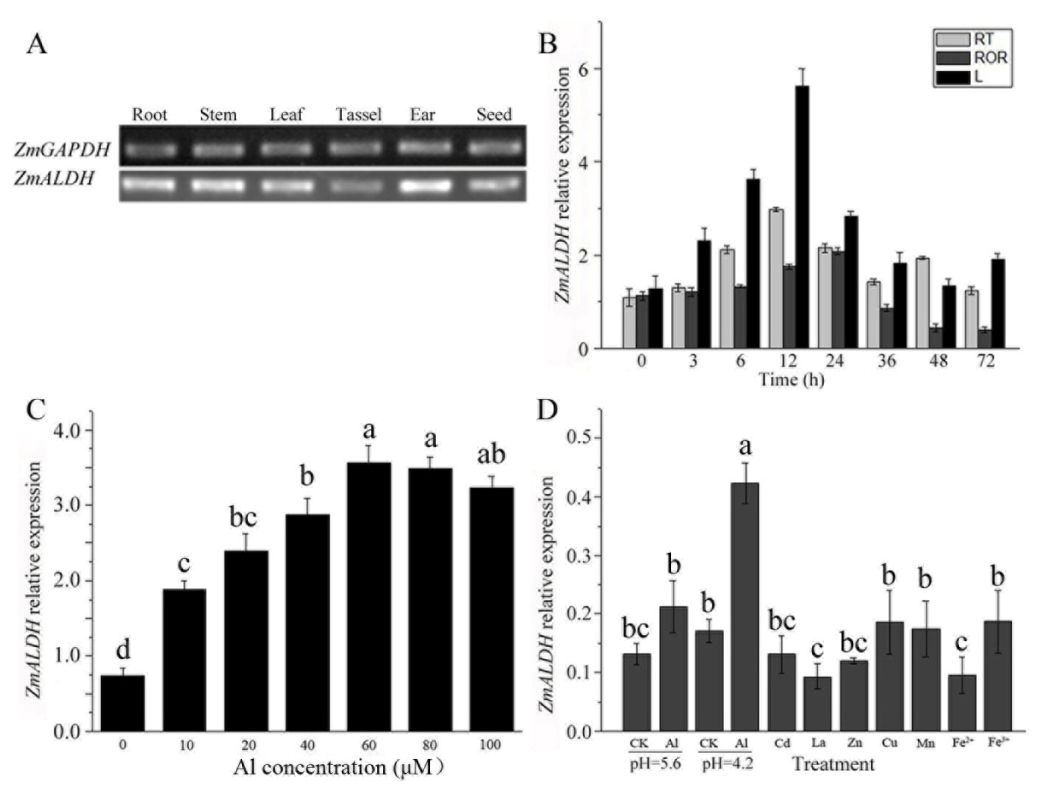
2.2. Expression Analysis of ZmALDH

To better understand the physiological function of ZmALDH, its expression patterns in different organs were examined by semi-quantitative RT-PCR. As shown in Figure 2A, ZmALDH was expressed in all tested organs, including the root, stem, leaf, tassel, ear, and seed, indicating that ZmALDH influences the growth and development of maize. To identify whether the transcription of ZmALDH was induced by Al stress at the seedling stage, its expression was measured by quantitative RT-PCR. The results revealed that ZmALDH was expressed in both leaves (L) and roots, and the expression level at the root tips (RT, apical 0–1 cm) was consistently higher than that in the rest of root (ROR) under Al stress (Figure 2B). The expression of ZmALDH increased initially until reaching a peak at 12 h and then decreased thereafter in the roots and leaves. A dose–response curve analysis showed that the expression of ZmALDH was induced by low concentrations of Al (10 μM) and increased until 60 μM AlCl3, after which expression levels were maintained at a steady level (Figure 2C).
To determine the Al-specificity of ZmALDH upregulation, the effects of various divalent and trivalent cations were also evaluated in the roots. As shown in Figure 2D, the expression of ZmALDH in the roots did not respond to low pH, cadmium (Cd), lanthanum (La), zinc (Zn), copper (Cu), manganese (Mn), or iron (Fe), except for Al.

2.3. Subcellular Localization

Prediction algorithms indicated that ZmALDH targets the cytoplasm or the chloroplast (Table S2). To determine its subcellular localization, we performed a transient assay with the ZmALDH-GFP fusion gene under the control of the CaMV35S promoter (35S:ZmALDH::GFP) in tobacco leaves and maize protoplasts. The control vector consisted of the CaMV35S promoter driving the expression of GFP alone (pCAMBIA2300-eGFP). As shown in Figure 3 and Figure S2, for the control vector, soluble GFP was localized to the cytoplasm and the nucleus in both cell types, as expected. However, the fluorescence of the 35S:ZmALDH::GFP chimera was associated with the cell and the nuclear periphery of the maize protoplasts and tobacco leaf cells, suggesting a plasma and nuclear membrane localization.

2.4. Root Growth Analysis of ZmALDH Transgenic Arabidopsis

To investigate the function of ZmALDH, the coding region of ZmALDH, driven by the CaMV 35S promoter, was stably introduced into wild-type (WT) Arabidopsis. Two highly expressed homozygous T3 lines (L4 and L6) were identified by PCR and RT-PCR (Figure S3), and used in subsequent analyses.
To determine whether ZmALDH overexpression could enhance Al tolerance in transgenic Arabidopsis, we examined the growth of the two transgenic lines (L4 and L6) and WT plants treated with 0, 50, and 100 μM AlCl3. In the absence of Al, the root growth of all plants was similar (Figure 4A,B). After treatment with 50 μM AlCl3, the relative root elongation (RRE) values for L4 and L6 were 78.96% and 73.92%, respectively, compared with 44.23% in WT (Figure 4C). At a high concentration of Al (100 μM), the RRE of all tested plants further decreased further (Figure 4C), but was still higher in the transgenic lines than in WT plants, implying that the root growth was more resistant to Al toxicity in transgenic plants than in WT (Figure 4).

2.5. ZmALDH Overexpression Reduces Al Accumulation

We further evaluated whether the increased resistance to Al toxicity observed in the transgenic lines (L4 and L6) was related to Al accumulation. We first visualized the Al uptake in roots by staining with hematoxylin, where darker blue-purple staining indicates greater Al absorption. In control conditions, staining in the root, including the root tip and basic root, was very weak. However, staining became darker after Al treatment (Figure 5A). Compared to the intensity in WT plants, L4 and L6 had weaker staining intensities, especially at the root tips (Figure 5A). Further, we quantified these differences by measuring the Al content by ICP-MS. In the absence of Al, WT, L4, and L6 plants did not exhibit any discernible differences and contained very low concentrations (~2.5 mg/g DW) of Al (Figure 5B). The Al contents of these three plants increased after exposure to Al, however, Al accumulation in WT plants was more than 1.8-fold higher than that in L4 and L6 plants. Al contents in leaves were similar in all materials and were not affected by Al treatment (Figure S4).

2.6. Comparative Analysis of the Oxidative Stress Response under Al Treatment

To determine oxidative damage induced by Al toxicity, we measured the production of H2O2 and the superoxide anion radical (O2). DAB staining was used to visualize the production of H2O2, where darker staining indicates more H2O2 production in the tissue. As shown in Figure 6A,B, WT, L4, and L6 plants contained a similarly low concentration of H2O2, approximately 1.13 μmol g−1 FW in the absence of Al. The H2O2 concentration increased in all plants after exposure to Al, whereas H2O2 in WT plants was 1.7-fold higher than that of L4 and L6 (Figure 6B). Similar to H2O2, the concentrations of O2 increased in all plants after Al treatment, whereas L4 and L6 accumulated only 60% of the O2 levels in the WT (Figure 6C). These results confirmed that the accumulation of ROS induced by Al stress in transgenic lines was less than that in WT plants.
Excessive production and accumulation of ROS in plants would directly trigger membrane lipid peroxidation, resulting in cell damage or death [10,30]. Therefore, we further measured the absorption of Evans blue and the accumulation of malondialdehyde (MDA). Levels of Evans blue uptake after 12 h of Al treatment in the root tips of WT plants were approximately 3-fold higher than those in L4 and L6 (Figure 7A,B). Meanwhile, the MDA contents in WT, L4, and L6 were similar to those in the control condition. After 12 h of Al stress, the accumulation of MDA in WT plants was 1.8-fold higher than that in L4 and L6 (Figure 7C). These results suggested that the overexpression of ZmALDH in Arabidopsis plants could reduce lipid peroxidation and cell damage caused by Al stress.

2.7. ZmALDH Enhanced the Tolerance to Oxidative Stress Induced by Al Toxicity

NAD(P)H, producedby ALDH catalytic oxidative, is an important substance for the maintenance of the balance of glutathione (GSH) in plants. The latter was indispensable for the AsA-GSH cycle, which is necessary to remove ROS effectively [19]. To investigate whether the increased Al tolerance of transgenic lines was related to the AsA-GSH cycle, we detected the contents of two antioxidants, including GSH and AsA. As shown in Figure 8, the GSH and AsA contents were maintained at a low level in the absence of Al. However, after exposure to 60 μM AlCl3 for 12 h, the GSH accumulation in WT plants and two transgenic lines increased 16.2%, 73.5%, and 66.5%, respectively, and AsA increased to 537 μg/g FW, 918 μg/g FW, and 922 μg/g FW, respectively. These results implied that the overexpression of ZmALDH could improve the contents of antioxidants associated with the AsA-GSH cycle.
Subsequently, GR and APX activity levels were measured. The activities of GR and APX increased upon Al exposure, and the levels in transgenic L4 and L6 plants were approximately 70% and 200%, respectively, greater than in the WT plants (Figure 9A,B). Consistent with these findings, a real-time PCR analysis confirmed that the transcript levels of AtGR1 and AtAPX1 were about 1.29- and 2.5-fold higher, respectively, in L4 and L6 transgenic lines than in WT plants after exposure to Al (Figure S5A,B). These results indicated that the Al tolerance of ZmALDH transgenic lines was related to the increased activity and expression levels of GR and APX in the AsA-GSH cycle.
The ROS scavenging ability is also linked to another enzymatic system. Hence, we further determined the transcript levels of the ROS scavengers as well as the activities of their products. The activities of SOD, POD, and CAT in WT, L4, and L6 plants were similar in the absence of Al. Under Al stress, activity levels of these ROS-scavenging enzymes were higher in L4 and L6 (by 50%, 100%, and 130%, respectively) than in the WT (Figure 10A–C). In addition, the transcript levels of AtSOD1 and AtPOD1 showed similar patterns in all lines in control conditions and were up-regulated in transgenic lines after exposure to Al (Figure 10D,E). However, the AtCAT1 expression trend was similar to that of AtAPX1 in WT and transgenic lines, regardless of Al treatment (Figure 10F). These results demonstrated that ZmALDH could reduce the oxidative damage induced by Al stress by promoting ROS scavenging.

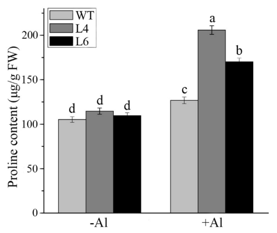
2.8. ZmALDH Promoted the Accumulation of Cellular Osmolytes

Plant ALDHs contribute to the synthesis of osmolytes, including sorbitol, myo-inositol, and proline, which have hydroxyl radical scavenging activity [31,32]. Therefore, we detected the proline content in WT and transgenic plants under control and Al stress conditions. As shown in Figure 11, the proline contents in all plants were similar under control conditions. After Al treatment, the levels of proline accumulation in L4 and L6 were 1.4-fold higher than those in WT plants, indicating that the overexpression of ZmALDH increased the accumulation of proline, potentially increasing Al tolerance in transgenic Arabidopsis.

3. Discussion

The ALDH superfamily is a group of enzymes able to catalyze the conversion of aldehydes to the corresponding acids [12,21]. ALDH genes are activated by environmental stresses [14,15,25] and have a certain influence on the stress response. VCAL68, a homolog of ALDH, is the only reported gene involved in the response to Al stress in plants [27]. However, the functions of ALDH genes in the response to Al stress are not well-characterized. In the present study, we cloned and characterized an aldehyde dehydrogenase gene from maize, ZmALDH, and investigated the role of ZmALDH under Al stress by its overexpression in Arabidopsis.
ZmALDH belongs to class 3 ALDHs, which are involved in the detoxification of aldehydes [14]. It had two Aldedh domains and was highly homologous to OsALDH3-1 and OsALDH3-2 (Figure 1), suggesting that ZmALDH exerts a similar function to that of this gene subfamily. In normal conditions, OsALDH3-1 and OsALDH3-2 are highly expressed in the young leaf and stem, with little expression in the young root [33]. However, ZmALDH was expressed in almost all tissues, with high expression in the root, stem, leaf, and ear (Figure 2A). This difference may be attributed to interspecific difference, or gene function evolution. Previous studies have shown that OsALDH3-1 and OsALDH3-2 are down-regulated by both drought and high salinity stresses [33]. Moreover, the maize genome contains ~28 ALDH genes, and the transcript level of ZmALDH is repressed by drought stress but strongly induced by salt stress [24,34]. In our study, ZmALDH was specifically up-regulated by Al stress and peaked at 12 h after Al treatment (Figure 2). This differed from the expression pattern of VCAL68, which exhibited expression peaks at 2 h and 24 h under Al stress [27]. These findings suggested that the response of ALDH genes to different stresses differs among plant species.
The subcellular localization indicates functional specialization. Plant ALDHs show wide variation in localization; for example, AtALDH3I1, AtALDH7B4, and OsALDH2B1 are located in chloroplasts, cytoplasmic [15], and nuclear compartments [26], respectively. However, ZmALDH in this study was localized in both the plasma membrane and nuclear membrane (Figure 3), different from any previous ALDHs. We speculated that Al-induced oxidative stress on the one hand leads to membrane lipid peroxidation and the production of toxic substances (such as ROS and aldehydes) [35,36], thereby activating the expression of ALDH genes and stimulating the synthesis of antioxidant enzymes and antioxidants, which were transported to the damaged plasma membrane to alleviate the oxidative stress [15,33]. On the other hand, an increase in the Al tolerance of transgenic lines was also related to a decrease in the Al content in roots, which is affected by the expression of ZmALDH. Thus, ZmALDH might have an influence on the crosstalk between the Al accumulation and oxidative stress response mechanisms.
Several studies have shown that Al-induced oxidative stress is an early process in toxicity responses in plants [9,37,38]. In this study, Evans blue and DAB staining revealed that Al stress caused plasma membrane damage and H2O2 accumulation (Figure 6 and Figure 7). ALDHs could decrease oxidative stress by scavenging hydroxyl radicals and cytotoxic aldehydes [39,40]. Similar to the Ath-ALDH3 response to cadmium stress [14], the contents of ROS (H2O2 and O2) and MDA in ZmALDH transgenic Arabidopsis decreased (Figure 6 and Figure 7), weakening of the oxidative stress induced by Al toxicity. Furthermore, under Al stress, the transgenic Arabidopsis with ZmALDH overexpression exhibited higher Al tolerance, including longer roots and less Al accumulation (Figure 5). This is perhaps due to the efficient conjugation of GSH with Al and/or ROS via overexpression of ZmALDH. Consequentially, the overexpressed transgenic plants showed lower Al content, lower ROS content, and less oxidative damage under Al stress. This is quite similar with the case of OsGSTU6 which improved the Cd tolerance of transgenic rice accompanied with low Cd content via ensuring the efficient conjugation of GSH with Cd and/or ROS [41]. These results suggested that ZmALDH contributed to Al tolerance and had a potential role in the detoxification of reactive aldehydes derived from lipid peroxidation.
Coincidentally, overexpression of some Al-inducible ROS-mediated and/or -related genes could promote Al tolerance [42]. For example, the overexpression of alternative oxidase gene (AOX) and MnSOD1 enhanced Al resistance in tobacco cells [43] and transgenic Brassica napus [44], respectively. Transgenic Arabidopsis plants overexpressing AtPrx64 exhibited tolerance to Al stress [45]. Overexpression of ZmAT6 in both maize and Arabidopsis could partially enhance Al tolerance via scavenging ROS [46]. Additionally, enhancing the activity of antioxidases in antioxidant pathways also contributes to resistance to Al toxicity in plants [47]. In our study, ZmALDH enhanced the Al tolerance of Arabidopsis by promoting the synthesis of GSH and AsA, enhancing the enzymatic activity of GR and APX, and increasing the transcript level of AtGR1 (Figure 8, Figure 9 and Figure S5). Furthermore, overexpression of ZmALDH could also increase the expression levels of AtSOD1 and AtPOD1, and improved the activity of these two antioxidant enzymes (Figure 10).
Under abiotic stress conditions, such as salt, drought [48], and Al stress [49], higher proline accumulation protects cells via osmotic adjustment and toxic free radical scavenging [50]. We obtained similar results, in which proline accumulation was slightly higher in transgenic lines than in WT plants (Figure 11). Together, these findings demonstrated that the overexpression of ZmALDH in Arabidopsis conferred tolerance to Al stress by enhancing its oxidative stress tolerance.

4. Materials and Methods

4.1. Plant Materials, Growth Conditions, and Treatment

The Al-tolerant maize inbred line 178 [29] and Arabidopsis thaliana (Columbia ecotype, Col-0) were used in this study. Seed treatment, germination, and culture conditions were performed as described in a previous study [46].
The metal treatments, time-course analysis, and manipulation of Al concentrations were performed as described previously [51]. The roots and/or the shoots from all the treated seedlings were immediately frozen in liquid nitrogen, and stored at −80 °C for RNA extraction.
For Al tolerance analysis of transgenic Arabidopsis, two-week-old WT plants and transgenic lines grown in 2% MGRL solution (pH 4.5) [50] were pre-treated with 0.5 mM CaCl2 for 30 min and then treated with or without 60 μM AlCl3 for 12 h. Root and shoot samples were collected, separately.

4.2. Cloning and Sequence Analysis of ZmALDH

Based on the reference sequence of B73 cDNA, the ZmALDH gene was amplified from 178 by specific primers (Table S3). A multiple sequence alignment of amino acid sequences (Table S4) was generated using DNAMAN (Lynnon). A phylogenetic tree was inferred using the neighbor-joining method by MEGA 6.0 [52].

4.3. RNA Isolation and Gene Expression Analysis

Total RNA extraction, semiquantitative RT-PCR and real-time PCR were performed as described previously [53]. ZmGAPDH and AtACT2 were used as the internal reference genes. Three biological replicates were performed for each experiment. The primers are listed in Table S3.

4.4. Subcellular Localization

The subcellular localization of the ZmALDH protein was predicted using several online websites (Table S2) and determined by transient expression in tobacco leaves and maize leaf protoplasts with a 35S:ZmALDH::GFP translational fusion cloned in the pCAMBIA2300 vector [46,54].

4.5. Generation of Transgenic Arabidopsis Plants

The p35S:ZmALDH vector was transformed into Arabidopsis ecotype Columbia-0 (Col-0) by the flowering dip method [55]. The transgenic plants were selected as described previously [56]. Finally, the positive homozygous seeds of the T3 generation were used for further experiments.

4.6. Measurement of Oxidative Damage

Oxidative stress induced by Al stress was evaluated by cell death and membrane lipid peroxidation. The former was estimated by determining Evans blue uptake [57] and the latter was detected by the concentration of malondialdehyde via the thiobarbituric acid method [8].

4.7. Determination of Reactive Oxygen Species (ROS)

The production of H2O2 was visualized by 3,3′-diaminobenzidine (DAB) staining according to previously described methods [17] and measured as described before [46]. The super oxygen free radical (O2) was detected by the hydroxylamine method [58].

4.8. Determination of Glutathione and Ascorbic Acid

The total GSH and AsA contents were determined according to methods of Hodges et al. [59] and Noctor and Foyer [60].

4.9. Enzymatic Activity and Gene Expression Analyses

Antioxidant enzyme extraction and the measurement of SOD, POD, CAT, and APX activity levels were carried out as previously described [10,46]. GR activity was determined by the decrease in absorbance at 340 nm due to NADPH oxidation [61].
The relative expression levels of enzyme genes described above were measured by RT-PCR, and the primers are listed in Table S1.

4.10. Measurement of Relative Root Growth

The sterilized seeds were first grown in 1/2 MS medium (pH 4.5) for 3 days. Then the seedlings were transplanted into 1/8 MS medium (pH 4.5) with or without 50 μM and 100 μM AlCl3 for 7 days, and the initial and final root lengths were recorded. The RRE was defined as the ratio of the net root growth length after treatment with Al to that of the control.

4.11. Al Content Assay

The accumulation of Al was visualized by hematoxylin staining [62]. The Al contents in WT and transgenic plants with or without Al treatment were measured by ICP-MS (PerkinElmer, NexlON 2000, Seer Green, UK).

4.12. Statistical Analysis and Reproducibility

All treatments were repeated at least three times. Statistical analyses, including Tukey’s test, were performed using SPSS 21.0 (IBM, Chicago, IL, USA, 2012).

5. Conclusions

ZmALDH could promote the Al tolerance of plants by enhancing the AsA-GSH cycle, increasing the transcript levels of antioxidant genes as well as activating their products, and mediating the accumulation of Al in roots.

Supplementary Materials

The following supporting information can be downloaded at: https://0-www-mdpi-com.brum.beds.ac.uk/article/10.3390/ijms23010477/s1.

Author Contributions

H.-M.D. carried out the experiments, analyzed the data, and drafted the manuscript; C.L., X.-W.J., C.-F.D., Y.Y., S.L. and W.-Z.H. provided general guidance; S.-Z.Z. designed the experiment and revised the manuscript. All authors have read and agreed to the published version of the manuscript.

Funding

This work was supported by the National Natural Science Foundation of China, grant number 32171951, the Applied Basic Research Programs of Science and Technology Department of Sichuan, grant number 2021YJ0300, and the Open Project Program of Panxi Crops Research and Utilization Key Laboratory of Sichuan Province, grant number SZ21ZZ05.

Institutional Review Board Statement

Not applicable.

Informed Consent Statement

Not applicable.

Data Availability Statement

All data generated or analyzed during this study are available within the article or upon request from the corresponding author.

Conflicts of Interest

The authors declare no conflict of interest.

References

  1. Kochian, L.V.; Hoekenga, O.A.; Pineros, M.A. How do crop plants tolerate acid soils? Mechanisms of aluminum tolerance and phosphorous efficiency. Annu. Rev. Plant Biol. 2004, 55, 459–493. [Google Scholar] [CrossRef] [PubMed]
  2. Kochian, L.V. Cellular mechanisms of aluminum toxicity and resistance in plants. Annu. Rev. Plant Biol. 1995, 46, 237–260. [Google Scholar] [CrossRef]
  3. Ma, J.F. Role of organic acids in detoxification of aluminum in higher plants. Plant Cell Physiol. 2000, 41, 383–390. [Google Scholar] [CrossRef] [PubMed] [Green Version]
  4. Zheng, S.J.; Yang, J.L. Target sites of aluminum phytotoxicity. Biol. Plantarum 2005, 49, 321–331. [Google Scholar] [CrossRef]
  5. Shen, R.; Ma, J.F.; Kyo, M.; Iwashita, T. Compartmentation of aluminium in leaves of an Al-accumulator, Fagopyrum esculentum Moench. Planta 2002, 215, 394–398. [Google Scholar] [CrossRef] [PubMed]
  6. Ma, J.F.; Ryan, P.R.; Delhaize, E. Aluminium tolerance in plants and the complexing role of organic acids. Trends Plant Sci. 2001, 6, 273–278. [Google Scholar] [CrossRef]
  7. Ryan, P.R.; Delhaize, E.; Jones, D.L. Function and mechanism of organic anion exudation from plant roots. Annu. Rev. Plant Physiol. Plant Mol. Biol. 2001, 52, 527–560. [Google Scholar] [CrossRef]
  8. Yamamoto, Y.; Kobayashi, Y.; Matsumoto, H. Lipid peroxidation is an early symptom triggered by aluminum, but not the primary cause of elongation inhibition in pea roots. Plant Physiol. 2001, 125, 199–208. [Google Scholar] [CrossRef] [Green Version]
  9. Tabaldi, L.A.; Cargnelutti, D.; Gonçalves, J.F.; Pereira, L.B.; Castro, G.Y.; Maldaner, J.; Rauber, R.; Rossato, L.V.; Bisognin, D.A.; Schetinger, M.R.C. Oxidative stress is an early symptom triggered by aluminum in Al-sensitive potato plantlets. Chemosphere 2009, 76, 1402–1409. [Google Scholar] [CrossRef]
  10. Achary, V.M.; Jena, S.; Panda, K.K.; Panda, B.B. Aluminium induced oxidative stress and DNA damage in root cells of Allium cepa L. Ecotoxicol. Environ. Saf. 2008, 70, 300–310. [Google Scholar] [CrossRef]
  11. Kirch, H.H.; Bartels, D.; Wei, Y.; Schnable, P.S.; Wood, A.J. The ALDH gene superfamily of Arabidopsis. Trends Plant Sci. 2004, 9, 371–377. [Google Scholar] [CrossRef] [Green Version]
  12. Singh, S.; Brocker, C.; Koppaka, V.; Chen, Y.; Jackson, B.C.; Matsumoto, A.; Thompson, D.C.; Vasiliou, V. Aldehyde dehydrogenases in cellular responses to oxidative/electrophilic stress. Free Radic. Biol. Med. 2013, 56, 89–101. [Google Scholar] [CrossRef] [Green Version]
  13. Møller, I.M.; Jensen, P.E.; Hansson, A. Oxidative modifications to cellular components in plants. Annu. Rev. Plant Biol. 2007, 58, 459–481. [Google Scholar] [CrossRef] [Green Version]
  14. Sunkar, R.; Bartels, D.; Kirch, H.H. Overexpression of a stress-inducible aldehyde dehydrogenase gene from Arabidopsis thaliana in transgenic plants improves stress tolerance. Plant. J. 2003, 35, 452–464. [Google Scholar] [CrossRef]
  15. Kotchoni, S.O.; Kuhns, C.; Ditzer, A.; Kirch, H.H.; Bartels, D. Over-expression of different aldehyde dehydrogenase genes in Arabidopsis thaliana confers tolerance to abiotic stress and protects plants against lipid peroxidation and oxidative stress. Plant. Cell Environ. 2006, 29, 1033–1048. [Google Scholar] [CrossRef]
  16. Sun, C.; Liu, L.; Yu, Y.; Liu, W.; Lu, L.; Jin, C.; Lin, X. Nitric oxide alleviates aluminum-induced oxidative damage through regulating the ascorbate-glutathione cycle in roots of wheat. J. Integr. Plant Biol. 2015, 57, 550–561. [Google Scholar] [CrossRef]
  17. Yu, Y.; Zhou, W.; Zhou, K.; Liu, W.; Liang, X.; Chen, Y.; Sun, D.; Lin, X. Polyamines modulate aluminum-induced oxidative stress differently by inducing or reducing H2O2 production in wheat. Chemosphere 2018, 212, 645–653. [Google Scholar] [CrossRef]
  18. Mittova, V.; Volokita, M.; Guy, M.; Tal, M. Activities of SOD and the ascorbate-glutathione cycle enzymes in subcellular compartments in leaves and roots of the cultivated tomato and its wild salt-tolerant relative Lycopersicon pennellii. Physiol. Plantarum 2000, 110, 42–51. [Google Scholar] [CrossRef]
  19. Li, Y.; Liu, Y.; Zhang, J. Advances in the research on the AsA-GSH cycle in horticultural crops. Front. Agric. China 2010, 4, 84–90. [Google Scholar] [CrossRef]
  20. Kenichi, O. Glutathione-associated regulation of plant growth and stress responses. Antioxid. Redox Signal. 2005, 7, 973–981. [Google Scholar]
  21. Perozich, J.; Nicholas, H.; Wang, B.C.; Lindahl, R.; Hempel, J. Relationships within the aldehyde dehydrogenase extended family. Protein Sci. 1999, 8, 137–146. [Google Scholar] [CrossRef] [PubMed] [Green Version]
  22. Frommel, J.; Koncitikova, R.; Kopecny, D.; Soural, M.; Sebela, M. Oxidation of imidazole- and pyrazole-derived aldehydes by plant aldehyde dehydrogenases from the family 2 and 10. Chem. Biol. Interact. 2019, 304, 194–201. [Google Scholar] [CrossRef] [PubMed]
  23. Bartels, D. Targeting detoxification pathways: An efficient approach to obtain plants with multiple stress tolerance? Trends Plant Sci. 2001, 6, 284–286. [Google Scholar] [CrossRef]
  24. Zhou, M.L.; Zhang, Q.; Zhou, M.; Qi, L.P.; Yang, X.B.; Zhang, K.X.; Pang, J.F.; Zhu, X.M.; Shao, J.R.; Tang, Y.X.; et al. Aldehyde dehydrogenase protein superfamily in maize. Funct. Integr. Genom. 2012, 12, 683–691. [Google Scholar] [CrossRef]
  25. An, X.; Duan, F.Y.; Guo, S.; Chen, F.J.; Yuan, L.X.; Gu, R.L. Transcriptional regulation of expression of the maize aldehyde dehydrogenase 7 gene (ZmALDH7B6) in response to abiotic stresses. J. Integr. Agr. 2014, 13, 1900–1908. [Google Scholar] [CrossRef] [Green Version]
  26. Ke, Y.G.; Yuan, M.; Liu, H.B.; Hui, S.G.; Qin, X.F.; Chen, J.; Zhang, Q.L.; LI, X.H.; Xiao, J.H.; Zhang, Q.F.; et al. The versatile functions of OsALDH2B1 provide a genic basis for growth-defense trade-offs in rice. Proc. Natl. Acad. Sci. USA 2020, 117, 3867–3873. [Google Scholar] [CrossRef]
  27. Inostroza-Blancheteau, C.; Reyes-Diaz, M.; Aquea, F.; Nunes-Nesi, A.; Alberdi, M.; Arce-Johnson, P. Biochemical and molecular changes in response to aluminium-stress in highbush blueberry (Vaccinium corymbosum L.). Plant Physiol. Biochem. 2011, 49, 1005–1012. [Google Scholar] [CrossRef]
  28. Xu, L.M.; Liu, C.; Cui, B.M.; Wang, N.; Zhao, Z.; Zhou, L.N.; Huang, K.F.; Ding, J.Z.; Du, H.M.; Jiang, W.; et al. Transcriptomic responses to aluminum (Al) stress in maize. J. Integr. Agr. 2018, 17, 1946–1958. [Google Scholar] [CrossRef] [Green Version]
  29. Xu, L.M.; Liu, W.; Cui, B.M.; Wang, N.; Ding, J.Z.; Liu, C.; Gao, S.B.; Zhang, S.Z. Aluminium tolerance assessment of 141 maize germplasms in a solution culture. Univers. J. Agric. Res. 2017, 5, 1–9. [Google Scholar] [CrossRef] [Green Version]
  30. Šimonovičová, M.; Huttová, J.; Mistrík, I.; Široká, B.; Tamás, L. Root growth inhibition by aluminum is probably caused by cell death due to peroxidase-mediated hydrogen peroxide production. Protoplasma 2004, 224, 91–98. [Google Scholar] [CrossRef]
  31. Chaudhuri, P.; Rashid, N.; Thapliyal, C. Osmolyte System and Its Biological Significance. In Cellular Osmolytes; Rajendrakumar, S.L., Dar, T., Eds.; Springer: Singapore, 2017; pp. 1–34. [Google Scholar]
  32. Hasanuzzaman, M.; Anee, T.I.; Bhuiyan, T.F.; Nahar, K.; Fujita, M. Emerging role of osmolytes in enhancing abiotic stress tolerance in rice. In Advances in Rice Research for Abiotic Stress Tolerance; Hasanuzzaman, M., Nahar, K., Fujita, M., Biswas, J.K., Eds.; Woodhead Publishing: New York, NY, USA, 2019; pp. 677–708. [Google Scholar]
  33. Gao, C.; Han, B. Evolutionary and expression study of the aldehyde dehydrogenase (ALDH) gene superfamily in rice (Oryza sativa). Gene 2009, 431, 86–94. [Google Scholar] [CrossRef]
  34. Jimenez-Lopez, J.C.; Gachomo, E.W.; Seufferheld, M.J.; Kotchoni, S.O. The maize ALDH protein superfamily: Linking structural features to functional specificities. BMC Struct. Biol. 2010, 10, 43. [Google Scholar] [CrossRef] [Green Version]
  35. Yamamoto, Y.; Kobayashi, Y.; Devi, S.R.; Rikiishi, S.; Matsumoto, H. Aluminum toxicity is associated with mitochondrial dysfunction and the production of reactive oxygen species in plant cells. Plant Physiol. 2002, 128, 63–72. [Google Scholar] [CrossRef]
  36. Chowra, U.; Yanase, E.; Koyama, H.; Panda, S.K. Aluminium-induced excessive ROS causes cellular damage and metabolic shifts in black gram Vigna mungo (L.) Hepper. Protoplasma 2017, 254, 293–302. [Google Scholar] [CrossRef]
  37. Richards, K.D.; Schott, E.J.; Sharma, Y.K.; Davis, K.R.; Gardner, R.C. Aluminum induces oxidative stress genes in Arabidopsis thaliana. Plant Physiol. 1998, 116, 409–418. [Google Scholar] [CrossRef] [Green Version]
  38. Sun, C.; Liu, L.; Zhou, W.; Lu, L.; Jin, C.; Lin, X. Aluminum Induces Distinct Changes in the Metabolism of Reactive Oxygen and Nitrogen Species in the Roots of Two Wheat Genotypes with Different Aluminum Resistance. J. Age Food Chem. 2017, 65, 9419–9427. [Google Scholar] [CrossRef]
  39. Chen, Y.; Mehta, G.; Vasiliou, V. Antioxidant defenses in the ocular surface. Ocul. Surf. 2009, 7, 176–185. [Google Scholar] [CrossRef] [Green Version]
  40. Rodriguez, S.M.; Andrade, M.O.; Gomes, A.P.; Damatta, F.M.; Baracat-Pereira, M.C.; Fontes, E.P. Arabidopsis and tobacco plants ectopically expressing the soybean antiquitin-like ALDH7 gene display enhanced tolerance to drought, salinity, and oxidative stress. J. Exp. Bot. 2006, 57, 1909–1918. [Google Scholar] [CrossRef] [Green Version]
  41. Jing, X.Q.; Zhou, M.R.; Nie, X.M.; Zhang, L.; Shi, P.T.; Shalmani, A.; Miao, H.; Li, W.Q.; Liu, W.T.; Chen, K.M. OsGSTU6 Contributes to Cadmium Stress Tolerance in Rice by Involving in Intracellular ROS Homeostasis. J. Plant Growth Regul. 2021, 40, 945–961. [Google Scholar] [CrossRef]
  42. Ezaki, B.; Gardner, R.C.; Ezaki, Y.; Matsumoto, H. Expression of aluminum-induced genes in transgenic Arabidopsis plants can ameliorate aluminum stress and/or oxidative stress. Plant Physiol. 2000, 122, 657–665. [Google Scholar] [CrossRef] [Green Version]
  43. Panda, S.K.; Sahoo, L.; Katsuhara, M.; Matsumoto, H. Overexpression of alternative oxidase gene confers aluminum tolerance by altering the respiratory capacity and the response to oxidative stress in tobacco cells. Mol. Biotechnol. 2013, 54, 551–563. [Google Scholar] [CrossRef]
  44. Basu, U.; Good, A.G.; Taylor, G.J. Transgenic Brassica napus plants overexpressing aluminum-induced mitochondrial manganese superoxide dismutase cDNA are resistant to aluminum. Plant Cell Environ. 2001, 24, 1269–1278. [Google Scholar] [CrossRef] [Green Version]
  45. Wu, Y.S.; Yang, Z.L.; How, J.Y.; Xu, H.N.; Chen, L.M.; Li, K.Z. Overexpression of a peroxidase gene (AtPrx64) of Arabidopsis thaliana in tobacco improves plant’s tolerance to aluminum stress. Plant Mol. Biol. 2017, 95, 157–168. [Google Scholar] [CrossRef]
  46. Du, H.; Huang, Y.; Qu, M.; Li, Y.; Hu, X.; Yang, W.; Li, H.; He, W.; Ding, J.; Liu, C.; et al. A maize ZmAT6 gene confers aluminum tolerance via reactive oxygen species scavenging. Front. Plant Sci. 2020, 11, 1016. [Google Scholar] [CrossRef]
  47. Irany, R.P.; Anny, C.D.L.; Tatiane, L.P.; Raquel, A.A.; Maria, D.C.P.B. Gene expression and antioxidant enzymatic activity in passion fruit exposed to aluminum. Afr. J. Agric. Res. 2018, 13, 115–120. [Google Scholar] [CrossRef] [Green Version]
  48. Patade, V.Y.; Bhargava, S.; Suprasanna, P. Salt and drought tolerance of sugarcane under iso-osmotic salt and water stress: Growth, osmolytes accumulation, and antioxidant defense. J. Plant Interact. 2011, 6, 275–282. [Google Scholar] [CrossRef] [Green Version]
  49. Pirzadah, T.B.; Malik, B.; Tahir, I.; Rehman, R.U.; Hakeem, K.R.; Alharby, H.F. Aluminium stress modulates the osmolytes and enzyme defense system in Fagopyrum species. Plant Physiol. Biochem. 2019, 144, 178–186. [Google Scholar] [CrossRef]
  50. Kaur, G.; Asthir, B. Proline: A key player in plant abiotic stress tolerance. Biol. Plantarum 2015, 59, 609–619. [Google Scholar] [CrossRef]
  51. Du, H.; Ryan, P.R.; Liu, C.; Li, H.; Hu, W.; Yan, W.; Huang, Y.; He, W.; Luo, B.; Zhang, X.; et al. ZmMATE6 from maize encodes a citrate transporter that enhances aluminum tolerance in transgenic Arabidopsis thaliana. Plant Sci. 2021, 311, 111016. [Google Scholar] [CrossRef] [PubMed]
  52. Tamura, K.; Stecher, G.; Peterson, D.; Filipski, A.; Kumar, S. MEGA6: Molecular evolutionary genetics analysis version 6. Mol. Biol. Evol. 2013, 30, 2725–2729. [Google Scholar] [CrossRef] [PubMed] [Green Version]
  53. Li, H.; Du, H.; Huang, K.; Chen, X.; Liu, T.; Gao, S.; Liu, H.; Tang, Q.; Rong, T.; Zhang, S. Identification, and functional and expression analyses of the CorA/MRS2/MGT-type magnesium transporter family in maize. Plant Cell Physiol. 2016, 57, 1153–1168. [Google Scholar] [CrossRef] [PubMed] [Green Version]
  54. Yoo, S.D.; Cho, Y.H.; Sheen, J. Arabidopsis mesophyll protoplasts: A versatile cell system for transient gene expression analysis. Nat. Protoc. 2007, 2, 1565–1572. [Google Scholar] [CrossRef] [PubMed] [Green Version]
  55. Clough, S.J.; Bent, A.F. Floral dip: A simplifed method for Agrobacterium-mediated transformation of Arabidopsis thaliana. Plant J. Cell Mol. Biol. 1998, 16, 735. [Google Scholar] [CrossRef] [PubMed] [Green Version]
  56. Li, H.; Wang, N.; Ding, J.; Liu, C.; Du, H.; Huang, K.; Cao, M.; Lu, Y.; Gao, S.; Zhang, S. The maize CorA/MRS2/MGT-type Mg transporter, ZmMGT10, responses to magnesium deficiency and confers low magnesium tolerance in transgenic Arabidopsis. Plant Mol. Biol. 2017, 95, 269–278. [Google Scholar] [CrossRef]
  57. Baker, C.J.; Mock, N.M. An improved methods for monitoring cell death in cell suspension and leaf disc assays using evans blue. Plant Cell Tissue Org. Cult. 1994, 39, 7–12. [Google Scholar] [CrossRef]
  58. Elstner, E.F.; Heupel, A. Inhibition of nitrite formation from hydroxylammoniumchloride: A simple assay for superoxide dismutase. Anal. Biochem. 1976, 70, 616–620. [Google Scholar] [CrossRef]
  59. Hodges, D.; Andrews, C.; Johnson, D.; Hamilton, R. Antioxidant compound responses to chilling stress in differentially sensitive inbred maize lines. Plant Physiol. 1996, 98, 685–692. [Google Scholar] [CrossRef]
  60. Noctor, G.; Foyer, C.H. Ascorbate and glutathione: Keeping active oxygen under control. Annu. Rev. Plant Physiol. Plant Mol. Biol. 1998, 49, 249–279. [Google Scholar] [CrossRef]
  61. Foyer, C.H.; Halliwell, B. The presence of glutathione and glutathione reductase in chloroplasts: A proposed role in ascorbic acid metabolism. Planta 1976, 133, 21–25. [Google Scholar] [CrossRef]
  62. Ribeiro, A.P.; de Souza, W.R.; Martins, P.K.; Vinecky, F.; Duarte, K.E.; Basso, M.F.; da Cunha, B.; Campanha, R.B.; de Oliveira, P.A.; Centeno, D.C.; et al. Overexpression of BdMATE gene improves aluminum tolerance in Setaria viridis. Front. Plant Sci. 2017, 8, 865. [Google Scholar] [CrossRef] [Green Version]
Figure 1. Sequence and phylogenetic analysis of ZmALDH and other known ALDH proteins: (A) Multiple sequence amino acid alignment (DNAMAN) of ZmALDH (AQK73130), OsALDH3-1 (XP_015623875), and OsALDH3-2 (XP_015623591); (B) Phylogenetic relationships of ZmALDH and previously published ALDH proteins. Unrooted NJ trees were generated using MEGA 6.0. Bootstrap values from 1000 replicates are indicated at each branch.
Ijms 23 00477 g001
Figure 2. Expression analysis of the ZmALDH gene: (A) Spatial expression patterns of ZmALDH in different tissues; (B) Transcript levels of ZmALDH in the maize seedlings of root tips (RT), the rest of the root (ROR), and the leaves (L) were quantified at 0, 3 h, 6 h, 12 h, 24 h, 36 h, 48 h, and 72 h after Al treatment (pH 4.2); (C) Expression analysis of ZmALDH under different concentrations of Al. Seedlings were treated for 6 h with 0 μM, 10 μM, 20 μM, 40 μM, 60 μM, 80 μM, and 100 μM Al, and then the roots were collected for RT-PCR; (D) Effect of other cations on ZmALDH expression. Seedlings were exposed for 6 h to a 0.5 mM CaCl2 solution (pH 4.2) containing 30 μM CdCl2, 2.0 μM CuCl2, 10 μM LaCl3, 100 μM ZnCl2, 200 μM MnSO4, 20 μM FeSO4·7H2O, 20 μM FeCl3, or 60 μM AlCl3, respectively. The roots were then collected for subsequent RT-PCR. Relative gene expression normalized to the ZmGAPDH. The values are presented as mean ± SD (n = 5) and different letters indicate significant difference at p < 0.05 (Tukey’s test).
Ijms 23 00477 g002
Figure 3. Subcellular localization of ZmALDH in tobacco leaves.
Ijms 23 00477 g003
Figure 4. Transgenic plants expressing ZmALDH are more resistant to Al: (A) Phenotype; (B) Root length, and (C) Relative root elongation of WT and transgenic lines under normal and Al stress conditions. Seedlings of WT and two ZmALDH transgenic lines (L4 and L6) were grown on a plate containing 0 μM, 50 μM, and 100 μM AlCl3 for 5 d. Scale bar: 1.0 cm. Values represent mean ± SD (n ≥ 15). Different letters indicate significant differences (p < 0.01) (Tukey’s test).
Ijms 23 00477 g004
Figure 5. Al accumulation in roots is reduced in transgenic Arabidopsis. (A) Hematoxylin staining after 12 h exposure to 0 and 60 μM AlCl3 in roots. Scale bar: 50 μm; (B) Al contents in lines (as in (A)) under Al stress for 12 h. Values represent mean ± SD (n ≥ 15). Different letters indicate significant differences (p < 0.01) (Tukey’s test).
Ijms 23 00477 g005
Figure 6. Overexpression of ZmALDH reduces the accumulation of superoxide (O2) and hydrogen peroxide (H2O2). Seedlings of WT, L4, and L6 were grown in the 2% MGRL solution for 14 d, and then plants were treated with 60 μM AlCl3 for 12 h, before roots were collected for subsequent experiments: (A) Histochemical visualization of H2O2 was performed with DAB staining. Scale bar: 250 μm. Contents of (B) H2O2 and (C) O2 in WT and transgenic lines after exposure to Al stress for 12 h. Values represent mean ± SD (n = 20). Different letters indicate significant differences (p < 0.01) (Tukey’s test).
Ijms 23 00477 g006
Figure 7. Effect of Al treatment on the lipid peroxidation and loss of plasma membrane integrity in roots. Seedling roots of wild–type plants and transgenic lines were collected after exposure to 60 μM AlCl3 for 12 h and used to evaluate (A) Evans blue staining, scale bar: 200 μm; (B) Evans blue uptake, and (C) Malondialdehyde (MDA) contents. Values represent mean ± SD (n = 20). Different letters indicate significant differences (p < 0.01) (Tukey’s test).
Ijms 23 00477 g007
Figure 8. Overexpression of ZmALDH increases the accumulation of total GSH and AsA in roots. The total contents of (A) GSH and (B) AsA in WT and transgenic Arabidopsis (L4 and L6) were detected after exposure to 60 μM AlCl3 for 12 h. Values represent mean ± SD (n = 20). Different letters indicate significant differences (p < 0.01) (Tukey’s test).
Ijms 23 00477 g008
Figure 9. Activity of GR and APX in wild–type and transgenic plants. The activity of (A) GR and (B) APXin wild–type plants and transgenic lines after exposure to Al for 12 h. Values are means ± SD (n = 20). Different letters indicate significant differences (p < 0.01) (Tukey’s test).
Ijms 23 00477 g009
Figure 10. Effect of Al on the activities and transcripts’ level of antioxidant enzymes. Two–week–old seedlings were treated with 60 μM AlCl3 for 12 h and the total activity of (A) SOD; (B) POD, and (C) CAT and transcripts’ level of (D) AtSOD1; (E) AtPOD1, and (F) AtCAT1 were analyzed. Relative gene expression was normalized to the AtACT2. Values are means ± SD (n = 20). Different letters indicate significant differences (p < 0.05) (Tukey’s test).
Ijms 23 00477 g010
Figure 11. Determination of free proline in wild–type plants and transgenic lines. Two-week-old seedlings were treated with or without 60 μM AlCl3 for 12 h, and the proline contents of roots were determined after that. Values are means ± SD (n = 20). Different letters indicate significant differences (p < 0.05) (Tukey’s test).
Ijms 23 00477 g011
Publisher’s Note: MDPI stays neutral with regard to jurisdictional claims in published maps and institutional affiliations.

