Article

The QseEF Two-Component System-GlmY Small RNA Regulatory Pathway Controls Swarming in Uropathogenic Proteus mirabilis

1
Department and Graduate Institute of Clinical Laboratory Sciences and Medical Biotechnology, College of Medicine, National Taiwan University, Taipei 10048, Taiwan
2
Department of Urology, National Taiwan University Hospital, Taipei 10002, Taiwan
3
Department of Nursing, National Taichung University of Science and Technology, Taichung City 404348, Taiwan
4
Department of Laboratory Medicine, National Taiwan University Hospital, Taipei 10002, Taiwan
*
Author to whom correspondence should be addressed.
These authors contributed equally to this work.
Int. J. Mol. Sci. 2022, 23(1), 487; https://0-doi-org.brum.beds.ac.uk/10.3390/ijms23010487
Submission received: 30 November 2021 / Revised: 23 December 2021 / Accepted: 28 December 2021 / Published: 1 January 2022
(This article belongs to the Special Issue Transcriptional Regulation and Its Misregulation in Human Diseases)

Abstract

:
Bacterial sensing of environmental signals through the two-component system (TCS) plays a key role in modulating virulence. In the search for the host hormone-sensing TCS, we identified a conserved qseEGF locus following glmY, a small RNA (sRNA) gene in uropathogenic Proteus mirabilis. Genes of glmY-qseE-qseG-qseF constitute an operon, and QseF binding sites were found in the glmY promoter region. Deletion of glmY or qseF resulted in reduced swarming motility and swarming-related phenotypes relative to the wild-type and the respective complemented strains. The qseF mutant had decreased glmYqseEGF promoter activity. Both glmY and qseF mutants exhibited decreased flhDC promoter activity and mRNA level, while increased rcsB mRNA level was observed in both mutants. Prediction by TargetRNA2 revealed cheA as the target of GlmY. Then, construction of the translational fusions containing various lengths of cheA 5′UTR for reporter assay and site-directed mutagenesis were performed to investigate the cheA-GlmY interaction in cheA activation. Notably, loss of glmY reduced the cheA mRNA level, and urea could inhibit swarming in a QseF-dependent manner. Altogether, this is the first report elucidating the underlying mechanisms for modulation of swarming motility by a QseEF-regulated sRNA GlmY, involving expression of cheA, rcsB and flhDC in uropathogenic P. mirabilis.

1. Introduction

Proteus mirabilis is an important pathogen of the urinary tract, especially in patients with indwelling urinary catheters [1]. Common strategies of pathogenesis employed by P. mirabilis include adherence via fimbriae [2,3], biofilm formation, flagella-mediated motility, immune modulation and urease production [4]. P. mirabilis exhibits a form of multicellular behavior termed swarming [5]. Swarming motility are the results of complex signal transduction and gene regulation [5]. It is generally believed that signals could be sensed and transmitted by two-component systems (TCSs). It is worth noting that the ability of P. mirabilis to express virulence factors is coupled to swarming differentiation [5]. In general, flagella are thought to assist in colonization and dissemination during P. mirabilis catheter-associated UTIs, and FlhDC is a master regulator that controls the expression of flagellum-related genes [6,7]. During swarmer cell differentiation, the expression level of flhDC rises dramatically [8].
Among multiple strategies for many bacteria to respond rapidly to changing environments is the regulation by the very versatile and adaptable regulatory small non-coding RNAs (sRNAs) [9,10,11]. Numerous cellular processes, such as motility [12], various stress responses and virulence factor expression are subject to the post-transcriptional control of sRNAs [9,10,11,13]. In many cases, the trans-acting sRNA mediated-regulation requires the chaperone protein Hfq to facilitate sRNA-mRNA interaction [14,15]. Two well-known ways for an sRNA to regulate translation are sequestering of ribosome binding site (rbs) and melting a secondary structure in the 5′ UTR to expose rbs, resulting in repression and activation of translation, respectively [16]. TCS and alternative σ factors have been shown to control sRNA expression [17,18]. In this regard, a PhoPQ TCS-regulated sRNA MgrR modulates expression of eptB, an LPS modification gene regulated by σE, to affect the sensitivity to antimicrobial peptides in response to low Mg2+ or the presence of antimicrobial peptides [19]. In addition, a σE-dependent sRNA, MicA, directly inhibited PhoPQ synthesis and consequently downregulated the PhoPQ regulons involved in pathogenicity, cell envelope composition and stress resistance [20].
Bacterial sensing of environmental signals plays a key role in regulating virulence and mediating bacterium host interactions. TCS is a common strategy used by bacteria to regulate gene expression in response to environmental cues. Enterohemorrhagic E. coli (EHEC) senses host hormones (epinephrine and norepinephrine) via the QseEF two-component system to activate actin polymerization and initiate formation of attaching and effacing (AE) lesions [21,22]. Deletion mutants of the qseEF are attenuated in virulence, as demonstrated for E. coli, Citrobacter rodentium, Salmonella and Yersinia pseudotuberculosis [23,24,25]. In addition, epinephrine can induce QseEF expression [21,24]. QseE is a sensor histidine kinase; QseF is a response regulator protein. The qseE and qseF genes are co-transcribed with qseG, a small outer membrane lipoprotein-encoded gene located between qseE and qseF [26]. The gene cluster glmY-qseE-qseG-qseF is conserved in Enterobacteriaceae [27]; however, previous work investigating QseEF or GlmY has been carried out mainly in EHEC and Salmonella. Although conserved across E. coli and Salmonella, the QseEF system has undergone specialization to regulate gene expression unique to each species.
Knowing the sensing of the stress hormones epinephrine (adrenaline) and norepinephrine (noradrenaline) through QseBC and QseEF TCSs plays an important role in modulating bacterial stress responses and virulence [24,28], we searched for the counterparts in uropathogenic P. mirabilis and identified QseEF homologue with high sequence identity but not QseBC. We then undertook an investigation to disclose the role of QseEF in P. mirabilis. Among the phenotypes screened, we found that P. mirabilis qseF mutant exhibited significantly reduced swarming ability and swarming-related phenotypic traits. Subsequently, the underlying mechanism of QseF-regulated swarming phenomenon was revealed to involve GlmY sRNA. For the first time, a pathway mediated by a two-component system through an sRNA was disclosed to be involved in swarming migration of uropathogenic P. mirabilis. The study provides a new insight into the underlying mechanisms of swarming motility in P. mirabilis.

2. Results

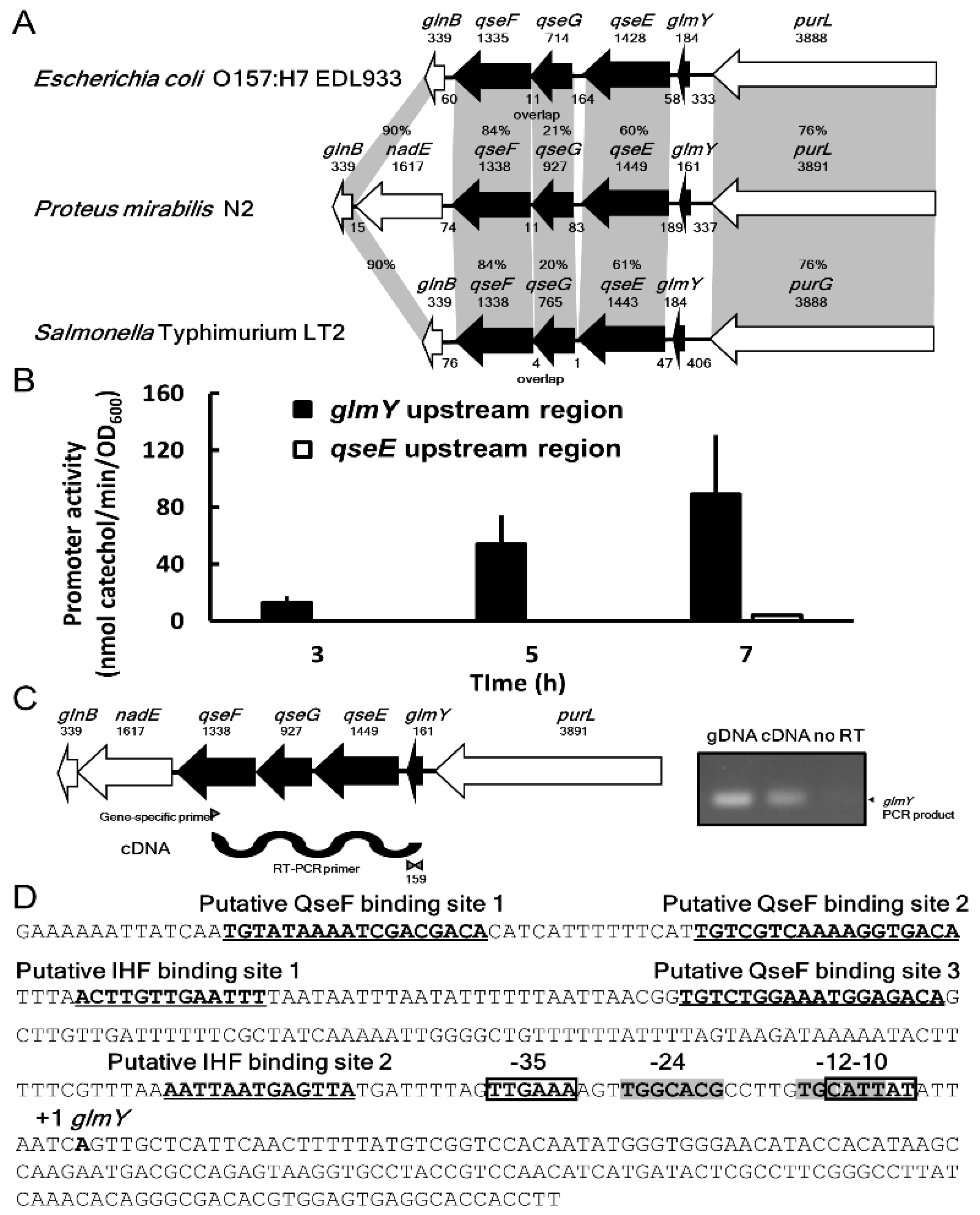
2.1. Identification of P. mirabilis qseEGF Gene Locus

Seeing that epinephrine and norepinephrine exist in urine, we sought QseBC and QseEF homologues in uropathogenic P. mirabilis N2. We identified a gene locus, purL-qseE-qseG-qseF-nadE-glnB, whose gene product with amino acid sequence identity of around 76, 60, 20, 84 and 90% to PurL, QseE, QseG, QseF and GlnB, respectively, with E. coli and Salmonella (both lacking nadE) (Figure 1A). We then tested whether epinephrine or norepinephrine has any effect on phenotypic traits of P. mirabilis. Neither hormone in the range 0.1–400 µM altered the phenotypes assayed (motility, biofilm formation, etc.) or showed any effect on expression of qseEF by real time RT-PCR.

2.2. Identification of P. mirabilis glmY and Co-Transcription of glmY, qseE, qseG and qseF

In view of conservation of the gene cluster glmY-glrK (qseE)-yfhG (qseG)-glrR (qseF)-glnB in Enterobacteriaceae [27] and modulation of motility [12] and pathogenesis [29,30] by GlmY, we searched the bacterial small RNA database (BSRD) for P. mirabilis GlmY counterpart and found it located upstream of qseE and downstream of purL (Figure 1A). We first examined the promoter activity in the upstream 548-bp region of qseE (nucleotide −553 to −6 of QseE start codon) by the xylE reporter assay. No promoter activity was found (Figure 1B). Then, we tested the glmY promoter activity using the upstream 379-bp DNA fragment (nucleotide −381 to −3 from the putative transcription start site) of glmY and found the promoter activity at 3, 5 and 7 h after incubation (Figure 1B). Furthermore, we demonstrated that glmY, qseE, qseG and qseF belong to a transcript by RT (reverse transcription)-PCR using qseF specific primer to obtain the cDNA for amplifying the glmY fragment (Figure 1C). The glmY PCR product of 159 bp was observed, but no product was produced in the no RT control (Figure 1C). These data indicate that glmY, qseE, qseG and qseF can share the same promoter upstream of glmY. The lack of promoter activity in the 548-bp DNA fragment upstream qseE results from the fragment containing only partial glmY promoter DNA sequences (201/379 bp). In summary, glmY, qseE, qseG and qseF are co-transcribed from the glmY promoter in P. mirabilis. In addition, we identified one overlapped σ7054, two IHF and three QseF putative binding sites in the upstream promoter region of glmY (Figure 1D).

2.3. Phenotypic Traits of glmY and qse Mutants

To investigate the roles of GlmY and QseEF TCS in P. mirabilis, we first generated isogenic mutants of glmY and qseF. Among the phenotypes assayed, both glmY and qseF mutant strains exhibited reduced swarming and swimming abilities relative to the wild-type and respective complemented strains (Figure 2A,D). Comparable growth of the wild-type and respective complemented strains was observed, and no growth defect was found in the mutant strains compared to the wild-type (data not shown). glmY and qseF mutants migrated much slower than the wild-type and respective complemented strains during the 8-h period after inoculation on the swarming plate. Mutants lacking qseE, qseG or qseEGF were also constructed with no growth defect compared to the wild-type bacteria. We found similar swarming and swimming patterns among qseF, qseE, qseG and qseEGF mutants (data not shown). Therefore, we investigated the QseEGF-mediated motility using qseF mutant to represent QseEF malfunction in the following experiments. Significantly reduced swarming-related phenotypes, including cell differentiation, hemolysin activity and flagellin level, were observed in the glmY and qseF mutants compared to the wild-type and respective complemented strains (Figure 2B,C,E). Transmission electron microscopy (TEM) also showed glmY and qseF mutant cells were shorter and had fewer flagella than the wild-type strain (Figure 2F).

2.4. Downregulation of glmYqseEGF Operon by qseF Deletion

Based on the presence of the putative QseF binding site in the glmYqseEGF promoter region (Figure 1D), we examined whether glmY expression is under the control of QseF by the reporter assay. Just as expected, qseF mutant had significantly lower glmY promoter activity compared to the wild-type and the qseF-complemented strain at 3, 5, 7 h after inoculation and incubation (Figure 3A).

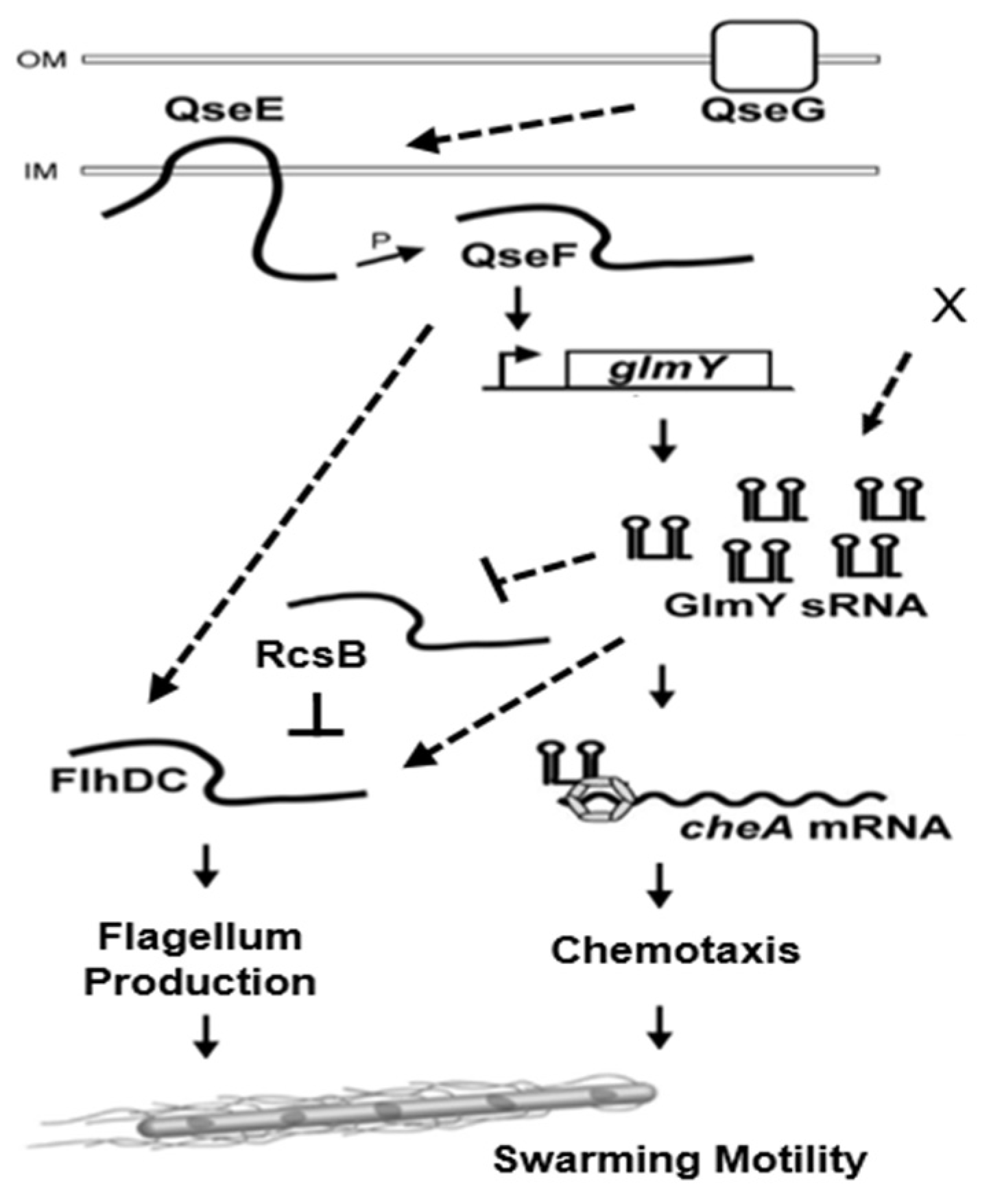
2.5. Altered Expression of flhDC, rcsB and cheA in qseF and glmY Mutants

With the notion that disruption of qseF, encoding a TCS transcriptional regulator QseF, impaired the swarming motility and related phenotypes in P. mirabilis, we thus examined the effect of qseF loss on flhDC expression by the reporter assay. The loss of qseF incurred significant reduction in flhDC promoter activity compared to the wild-type and the complemented strains at 3, 5, 7 h after inoculation (Figure 3B). Since QseF is a transcriptional regulator, we then tried to find the consensus QseF binding site (TGTCN10GACA) [31] in the promoter region of flhDC but failed. Since QseF could regulate glmY expression (Figure 3A) and both qseF and glmY mutants displayed decreased swarming and swimming motility (Figure 2A,D), we examined the promoter activity of flhDC in the glmY mutant. As is the case for qseF mutant, glmY mutant had a lower flhDC promoter activity than the wild-type and the complemented strains (Figure 3B). The mRNA level of flhDC in qseF and glmY mutants was consistent with the results of the reporter assay (Figure 3C).
Given the positive regulation of swarming and flhDC expression by both QseF and GlmY, the regulatory hierarchy from QseF to GlmY and the absence of QseF binding site in flhDC promoter region, we surmised that QseF may modulate swarming through GlmY to affect flhDC expression. How could GlmY, an sRNA, regulate both promoter activity and mRNA level of flhDC (Figure 3B,C)? In this respect, GlmY may modulate expression of a regulator which affects flhDC transcription directly and thus flhDC mRNA level. The direct regulation of flhDC mRNA level by GlmY may also contribute to the altered mRNA amount. As for flhDC regulator, P. mirabilis RcsB could modulate swarming by direct regulation of flhDC expression in a negative way [8,32] and rcsB overexpression led to a reduction of flhDC expression [33]. Therefore, we tested the effect of glmY mutation on rcsB mRNA level and found glmY mutant had increased rcsB mRNA level compared to the wild-type and glmY-complemented strain (Figure 3C). In view of regulation of glmY by QseF, qseF mutant exhibited a similar increase in rcsB mRNA level as the glmY mutant relative to the wild-type and the complemented strain (Figure 3C). On the other hand, we tried to identify whether GlmY could target flhDC or other mRNAs to modulate motility of P. mirabilis. We used the full sequence of GlmY as input to search for GlmY targets on the TargetRNA2 website. The tool did report a region in the 5′UTR of the cheA (but not flhDC or rcsB) as a candidate target with lower energy score and the p-value less than 0.05. The prediction shows interaction of GlmY (positions 17 to 28) with a 5′UTR region of cheA mRNA spanning −61 to −50 positions from the translation start site of cheA mRNA. CheA kinase, encoded by cheA, belongs to a family of two-component sensors responsible for bacterial chemotaxis [34]. As P. mirabilis GlmY positively regulated swarming (Figure 2A) and small RNAs exert positive regulation of virulence primarily at the level of mRNA stabilization [35], we assessed whether GlmY affects cheA mRNA level. As expected, the amount of cheA mRNA was reduced significantly in glmY and qseF mutants compared to the wild-type and respective complemented strains (Figure 3C).

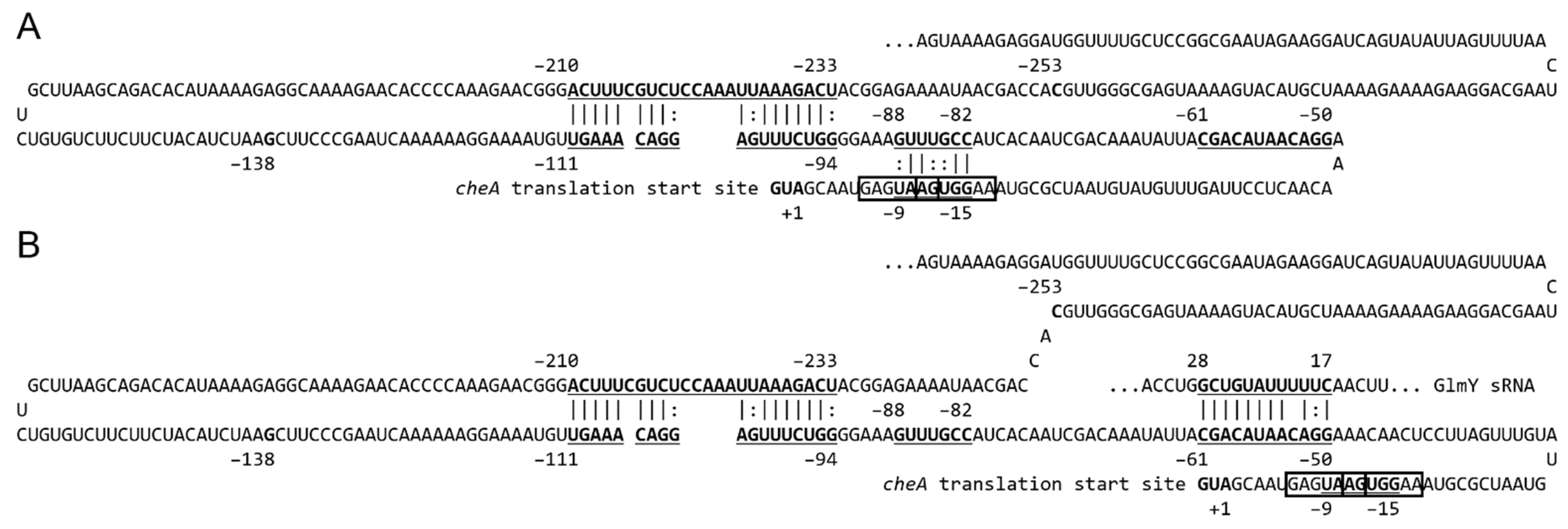
2.6. GlmY Activates cheA Expression at the Post-Transcriptional Level

To investigate the role of interaction between GlmY and cheA 5′UTR in cheA expression and thus swarming, we generated plasmids carrying the individual lac promoter-driven xylE translational fusion containing different lengths (520, 253, 138 and 61 bp) of cheA 5′UTR and the first 27 bp of cheA ORF (Figure 4A). The individual fusion plasmid was transformed into the wild-type strain and glmY mutant to probe the essential region of cheA 5′UTR requiring GlmY for activating cheA translation by monitoring the xylE activity. While the glmY mutant carrying the plasmid of 520, 253 or 138 bp-5′UTR translational fusion exhibited significantly lower xylE activity than the wild-type strain carrying the respective fusion plasmid, albeit to a lesser extent for the 138 bp-fusion plasmid (Figure 4B), the xylE activity of the wild-type strain and the glmY mutant harboring the 61 bp-fusion plasmid was comparable (Figure 4B). The results indicate that there is no need for GlmY to activate cheA translation when only the 61 bp-5′UTR of cheA is present. The data indicate all the 520, 253 and 138-bp fragments of cheA 5′UTR had constrained structures requiring GlmY for releasing to activate cheA translation.
To further demonstrate whether GlmY affected cheA expression via direct base-pairing, we performed site-directed mutagenesis to inactivate the interaction of GlmY and cheA 5′UTR. We introduced an 8-bp mutation into the predicted pairing regions of the cheA 5′UTR (cheAm) and glmY (GlmYm) (Figure 4C) using the cheA 5′UTR (253 bp)-xylE translational fusion plasmid (pcheA) and glmY-containing pBAD33 (pBglmY) as templates to generate pcheAm and pBglmYm, respectively. The wild-type P. mirabilis harboring the pcheAm exhibited significantly lower XylE activity compared to that harboring the wild-type cheA 5′UTR-xylE plasmid (pcheA) (Figure 4D). The glmY mutant carrying pBglmY and pcheAm showed significantly lower XylE activity than the mutant carrying pBglmY and pcheA (0.4 vs. 1 in Figure 4D). GlmY with the 8-bp substitution (GlmYm) could not effectively enhance the expression of cheA-xylE fusion to the extent of wild-type GlmY (0.6 vs. 1) in the glmY mutant (Figure 4D). In addition, introduction of compensating mutations into the cheA 5′UTR could restore the ability of GlmYm to activate the cheA-xylE fusion to 80% of the wild-type GlmY and cheA interaction (pBglmYm-pcheAm vs. pBglmY-pcheA in Figure 4D).
These results prove the role of cheA 5′ UTR in interaction with GlmY and the 8-bp direct pairing between GlmY and cheA 5′ UTR critical for facilitating expression of cheA. For the first time, this work identifies cheA as a novel target of GlmY for modulating swarming motility of P. mirabilis.

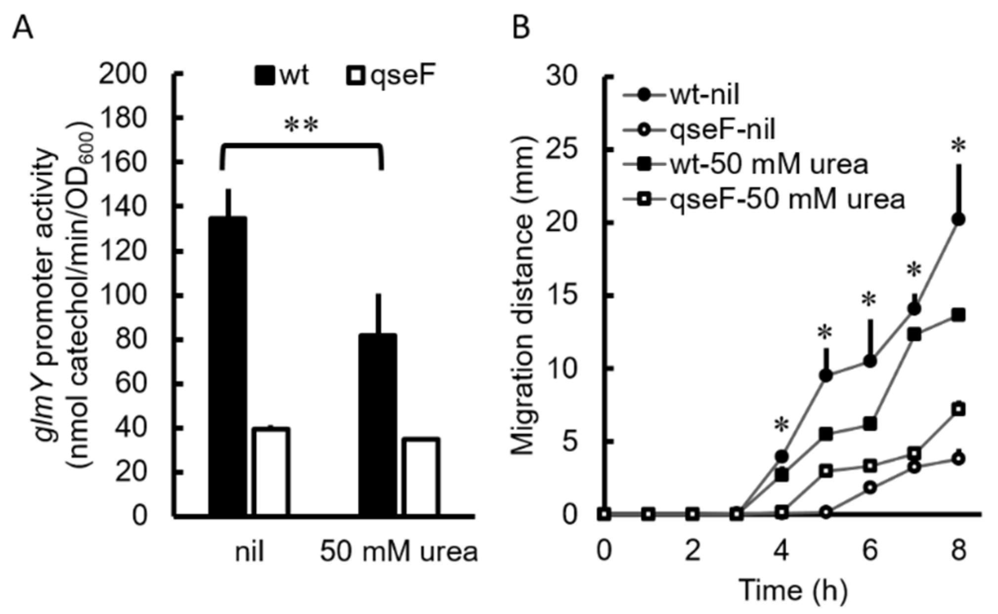
2.7. Urea Inhibited Promoter Activity of the glmYqseEGF Operon in P. mirabilis

Based on the fact that P. mirabilis swarming was subject to positive regulation of the QseF-GlmY pathway, it was tempting to determine whether urea affect the expression of glmYqseEGF operon in the urinary tract, an environment containing a lot of urea. Therefore, the XylE activities of the glmY-xylE reporter plasmid-transformed wild-type and the qseF mutant were monitored in the presence of urea. The promoter activity of the glmY operon was reduced by urea at 50 mM in the wild-type but not the qseF mutant after incubation for 5 h on the LB agar plate (Figure 5A). We then examined the swarming motility of the wild-type and qseF mutant in the presence of urea or not. The swarming motility of the wild-type but not the qseF mutant was decreased by urea at 50 mM (Figure 5B). The results suggest that urea could be a negative signal for expression of P. mirabilis glmYqseEGF operon in the urinary tract.

3. Discussion

For the first time, in this study, a TCS regulator QseF participating in modulation of swarming motility through GlmY (an sRNA) and the underlying mechanisms were revealed in uropathogenic P. mirabilis. GlmY regulated swarming through direct GlmY-cheA interaction together with GlmY-rcsB- and/or GlmY-mediated flhDC expression (Figure 6), whereby affecting swarming-related chemotaxis system and flagellum production (Figure 6). The chemotaxis system plays an essential role in flagellar function and swarm cell differentiation, thus important for P. mirabilis to display a vigorous swarming pattern [34,36]. Given decreased not abolished GlmY expression upon loss of qseF (Figure 3A), this indicates GlmY is subject to QseF-independent control. Moreover, the finding that loss of glmY or qseF resulting in a similar motility ability, expression of flhDC and rcsB and flhDC-associated phenotypes (cell length and flagellin level) (Figure 2 and Figure 3) indicates QseF could have alternative regulation of flhDC expression bypassing GlmY (Figure 6). In this regard, our preliminary data showed introduction of GlmY-expressing plasmid into qseF mutant could not restore flhDC mRNA to the wild-type level.
An ever-increasing number and variety of sRNAs are being identified to serve regulatory functions for bacteria to respond to environmental cues and thrive in diverse habitats [9,10,11,37]. E. coli GlmY has been coopted to modulate the expression of virulence and be involved in cellular metabolism and architecture, including for biosynthesis of LPS [38,39,40], a permeability barrier and a major virulence determinant in pathogenic bacteria [41]. GlmY fine-tunes expression of type III secretion system and its effectors to promote bacterial attachment and subsequent actin rearrangement on host cells through post-transcriptional control of EspFu and the locus-of-enterocyte-effacement (LEE) [29]. In addition, GlmY and GlmZ participate in gene expression of curli adhesion, acid resistance and also tryptophan metabolism [42]. Despite similar genomic gene arrangement of glmYqseEGF, there are discrepancies of the QseF and GlmY-related regulation between P. mirabilis and E. coli. First, a Rho-independent terminator exists in the end of E. coli GlmY [43] but not in that of P. mirabilis. Second, neither promoter prediction nor reporter assay showed a promoter present in the intergenic region between glmY and qseE of P. mirabilis, which is not the case for E. coli, initiating qseE transcription in the glmY-qseE intergenic region [31]. Third, E. coli QseEF is involved in regulating genes required for pedestal formation but not motility [21]. There was no difference in motility and flagellar expression as seen by Western blotting between the wild-type E. coli strain and the qseE mutant [21], contrary to the similar defect of both P. mirabilis qseF and qseE mutants in swarming and swimming abilities. E. coli glmY and qseE are independently transcribed from different promoters [31], while we demonstrated that glmY and qseEGF constitute an operon by RT-PCR assay (Figure 1C). In this regard, the presence of the conserved RNase E-cleavage motif GCCUUAU in GlmY of P. mirabilis [38] indicates the glmYqseEGF transcript could be processed to produce effective GlmY for its function.
We found swarming, swimming and swarming-related phenotypes (cell length, haemolysin activity and flagellin level) were all reduced in both qseF and glmY mutants (Figure 2). Both flhDC promoter activity and mRNA level were downregulated in qseF and glmY mutants (Figure 3). Because no QseF binding site in the promoter region of flhDC, we speculated QseF may exert its effect on flhDC expression through GlmY. TargetRNA2 tool, focusing its search for an sRNA-mRNA interaction in a neighborhood around the rbs of the mRNA, revealed no GlmY binding site in flhDC and rcsB mRNAs. We then extended mRNA searching by IntaRNA for GlmY-interacting sites and binding sites of −101 to −92 and −284 to −271 from AUG (both outside rbs) for flhDC and rcsB, respectively, were identified. Based on the GlmY target prediction, there are two possibilities for upregulation of flhDC mRNA level by GlmY. One is by direct interaction with flhDC mRNA; the other is indirectly through rcsB. It also can not be ruled out that both modes of action coexist. Further studies are needed to investigate whether GlmY directly interacts with mRNAs of flhDC and rcsB. For example, the flhDC (or rcsB) mRNA amount of the glmY mutant harboring wild-type or interacting site-mutated glmY on a plasmid will be determined to assess the GlmY-flhDC (or rcsB) mRNA interaction. It is noteworthy that overexpression of glmY in the same E. coli strain either had no effect or resulted in motility repression [12,44]. It could be due to different plasmid vectors and assay conditions used.
Translational fusion assay indicated that regions of −520, −253 and −138 to +27 from AUG of cheA mRNA contain constrained secondary structures needed to be resolved by GlmY for cheA translation, albeit to a lesser extent for the region of −138 to +27 (Figure 4B). To elucidate how cheA 5′UTR interacts with GlmY, we uploaded sequences from −253 to −138 and −138 to +27 of cheA mRNA to the IntaRNA webserver and interaction between regions of −233 to −211 and −111 to −94 from AUG was revealed (Figure 7). Likewise, using sequences from −138 to −61 and −61 to +27 of cheA mRNA as input fragments showed interaction between regions of −88 to −82 and −15 to −9 by IntaRNA (Figure 7). Inspection of the sequence of −15 to −9 disclosed the existence of two overlapped putative ribosome binding sites (rbs), aagguga (gagguga in E. coli) and gaaugag (gaagga in E. coli) for translation of long cheA and short cheA of E. coli [45], respectively (Figure 7). Furthermore, IntaRNA revealed interaction of cheA −61 to −50 from AUG with GlmY +17 to +28 (Figure 7) by using full-length GlmY and −253 to +27 of cheA as the input fragments. These data suggest interaction between regions of −88 to −82 and −15 to −9 from AUG could inhibit cheA translation by hiding rbs. Hence GlmY is required for releasing the secondary structure to assist cheA translation. The reason for the less extent of translation affected in the absence of glmY using the translational fusion comprising −138 to +27 of cheA compared to the −253 to +27 of cheA fusion could be ascribed to the interaction between regions of −233 to −211 and −111 to −94 from AUG (Figure 7), thereby affecting the ease for GlmY to uncover the rbs for translation. It is interesting to know that GlmY not only activated cheA translation but also maintained cheA mRNA level. This is in line with that sRNA-mediated stability control is the crucial element of activation of trans-encoded mRNAs [35].
EHEC regulated pathogenesis and motility by sensing epinephrine or norepinephrine through QseBC and QseEF two-component signaling systems [21,22]. The membrane kinase QseC autophosphorylates and phosphorylates the QseB response regulator initiating a signaling cascade that activates QseEF to trigger expression of LEE genes, leading to AE lesions on intestinal epithelial cells. In P. mirabilis genome, only QseEF homologue of high similarity was found, with no homologue of QseBC existing. We found QseF participated in swarming regulation and deletion of qseE or qseG also had decreased swarming motility. Previous study has revealed the phosphorylation state of QseE and QseF is governed by interaction with QseG in response to epinephrine for post-transcriptional regulation of virulence genes through GlmY [46]. In addition, QseEGF has been shown to modulate transcription of phoPQ, linking to the virulence regulation [47]. The similarity of the transcriptome profiles of qseE, qseF and qseG mutants also indicates that these proteins work together [47]. Therefore, P. mirabilis QseEF likely should exert functions other than swarming to affect virulence. Accordingly, our preliminary data showing qseF mutant had a significantly impaired ability to colonize mouse bladders and kidneys compared to the wild-type. The investigation of virulence traits such as cytotoxicity, urothelial cell invasion and survival in macrophages is underway.
We found that urea could serve as a negative signal of QseEF for swarming (Figure 5). In this way, the expression of QseF will be inhibited in the urine (rich in urea) and the role of QseF in facilitating swarming will be neglected. In view of our preliminary data showing a significant difference between the wild-type and qseF mutant in colonization of mouse bladders and kidneys, it is tempting to surmise there is other cues in the urine to increase expression of qseF. In this aspect, our unpublished transcriptome data reveal mRNA levels of fatty acid synthetic genes increase but those of degradative genes decrease in the qseF mutant relative to the wild-type. This indicates QseF may sense fatty acid and be involved in fatty acid metabolism. Interestingly, we found oleic acid (0.01%) is a positive signal for qseF expression (data not shown) and swarming motility of P. mirabilis [48]. The oleic acid concentration used is an attainable concentration in the urine according to the Human Metabolome Database (https://hmdb.ca/metabolites?utf8=%E2%9C%93&quantified=1&urine=1&filter=true, accessed on 29 November 2021). Given QseE is also phosphatase [23], it is reasonable to infer that the phosphatase function of QseE prevents QseF activation under the presence of urea, whereas its kinase function triggers QseF regulon expression under appropriate concentrations of oleic acid. A finely tuned balance in these opposing activities should determine the regulon response of QseF as the case of the bifunctional DevS kinase responsive to environmental oxygen in Mycobacterium tuberculosis [49].
Since catheter-associated UTI (CAUTI) is a major health concern, research directed at understanding the pathogenesis is warranted and should lead to improved diagnosis, prevention and treatment. P. mirabilis is notorious for causing CAUTIs. In this work, we demonstrated a new regulatory pathway involving an sRNA, GlmY, participating in swarming motility of P. mirabilis during which expression of several virulence genes is increased. It is believed that P. mirabilis swarming up catheters is primed to infect the urinary tract [50], so elucidating the swarming mechanisms could provide new approaches in the development of intervention strategies and facilitate the discovery of novel therapeutics.

4. Materials and Methods

4.1. Bacterial Strains, Plasmids, Reagents and Growth Conditions

The bacterial strains and plasmids used in this study are listed in Table S1 in the Supplementary Materials. The bacterial strains used are a clinical isolate from a patient of UTI (the wild-type N2), its derived mutants and respective complemented strains. All chemicals were obtained from the Sigma-Aldrich unless otherwise indicated, and primer sequences are given in Table S2. Bacteria were stored at −80 °C and routinely cultured in Luria-Bertani (LB) broth at 37 °C. The LSW- agar plate [15] was used to prevent the phenotypic expression of swarming motility for selecting mutant clones and colony counting.

4.2. Construction of P. mirabilis Mutants and Complemented Strains

Sequences flanking the qseF gene was amplified by PCR using the primer pairs qseF-upF/XbaI-qseF-upR and XbaI-qseF-dnF/qseF-dnR for qseF mutant, and cloned into pGEM-T Easy (Promega) to generate pGqseF-up and pGqseF-dn. pGqseF-up was digested with SalI/XbaI, and the qseF upstream sequence-containing fragment was ligated to SalI/XbaI-digested pGqseF-dn to produce the pGqseF-updn plasmid, which contains both upstream and downstream sequences of qseF. A Kmr cassette was inserted in the XbaI-digested pGqseF-updn plasmid to generate pGqseF-updn-Km. The DNA fragment containing the Kmr cassette-disrupted combined upstream and downstream sequences of qseF was cleaved by SalI/SphI from pGqseF-updn-Km, and ligated into SalI/SphI-cleaved pUT-Km1 to generate pUTqseF-Km. For glmY mutant, pUTglmY-Km was constructed in a similar way except using primer pairs glmY-upF/XbaI-glmY-upR and glmY-dnF/glmY-dnR. For gene inactivation by homologous recombination, pUTqseF-Km or pUTglmY-Km was transferred to wild-type P. mirabilis N2 by conjugation. Transconjugants were spread on LSW- plates containing tetracycline (20 μg/mL) and kanamycin (100 μg/mL), and confirmation of mutants with double-crossover events by colony PCR and Southern blot hybridization were performed. For complementation of mutants, the fragments containing full-length qseF gene or glmY was amplified by PCR using primer pairs qseF-comF/qseF-comR or glmY-comF/glmY-comR, and cloned into pGEM-T easy to generate the plasmid pGEM-qseF or pGEM-glmY. qseF is driven by lac promoter. glmY is driven by its own promoter (382 bp), divergent from lac promoter, to ensure expression of the correct transcript. pGEM-qseF or pGEM-glmY was then transformed into the respective mutant to generate the complemented strain.

4.3. Swarming and Swimming Assays

The swarming migration assays were performed as described previously [48]. The overnight bacterial cultures (5 µL) were inoculated onto the center of LB swarming plates containing 1.5% (wt/vol) agar, which were then incubated, and the swarming migration distance was measured by monitoring the swarm fronts of the bacterial cells at 1-h intervals. For swimming assays, the overnight culture was stabbed into the center of the swimming plates containing 0.3% agar and migration distance was recorded after incubation for 16 h at 37 °C.

4.4. Measurement of the Haemolysin Activity and Cell Length

The overnight bacterial cultures (120 μL) were inoculated onto the surface of LB swarming plates, which were then incubated at 37 °C for 5 h. The hemolysin activity and cell length were determined as described previously [48]. Cell-associated haemolytic activity was determined by incubation of a 20-μL cell suspension (OD600nm = 0.5) in a 980-μL solution of 0.85% NaCl, 20 mM CaCl2 and 2% washed sheep erythrocytes at 42 °C for 15 min. After centrifugation, the amount of haemoglobin released by lysis was measured by the increase of the optical density of the assay supernatant at 543 nm.

4.5. Measurement of the Flagellin Level

Flagellin levels were determined as described previously by SDS-PAGE and Coomassie brilliant blue staining [15]. Western blotting was performed to confirm the flagellin band. The flagellin samples on the SDS-PAGE gel were transferred to an Hybond-P membrane (GE Healthcare, Chicago, IL, USA). The blot was incubated with mouse polyclonal antiserum against FlaA, followed by sheep anti-mouse IgG conjugated with horseradish peroxidase (GE Healthcare, Chicago, IL, USA), and then developed using enhanced chemiluminescence detection reagents (PerkinElmer, Waltham, MA, USA).

4.6. Transmission Electron Microscopy

Transmission electron microscopy (TEM) was performed as previously described [3] by using 1% phosphotungstic acid (PTA)-stained bacteria on a carbon-coated grid and TEM pictures were obtained with a Hitachi H-7100 electron microscope (Hitachi High-Tech America, Pleasanton, CA, USA).

4.7. Real Time Reverse Transcription PCR (RT-PCR)

To study the effect of qseF or glmY deletion on the mRNA amount of motility-related genes, overnight LB cultures (100 μL) of the wild-type, mutants and the complemented strains were spread on the LB agar and incubated for 5 h at 37 °C. Total RNA was extracted, and real time RT-PCR was carried out as described previously [3] to measure the mRNA level using primer pairs listed in Table S2. The levels of RNAs were normalized against housekeeping gene gyrB mRNA.

4.8. Transcriptional and Translational Reporter Assays

For transcriptional reporter assay, the promoter region of the gene was amplified by SphI and PstI-included primers and cloned into pGEM-T Easy. These promoter-containing plasmids were cut by SphI and PstI, and the promoter-containing fragment was ligated, respectively to the xylE-containing pACYC184-xylE digested by SphI and PstI to construct the transcriptional reporter plasmid. The XylE activity of the transcriptional reporter plasmid-transformed wild-type and mutants was measured as described previously [3]. For translational reporter assay, the 5′UTR (520, 253, 138 or 61 bp) with 27 bp coding region (5′UTR-27) of cheA gene was amplified by SacI and BglII-included primers and cloned into pGEM-T Easy to generate the plasmid pGcheA-5′UTR. The fragment containing full-length xylE gene was amplified by PCR using primer pairs BglII-xylE-F and xylE-R, and cloned into pGEM-T Easy to generate the plasmid pGxylE. pGcheA-5′UTR was digested with SacI and BglII, and the cheA 5′UTR-27 sequence-containing fragment was ligated to SacI/BglII-digested pGxylE to produce the translational reporter plasmid pcheA of in-frame cheA-xylE fusion driven by lac promoter. The wild-type and mutants transformed with the translational reporter plasmid were grown overnight in LB broth containing ampicillin (100 μg/mL). Then, the cultures (100 μL) were spread onto the LB agar plate and incubated for 5 h at 37 °C before the XylE activity was measured.

4.9. Site-Directed Mutagenesis

The translational reporter plasmid pGEM-cheA-xylE (pcheA) and the glmY-harboring pBAD plasmid (pBglmY)) containing mutations in the putative GlmY- and cheA 5′UTR-interacting site, respectively, were generated (pcheAm and pBglmYm) by a KOD-Plus-mutagenesis kit (Toyobo, Osaka, Japan) using primers listed in Table S2 according to the manufacturer’s protocol. The inverse PCR products of pcheA and pBglmY were digested by DpnI to remove the template plasmid DNA, self-ligation of PCR products was performed using T4 polynucleotide kinase and ligase and then DNA sequencing was performed to confirm the DNA sequence of the mutated sites. We introduced pcheA or pcheAm into the wild-type and compared the XylE activity to confirm the essential GlmY-interacting site in cheA 5′UTR. Additionally, we transformed the combinations of plasmids pBglmY-pcheA, pBglmY-pcheAm, pBglmYm-pcheA or pBglmYm-pcheAm into the glmY mutant and determined the XylE activity to evaluate the direct interaction of cheA 5′UTR and GlmY.

Supplementary Materials

The following supporting information can be downloaded at: https://0-www-mdpi-com.brum.beds.ac.uk/article/10.3390/ijms23010487/s1.

Author Contributions

Conceptualization, S.-J.L.; investigation, W.-Y.L., Y.-L.T., P.-Y.S. and T.-S.L.; formal analysis, W.-Y.L., Y.-L.T. and P.-H.Y.; resources, Y.-J.L.; data curation, W.-Y.L., Y.-L.T., Y.-J.L., P.-H.Y. and S.-J.L.; supervision, S.-J.L.; writing—original draft preparation, S.-J.L. and W.-Y.L.; writing—review and editing, S.-J.L.; project administration, S.-J.L.; funding acquisition, S.-J.L. and Y.-J.L. All authors have read and agreed to the published version of the manuscript.

Funding

This research was funded by the Ministry of Science and Technology (MOST), Taiwan (grant number: MOST 105-2320-B-002-050-MY3 to S.-J.L.).

Institutional Review Board Statement

Not applicable.

Informed Consent Statement

Not applicable.

Data Availability Statement

Not applicable.

Acknowledgments

We thank the Graduate Institute of Anatomy and Cell Biology, College of Medicine, National Taiwan University for providing assistance in TEM examination.

Conflicts of Interest

The authors declare no conflict of interest. The funders had no role in the design of the study, in the collection, analyses or interpretation of data, in the writing of the manuscript or in the decision to publish the results.

References

  1. Norsworthy, A.N.; Pearson, M.M. From Catheter to Kidney Stone: The Uropathogenic Lifestyle of Proteus mirabilis. Trends Microbiol. 2017, 25, 304–315. [Google Scholar] [CrossRef] [Green Version]
  2. Jansen, A.M.; Lockatell, V.; Johnson, D.E.; Mobley, H.L. Mannose-resistant Proteus-like fimbriae are produced by most Proteus mirabilis strains infecting the urinary tract, dictate the in vivo localization of bacteria, and contribute to biofilm formation. Infect. Immun. 2004, 72, 7294–7305. [Google Scholar] [CrossRef] [PubMed] [Green Version]
  3. Tsai, Y.L.; Chien, H.F.; Huang, K.T.; Lin, W.Y.; Liaw, S.J. cAMP receptor protein regulates mouse colonization, motility, fimbria-mediated adhesion, and stress tolerance in uropathogenic Proteus mirabilis. Sci. Rep. 2017, 7, 7282. [Google Scholar] [CrossRef] [Green Version]
  4. Nielubowicz, G.R.; Mobley, H.L. Host-pathogen interactions in urinary tract infection. Nat. Rev. Urol. 2010, 7, 430–441. [Google Scholar] [CrossRef] [PubMed]
  5. Armbruster, C.E.; Mobley, H.L. Merging mythology and morphology: The multifaceted lifestyle of Proteus mirabilis. Nat. Rev. Microbiol. 2012, 10, 743–754. [Google Scholar] [CrossRef]
  6. Jacobsen, S.M.; Stickler, D.J.; Mobley, H.L.; Shirtliff, M.E. Complicated catheter-associated urinary tract infections due to Escherichia coli and Proteus mirabilis. Clin. Microbiol. Rev. 2008, 21, 26–59. [Google Scholar] [CrossRef] [PubMed] [Green Version]
  7. Fraser, G.M.; Hughes, C. Swarming motility. Curr. Opin. Microbiol. 1999, 2, 630–635. [Google Scholar] [CrossRef]
  8. Clemmer, K.M.; Rather, P.N. Regulation of flhDC expression in Proteus mirabilis. Res. Microbiol. 2007, 158, 295–302. [Google Scholar] [CrossRef]
  9. Gottesman, S.; Storz, G. Bacterial small RNA regulators: Versatile roles and rapidly evolving variations. Cold Spring Harb. Perspect. Biol. 2011, 3, a003798. [Google Scholar] [CrossRef] [Green Version]
  10. Waters, L.S.; Storz, G. Regulatory RNAs in bacteria. Cell 2009, 136, 615–628. [Google Scholar] [CrossRef] [PubMed] [Green Version]
  11. Guillier, M.; Gottesman, S.; Storz, G. Modulating the outer membrane with small RNAs. Genes Dev. 2006, 20, 2338–2348. [Google Scholar] [CrossRef] [Green Version]
  12. Bak, G.; Lee, J.; Suk, S.; Kim, D.; Young Lee, J.; Kim, K.S.; Choi, B.S.; Lee, Y. Identification of novel sRNAs involved in biofilm formation, motility, and fimbriae formation in Escherichia coli. Sci. Rep. 2015, 5, 15287. [Google Scholar] [CrossRef] [PubMed] [Green Version]
  13. Kulesus, R.R.; Diaz-Perez, K.; Slechta, E.S.; Eto, D.S.; Mulvey, M.A. Impact of the RNA chaperone Hfq on the fitness and virulence potential of uropathogenic Escherichia coli. Infect. Immun. 2008, 76, 3019–3026. [Google Scholar] [CrossRef] [Green Version]
  14. Chao, Y.; Vogel, J. The role of Hfq in bacterial pathogens. Curr. Opin. Microbiol. 2010, 13, 24–33. [Google Scholar] [CrossRef] [PubMed]
  15. Wang, M.C.; Chien, H.F.; Tsai, Y.L.; Liu, M.C.; Liaw, S.J. The RNA chaperone Hfq is involved in stress tolerance and virulence in uropathogenic Proteus mirabilis. PLoS ONE 2014, 9, e85626. [Google Scholar] [CrossRef]
  16. Vogel, J.; Luisi, B.F. Hfq and its constellation of RNA. Nat. Rev. Microbiol. 2011, 9, 578–589. [Google Scholar] [CrossRef] [PubMed] [Green Version]
  17. Valverde, C.; Haas, D. Small RNAs controlled by two-component systems. Adv. Exp. Med. Biol. 2008, 631, 54–79. [Google Scholar] [PubMed]
  18. Vogel, J. A rough guide to the non-coding RNA world of Salmonella. Mol. Microbiol. 2009, 71, 1–11. [Google Scholar] [CrossRef]
  19. Overgaard, M.; Kallipolitis, B.; Valentin-Hansen, P. Modulating the bacterial surface with small RNAs: A new twist on PhoP/Q-mediated lipopolysaccharide modification. Mol. Microbiol. 2009, 74, 1289–1294. [Google Scholar] [CrossRef]
  20. Coornaert, A.; Lu, A.; Mandin, P.; Springer, M.; Gottesman, S.; Guillier, M. MicA sRNA links the PhoP regulon to cell envelope stress. Mol. Microbiol. 2010, 76, 467–479. [Google Scholar] [CrossRef] [Green Version]
  21. Reading, N.C.; Torres, A.G.; Kendall, M.M.; Hughes, D.T.; Yamamoto, K.; Sperandio, V. A novel two-component signaling system that activates transcription of an enterohemorrhagic Escherichia coli effector involved in remodeling of host actin. J. Bacteriol. 2007, 189, 2468–2476. [Google Scholar] [CrossRef] [Green Version]
  22. Njoroge, J.; Sperandio, V. Enterohemorrhagic Escherichia coli virulence regulation by two bacterial adrenergic kinases, QseC and QseE. Infect. Immun. 2012, 80, 688–703. [Google Scholar] [CrossRef] [Green Version]
  23. Moreira, C.G.; Sperandio, V. Interplay between the QseC and QseE bacterial adrenergic sensor kinases in Salmonella enterica serovar Typhimurium pathogenesis. Infect. Immun. 2012, 80, 4344–4353. [Google Scholar] [CrossRef] [Green Version]
  24. Moreira, C.G.; Russell, R.; Mishra, A.A.; Narayanan, S.; Ritchie, J.M.; Waldor, M.K.; Curtis, M.M.; Winter, S.E.; Weinshenker, D.; Sperandio, V. Bacterial adrenergic sensors regulate virulence of enteric pathogens in the gut. mBio 2016, 7, e00826-16. [Google Scholar] [CrossRef] [Green Version]
  25. Cameron, E.A.; Gruber, C.C.; Ritchie, J.M.; Waldor, M.K.; Sperandio, V. The QseG lipoprotein impacts the virulence of enterohemorrhagic Escherichia coli and Citrobacter rodentium and regulates flagellar phase variation in Salmonella enterica serovar Typhimurium. Infect. Immun. 2018, 86, e00936-17. [Google Scholar] [CrossRef] [Green Version]
  26. Reading, N.C.; Rasko, D.A.; Torres, A.G.; Sperandio, V. The two-component system QseEF and the membrane protein QseG link adrenergic and stress sensing to bacterial pathogenesis. Proc. Natl. Acad. Sci. USA 2009, 106, 5889–5894. [Google Scholar] [CrossRef] [PubMed] [Green Version]
  27. Gopel, Y.; Luttmann, D.; Heroven, A.K.; Reichenbach, B.; Dersch, P.; Gorke, B. Common and divergent features in transcriptional control of the homologous small RNAs GlmY and GlmZ in Enterobacteriaceae. Nucleic Acids Res. 2011, 39, 1294–1309. [Google Scholar] [CrossRef] [Green Version]
  28. Lustri, B.C.; Sperandio, V.; Moreira, C.G. Bacterial chat: Intestinal metabolites and signals in host-microbiota-pathogen interactions. Infect. Immun. 2017, 85, e00476-17. [Google Scholar] [CrossRef] [PubMed] [Green Version]
  29. Gruber, C.C.; Sperandio, V. Posttranscriptional control of microbe-induced rearrangement of host cell actin. mBio 2014, 5, e01025-13. [Google Scholar] [CrossRef] [Green Version]
  30. Bennett, A.M.; Shippy, D.C.; Eakley, N.; Okwumabua, O.; Fadl, A.A. Functional characterization of glucosamine-6-phosphate synthase (GlmS) in Salmonella enterica serovar Enteritidis. Arch. Microbiol. 2016, 198, 541–549. [Google Scholar] [CrossRef] [PubMed]
  31. Reichenbach, B.; Gopel, Y.; Gorke, B. Dual control by perfectly overlapping sigma 54- and sigma 70- promoters adjusts small RNA GlmY expression to different environmental signals. Mol. Microbiol. 2009, 74, 1054–1070. [Google Scholar] [CrossRef]
  32. Howery, K.E.; Clemmer, K.M.; Rather, P.N. The Rcs regulon in Proteus mirabilis: Implications for motility, biofilm formation, and virulence. Curr. Genet. 2016, 62, 775–789. [Google Scholar] [CrossRef]
  33. Liu, M.C.; Lin, S.B.; Chien, H.F.; Wang, W.B.; Yuan, Y.H.; Hsueh, P.R.; Liaw, S.J. 10′(Z),13′(E)-heptadecadienylhydroquinone inhibits swarming and virulence factors and increases polymyxin B susceptibility in Proteus mirabilis. PLoS ONE 2012, 7, e45563. [Google Scholar] [CrossRef]
  34. Burkart, M.; Toguchi, A.; Harshey, R.M. The chemotaxis system, but not chemotaxis, is essential for swarming motility in Escherichia coli. Proc. Natl. Acad. Sci. USA 1998, 95, 2568–2573. [Google Scholar] [CrossRef] [Green Version]
  35. Podkaminski, D.; Vogel, J. Small RNAs promote mRNA stability to activate the synthesis of virulence factors. Mol. Microbiol. 2010, 78, 1327–1331. [Google Scholar] [CrossRef] [PubMed]
  36. Verstraeten, N.; Braeken, K.; Debkumari, B.; Fauvart, M.; Fransaer, J.; Vermant, J.; Michiels, J. Living on a surface: Swarming and biofilm formation. Trends Microbiol. 2008, 16, 496–506. [Google Scholar] [CrossRef] [PubMed]
  37. Valentin-Hansen, P.; Johansen, J.; Rasmussen, A.A. Small RNAs controlling outer membrane porins. Curr. Opin. Microbiol. 2007, 10, 152–155. [Google Scholar] [CrossRef]
  38. Gopel, Y.; Papenfort, K.; Reichenbach, B.; Vogel, J.; Gorke, B. Targeted decay of a regulatory small RNA by an adaptor protein for RNase E and counteraction by an anti-adaptor RNA. Genes Dev. 2013, 27, 552–564. [Google Scholar] [CrossRef] [Green Version]
  39. Gopel, Y.; Khan, M.A.; Gorke, B. Menage a trois: Post-transcriptional control of the key enzyme for cell envelope synthesis by a base-pairing small RNA, an RNase adaptor protein, and a small RNA mimic. RNA Biol. 2014, 11, 433–442. [Google Scholar] [CrossRef] [PubMed]
  40. Urban, J.H.; Vogel, J. Two seemingly homologous noncoding RNAs act hierarchically to activate glmS mRNA translation. PLoS Biol. 2008, 6, e64. [Google Scholar] [CrossRef]
  41. Klein, G.; Raina, S. Regulated assembly of LPS, its structural alterations and cellular response to LPS defects. Int. J. Mol. Sci. 2019, 20, 356. [Google Scholar] [CrossRef] [Green Version]
  42. Gruber, C.C.; Sperandio, V. Global analysis of posttranscriptional regulation by GlmY and GlmZ in enterohemorrhagic Escherichia coli O157:H7. Infect. Immun. 2015, 83, 1286–1295. [Google Scholar] [CrossRef] [Green Version]
  43. Urban, J.H.; Papenfort, K.; Thomsen, J.; Schmitz, R.A.; Vogel, J. A conserved small RNA promotes discoordinate expression of the glmUS operon mRNA to activate GlmS synthesis. J. Mol. Biol. 2007, 373, 521–528. [Google Scholar] [CrossRef]
  44. De Lay, N.; Gottesman, S. A complex network of small non-coding RNAs regulate motility in Escherichia coli. Mol. Microbiol. 2012, 86, 524–538. [Google Scholar] [CrossRef] [PubMed] [Green Version]
  45. McNamara, B.P.; Wolfe, A.J. Coexpression of the long and short forms of CheA, the chemotaxis histidine kinase, by members of the family Enterobacteriaceae. J. Bacteriol. 1997, 179, 1813–1818. [Google Scholar] [CrossRef] [PubMed] [Green Version]
  46. Göpel, Y.; Görke, B. Interaction of lipoprotein QseG with sensor kinase QseE in the periplasm controls the phosphorylation state of the two-component system QseE/QseF in Escherichia coli. PLoS Genet. 2018, 14, e1007547. [Google Scholar] [CrossRef] [PubMed]
  47. Reading, N.C.; Rasko, D.; Torres, A.G.; Sperandio, V. A transcriptome study of the QseEF two-component system and the QseG membrane protein in enterohaemorrhagic Escherichia coli O157:H7. Microbiology 2010, 156, 1167–1175. [Google Scholar] [CrossRef] [PubMed] [Green Version]
  48. Liaw, S.J.; Lai, H.C.; Wang, W.B. Modulation of swarming and virulence by fatty acids through the RsbA protein in Proteus mirabilis. Infect. Immun. 2004, 72, 6836–6845. [Google Scholar] [CrossRef] [Green Version]
  49. Kaur, K.; Kumari, P.; Sharma, S.; Sehgal, S.; Tyagi, J.S. DevS/DosS sensor is bifunctional and its phosphatase activity precludes aerobic DevR/DosR regulon expression in Mycobacterium tuberculosis. FEBS J. 2016, 283, 2949–2962. [Google Scholar] [CrossRef] [Green Version]
  50. Schaffer, J.N.; Pearson, M.M. Proteus mirabilis and urinary tract infections. Microbiol. Spectr. 2015, 3, 1–39. [Google Scholar] [CrossRef] [Green Version]
Figure 1. The glmYqseEGF gene locus in P. mirabilis N2. (A) The P. mirabilis glmYqseEGF gene locus corresponds to the similar locus in E. coli O157:H7 EDL933 and Salmonella Typhimurium LT2 with corresponding genes in shadows. An amino acid sequence analysis of the locus in these strains was performed using position-specific iterative BLAST. The percent amino acid identities of PurL, QseE, QseG, QseF and GlnB between P. mirabilis N2 and E. coli O157:H7 EDL933 or Salmonella Typhimurium LT2 are shown. The number above each arrow represents the gene length (bp). The intergenic space or overlap in terms of base pairs is also shown. (B) The promoter activities of glmY (−381 to −3 from putative glmY transcriptional start site) and qseE (−553 to −6 from qseE start codon) upstream region. (C) The glmY-qseE-qseG-qseF constitute an operon by gene-specific reverse transcription PCR. cDNA was synthesized by the qseF-specific primer and then the glmY DNA fragment was amplified by PCR. no RT, negative control; gDNA (genomic DNA), positive control. (D) The sequence of glmYqseEGF promoter region in P. mirabilis N2. The putative QseF and IHF binding sites are bold and underlined. The conserved σ70 and σ54 binding sites are indicated in boxes and shadows, respectively.
Ijms 23 00487 g001
Figure 2. GlmY and QseF regulated swarming and related phenotypes. Swarming motility (A), cell length (B), hemolysin activity (C) and swimming motility (D) were determined in the wild-type, mutants of qseF and glmY and complemented strains. Swarming migration was monitored at 1-h intervals, and swimming motility was determined after incubation for 16 h. Cell length and hemolysin activity were determined at 5 h after incubation on the LB agar plate. The data are the averages and standard deviations of three independent experiments. Significant difference of qseF or glmY mutant from the wild-type at 4, 5, 6, 7 and 8 h is indicated (* p < 0.05; ** p < 0.01; *** p < 0.001 by the Student’s t test) in (A). Significant difference from the wild-type is indicated (* p < 0.05; *** p < 0.001 by the Student’s t test) in (BD). (E) Analysis of flagellin expression by SDS-PAGE (upper panel) and Western blotting (lower panel). The flagellin level of the wild-type, mutants of qseF and glmY and complemented strains were examined at 5 h after seeding on the swarming plates by the SDS-PAGE and Western blotting as described in Materials and Methods. The representative picture of three independent experiments is shown. M, molecular weight marker. (F) TEM pictures of wild-type and mutants of qseF and glmY. Bacterial cultures were applied onto a carbon-coated grid, cells were stained with 1% PTA and TEM pictures were taken. Flagella are indicated by arrows. wt, wild-type; qseF, qseF mutant; glmY, glmY mutant; qseFc, qseF-complemented strain; glmYc, glmY-complemented strain.
Ijms 23 00487 g002
Figure 3. QseF-controlled GlmY regulated expression of flhDC, rcsB and cheA. (A) The glmY promoter activity in the wild-type, qseF mutant and qseF-complemented strain. (B) The flhDC promoter activity in the wild-type, mutants of qseF and glmY and the complemented strains. (C) The mRNA level of flhDC, rcsB and cheA in the wild-type, mutants of qseF and glmY and the complemented strains. In (A,B), bacteria cultures were spread on LB agar plate and XylE promoter activities were determined as described in Materials and Methods after incubation for 3, 5 and 7 h at 37 °C. In (C), the mRNA amount was measured at 5 h after inoculation on the LB agar plate. The value for the wild-type was set at 1, and other data are presented relative to this value. The data are the averages and standard deviations of three independent experiments. Significant difference from the wild-type is indicated (* p < 0.05; ** p < 0.01; *** p < 0.001 by the Student’s t test). wt, wild-type; qseF, qseF mutant; glmY, glmY mutant; qseFc, qseF- complemented strain; glmYc, glmY-complemented strain.
Ijms 23 00487 g003
Figure 4. GlmY activated cheA expression at the post-transcriptional level. (A) Schematic representation of the different constructs used in the cheA-xylE translational reporter assay. The various lengths of cheA 5′UTR (520, 253, 138 and 61 bp before AUG plus 27 bp after AUG were in frame ligated to xylE gene to obtain the translational fusion under the control of lac promoter in the pGEM-T easy vector. (B) The effect of various lengths of cheA 5′UTR (as shown in (A)) on XylE activity of the cheA 5′UTR-xylE translational fusion in the wild-type and glmY mutant. The activity of XylE in the translational cheA 5′UTR-xylE reporter plasmid-transformed P. mirabilis strains was determined using the reporter assay at 5 h after incubation on an LB agar plate. The value obtained for the wild-type strain at a length of 5′UTR was set at 1. The data are the averages and standard deviations of three independent experiments. Significant difference from the wild-type is indicated (* p  < 0.05; ** p < 0.01 by the Student’s t test). (C) The predicted region of base-pairing between GlmY and the 5′ UTR of the cheA mRNA. The interaction site of cheA and GlmY are underlined and the substitutions present in GlmY (GlmYm) and cheA 5′ UTR (cheAm) are shown. (i). Wild-type GlmY and cheA 5′ UTR; (ii) wild-type GlmY and mutated cheA 5′ UTR; (iii) wild-type cheA 5′ UTR and mutated GlmY; (iv) mutated GlmY and the compensatory mutations in cheA 5′ UTR. (D) Analysis of GlmY interaction with cheA 5′ UTR for cheA expression. The cheA 5′UTR (253 bp)-xylE translational fusion plasmid (pcheA) and glmY-containing pBAD33 (pBglmY) were used as templates for introducing an 8-bp substitution (shown in (C)) into GlmY and cheA 5′ UTR by site-directed mutagenesis to produce pcheAm and pBglmYm, respectively. The pcheA and pcheAm were introduced into wild-type P. mirabilis separately, while glmY mutant was transformed with combinations of pBglmY-pcheA, pBglmY-pcheAm, pBglmYm-pcheA or pBglmYm-pcheAm. Then, the activities of XylE in the various glmY mutants (in the presence of 0.2% arabinose) and wild-types were determined at 5 h after incubation on an LB agar plate. The relative XylE activity was shown with the value of the pcheA-harbored wild-type or glmY mutant carrying pBglmY and pcheA set at 1, respectively. Significant difference from the wild-type or glmY mutant set at 1 is indicated (* p < 0.05; ** p < 0.01 by the Student’s t test). wt, wild-type; glmY, glmY mutant.
Ijms 23 00487 g004
Figure 5. Urea inhibited glmY expression through QseF. (A) glmY promoter activity in the wild-type and qseF mutant in the presence and absence of urea. Bacterial cultures were spread on an LB agar plate with or without 50 mM of urea, and XylE promoter activities were determined at 5 h after incubation at 37 °C. (B) Swarming motility of the wild-type and qseF mutant in the presence and absence of 50 mM of urea was monitored at 1-h intervals. The data are the averages and standard deviations of three independent experiments. Significant difference between presence and absence of urea is indicated (* p < 0.05; ** p < 0.01 by the Student’s t test). wt, wild-type; qseF, qseF mutant; nil, no urea.
Ijms 23 00487 g005
Figure 6. Summary of the swarming regulation by the QseEF-GlmY pathway involving expression of rcsB, flhDC and cheA. The sensor kinase QseE senses specific signals and transfers phosphoryl group to response regulator QseF. Then, QseF activates expression of GlmY, followed by cheA activation and rcsB inhibition. RcsB downregulation and CheA upregulation facilitate flagellum production and swarmer cell differentiation, respectively, thereby enhancing swarming motility. QseG could help activation of QseEF and unknown factor X facilitates GlmY expression. GlmY may interact with flhDC and modulate its expression. In addition, QseF could affect flhDC expression in a GlmY-independent way. Arrow, positive effect; line with a vertical bar, negative effect; dotted line, direct effect needed to be investigated.
Ijms 23 00487 g006
Figure 7. A model for regulation of cheA by GlmY sRNA. (A) The model structure for the 5′ UTR of cheA mRNA in the absence of GlmY. The expression of cheA is silenced at the post-transcriptional level by pairing −111 to −94 with −233 to −210 from AUG of cheA 5′ UTR and −88 to −82 from AUG with the putative ribosome binding site of cheA. (B) The putative structure for the 5′ UTR of cheA mRNA in the presence of GlmY. GlmY pairs with −61 to −50 from AUG of cheA 5′ UTR freeing the ribosome binding site and causing the cheA mRNA to be translated. Two overlapped putative ribosome binding sequences (AAGGUGA and GAAUGAG) are indicated in boxes, and the RNA-RNA base-pairing sequences predicted by intaRNA are underlined.
Ijms 23 00487 g007
Publisher’s Note: MDPI stays neutral with regard to jurisdictional claims in published maps and institutional affiliations.

