Article

Glucosylceramide Changes Bacterial Metabolism and Increases Gram-Positive Bacteria through Tolerance to Secondary Bile Acids In Vitro †

1
The United Graduate School of Agricultural Sciences, Kagoshima University, Korimoto 1-21-24, Kagoshima 890-0065, Japan
2
Faculty of Agriculture, Saga University, Honjo-cho, 1, Saga 840-8502, Japan
3
Graduate School of Health Sciences, Saga University, Honjo-cho, 1, Saga 840-8502, Japan
4
Department of Health and Nutrition Sciences, Nishikyushu University, Ozaki, 4490-9, Saga 842-8585, Japan
5
Division of Systems Bioengineering, Department of Bioscience and Biotechnology, Faculty of Agriculture, Graduate School, Kyushu University, Motooka 744, Nishi-ku, Fukuoka 819-0395, Japan
*
Author to whom correspondence should be addressed.
This research was part of the dissertation submitted by the first author in partial fulfillment of the Ph.D. degree.
Int. J. Mol. Sci. 2022, 23(10), 5300; https://0-doi-org.brum.beds.ac.uk/10.3390/ijms23105300
Submission received: 16 April 2022 / Revised: 5 May 2022 / Accepted: 6 May 2022 / Published: 10 May 2022

Abstract

:
Glucosylceramide is present in many foods, such as crops and fermented foods. Most glucosylceramides are not degraded or absorbed in the small intestine and pass through the large intestine. Glucosylceramide exerts versatile effects on colon tumorigenesis, skin moisture, cholesterol metabolism and improvement of intestinal microbes in vivo. However, the mechanism of action has not yet been fully elucidated. To gain insight into the effect of glucosylceramide on intestinal microbes, glucosylceramide was anaerobically incubated with the dominant intestinal microbe, Blautia coccoides, and model intestinal microbes. The metabolites of the cultured broth supplemented with glucosylceramide were significantly different from those of broth not treated with glucosylceramide. The number of Gram-positive bacteria was significantly increased upon the addition of glucosylceramide compared to that in the control. Glucosylceramide endows intestinal microbes with tolerance to secondary bile acid. These results first demonstrated that glucosylceramide plays a role in the modification of intestinal microbes.

1. Introduction

The importance of diet in health has long been recognized worldwide. The Chinese ancient document, the Shurai, written in 800 BC, describes medical doctors who specialized in foods, and the ancient Greek medical doctor, Hippocrates (460–370 BC), described the importance of food in medicine. After the scientific approach was established, the relationship between health and intestinal microbes was studied by Mechnikov, a doctor who, in 1907, proposed that intestinal microbes lower the pH of the intestinal environment and prevent intestinal decay and aging [1]. These microbes were later classified as probiotics. In 1998, Gibson proposed that some substances promote the growth of preferable intestinal bacteria and termed them prebiotics [2]. Fructooligosaccharides [3], galactooligosaccharides [4], beta-glucan [5], polyunsaturated fatty acids [6], catechin [7,8,9], anthocyanins [10], pectin [11], alginate [3], and enzymes [12,13,14] have been proposed as prebiotics.
Sphingolipids are lipids composed of a sphingoid base, an amide-linked fatty acid moiety, and other sensory bases. Since the discovery by Thudichum in 1874, sphingolipids have attracted attention because of their relevance to several neural disorders, such as Gaucher disease, Niemann–Pick, Fabry, Tay–Sachs, Krabbe, and Sandohoff diseases in the 1930s and their chemical structures were determined [15]. In the late 1980s, sphingolipids emerged as anti-cancer agents. For example, the degraded products of sphingolipids, including sphingosine and lysosphingolipids, inhibit protein kinase and have anti-tumor effects [16] and ceramide formed through the subcellular localization regulates stress responses [17].
Glucosylceramide is one of the sphingolipids composed of a sugar moiety and ceramide. In glucosylceramides from koji, Aspergillus oryzae [18], A. luchuensis [19,20,21], and Cryptococcus neoformans [22], a simple β-glucose is covalently attached to the 1-hydroxyl base of ceramide. Glucosylceramide has been detected in several species, including crop plants and fungi, as well as fermented foods [23,24,25]. Glucosylceramide prevents aberrant crypt foci [26,27,28,29,30], improves lipid metabolism [31] and skin barrier function [32,33]. To explain the underlying mechanism, glucosylceramide was proposed to prevent the production of inflammatory cytokines in the small intestine [34]. Glucosylceramide-derived sphingoid bases induce apoptosis [35,36], whereas feeding with glucosylceramide increases intestinal Blautia coccoides, Bacteroides sartorii, and Hathewaya histolytica levels in vivo [23]. Most food components alter their chemical forms in the small intestine through the action of enzymes. However, the majority of the chemical form of glucosylceramide is not altered by enzymes in the small intestine [23]. Indeed, the enzymatic degradation and absorption of glucosylceramide are limited [37,38]. Several intestinal microbes have been shown to degrade glucosylceramide, however, some glucosylceramide remains in its chemical form throughout the colon [39].
Bile acids are synthesized in a glycine- or taurine-conjugated form in the liver, pooled in the bladder, secreted into the duodenum, and degraded into non-ester forms by intestinal microbes [40]. Bile acids facilitate the saponification and absorption of lipids in the small intestine. However, they also disrupt the membrane structure of intestinal microbes. In particular, when cholic acid is dehydroxylated at the 7α-position by intestinal microbes and becomes secondary cholic acid, namely deoxycholic acid, these products exert strong antimicrobial activity and affect intestinal microbial flora in vivo [41]. Secondary bile acids also induce colon cancer [42] and are associated with nonalcoholic steatohepatitis [43].
To gain insight into the role of glucosylceramide in vivo, glucosylceramide was added to intestinal microbes in vitro and cultured anaerobically, and its effects on intestinal microbes and metabolites were analyzed. We found that glucosylceramide alters the metabolism of and production of lactic acid by Blautia coccoides, increases the ratio of Gram-positive bacteria and endows tolerance to deoxycholic acid in certain bacteria such as Lactobacillus delbrueckii, Streptococcus mutans, Clostridium butyricum, Blautia coccoides and Enterococcus faecalis. These novel findings provide information on the mechanism of action of glucosylceramide on intestinal microbes in vivo.

2. Materials and Methods

2.1. Strains

The following 15 strains were used as the cell-mock-1, which are bacteria residing with human beings commercially available from National Institute of Technology and Evaluation, Japan (Kisarazu, Japan, https://www.nite.go.jp/data/000107977.pdf, accessed on 5 May 2022). Bacillus subtilis (NBRC13719), Bifidobacterium pseudocatenulatum (NBRC113353), Clostridium butyricum (NBRC13949), Corynebacterium striatum (NBRC15291), Cutibacterium acnes subsp. acnes (NBRC107605), Lactobacillus delbrueckii subsp. delbrueckii (NBRC3202), Streptococcus mutans (NBRC13955), Staphylococcus epidermidis (NBRC100911), Acinetobacter radioresistens (NBRC102413), Bacteroides uniformis (NBRC113350), Enterocluster clostridioforme (NBRC113352), Comamonas terrigena (NBRC13299), Escherichia coli (NBRC3301), Parabacteroides distasonis (NBRC113806), and Pseudomonas putida (NBRC14164). Enterococcus faecalis (NBRC 100481) was commercially purchased from the National Institute of Technology and Evaluation. Blautia coccoides (JCM1395) was commercially purchased from the Japan Collection of Microorganisms, RIKEN BRC (Wako, Kyoto, Japan).

2.2. Materials

Deoxycholic acid was purchased from Nacalai Tesque, Inc. (Kyoto, Japan) and glucosylceramide from soybeans was purchased from Nagara Science Co., Ltd. (Gifu, Japan).

2.3. Bacterial Cultures

Lactobacillus delbrueckiii subsp. lactis was cultured in MRS broth (FUJIFILM Wako Pure Chemical Industries, Ltd., Osaka, Japan). Escherichia coli, Bacteroides uniformis, and Blautia coccoides were cultured in GAM broth (Nissui, Tokyo, Japan). Staphylococcus epidermidis was cultured in No. 702 broth (FUJIFILM Wako Pure Chemical Industries, Ltd., Osaka, Japan). Clostridium butyricum was cultured in CM0149 broth (Kanto Chemical Co. Inc., Tokyo, Japan). Corynebacterium striatum was cultured in Trypic Soy broth (Becton Dickinson and company, Sunnyvale, CA, USA). Streptococcus mutans was cultured in broth containing Tryptic soy broth of 30 g/L and yeast extract of 3 g/L. Other bacteria were cultured in the yeast casitone fatty acids (YCFA) medium and vitamin solution prepared as previously described [44]. Bacterial growth was monitored at 600 nm using a spectrophotometer (UV-1800, Shimadzu Co., Kyoto, Japan).
A glass vial (10 mL, 45 mm × 22 mm, Agilent Technologies Inc., Santa Clara, CA, USA) was used for anaerobic culture. Nitrogen gas infusion for 5 min was used to purge air. The vial was then locked using a PTFE/butyl cap and autoclaved. All bacteria were grown at 37 °C for 24 h under anaerobic conditions using resarulin (final concentration 1 μg/mL) as an indicator of an anaerobic environment.

2.4. Coculture of Glucosylceramide and Microbes

Microbes were inoculated into appropriate media and incubated at 37 °C for total 48 h. Cell-mock-1 was incubated as a mixture of 15 bacteria. Other bacteria were incubated individually. Considering that the deoxycholic acid concentration in the colon is 0.046–0.21 mM [40], the final concentrations of deoxycholic acid in the broths were adjusted to 0–2.0 mM. Deoxycholic acid (2 mM in distilled water) was diluted to the appropriate concentration, according to the experimental conditions. Using a syringe, 100 μL of pre-configured deoxycholic acid (24 h from the start of the culture), 25 μL of the glucosylceramide solution (20 μg/mL dissolved in ethanol or DMSO), vitamin solution (10 μL/mL) and a certain volume of the bacterial culture broth (OD600 = 0.1) were added individually to the sterilized broth. The total volume of the liquid in the vial was 5 mL.

2.5. Analysis of Metabolites of Cultured Broth Using Gas Chromatography

The cultured broth was freeze-dried, and water-soluble metabolites were extracted and analyzed using gas chromatography with flame ionization detection (GC-FID), as previously described [45].

2.6. Analysis of Lactic Acid

The lactic acid concentration in the cultured broth was analyzed using high-performance liquid chromatography (HPLC), as previously described [46].

2.7. Next-Generation Sequencing Analysis of Cell-Mock-1 Incubated with Glucosylceramide

Cultured broth (5 mL) was collected and frozen at −27 °C. Next-generation sequencing (NGS) analysis was performed using the Techno Suruga Laboratory (Shimizu, Japan). Briefly, bacterial DNA was extracted using the MORA-EXTRACT kit (Kyokuto Pharmaceutical, Tokyo, Japan) and FastPrep-24 5G (MP Biomedicals, Irvine, CA, USA). Conditions for PCR amplification of 16S rDNA including primers are described by Takahashi et al. [47]. The PCR products were quantified, and deletion of primer sequences and determination of the sequences was performed using the MiSeq system (Illumina, San Diego, CA, USA) and MiSeq Reagent Kit v3 (600 cycles) (Illumina, San Diego, CA, USA). Fastq-join [48] was used to join pair-ends, and the FASTX-Toolkit [49] was used to filter sequences with more than 99% quality values (>20). QIIME ver. 1.8.0 [50] was used for the deletion of chimeric sequences detected by Usearch6.1.544_i86. The RDP MultiClassifier ver. 2.11 was used to search for homologous sequences with a confidence of more than 0.8.

2.8. Statistical Analysis

IBM SPSS Statistics (ver. 23.0; IBM Inc., Armonk, NY, USA) and SIMCA-P+, ver. 13.0 (Umetrics, Umeå, Sweden) were used for statistical analyses. Normality of the data was verified using the Shapiro–Wilk test. Significance of differences between the means was verified using a one-sided unpaired Student’s t-test. Hierarchical clustering and principal component analysis were used to group the operational taxonomic units (OTUs) of the NGS analyses. Hierarchical clustering, principal component analysis, and partial least squares-discriminant analysis (PLS-DA) were used to analyze the data obtained by GC-mass spectrometry (MS). Hierarchical clustering was performed using MetaboAnalyst ver. 5.0 (https://www.metaboanalyst.ca/home.xhtml, accessed on 5 May 2022) [51]. The OTU counts of the 16 bacterial species were divided by the total OTU count. IC50 values were calculated using the ImageJ software. Curve fitting (four-variable logistic curve) with the ImageJ software was used to create approximate dose-response curve equations. The concentration of deoxycholic acid at a 50% reduction in bacterial concentration was considered the semi-inhibitory concentration of the bacteria. Each experiment was repeated three times, and the average value was calculated.

3. Results

3.1. Glucosylceramide Addition Alters Metabolism of Blautia coccoides

As most glucosylceramide reaches the colon without enzymatic degradation, glucosylceramide was directly added to the in vitro culture of intestinal microbes and analyzed as a model large intestine. The effect of culturing Blautia coccoides, a major intestinal microbe involved in the decrease in visceral fat accumulation [52], with purified glucosylceramide was analyzed in vitro. To determine the effect of glucosylceramide, metabolites of B. coccoides were incubated with or without the addition of glucosylceramide. Considering that glucosylceramide is contained in crops and fermented foods at a concentration of 0.94–2.2 mg/g [19,24,25,53], the concentration was set at a final concentration of 20 μg/mL, since it can be hypothesized that 25 g of crops or fermented foods reach the large intestine, which has an average capacity of 1065 cc [54] (corresponds to 21 μg/mL in the large intestine if the food contains 10 % water). The cocultured metabolites were analyzed and compared (n = 8, Supplementary Table S1). Consequently, 32 peaks were identified (Table 1). Based on these data, PLS-DA was performed and corrected using an internal standard approach (Figure 1A), which revealed that the control and glucosylceramide groups were separated (Figure 1C). The permutation test results demonstrated that this was not an overfitting model (Figure 1D). Important components that led to the separation of the two groups, as indicated by a VIP score greater than 1.0, were chosen. Substances, such as citric acid, threonine, lysine, lactic acid, valine, proline, and isoleucine, contributed to the separation of the two groups (Table 2). A one-sided unpaired Student’s t-test revealed a substantial increase in citric acid content in the glucosylceramide group compared with that of the control group (p < 0.01). To further elucidate the effect of glucosylceramide on the physiology of B. coccoides, the short-chain fatty acids produced by B. coccoides were analyzed. Lactic acid levels were significantly increased in the broth supplemented with glucosylceramide compared with that of the control broth (Figure 1E). Taken together, these results indicate that glucosylceramide addition alters the metabolism of B. coccoides.

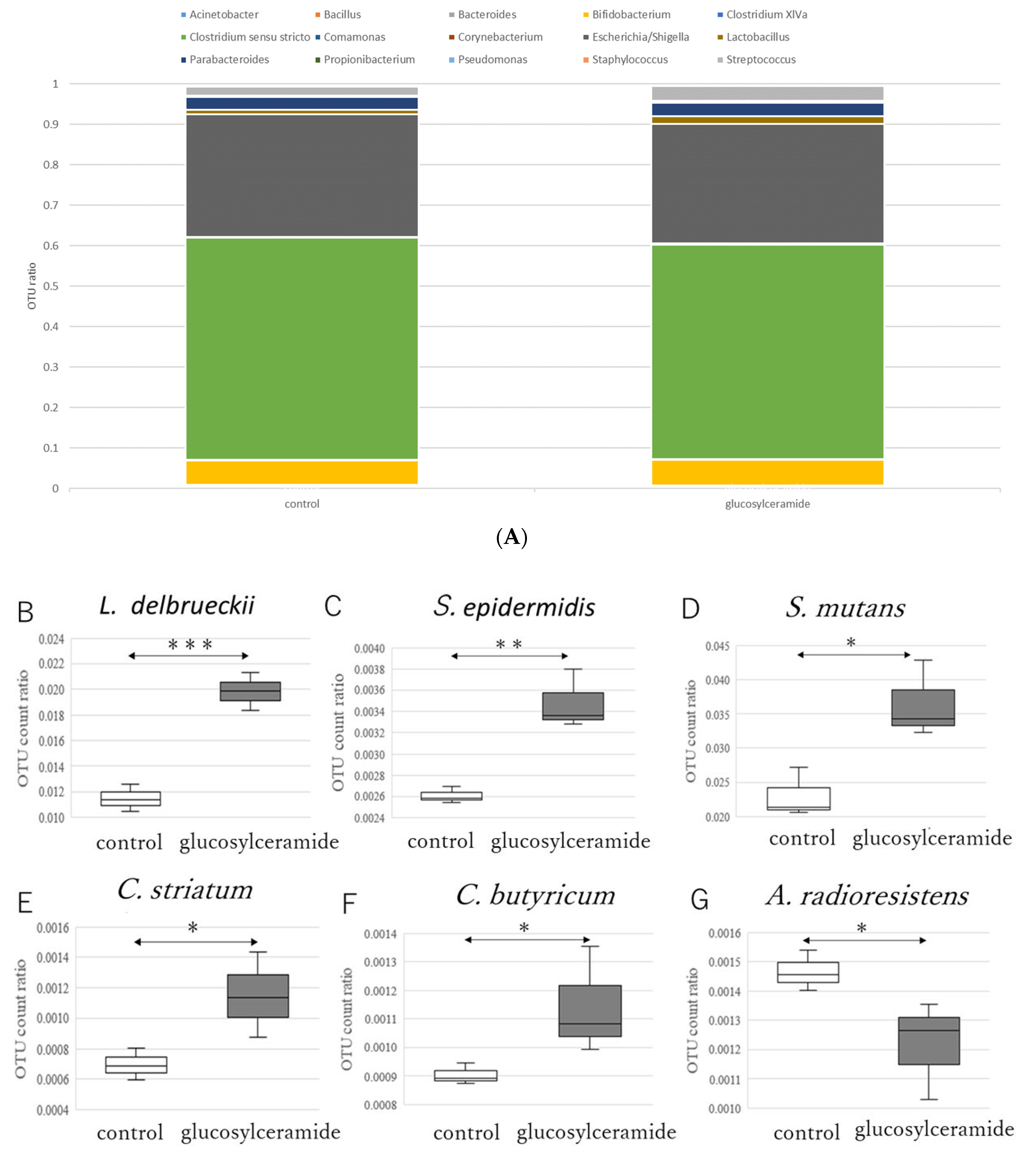
3.2. Glucosylceramide Addition Increases Gram-Positive Intestinal Microbes

These results clearly indicate that glucosylceramide alters the metabolism of the intestinal microbes in this system. Next, to determine which bacteria were increased in response to glucosylceramide, NGS analysis was performed after incubation of model intestinal microbes (cell-mock-1), consisting of known ratios of bacteria that reside within human beings with glucosylceramide (Supplementary Table S2). Cell-mock-1 is a composite of bacteria Bacillus subtilis, Bifidobacterium pseudocatenulatum, Clostridium butyricum, Corynebacterium striatum, Cutibacterium acnes subsp. acnes, Lactobacillus delbrueckii subsp. delbrueckii, Streptococcus mutans, Staphylococcus epidermidis, Acinetobacter radioresistens, Bacteroides uniformis, Clostridium clostridioforme, Comamonas terrigena, Escherichia coli, Parabacteroides distasonis, and Pseudomonas putida. These bacteria were incubated in YCFA medium as a mixed state anaerobically with or without glucosylceramide. From the incubated culture, genomes were extracted and analyzed using NGS. As a result, relative OTU counts of Clostridium sensu stricto and Escherichia/Shigella were dominant (Figure 2A), maybe because of the anaerobic condition. These dominant bacteria were not significantly affected by glucosylceramide. In contrast, relative OTU counts of minor Gram-positive bacteria, such as Lactobacillus delbrueckii (Figure 2B), Staphylococcus epidermidis (Figure 2C), Streptococcus mutans (Figure 2D), Corynebacterium striatum (Figure 2E), and Clostridium butyricum (Figure 2F), were significantly increased in glucosylceramide-treated cultures compared with those in control cultures (p < 0.05). In contrast, the Gram-negative bacterium, Acinetobacter radioresistens (Figure 2G), decreased in glucosylceramide-treated cultures compared with that in control cultures (p < 0.05).

3.3. Glucosylceramide Confers Tolerance of Intestinal Microbes to Secondary Bile Acids

Considering that certain intestinal bacteria have a tolerance to bile acids [55], we hypothesized that these bacteria increased growth in the existence of glucosylceramide because they acquired tolerance to deoxycholic acid. Therefore, tolerance of these bacteria towards deoxycholic acid was investigated.
With the addition of glucosylceramide, L. delbrueckii showed increased growth at 1.0–2.0 mM deoxycholic acid relative to that in the control (Figure 3A). Glucosylceramide-supplemented L. delbrueckii displayed an increased half-maximal inhibitory concentration value compared with that of the control, although the difference was not statistically significant (Table 3).
The tolerance of another bacterium, S. epidermidis, to deoxycholic acid was also investigated. There was no significant difference in the growth or half-maximal inhibitory concentration between the glucosylceramide-treated and control S. epidermidis (Figure 3B and Table 3). Growths of S. mutans, C. striatum and C. butyricum in the presence of deoxycholic acid were also partially rescued with glucosylceramide (Figure 3C–E). IC50 of S. mutans was significantly increased (p < 0.01, Table 3). The growth of E. coli was not inhibited by deoxycholic acid (Figure 3F) and it was difficult to measure the effect of glucosylceramide. The glucosylceramide effect was not observed in Bacteroides uniformis (Figure 3G). Furthermore, the tolerances of other major intestinal microbes, including Blautia coccoides and Enterococcus faecalis, against deoxycholic acid were investigated. The growth of B. coccoides was rescued with glucosylceramide at 1 mM deoxycholic acid concentration (Figure 3H). Similarly, the growth of E. faecalis was partially rescued with glucosylceramide (Figure 3I). The IC50 value for E. faecalis was also significantly higher after glucosylceramide addition than that in the control (Table 3). These results suggest that glucosylceramide increased the tolerance of certain intestinal microbes to deoxycholic acid.

4. Discussion

Although we have long known that glucosylceramide is present in foods such as crops and fermented foods at 0.94–2.2 mg/g [19,24,25,53], and the majority of the molecule passes through the small intestine and reaches the colon, the mechanism of action of glucosylceramide on intestinal microbes has not yet been reported. In this study, we first report the in vitro prebiotic effects of glucosylceramide. Using metabolome analysis followed by PLS-DA, we demonstrate that adding glucosylceramide to the major intestinal bacterium B. coccoides has a significant metabolic effect. Furthermore, the addition of glucosylceramide increased the number of Gram-positive bacteria through tolerance to deoxycholic acid. These results provide the first evidence of a mechanism underlying the action of glucosylceramide on intestinal microbes.
This research has certain limitations and weaknesses. Adding pure components to monocultures does not necessarily reflect what occurs in the complex microbiota of the gut, since the actual gut is filled with various substances and microbes. Moreover, a certain amount of glucosylceramide, although very small [38], might be degraded and absorbed in the small intestine. This research did not consider the absorption of glucosylceramide into the epithelial cells or mucosa in the intestine into account. However, it has a strong aspect as relative to in vivo studies. During the passage through the intestine, glucosylceramide might interact with many bacteria. Some bacteria might attach to the epithelial cells of the intestine, and might not be detected in the feces. On the contrary, in vitro studies generates the direct interaction knowledge between glucosylceramide and bacteria. Therefore, the knowledge obtained through in vitro study should mutually complement in vivo studies to reach a comprehensive understanding of the interaction between substances and intestinal microbes.
Secondary bile acids are formed in the large intestine by intestinal microbes and disrupt the membranes of bacteria, killing them [41]. In the present study, glucosylceramide attenuated these effects. Therefore, glucosylceramide that reaches the large intestine may protect the intestinal microbes from secondary bile acids.
Blautia species are one of the most dominant intestinal epithelial barrier-associated bacteria, which belongs to the Lachnospiraceae family and Firmicutes phylum [56]. Blautia species are strictly anaerobic, non-motile bacteria with spherical or oval morphologies. They can assimilate various carbohydrates such as glucose, fructose, lactose, mannose, arabinose and xylose, CO and H2/CO2, and produces acetic acid, succinic acid, lactic acid and ethanol. It is suggested from gene expression analysis that the tendency of assimilation of carbohydrates of Blautia species is lower than that of lactic acid bacteria. There is a negative correlation between the abundance of Blautia and the markers of obesity-related metabolic disorders when fed with maize-derived non-digestible feruloylated oligo- and polysaccharides [57], potato fibers [58] and soy milk [59]. Intestinal microbes of Japanese people contained a high abundance of Blautia and Bifidobacterium and a low abundance of Bacteroides [60,61]. Considering that Blautia has a strong taxonomic association with twin inheritance [62], there might be a genetic preference of Blautia towards the intestine of specific genetic background. Blautia species are also linked to abnormal Paneth cell counts [63], Crohn’s disease and primary sclerosing cholangitis [64]. B. coccoides in the intestine increases with glucosylceramide in an animal model [23], but decreases with pectins and flavanones in humans [65]. Blautia species produce useful secondary metabolites such as chrysin, apigenin, desmethyllicaritin, 3′desmethyllarctigenin, bisdemethylcurcmin, demethyldemethoxycurcumin and ceramide in the intestine [54].
Promoted survival of Gram-positive bacteria elucidated in this study suggests the clinical benefits of glucosylceramide. For example, feeding of L. brueckii subsp. delbrueckii improves immune health in an animal model [66] and inhibits the growth of Klebsiella pneumonia [67]. C. butyricum also functions as probiotics by inducing interleukin-10-producing macrophages in inflamed mucosa via the Toll-like receptor 2/myeloid differentiation primary response gene 88 pathway [68] and has many clinical applications [69]. Although pathogenicity is reported for C. striatum [70], these events are not reported in the intestine. Since S. mutans [71] and S. epidermidis [72] form biofilms and are opportunistic pathogens, the health benefits of the modulation of intestinal microbes through the ingestion of glucosylceramide should be considered comprehensively. Still, this study suggests that the ingestion of glucosylceramide, on the whole, might promote the survival of beneficial intestinal microbes, which is a target of future studies.
This study did not elucidate how or why glucosylceramide increased the ratio of Gram-positive bacteria. Since Gram-positive bacteria have a thick peptidoglycan layer, they may be better suited for the attachment of glucosylceramide and its incorporation into their structures as compared with that of Gram-negative bacteria. Consistent with this hypothesis, Lacticaseibacillus casei was reported to bind glucosylceramide [73]. In addition, several other lactic acid bacteria have been shown to bind sphingolipids containing carbohydrate moieties, including Lactobacillus johnsonii to asialoganglioside [74] and Propionibacterium freudenreichii to lactosylceramide [75]. Therefore, it can be hypothesized that glucosylceramide bound to the surface of Gram-positive bacteria and endowed tolerance to deoxycholic acid. The effect of glucosylceramide on other intestinal bacteria should also be investigated.
This study proposes a potential mechanism for the observed effects in vivo [26,27,28,29,30]. Our results suggest that glucosylceramide protects intestinal microbes, especially Gram-positive bacteria, increases the production of substances such as citric acid, and as a result, might exert various health effects. Consistent with this hypothesis, citric acid improves several nutritional profiles in quails [76]. However, this hypothesis requires further investigation.
Our analysis of bacterial tolerance to deoxycholic acid was generally consistent with our results obtained from NGS analysis. However, S. epidermidis did not show significant recovery of tolerance to deoxycholic acid with glucosylceramide, which was inconsistent with the NGS analysis. The tolerance analysis may have lacked sufficient sensitivity to detect the difference in S. epidermidis tolerance towards deoxycholic acid, which can be elucidated using a more precise analysis tool in future studies.
Several substances have been proposed as potential prebiotics. However, there are no reports of the effects of glucosylceramide on intestinal microbes. Based on our results, glucosylceramide can be hypothesized as a type of prebiotic.
Feeding of fructooligosaccharide and galactooligosaccharide decreased butyrate, increased Actinobacteria, Bifidobacterium, Acidaminococcosu and decreased Salmonella, Coprococcus, Turicbacter, Enterobacter and Phascolactobacterium in vivo [77]. Therefore, it could be hypothesized that glucosylceramide might function differently from oligosaccharides, which is a target of future studies.
Sphingolipid synthesis was shown to improve the survival of intestinal Bacteroides [78]. Therefore, it is suggested that exogenous glucosylceramide also improves the survival of these intestinal bacteria, which is a target of future studies.
Glucosylceramide in the luminal side of the intestine, which was investigated in this study, might affect that in the epithelial cells. Glucosylceramide is shown to protect CaCO2 cells treated with lipopolysaccharide in an in vitro intestinal tract model [79]. Furthermore, glucosylceramide production maintains colon integrity when challenged with Bacteroides fragilis toxin [80]. Together with the results shown in this study that glucosylceramide protects intestinal microbes, glucosylceramide exerts its effects both in the luminal space of the intestine and the epithelial cells.
In conclusion, we demonstrate that the addition of glucosylceramide induces changes in the metabolism of B. coccoides and increases the number of Gram-positive bacteria in an intestine-simulating environment through tolerance to deoxycholic acid. These results first indicate the prebiotic-like function of glucosylceramide.

Supplementary Materials

The following supporting information can be downloaded at: https://0-www-mdpi-com.brum.beds.ac.uk/article/10.3390/ijms23105300/s1.

Author Contributions

H.K., T.Y. and J.N. conceived and designed the experiments. H.D., A.O. and K.T. performed the experiments. H.K. and H.D. wrote the manuscript. J.N. revised the manuscript. All authors have read and agreed to the published version of the manuscript.

Funding

Part of this research was financially supported by JSPS KAKENHI 20K05809.

Institutional Review Board Statement

Not applicable.

Informed Consent Statement

Not applicable.

Data Availability Statement

Inquiries can be directed to the corresponding author.

Acknowledgments

All authors have provided consent. This research was part of the dissertation submitted by the first author in partial fulfillment of the Ph.D. degree. Tomoya Sato, Yuka Kitajima, Mayu Nagatome and Moemi Mansho at Graduate School of Health Sciences, Saga University contributed to the experiments. We would like to acknowledge Satoru Fukiya at Hokkaido University for the manipulation of anaerobic cultures.

Conflicts of Interest

The authors declare no potential conflict of interest.

References

  1. Lilly, D.M.; Stillwell, R.H. Probiotics: Growth-promoting factors produced by microorganisms. Science 1965, 147, 747–748. [Google Scholar] [CrossRef] [PubMed]
  2. Gibson, G.R. Dietary modulation of the human gut microflora using prebiotics. Br. J. Nutr. 1998, 80, S209–S212. [Google Scholar] [CrossRef] [PubMed]
  3. Wang, Y.; Han, F.; Hu, B.; Li, J.; Yu, W. In vivo prebiotic properties of alginate oligosaccharides prepared through enzymatic hydrolysis of alginate. Nutr. Res. 2006, 26, 597–603. [Google Scholar] [CrossRef]
  4. Kukkonen, K.; Savilahti, E.; Haahtela, T.; Juntunen-Backman, K.; Korpela, R.; Poussa, T.; Tuure, T.; Kuitunen, M. Probiotics and prebiotic galacto-oligosaccharides in the prevention of allergic diseases: A randomized, double-blind, placebo-controlled trial. J. Allergy Clin. Immunol. 2007, 119, 192–198. [Google Scholar] [CrossRef] [PubMed]
  5. Joyce, S.A.; Kamil, A.; Fleige, L.; Fleige, L.; Gahan, C.G. The cholesterol-lowering effect of oats and oat beta glucan: Modes of action and potential role of bile acids and the microbiome. Front. Nutr. 2019, 6, 171. [Google Scholar] [CrossRef] [PubMed]
  6. Kankaanpää, P.; Yang, B.; Kallio, H.; Lsolauri, E.; Saiminen, S. Effects of polyunsaturated fatty acids in growth medium on lipid composition and on physicochemical surface properties of lactobacilli. Appl. Environ. Microbiol. 2004, 70, 129–136. [Google Scholar] [CrossRef]
  7. Guo, T.; Song, D.; Cheng, L.; Xin, Z. Interactions of tea catechins with intestinal microbiota and their implication for human health. Food Sci. Biotechnol. 2019, 28, 1617–1625. [Google Scholar] [CrossRef]
  8. Ito, Y.; Ichikawa, T.; Morohoshi, Y.; Nakamura, T.; Saegusa, Y.; Ishihara, K. Effect of tea catechins on body fat accumulation in rats fed a normal diet. Biomed. Res. 2008, 29, 27–32. [Google Scholar] [CrossRef]
  9. Jung, E.S.; Park, H.M.; Hyun, S.M.; Shon, J.C.; Singh, D.; Liu, K.-H.; Whon, T.W.; Bae, J.-W.; Hwang, J.S.; Lee, C.H. The green tea modulates large intestinal microbiome and exo/endogenous metabolome altered through chronic UVB-exposure. PLoS ONE 2017, 12, e0187154. [Google Scholar] [CrossRef]
  10. Hidalgo, M.; Oruna-Concha, M.J.; Kolida, S.; Walton, G.E.; Kallithraka, S.; Spencer, J.P.E.; Gibson, G.R.; Pascual-Teresa, S.D. Metabolism of anthocyanins by human gut microflora and their influence on gut bacterial growth. J. Agric. Food Chem. 2012, 60, 3882–3890. [Google Scholar] [CrossRef]
  11. Naqash, F.; Masoodi, F.A.; Rather, S.A.; Wani, S.M.; Gani, A. Emerging concepts in the nutraceutical and functional properties of pectin—A review. Carbohydr. Polym. 2017, 168, 227–239. [Google Scholar] [CrossRef] [PubMed]
  12. Yang, Y.; Kumrungsee, T.; Kato, N.; Fukuda, S.; Kuroda, M.; Yamaguchi, S. Supplemental Aspergillus lipase and protease preparations display powerful bifidogenic effects and modulate the gut microbiota community of rats. Fermentation 2021, 7, 294. [Google Scholar] [CrossRef]
  13. Yang, Y.; Kumrungsee, T.; Kato, N.; Fukuda, S.; Kuroda, M.; Yamaguchi, S. Aspergillus-derived cellulase preparation exhibits prebiotic-like effects on gut microbiota in rats. Fermentation 2022, 8, 71. [Google Scholar] [CrossRef]
  14. Yang, Y.; Iwamoto, A.; Kumrungsee, T.; Okazaki, Y.; Kuroda, M.; Yamaguchi, S.; Kato, N. Consumption of an acid protease derived from Aspergillus oryzae causes bifidogenic effect in rats. Nutr. Res. 2017, 44, 60–66. [Google Scholar] [CrossRef]
  15. Platt, F.M. Sphingolipid lysosomal storage disorders. Nature 2014, 510, 68–75. [Google Scholar] [CrossRef]
  16. Hannun, Y.A.; Bell, R.M. Functions of sphingolipids and sphingolipid breakdown products in cellular regulation. Science 1989, 243, 500–507. [Google Scholar] [CrossRef]
  17. Hannun, Y.A.; Obeid, L.M. Principles of bioactive lipid signalling: Lessons from sphingolipids. Nat. Rev. Mol. Cell Biol. 2008, 9, 139–150. [Google Scholar] [CrossRef]
  18. Fujino, Y.; Ohnishi, M. Structure of cerebroside in Aspergillus oryzae. Biochim. Biophys. Acta 1976, 486, 161–171. [Google Scholar] [CrossRef]
  19. Hirata, M.; Tsuge, K.; Jayakody, L.N.; Urano, Y.; Sawada, K.; Inaba, S.; Nagao, K.; Kitagaki, H. Structural determination of glucosylceramides in the distillation remnants of shochu, the Japanese traditional liquor, and its production by Aspergillus kawachii. J. Agric. Food Chem. 2012, 60, 11473–11482. [Google Scholar] [CrossRef]
  20. Hamajima, H.; Fujikawa, A.; Yamashiro, M.; Ogami, T.; Kitamura, S.; Tsubata, M.; Tan, S.; Matsunaga, H.; Sawada, K.; Kumagai, S.; et al. Chemical analysis of the sugar moiety of monohexosylceramide contained in koji, Japanese traditional rice fermented with Aspergillus. Fermentation 2016, 2, 2. [Google Scholar] [CrossRef]
  21. Tani, Y.; Amaishi, Y.; Funatsu, T.; Ito, M.; Itonori, S.; Hata, Y.; Ashida, H.; Yamamoto, K. Structural analysis of cerebrosides from Aspergillus fungi: The existence of galactosylceramide in A. oryzae. Biotechnol. Lett. 2014, 36, 2507–2513. [Google Scholar] [CrossRef] [PubMed]
  22. Del Poeta, M.; Nimrichter, L.; Rodrigues, M.L.; Luberto, C. Synthesis and biological properties of fungal glucosylceramide. PLoS Pathog. 2014, 10, e1003832. [Google Scholar] [CrossRef] [PubMed]
  23. Hamajima, H.; Matsunaga, H.; Fujikawa, A.; Sato, T.; Mitsutake, S.; Yanagita, T.; Nagao, K.; Nakayama, J.; Kitagaki, H. Japanese traditional dietary fungus koji Aspergillus oryzae functions as a prebiotic for Blautia coccoides through glycosylceramide: Japanese dietary fungus koji is a new prebiotic. Springer Plus 2016, 5, 1321. [Google Scholar] [CrossRef]
  24. Takahashi, K.; Izumi, K.; Nakahata, E.; Hirata, M.; Sawada, K.; Tsuge, K.; Nagao, K.; Kitagaki, H. Quantitation and structural determination of glucosylceramides contained in sake lees. J. Oleo Sci. 2014, 63, 15–23. [Google Scholar] [CrossRef] [PubMed]
  25. Takakuwa, N.; Saito, K.; Ohnishi, M.; Oda, Y. Determination of glucosylceramide contents in crop tissues and by-products from their processing. Bioresour. Technol. 2005, 96, 1089–1092. [Google Scholar] [CrossRef]
  26. Schmelz, E.M.; Sullards, M.C.; Dillehay, D.L.; Merrill, A.H., Jr. Colonic cell proliferation and aberrant crypt foci formation are inhibited by dairy glycosphingolipids in 1,2-dimethylhydrazine-treated CF1 mice. J. Nutr. 2000, 130, 522–527. [Google Scholar] [CrossRef]
  27. Symolon, H.; Schmelz, E.M.; Dillehay, D.L.; Merrill, A.H., Jr. Dietary soy sphingolipids suppress tumorigenesis and gene expression in 1, 2-dimethylhydrazine-treated CF1 mice and Apc Min/+ mice. J. Nutr. 2004, 134, 1157–1161. [Google Scholar] [CrossRef]
  28. Fujiwara, K.; Yazama, H.; Donishi, R.; Koyama, S.; Fukuhara, T.; Takeuchi, H. Inhibitory effects of glucosylceramide on tumorigenesis induced by a carcinogen in mice. Laryngoscope 2020, 130, E593–E597. [Google Scholar] [CrossRef]
  29. Yazama, H.; Kitatani, K.; Fujiwara, K.; Kato, M.; Hashimoto-Nishimura, M.; Kawamoto, K.; Hasegawa, K.; Kitano, H.; Bielawska, A.; Bielawski, J.; et al. Dietary glucosylceramides suppress tumor growth in a mouse xenograft model of head and neck squamous cell carcinoma by the inhibition of angiogenesis through an increase in ceramide. Int. J. Clin. Oncol. 2015, 20, 438–446. [Google Scholar] [CrossRef]
  30. Fujiwara, K.; Kitatani, K.; Fukushima, K.; Yazama, H.; Umehara, H.; Kikuchi, M.; Igarashi, Y.; Kitano, H.; Okazaki, T. Inhibitory effects of dietary glucosylceramides on squamous cell carcinoma of the head and neck in NOD/SCID mice. Int. J. Clin. Oncol. 2011, 16, 133–140. [Google Scholar] [CrossRef]
  31. Hossain, Z.; Sugawara, T.; Aida, K.; Hirata, T. Effect of dietary glucosylceramide from sea cucumber on plasma and liver lipids in cholesterol-fed mice. Fish. Sci. 2011, 77, 1081–1085. [Google Scholar] [CrossRef]
  32. Tsuji, K.; Mitsutake, S.; Ishikawa, J.; Takagi, Y.; Akiyama, M.; Shimizu, H.; Tomiyama, T.; Igarashi, Y. Dietary glucosylceramide improves skin barrier function in hairless mice. J. Dermatol. Sci. 2006, 44, 101–107. [Google Scholar] [CrossRef] [PubMed]
  33. Miyagawa, M.; Fujikawa, A.; Nagadome, M.; Kohama, K.; Ogami, T.; Kitamura, S.; Kitagaki, H. Glycosylceramides purified from the Japanese traditional non-pathogenic fungus Aspergillus and koji increase the expression of genes involved in tight junctions and ceramide delivery in normal human epidermal keratinocytes. Fermentation 2019, 5, 43. [Google Scholar] [CrossRef]
  34. Yamashita, S.; Seino, T.; Aida, K.; Kinoshita, M. Effects of plant sphingolipids on inflammatory stress in differentiated caco-2 cells. J. Oleo Sci. 2017, 66, 1337–1342. [Google Scholar] [CrossRef] [PubMed]
  35. Sugawara, T.; Kinoshita, M.; Ohnishi, M.; Miyazawa, T. Apoptosis induction by wheat-flour sphingoid bases in DLD-1 human colon cancer cells. Biosci. Biotechnol. Biochem. 2002, 66, 2228–2231. [Google Scholar] [CrossRef] [PubMed]
  36. Aida, K.; Kinoshita, M.; Sugawara, T.; Ono, J.; Miyazawa, T.; Ohnishi, M. Apoptosis inducement by plant and fungus sphingoid bases in human colon cancer cells. J. Oleo Sci. 2004, 53, 503–510. [Google Scholar] [CrossRef]
  37. Nilsson, Å. Metabolism of cerebroside in the intestinal tract of the rat. Biochim. Biophys. Acta BBA-Lipids Lipid Metab. 1969, 187, 113–121. [Google Scholar] [CrossRef]
  38. Sugawara, T.; Tsuduki, T.; Yano, S.; Hirose, M.; Duan, J.; Aida, K.; Ikeda, I.; Hirata, T. Intestinal absorption of dietary maize glucosylceramide in lymphatic duct cannulated rats. J. Lipid Res. 2010, 51, 1761–1769. [Google Scholar] [CrossRef]
  39. Kawata, M.; Tsukamoto, A.; Isozaki, R.; Nobukawa, S.; Kawahara, N.; Akutsu, S.; Suzuki, M.; Asanuma, N. Glucerabacter Canisensis gen. nov., sp. nov., isolated from dog feces and its effect on the hydrolysis of plant glucosylceramide in the intestine of dogs. Arch. Microbiol. 2018, 200, 505–515. [Google Scholar] [CrossRef]
  40. Zwicker, B.L.; Agellon, L.B. Transport and biological activities of bile acids. Int. J. Biochem. Cell Biol. 2013, 45, 1389–1398. [Google Scholar] [CrossRef]
  41. Yokota, A.; Fukiya, S.; Islam, K.B.M.S.; Ooka, T.; Ogura, Y.; Hayashi, T.; Hagio, M.; Ishizuka, S. Is bile acid a determinant of the gut microbiota on a high-fat diet? Gut Microbes 2012, 3, 455–459. [Google Scholar] [CrossRef] [PubMed]
  42. Zeng, H.; Umar, S.; Rust, B.; Lazarova, D.; Bordonaro, M. Secondary bile acids and short chain fatty acids in the colon: A focus on colonic microbiome, cell proliferation, inflammation, and cancer. Int. J. Mol. Sci. 2019, 20, 1214. [Google Scholar] [CrossRef] [PubMed]
  43. Mouzaki, M.; Wang, A.Y.; Bandsma, R.; Comelli, E.M.; Arendt, B.M.; Zhang, L.; Fung, S.; Fischer, S.E.; Mcgilvray, L.G.; Allard, J.P. Bile acids and dysbiosis in non-alcoholic fatty liver disease. PLoS ONE 2016, 11, e0151829. [Google Scholar] [CrossRef] [PubMed]
  44. Duncan, S.H.; Hold, G.L.; Harmsen, H.; Stewart, C.S.; Flint, H.J. Growth requirements and fermentation products of Fusobacterium prausnitzii, and a proposal to reclassify it as Faecalibacterium prausnitzii gen. nov., comb. nov. Int. J. Syst. Evol. Microbiol. 2002, 52, 2141–2146. [Google Scholar] [CrossRef]
  45. Kusaba, Y.; Otsuka, A.; Dai, H.; Inaba, S.; Kitagaki, H. Induction of chromosomal aneuploids from brewery shochu yeast with novel brewery characteristics. Fermentation 2022, 8, 62. [Google Scholar] [CrossRef]
  46. Motomura, S.; Horie, K.; Kitagaki, H. Mitochondrial activity of sake brewery yeast affects malic and succinic acid production during alcoholic fermentation. J. Inst. Brew. 2012, 118, 22–26. [Google Scholar] [CrossRef]
  47. Takahashi, S.; Tomita, J.; Nishioka, K.; Hisada, T.; Nishijima, M. Development of a prokaryotic universal primer for simultaneous analysis of bacteria and archaea using next-generation sequencing. PLoS ONE 2014, 9, e105592. [Google Scholar] [CrossRef]
  48. Aronesty, E. Comparison of sequencing utility programs. Open Bioinform. J. 2013, 7, 1–8. [Google Scholar] [CrossRef]
  49. Gordon, A.; Hannon, G.J. FASTX-Toolkit FASTQ/A Short-Reads Preprocessing Tools [Software]. Available online: http://hannonlab.cshl.edu/fastx_toolkit/index.html (accessed on 5 May 2022).
  50. Caporaso, J.G.; Kuczynski, J.; Stombaugh, J.; Bittinger, K.; Bushman, F.D.; Costello, E.K.; Fierer, N.; Pena, A.G.; Goodrich, J.K.; Gordon, J.I.; et al. QIIME allows analysis of high-throughput community sequencing data. Nat. Methods 2010, 7, 335–336. [Google Scholar] [CrossRef]
  51. Chong, J.; Soufan, O.; Li, C.; Caraus, L.; Li, S.; Bourque, G.; Wishart, D.S.; Xia, J. MetaboAnalyst 4.0: Towards more transparent and integrative metabolomics analysis. Nucleic Acids Res. 2018, 46, W486–W494. [Google Scholar] [CrossRef]
  52. Ozato, N.; Saito, S.; Yamaguchi, T.; Katashima, M.; Tokuda, I.; Sawada, K.; Katsuragi, Y.; Kakuta, M.; Imoto, S.; Ihara, K.; et al. Blautia genus associated with visceral fat accumulation in adults 20–76 years of age. NPJ Biofilms Microbiomes 2019, 5, 28. [Google Scholar] [CrossRef] [PubMed]
  53. Sawada, K.; Sato, T.; Hamajima, H.; Jayakody, L.N.; Hirata, M.; Yamashiro, M.; Tajima, M.; Mitsutake, S.; Nagao, K.; Tsuge, K.; et al. Glucosylceramide contained in koji mold-cultured cereal confers membrane and flavor modification and stress tolerance to Saccharomyces cerevisiae during coculture fermentation. Appl. Environ. Microbiol. 2015, 81, 3688–3698. [Google Scholar] [CrossRef] [PubMed]
  54. Olsson, O. The capacity of the large intestine. Acta Radiol. 1952, 37, 348–355. [Google Scholar] [CrossRef] [PubMed]
  55. Dey, D.K.; Koo, B.G.; Sharma, C.; Kang, S.C. Characterization of Weissella confusa DD_A7 isolated from kimchi. LWT 2019, 111, 663–672. [Google Scholar] [CrossRef]
  56. Liu, X.; Mao, B.; Gu, J.; Wu, J.; Cui, S.; Wang, G.; Zhao, J.; Zhang, H.; Chen, W. Blautia—A new functional genus with potential probiotic properties? Gut Microbes 2021, 13, 1875796. [Google Scholar] [CrossRef]
  57. Yang, J.; Bindels, L.B.; Segura Munoz, R.R.; Martínez, I.; Walter, J.; Ramer-Tait, A.E.; Rose, D.J. Disparate metabolic responses in mice fed a high-fat diet supplemented with maize-derived non-digestible feruloylated oligo- and polysaccharides are linked to changes in the gut microbiota. PLoS ONE 2016, 11, e0146144. [Google Scholar] [CrossRef]
  58. Panasevich, M.R.; Kerr, K.R.; Dilger, R.N.; Fahey, G.C.; Guérin-Deremaux, L., Jr.; Lynch, G.L.; Wils, D.; Suchodolski, J.S.; Steer, J.M.; Dowd, S.E.; et al. Modulation of the faecal microbiome of healthy adult dogs by inclusion of potato fibre in the diet. Br. J. Nutr. 2015, 113, 125–133. [Google Scholar] [CrossRef]
  59. Lee, S.M.; Han, H.W.; Yim, S.Y. Beneficial effects of soy milk and fiber on high cholesterol diet-induced alteration of gut microbiota and inflammatory gene expression in rats. Food Funct. 2015, 6, 492–500. [Google Scholar] [CrossRef]
  60. Odamaki, T.; Kato, K.; Sugahara, H.; Hashikura, N.; Takahashi, S.; Xiao, J.Z.; Abe, F.; Osawa, R. Age-related changes in gut microbiota composition from newborn to centenarian: A cross-sectional study. BMC Microbiol. 2016, 16, 90. [Google Scholar] [CrossRef]
  61. Nakayama, J.; Watanabe, K.; Jiang, J.; Matsuda, K.; Chao, S.H.; Haryono, P.; La-Ongkham, O.; Sarwoko, M.A.; Sujaya, I.N.; Zhao, L.; et al. Diversity in gut bacterial community of school-age children in Asia. Sci. Rep. 2015, 5, 8397. [Google Scholar] [CrossRef]
  62. Bonder, M.J.; Kurilshikov, A.; Tigchelaar, E.F.; Mujagic, Z.; Imhann, F.; Vila, A.V.; Deelen, P.; Vatanen, T.; Schirmer, M.; Smeekens, S.P.; et al. The effect of host genetics on the gut microbiome. Nat. Genet. 2016, 48, 1407–1412. [Google Scholar] [CrossRef] [PubMed]
  63. Liu, T.-C.; Gurram, B.; Baldridge, M.; Head, R.; Lam, V.; Luo, C.; Cao, Y.; Simpson, P.; Hayward, M.; Holte, M.; et al. O-011 Paneth cell phenotypes define a subtype of pediatric Crohn’s disease through alterations in host–microbial interactions. Inflamm. Bowel Dis. 2016, 22, S4. [Google Scholar] [CrossRef]
  64. Torres, J.; Bao, X.; Goel, A.; Colombel, J.-F.; Pekow, J.; Jabri, B.; Williams, K.M.; Castillo, A.; Odin, J.A.; Meckel, K.; et al. The features of mucosa-associated microbiota in primary sclerosing cholangitis. Aliment. Pharmacol. Ther. 2016, 43, 790–801. [Google Scholar] [CrossRef] [PubMed]
  65. Cuervo, A.; Valdés, L.; Salazar, N.; de los Reyes-Gavilán, C.G.; Ruas-Madiedo, P.; Gueimonde, M.; González, S. Pilot study of diet and microbiota: Interactive associations of fibers and polyphenols with human intestinal bacteria. J. Agric. Food Chem. 2014, 62, 5330–5336. [Google Scholar] [CrossRef]
  66. Suda, Y.; Kagawa, K.; Fukuyama, K.; Elean, M.; Zhou, B.; Tomokiyo, M.; Islam, M.A.; Rajoka, M.; Kober, A.; Shimazu, T.; et al. Soymilk-fermented with Lactobacillus delbrueckii subsp. delbrueckii TUA4408L improves immune-health in pigs. Benef. Microbes 2022, 13, 61–72. [Google Scholar] [CrossRef]
  67. Mogna, L.; Deidda, F.; Nicola, S.; Amoruso, A.; Del Piano, M.; Mogna, G. In vitro inhibition of Klebsiella pneumoniae by Lactobacillus delbrueckii subsp. delbrueckii LDD01 (DSM 22106): An innovative strategy to possibly counteract such infections in humans? J. Clin. Gastroenterol. 2016, 50, S136–S139. [Google Scholar] [CrossRef]
  68. Kanai, T.; Mikami, Y.; Hayashi, A. A breakthrough in probiotics: Clostridium butyricum regulates gut homeostasis and anti-inflammatory response in inflammatory bowel disease. J. Gastroenterol. 2015, 50, 928–939. [Google Scholar] [CrossRef]
  69. Tomita, Y.; Ikeda, T.; Sakata, S.; Saruwatari, K.; Sato, R.; Iyama, S.; Jodai, T.; Akaike, K.; Ishizuka, S.; Saeki, S.; et al. Association of probiotic Clostridium butyricum therapy with survival and response to immune checkpoint blockade in patients with lung cancer. Cancer Immunol. Res. 2020, 8, 1236–1242. [Google Scholar] [CrossRef]
  70. Boltin, D.; Katzir, M.; Bugoslavsky, V.; Yalashvili, I.; Brosh-Nissimov, T.; Fried, M.; Elkayam, O. Corynebacterium striatum--a classic pathogen eluding diagnosis. Eur. J. Intern. Med. 2009, 20, e49–e52. [Google Scholar] [CrossRef]
  71. Krzyściak, W.; Pluskwa, K.K.; Jurczak, A.; Koscielniak, D. The pathogenicity of the Streptococcus genus. Eur. J. Clin. Microbiol. Infect. Dis. 2013, 32, 1361–1376. [Google Scholar] [CrossRef]
  72. O’Gara, J.P.; Humphreys, H. Staphylococcus epidermidis biofilms: Importance and implications. J. Med. Microbiol. 2001, 50, 582–587. [Google Scholar] [CrossRef] [PubMed]
  73. Yamamoto, K.; Miwa, T.; Taniguchi, H.; Nagano, T.; Shimarmura, K.; Tanaka, T.; Kumagai, H. Binding specificity of Lactobacillus to glycolipids. Biochem. Biophys. Res. Commun. 1996, 228, 148–152. [Google Scholar] [CrossRef] [PubMed]
  74. Neeser, J.R.; Granato, D.; Rouvet, M.; Servin, A.; Teneberg, S.; Karlsson, K.-A. Lactobacillus johnsonii La1 shares carbohydrate-binding specificities with several enteropathogenic bacteria. Glycobiology 2000, 10, 1193–1199. [Google Scholar] [CrossRef] [PubMed]
  75. Strömberg, N.; Ryd, M.; Lindberg, A.A.; Karlsson, K.-A. Studies on the binding of bacteria to glycolipids. Two species of Propionibacterium apparently recognize separate epitopes on lactose of lactosylceramide. FEBS Lett. 1988, 232, 193–198. [Google Scholar] [CrossRef]
  76. Fikry, A.M.; Attia, A.I.; Ismail, I.E.; Alagawany, M.; Reda, F.M. Dietary citric acid enhances growth performance, nutrient digestibility, intestinal microbiota, antioxidant status, and immunity of Japanese quails. Poult. Sci. 2021, 100, 101326. [Google Scholar] [CrossRef]
  77. Liu, F.; Li, P.; Chen, M.; Luo, Y.; Prabhakar, M.; Zheng, H.; He, Y.; Qi, Q.; Long, H.; Zhang, Y.; et al. Fructooligosaccharide (FOS) and Galactooligosaccharide (GOS) increase bifidobacterium but reduce butyrate producing bacteria with adverse glycemic metabolism in healthy young population. Sci. Rep. 2017, 7, 11789. [Google Scholar] [CrossRef]
  78. An, D.; Na, C.; Bielawski, J.; Hannun, Y.A.; Kasper, D.L. Membrane sphingolipids as essential molecular signals for Bacteroides survival in the intestine. Proc. Natl. Acad. Sci. USA 2011, 108, 4666–4671. [Google Scholar] [CrossRef]
  79. Yamashita, S.; Soga, M.; Nguma, E.; Kinoshita, M.; Miyazawa, T. Protective mechanism of rice-derived lipids and glucosylceramide in an in vitro intestinal tract model. J. Agric. Food Chem. 2021, 69, 10206–10214. [Google Scholar] [CrossRef]
  80. Patterson, L.; Allen, J.; Posey, I.; Shaw, J.J.P.; Costa-Pinheiro, P.; Walker, S.J.; Gademsey, A.; Wu, X.; Wu, S.; Zachos, N.C.; et al. Glucosylceramide production maintains colon integrity in response to Bacteroides fragilis toxin-induced colon epithelial cell signaling. FASEB J. 2020, 34, 15922–15945. [Google Scholar] [CrossRef]
Figure 1. Metabolite analysis of the Blautia coccoides culture treated with glucosylceramide. (A) Score plot of principal component analysis (PCA) of metabolites of B. coccoides treated with or without 20 μg/mL glucosylceramide. (B) Loading plot of metabolites of B. coccoides treated with or without 20 μg/mL glucosylceramide. The substances corresponding to the numbers are listed in Table 1. (C) Partial least squares-discriminant analysis (PLS-DA) of metabolites of B. coccoides treated with or without 20 μg/mL glucosylceramide. (D) The validated plot of PLS-DA of metabolites of B. coccoides treated with or without 20 μg/mL glucosylceramide after a permutation test (n = 200). (E) Lactic acid concentration (mg/mL) of B. coccoides cultures (24 h) treated with or without 20 μg/mL glucosylceramide and 2 mM DCA from the start of the culture in YCFA medium. Statistical difference of the means was verified using one-sided unpaired Student’s t-test (n = 3, * p < 0.05).
Ijms 23 05300 g001
Figure 2. Relative OTU ratios of microbes treated with glucosylceramide. (A) Average relative OTU ratios of all microbes treated with glucosylceramide. Relative OTU ratios of (B) L. delbrueckii, (C) S. epidermidis, (D) S. mutans, (E) C. striatum, (F) C. butyricum and (G) A. radioresistens treated with glucosylceramide. Data are expressed as means ± standard errors. Data normality was verified using the Shapiro–Wilk test. Statistical differences in means of data with normalities were analyzed using a one-sided unpaired Student’s t-test (n = 3; * p < 0.05, ** p < 0.01, *** p < 0.001).
Ijms 23 05300 g002
Figure 3. Growth of bacteria supplemented with various concentrations of deoxycholic acid (DCA) and glucosylceramide. OD600 values for (A) L. delbrueckii, (B) S. epidermidis, (C) S. mutans, (D) C. striatum, (E) C. butyricum, (F) E. coli, (G) B. uniformis, (H) B. coccoides and (I) E. faecalis. Values are mean ± standard errors of triplicate independent cultures. Statistical differences of the means were evaluated by one-sided unpaired Student’s t-test (n = 3, * p < 0.05, ** p < 0.01).
Ijms 23 05300 g003
Table 1. Substances detected by GC-FID.
No.Retention Time (min)Peak Name
16.923Lactic acid
26.994Unknown001
37.086Unknown002
47.595Alanine1
58.813Isoleucine1
69.191Valine2
711.896Methionine1
812.016Aspartic acid1
912.285Unknown003
1013.244L-proline
1113.341Butanoic acid
1213.378Glutamic acid1
1313.504Unknown004
1413.613Phenylalanine1
1513.797Unknown005
1613.994Unknown006
1714.398Glutamic acid2
1814.494Phenylalanine2
1914.819Unknown007
-15.587Ribitol(IS)
2016.126Phosphoric acid
2116.835Unknown008
2217.029Fructose1
2317.142Unknown009
2417.41Tyrosine
2517.595Unknown010
2618.2Gluconic acid
2718.527Unknown011
2819.364M-inosito1
2920.277Tryptophan2
3020.455Unknown012
3120.672Unknown013
3220.779Unknown014
Identification of peaks obtained with GC-FID. Numbers indicate the order of substances. Retention times indicate the time the substance appears during the GC-FID. Unidentified peaks were termed unknown.
Table 2. Substances with VIP scores greater than 1.0 in the PLS-DA model.
Peak NameVIP ScoreCoefficient
Citric acid3.180.227 **
Threonine21.82−0.130
Lysine11.690.121
Unknown31.600.114
Unknown11.510.108
Lactic acid21.270.091
Valine21.170.083
Proline, isolecine11.110.079
VIP scores were computed using the same data that were used to build the partial least squares-discriminant analysis (PLS-DA) model. The table includes components with a VIP score > 1.0. A No. with multiple component names represents components with peaks that were observed at the same retention time. Peaks whose chemicals could not be identified were labeled ‘Unknown’. One-sided unpaired Students t-test; n = 4, ** p < 0.01.
Table 3. IC50 of bacteria towards deoxycholic acid with or without glucosylceramide addition.
IC50 (mM)
BacteriaControlGlucosylceramidep Value
L.delbrueckii1.17 ± 0.161.47 ± 0.070.078
S. epidermidis0.343 ± 0.010.337 ± 0.000.131
S. mutans0.175 ± 0.0020.251 ± 0.0140.003 **
C. striatum0.369 ± 0.0140.393 ± 0.0200.197
C. butyricum0.088 ± 0.0030.117 ± 0.0140.053
E. coli---
B. uniformis0.21 ± 0.020.22 ± 0.020.344
B. coccoides0.67 ±0.070.81 ± 0.060.086
E. faecalis0.06 ± 0.000.08 ± 0.000.007 **
IC50 values were calculated independently for each bacterial culture. The IC50 values of the glucosylceramide group are included for comparison with that of the control group. Data are expressed as means ± standard errors. One-sided unpaired Student’s t-test; n ≥ 3; ** p < 0.01. IC50 of E. coli was not described because its growth increased with deoxycholic acid and it was difficult to calculate IC50.
Publisher’s Note: MDPI stays neutral with regard to jurisdictional claims in published maps and institutional affiliations.

