Article

Polyamine Oxidase Expression Is Downregulated by 17β-Estradiol via Estrogen Receptor 2 in Human MCF-7 Breast Cancer Cells

Department of Biochemistry, College of Life Science and Biotechnology, Yonsei University, Seoul 03722, Korea
*
Author to whom correspondence should be addressed.
Int. J. Mol. Sci. 2022, 23(14), 7521; https://0-doi-org.brum.beds.ac.uk/10.3390/ijms23147521
Submission received: 31 May 2022 / Revised: 2 July 2022 / Accepted: 4 July 2022 / Published: 7 July 2022
(This article belongs to the Special Issue The Role of Estrogen Receptors in Health and Disease 2.0)

Abstract

:
Polyamine levels decrease with menopause; however, little is known about the mechanisms regulated by menopause. In this study, we found that among the genes involved in the polyamine pathway, polyamine oxidase (PAOX) mRNA levels were the most significantly reduced by treatment with 17β-estradiol in estrogen receptor (ESR)-positive MCF-7 breast cancer cells. Treatment with 17β-estradiol also reduced the PAOX protein levels. Treatment with selective ESR antagonists and knockdown of ESR members revealed that estrogen receptor 2 (ESR2; also known as ERβ) was responsible for the repression of PAOX by 17β-estradiol. A luciferase reporter assay showed that 17β-estradiol downregulates PAOX promoter activity and that 17β-estradiol-dependent PAOX repression disappeared after deletions (−3126/−2730 and −1271/−1099 regions) or mutations of activator protein 1 (AP-1) binding sites in the PAOX promoter. Chromatin immunoprecipitation analysis showed that ESR2 interacts with AP-1 bound to each of the two AP-1 binding sites. These results demonstrate that 17β-estradiol represses PAOX transcription by the interaction of ESR2 with AP-1 bound to the PAOX promoter. This suggests that estrogen deficiency may upregulate PAOX expression and decrease polyamine levels.

1. Introduction

Polyamines are polycationic alkylamines and include spermidine, spermine, and the precursor diamine putrescine. These small water-soluble molecules are fully protonated in cells owing to the presence of primary and secondary amino groups. They can easily bind to a range of negatively charged molecules, including RNA, DNA, and certain types of proteins and phospholipids. Thus, polyamines are involved in gene regulation, protein and nucleic acid synthesis, and DNA stability and play a role in many physiological processes, including cell proliferation and differentiation [1,2,3].
Polyamines are tightly controlled and regulated by enzymes involved in the anabolism and catabolism of polyamines [4,5]. Ornithine decarboxylase 1 (ODC1) catalyzes the first rate-limiting step of polyamine biosynthesis, which converts ornithine to putrescine. S-adenosylmethionine (SAM) decarboxylase 1 (AMD1) catalyzes the conversion of SAM into decarboxylated SAM. Putrescine promotes the conversion of the AMD1 proenzyme into the active enzyme and enhances AMD1 activity by binding to the allosteric site of AMD1. Spermidine synthase (SRM) converts putrescine into spermidine, and spermine synthase (SMS) converts spermidine into spermine. Spermidine/spermine N1-acetyltransferase 1 (SAT1), polyamine oxidase (PAOX), and spermine oxidase (SMOX) are major enzymes that catalyze the backward conversion of polyamines. SAT1 is a rate-limiting enzyme of polyamine catabolism that catalyzes the N1-acetylation of spermidine or spermine. PAOX is a flavoenzyme that catalyzes the oxidative deamination of N1-acetyl-spermine or N1-acetyl-spermidine to produce spermidine or putrescine. Spermine oxidase (SMOX) is also a flavoenzyme that produces spermidine by the direct oxidation of spermine. The intracellular levels of polyamines are affected by absorption, excretion, biosynthesis, and degradation.
Several factors modulate the regulation of polyamine levels. Certain environmental conditions such as drought, starvation, and heat and cold shock, which trigger a polyamine-stress response, increase polyamine production [6]. In contrast, smoking [7] and alcohol consumption [8] reduce polyamine synthesis by decreasing stress. Polyamine levels are known to be high in several cancers, such as breast, lung, liver, ovarian, colon, and pancreatic cancers [4]. In contrast, kidney diseases such as chronic renal failure, diabetic nephropathy, and chronic glomerulonephritis are characterized by low polyamine levels [9]. In addition, aging gradually decreases polyamine levels in various organs [10,11,12]. Spermidine levels decrease from adulthood to old age in several regions of the brain, including the basal ganglia [12].
Menopause is characterized by a decrease in the secretion of the ovarian hormone estrogen as the ovaries are depleted and the permanent cessation of ovarian follicular activity [13,14]. Estrogen deficiency due to menopause has diverse implications, including a decline in dermal collagen content [15], cardiovascular deterioration [16], hair loss [17], nervous system imbalances [18], and loss of bone density [19,20].
Estrogen is known to affect polyamine metabolism. It has also been reported that polyamine levels decrease during menopause [21]. Estrogen stimulation increases the synthesis and accumulation of polyamines in ovariectomized rats [22]. The reduction in polyamines is responsible for various diseases, such as hair loss [23,24], ischemic brain [25], neurodegenerative diseases [26], collagen-induced arthritis [27], and osteoporosis [28]. Some disorders associated with polyamine deficiency overlap with the clinical implications of menopause.
Previous studies have shown a clear association between estrogen deficiency and reduced polyamines [29,30]. However, the molecular mechanism by which decreased estrogen levels lower polyamine levels has not been elucidated. Therefore, we sought to identify the enzyme via which estrogen deficiency manifests polyamine reduction, as well as to analyze the regulation of the target enzyme by estrogen. To this end, we investigated the effects of 17β-estradiol (E2) on the expression of enzymes involved in the polyamine metabolic pathway in estrogen receptor (ESR)-positive MCF-7 breast cancer cells. We found that PAOX expression was most strongly affected by E2. The estrogen receptor responsible for the repression of PAOX expression by E2 was also determined. The E2-responsive regions of the PAOX promoter were identified using a luciferase reporter assay. A transcription factor that binds to the E2-responsive regions of the PAOX promoter and the interaction of the transcription factor with the E2-estrogen receptor were analyzed using chromatin immunoprecipitation. Based on these results, we propose a molecular mechanism for estrogen-mediated PAOX repression.

2. Results

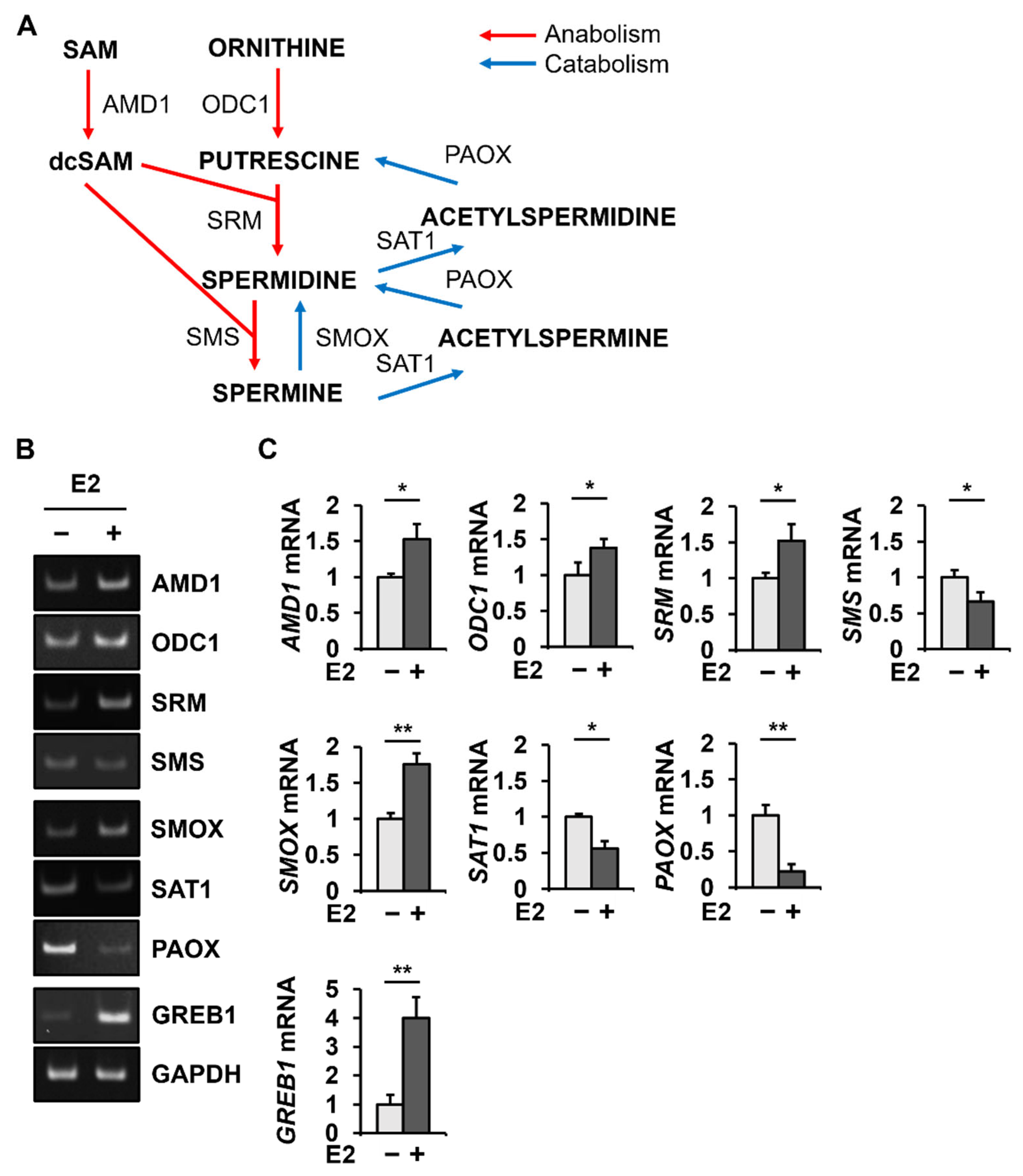
2.1. E2 Changes PAOX mRNA Levels Most among the Genes Involved in Polyamine Metabolism

The anabolic and catabolic enzymes involved in the polyamine metabolic pathway are shown (Figure 1A). To detect changes in polyamine metabolic enzymes due to E2 treatment, the mRNA levels of polyamine metabolic enzymes were analyzed in MCF-7 cells treated with or without E2 by conventional PCR (Figure 1B) and quantitative PCR (Figure 1C). The data from the conventional PCR were consistent with the quantitative PCR data. The quantitative PCR revealed that the level of GREB1 mRNA, which is known to be induced by E2 [31], increased to 400.1 ± 72.2% in MCF-7 cells. E2 treatment in MCF-7 cells increased the mRNA levels of AMD1 (154.9 ± 21.6%), ODC1 (138.0 ± 12.4%), SRM (152.6 ± 22.6%), and SMOX (176.0 ± 15.1%) but decreased the mRNA levels of SMS (66.5 ± 12.7%), SAT1 (56.1 ± 10.5%), and PAOX (22.6 ± 9.4%).
Among the genes encoding enzymes involved in polyamine metabolism, mRNA levels of PAOX showed the most significant change (4.3-fold decrease) after E2 treatment (Figure 1C). Therefore, changes in PAOX expression at the protein level were investigated in MCF-7 cells with or without E2 treatment. As expected, GREB1 levels were significantly increased to 2708.2 ± 164.2% by E2 treatment (Figure 2). PAOX polypeptides were detected as a major band of 56 kDa and a minor band of 35 kDa, suggesting isoform 1 (GenBank NP_690875.1) and isoform 2 (GenBank NP_997010.1), respectively (Figure 2A). Consistent with the E2-dependent decrease in PAOX mRNA levels, a significant decrease in levels of both PAOX polypeptides (16.9 ± 1.3% for the major 56-kda band) was observed after E2 treatment (Figure 2B).

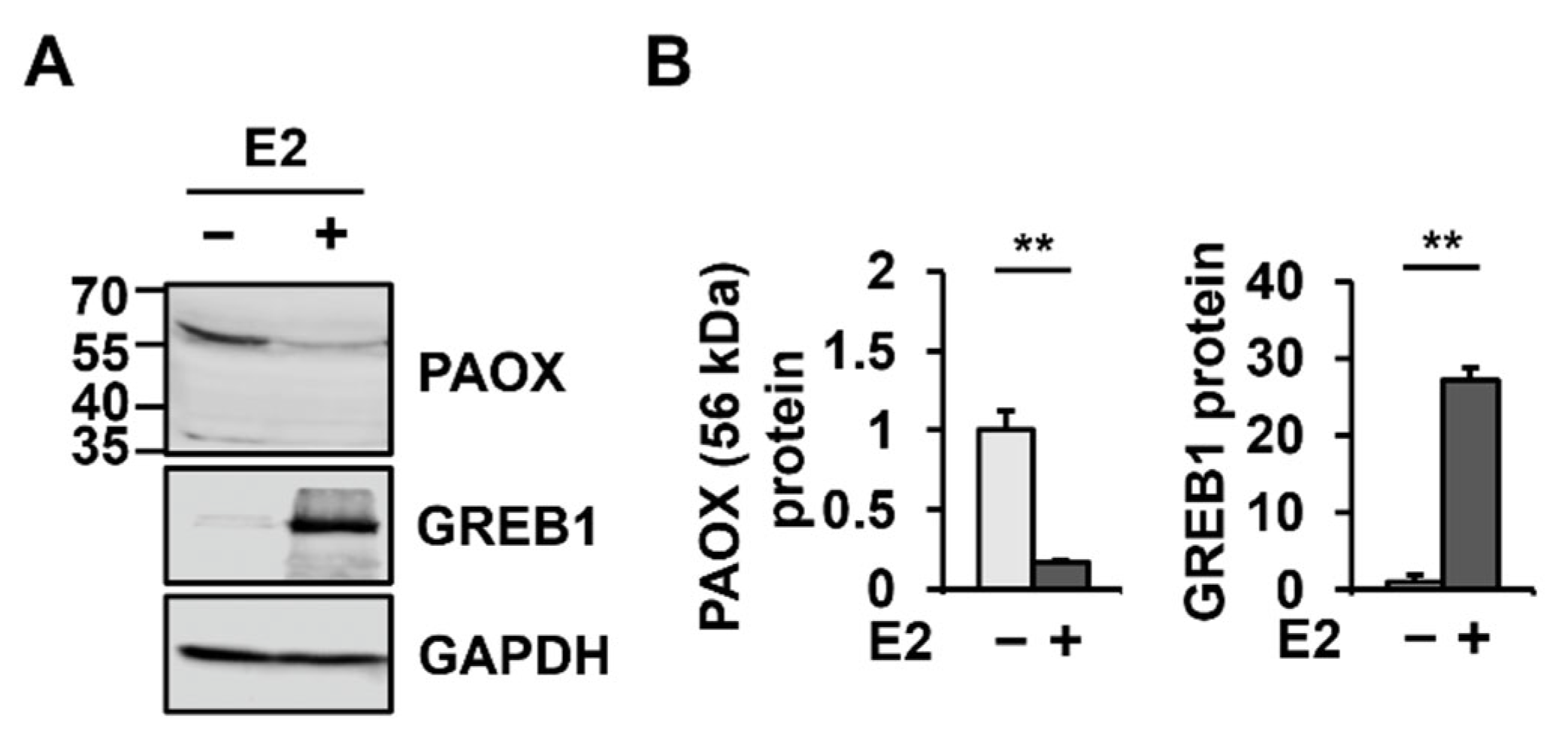
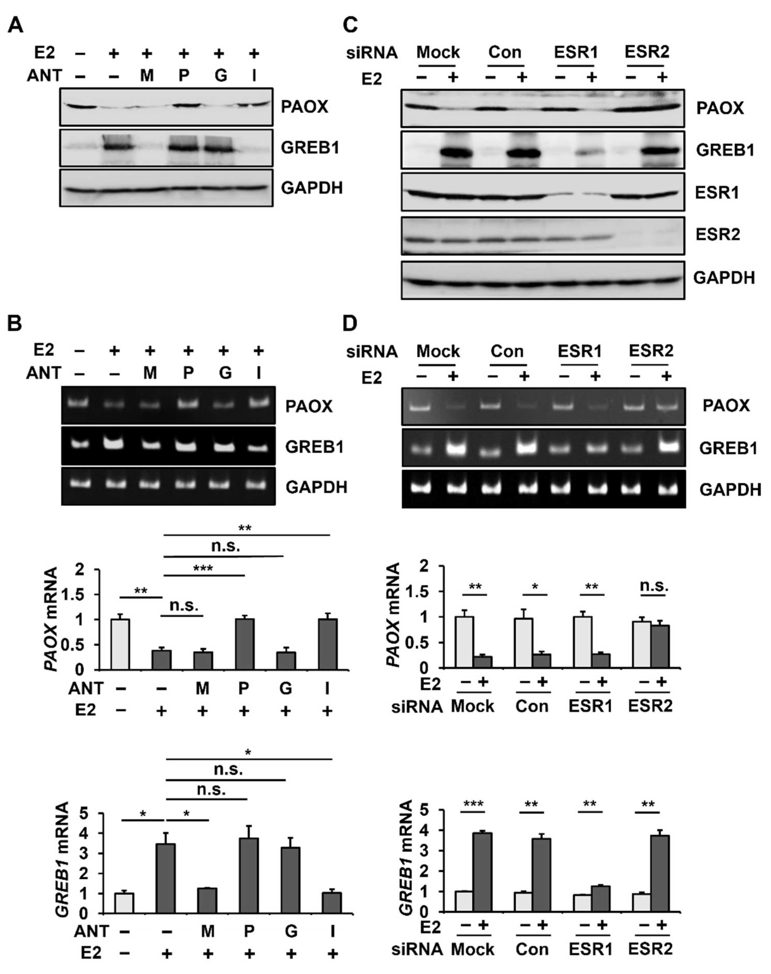
2.2. E2 Decreases PAOX Expression via the ESR2

To understand the estrogen receptor signaling pathway involved in E2-mediated PAOX downregulation, we analyzed E2-mediated PAOX expression in MCF-7 cells in the presence of MPP (antagonist of ESR1 (also known as ERα)), PHTPP (antagonist of ESR2 (also known as ERβ)), G-15 (antagonist of G Protein-Coupled Estrogen Receptor 1 (also known as GPER1)), and ICI 182,780 (non-specific ESR antagonist). E2-mediated upregulation of GREB1, which is known to involve ESR1 [31], was blocked by MPP and ICI 182,780. However, the E2-dependent downregulation of PAOX was hampered by PHTPP and ICI 182,780 but was not affected by MPP and G-15 (Figure 3A). E2-dependent decrease in PAOX mRNA levels was also blocked by PHTPP and ICI 182,780 but not MPP and G-15 (Figure 3B). These results suggest that E2 reduces PAOX expression through ESR2 in MCF-7 cells.
To clearly define the estrogen receptor involved in the E2-dependent downregulation of PAOX expression, we analyzed E2-mediated PAOX expression in MCF-7 cells with ESR1 or ESR2 knockdown using siRNA. The transfection of ESR1 or ESR2 siRNA into MCF-7 cells reduced the protein expression of ESR1 and ESR2, respectively (Figure 3C). ESR2 knockdown alleviated the decrease in PAOX induced by E2, while mock transfection or transfection with scrambled siRNA (control) or ESR1 siRNA did not alter the E2-mediated decrease in PAOX (Figure 3C). As previously reported [32], ESR1 knockdown severely blocked the increase in GREB1 by E2. In addition, it was also confirmed that E2-mediated PAOX repression and GREB1 induction were inhibited by ESR2 knockdown and ESR1 knockdown at the mRNA level, respectively (Figure 3D).

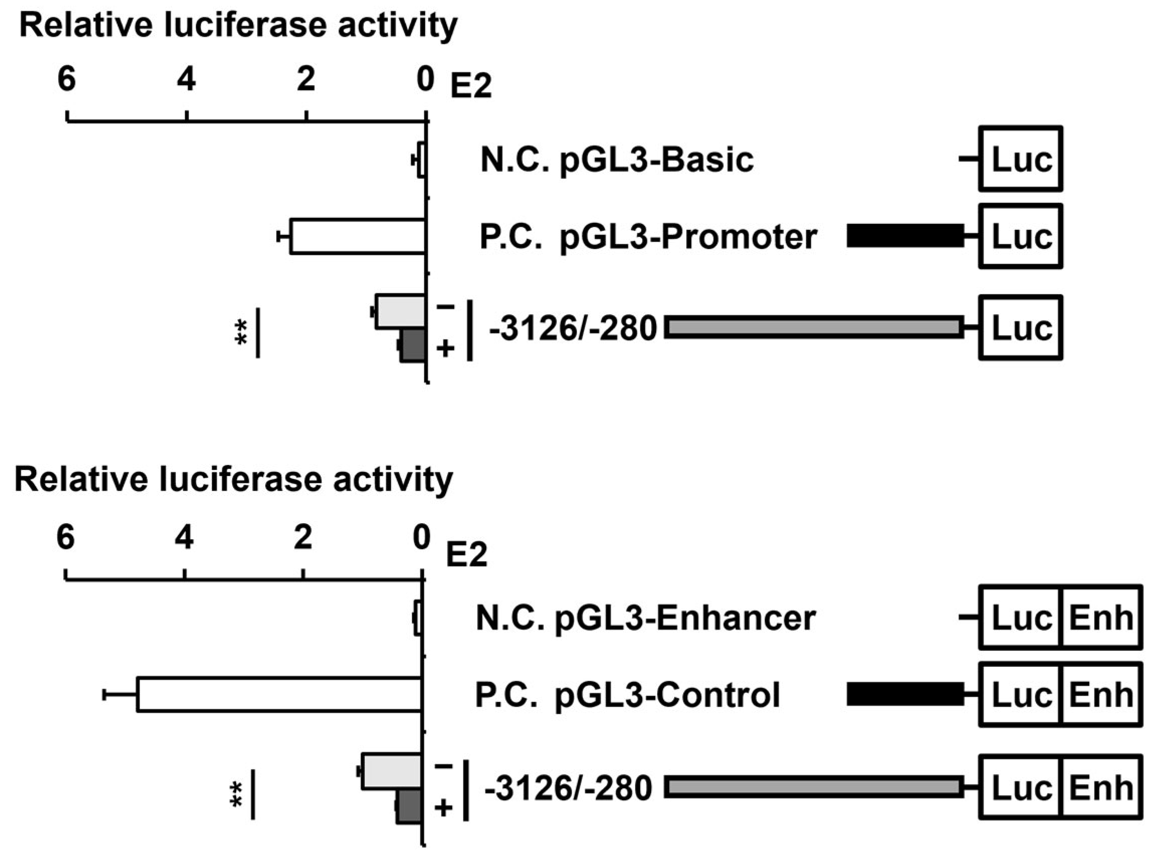
2.3. E2 Reduces the Activity of the PAOX Promoter in an ESR2-Dependent Manner

To understand whether the decrease in PAOX mRNA and protein levels by E2 is due to the decrease in PAOX transcriptional activity, we established constructs in which firefly luciferase cDNA was fused to the PAOX promoter (nucleotide positions −3126 to −280 from the start codon), and changes in PAOX promoter activity by E2 treatment in MCF-7 cells were measured by luciferase activity. Luciferase activity is driven by the PAOX promoter without an enhancer (pGL3-Basic-PAOX promoter (−3126/−280)) and was reduced to 49.9 ± 5.7% by treatment with E2 (Figure 4). Luciferase activity driven by the PAOX promoter in the presence of the SV40 enhancer (pGL3-Enhancer-PAOX promoter (−3126/−280)) was also reduced to 41.3 ± 1.6% by E2, a slightly larger difference in comparison to the absence of the SV40 enhancer. Therefore, it was confirmed that PAOX promoter activity is suppressed by E2. For subsequent analysis, we used a PAOX-promoter-driven luciferase construct containing the SV40 enhancer, where the effect of E2 can be better observed.
We then analyzed whether the reduction in PAOX promoter activity by E2 was ESR2-dependent by treatment with an ESR antagonist and siRNA in a PAOX promoter (−3126/−280)-driven reporter construct. As expected, the reduction in PAOX promoter activity by E2 was restored by PHTPP and ICI 182,780 but was not significantly altered by MPP and G-15 (Supplementary Figure S1A). In addition, transfection with ESR2 siRNA reversed the decrease in PAOX promoter activity by E2, but transfection with ESR1 siRNA did not (Supplementary Figure S1B).

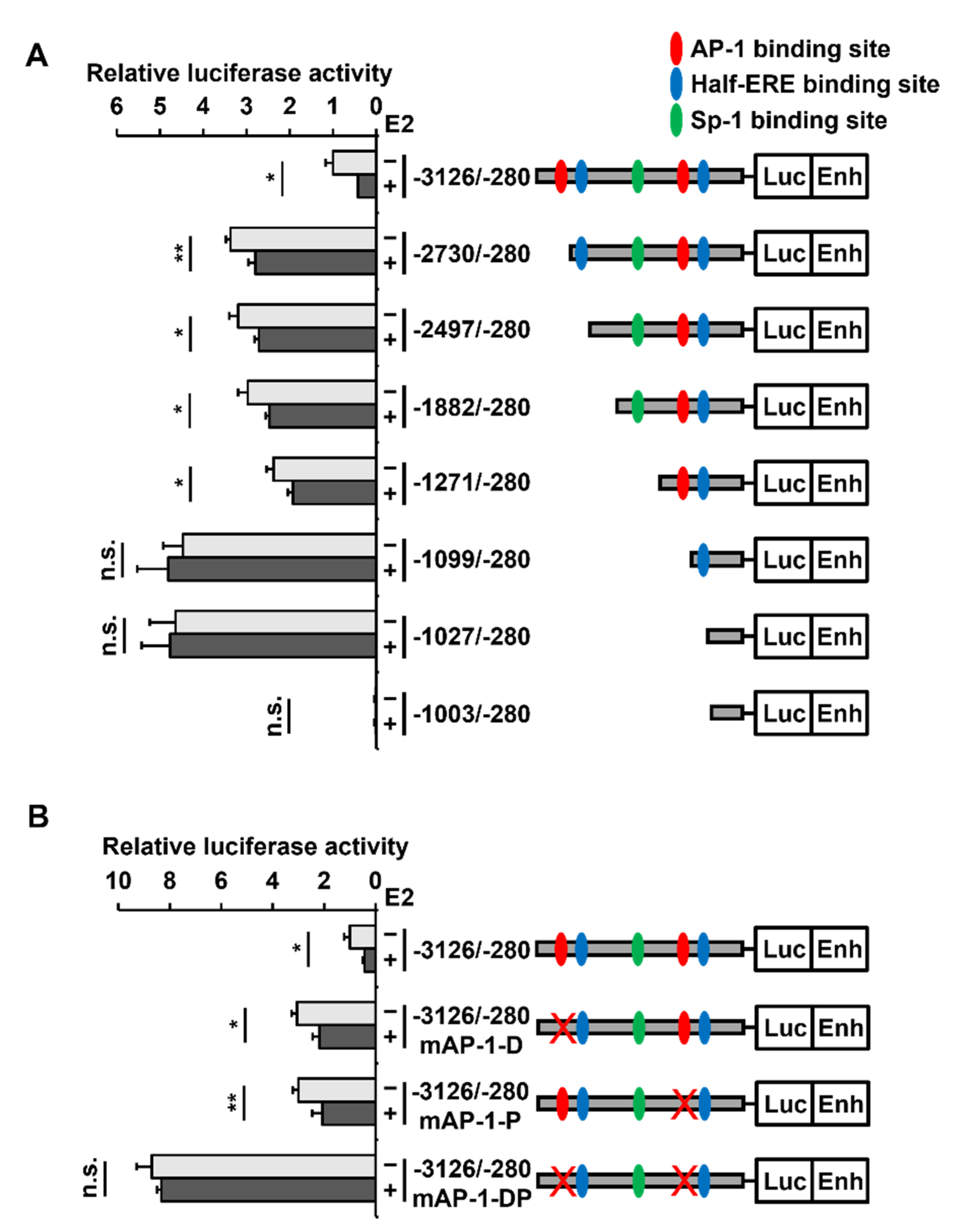
2.4. Two AP-1 Sites within the PAOX Promoter Are Involved in the E2-Mediated PAOX Repression

Using a transcription factor binding site search program (Jaspar database), two AP-1 at −2747/−2741 (TGATTCA) and −1158/−1152 (GGAGTCA), two half-ERE at −2516/−2511 (AGGTCA) and −1059/−1054 (AGGTCA), and one SP-1 at −1325/−1317 (TCGGGTGGGT) sites were identified in the PAOX promoter (−3126/−280) (Figure 5A). Luciferase analysis of pGL3-Enhancer–PAOX promoter (−3126/−280) derivatives with serial 5’-truncation of the PAOX promoter revealed that when the −3126/−2730 and −1271/−1099 regions including each of the two AP-1 binding sites in the PAOX promoter were deleted, promoter activity was increased gradually. In addition, these deletions gradually reduced the extent of the E2-dependent decrease in PAOX promoter (−3126/−280) activity. The −1099/−280 and −1027/−280 fragments of the PAOX promoter lacking two AP-1 sites no longer showed the E2-dependent decrease in promoter activity. The −1003/−280 fragment of the PAOX promoter did not show any promoter activity. These findings suggested that the two putative half-ERE sites and a putative SP-1 binding site in the PAOX promoter were not involved in the E2-dependent repression of the PAOX promoter.
To confirm the importance of AP-1 sites in E2-induced PAOX repression, site-directed mutagenesis at each of the two AP-1 binding sites, and subsequently at both the sites in the PAOX promoter, was performed, and the promoter activity of the reporter constructs was then measured. Luciferase analysis of the constructs showed that the repression of the PAOX promoter by E2 was reduced when the distal or proximal AP-1 binding site was mutated. In addition, when both AP-1 binding sites were mutated, the PAOX promoter activity was no longer altered by E2 (Figure 5B).

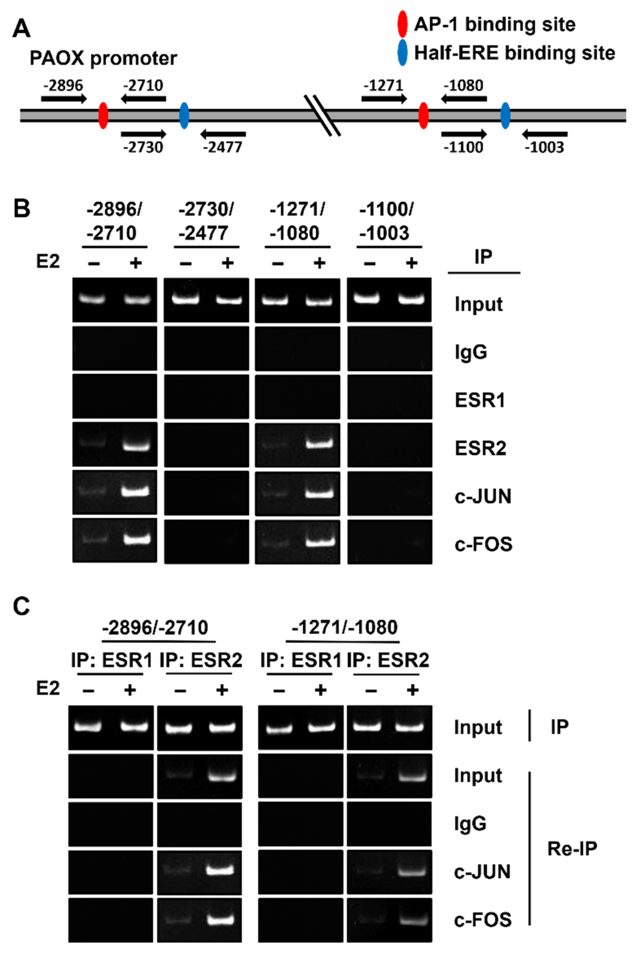
2.5. ESR2 Binds to the AP-1 Binding Sites of the PAOX Promoter

We showed that both ESR2 and AP-1 binding sites of the PAOX promoter are important for the E2-dependent repression of PAOX promoter activity. Therefore, the binding of c-JUN and c-FOS, constituting AP-1, and of ESR2 to the two AP-1 binding sites of the PAOX promoter was examined using chromatin immunoprecipitation (ChIP) analysis. ChIP reactions involving ESR1 and two putative half-ERE binding sites were used as negative controls. PCR products encompassing each of the two AP-1 binding sites were observed in all samples immunoprecipitated with the ESR2, c-JUN, or c-FOS antibodies in an E2-dependent manner but not in the sample immunoprecipitated with the ESR1 antibody (Figure 6A). In addition, no PCR products for the two half-ERE binding sites were observed in samples precipitated with ESR1, ESR2, c-JUN, or c-FOS antibodies. Therefore, c-JUN, c-FOS, and ESR2 were found to specifically bind to each of the two AP-1 binding sites in the PAOX promoter.
To analyze whether ESR2 binds to the AP-1 (c-JUN and c-FOS) bound to each of the two AP-1 binding sites of the PAOX promoter in an E2-dependent manner, Re-Immunoprecipitation (Re-IP) analysis was performed by immunoprecipitation of chromatin extracts first with anti-ESR1 or anti-ESR2 antibodies and secondarily with c-JUN or c-FOS antibodies. PCR products for each of the two AP-1 binding sites were observed in immunoprecipitates with the ESR2 antibody and then the c-FOS or c-JUN antibody in an E2-dependent manner (Figure 6B). PCR products for AP-1 binding sites were not detected in any immunoprecipitates with the ESR1 antibody. Therefore, we concluded that ESR2 binds to AP-1 (c-JUN and c-FOS) and is bound to each of the two AP-1 binding sites of the PAOX promoter in an E2-dependent manner.

3. Discussion

Herein, we examined changes in the expression of polyamine metabolic enzymes at the RNA level in ER-positive MCF-7 breast cancer cells, with and without the influence of E2. We found that E2 upregulates the mRNA levels of anabolic enzymes of polyamine metabolism, including AMD1, ODC1, and SRM but not SMS, which catalyzes the conversion of spermidine to spermine, and downregulates the mRNA levels of catabolic enzymes such as SAT1, and PAOX but not SMOX, which catalyzes the direct conversion of spermine to spermidine. We also found that, among polyamine metabolic enzymes, PAOX was most significantly altered by E2 in MCF-7 cells at the mRNA level. Furthermore, PAOX was reduced by E2 at both the protein and mRNA levels. As PAOX converts N1-acetylspermine and N1-acetylspermidine produced by SAT1 to spermidine or putrescine by oxidation, decreased PAOX expression results in increased polyamine and decreased H2O2 levels [33,34].
Estrogen transmits signals through three ESRs, namely, ESR1, ESR2, and the G-protein-coupled estrogen receptor 1 (GPER1) [35,36,37]. Once E2 binds to ESR1 or ESR2 in the cytoplasm, the ESRs undergo a structural change that induces receptor dimerization [38]. These E2–ESR complexes translocate to the nucleus and play a role in gene regulation by binding to the estrogen response element (ERE)-binding sites or transcription factors such as AP-1 and SP-1 [39,40,41,42]. In contrast to the ESR1 and ESR2, the GPER1 shows characteristics of G-protein-coupled receptors that act via the activation of the heterotrimeric G protein [43]. The GPER1 is anchored in the plasma membrane, and unlike nuclear estrogen receptors, signaling pathways involving GPER1 activation occur through a variety of secondary messengers via G-protein activation [44].
ESRs can regulate the expression of target genes by directly binding to EREs identified in estrogen-responsive promoters [32]. In addition, ESRs can regulate gene expression through protein–protein interactions with other transcription factors, such as the SP-1 and AP-1 [39,40,41], and co-regulators that have chromatin remodeling functions [45,46]. Approximately one-third of estrogen target genes in humans do not have an ERE or an ERE-like region [39].
ESRs can activate or repress the transcription of target genes. Molecular mechanisms that increase target gene expression through E2-ESR have been studied more than those that repress expression [47]. However, because nearly 70% of E2-regulated genes are repressed in MCF-7 cells [48], estrogen-mediated repression of target genes appears to be more common than activation. In estrogen-stimulated MCF-7 cells, the 6024 ESR1 and 9702 ESR2 binding sites, where the ESR1, ESR2, or both ER subtypes can bind, were identified using ChIP-Seq analysis [49]. Based on these results, it is expected that ESR2 is involved in the regulation of more genes than ESR1, at least in MCF-7 cells.
We established that ESR2 is responsible for E2-dependent PAOX expression repression and reporter assays using antagonists and siRNAs; however, the mechanism by which the E2–ESR2 complex downregulates PAOX expression was unclear. When we deleted the two AP-1 binding sites of the PAOX promoter, PAOX promoter activity in the absence of E2 gradually increased, consistent with a previous report [50]. More importantly, the deletion or mutation of the two AP-1 binding sites of the PAOX promoter gradually leads to the loss of E2-dependent promoter activity repression, while the deletion of two putative half-ERE and SP-1 binding sites did not affect promoter activity. ChIP and Re-IP assays revealed that the ESR2 as well as the c-FOS, and the c-JUN bound to each of the two AP-1 binding sites in the PAOX promoter, but the ESR2 did not bind to either of the two putative half-ERE sites. We confirmed that the E2–ESR2 complex binds to AP-1 bound to the two AP-1 binding sites of the PAOX promoter, thereby repressing PAOX transcription in MCF-7 cells.
The molecular mechanism of target gene downregulation by E2–ESR2 has been reported previously. For example, TNFAIP1 is downregulated by the binding of the E2–ESR2 directly to the ERE site on its promoter in primary hippocampal cells [51]. In addition, ESR1 expression is repressed by the indirect binding of E2–ESR2 to SP-1 in MCF-7 cells [52]. As a novel finding, we showed that the E2–ESR2 complex downregulates the expression of PAOX by binding to the AP-1 bound to AP-1 binding sites. Therefore, to the best of our knowledge, our finding is the first example of E2 repressing a target gene through ESR2–AP-1 interaction, independent of ERE.
In this study, we found that E2 upregulated the expression of most anabolic enzymes and downregulated the expression of most catabolic enzymes involved in polyamine metabolism. In addition, we demonstrated that PAOX expression, which was most significantly altered by E2 in MCF-7 cells, was repressed by the binding of the E2–ESR2 complex to AP-1 bound to the two AP-1 binding sites of the PAOX promoter.
Our data suggest that an increase in estrogen level would lead to an increase in the expression of polyamine anabolic enzymes, including the AMD1, ODC1, and SRM, and a decrease in the expression of polyamine catabolic enzymes, including SAT1 and PAOX, which leads to an increase in polyamine levels. This finding is consistent with a previous report that polyamine levels were increased upon E2 treatment in MCF-7 cells [53]. This estrogen-dependent change in polyamine levels may help to understand the hormone therapy of breast cancer and side effects of menopause, from the perspective of polyamine. A selective estrogen receptor modulator (SERM), such as tamoxifen, can reduce the incidence of hormone-receptor-positive breast cancer and the likelihood of cancer recurrence [54]. SERM treatment reduces polyamine levels in MCF-7 cells [55]. It is well documented that polyamines play a role in tumorigenesis, at least by increasing cell proliferation and suppressing apoptosis [4]. For example, the ODC1 inhibitor α-difluoromethylornithine is known to prevent the growth-promoting effect of E2 [56,57]. Moreover, sensitivity to the growth-promoting effects of E2 was reduced by about a third in MCF-7 cells overexpressing ODC1 compared with vector-transfected cells [58]. These results suggest that elevated polyamine levels contribute to estrogen-induced oncogenic phenotypes in breast cancer cells. In addition, a decrease in estrogen during menopause leads to a decrease in polyamine levels. Polyamine reduction is a common occurrence in postmenopausal women and has many clinical implications [59,60,61,62]. For example, polyamines effectively prevent bone loss in ovariectomized mice [20]. In addition, polyamine reduction leads to increased H2O2 and ROS levels, which, as previously mentioned, contribute to inflammatory diseases [63]. Therefore, we propose that preventing polyamine decline, for example by repressing or inhibiting PAOX, could be a potential treatment for alleviating postmenopausal symptoms.

4. Methods and Materials

4.1. Reagents and Antibodies

The ESR1 antagonist 1,3-Bis(4-hydroxyphenyl)-4-methyl-5-[4-(2-piperidinylethoxy)phenol]-1H-pyrazole dihydrochloride (MPP), ESR2 antagonist 4-[2-Phenyl-5,7-bis(trifluoromethyl)pyrazolo [1,5-a]pyrimidin-3-yl]phenol (PHTPP), the GPER1 antagonist (3aS*,4R*,9bR*)-4-(6-Bromo-1,3-benzodioxol-5-yl)-3a,4,5,9b-3H-cyclopenta[c]quinoline (G-15), and the non-specific ESR antagonist 7α,17β-[9-[(4,4,5,5,5-Pentafluoropentyl)sulfinyl]nonyl]estra-1,3,5(10)-triene-3,17-diol (ICI 182,780) were purchased from Tocris (Bristol, UK). The water-soluble β-estradiol (E2) and 2-hydroxypropyl-β-cyclodextrin (carrier for β-estradiol) were purchased from Sigma-Aldrich (St. Louis, MO, USA). The following antibodies were used: anti-PAOX (MBS3211539), obtained from MyBioSource (San Diego, CA, USA); anti-ESR1 (sc-8002) and anti-ESR2 (sc-373885), obtained from Santa Cruz Biotechnology (Cambridge, UK); anti-GREB1 (ab-72997), obtained from Cell Signaling (Danvers, MA, USA); anti-c-JUN (2250S) and anti-c-FOS (9165S), obtained from Abcam (Cambridge, UK); anti-GAPDH (AbC-2003), obtained from AbClone (Seoul, Korea); and the horseradish peroxidase-conjugated goat anti-mouse IgG and goat anti-rabbit IgG described previously [64].

4.2. Cell Culture

The MCF-7 cells were grown in DMEM (Hyclone, South Logan, UT, USA) supplemented with 10% fetal bovine serum (FBS; Gibco of Thermo Fisher Scientific, Waltham, MA, USA), 100 U/mL of penicillin, and 100 μg/mL of streptomycin at 37 °C under 5% CO2 and 95% air.

4.3. RNA Isolation and PCR

The total RNA was isolated from MCF-7 cells using TRIZOL (Invitrogen, Thermo Fisher Scientific), as described previously [65]. The cDNA mixtures were synthesized from the total RNA by reverse transcription-PCR using oligo dT15 primers and the AMV RT system (Promega, Madison, WI, USA) according to the manufacturer’s instructions. Conventional PCR was performed in a final volume of 10 μL containing 1 pM each of the 5′ primer and 3′ primer, 0.2 mM dNTPs, 1× Taq PCR buffer, 50 U/mL Taq polymerase, and cDNAs synthesized from 0.1 μg total RNA. PCR was performed with 25–30 cycles; the cycling conditions were as follows: denaturation at 94 °C for 30 s, annealing at the appropriate annealing temperature (Supplementary Table S1) for 30 s, and extension at 72 °C for 30 s. The PCR products were detected by a 5% PAGE analysis and visualized by ethidium bromide staining. Real-time PCR was conducted using a QuantiTect SYBR Green PCR kit (Qiagen, Hilden, Germany) and QuantStudio 3 Real-Time PCR system (Applied Biosystems, Foster City, CA, USA). The expression of the target genes was normalized to that of GAPDH expression.

4.4. Western Blotting

The cells were harvested using an SDS sample buffer (50 mM Tris-HCl, pH 6.8, 2% SDS, 0.1% bromophenol blue, and 10% glycerol) on ice for 15 min and centrifuged at 18,000 × g for 15 min after brief sonication. The cell lysate samples were boiled after the addition of 100 mM β-mercaptoethanol for 4 min and resolved by SDS-PAGE. Proteins in the gel were blotted onto polyvinylidene fluoride (PVDF) membranes (Millipore, Billerica, MA, USA). The blots were incubated with the indicated primary and secondary antibodies. Immunoreactive signals were detected using the Immobilon Western HRP substrate (Millipore, Billerica, MA, USA) and an LAS-3000 detector (Fujifilm, Tokyo, Japan) as previously described [66].

4.5. Treatment of E2 to MCF-7 Cells

The subconfluent MCF-7 cells were incubated in phenol-red free DMEM (PRF-DMEM; HyClone) containing 10% charcoal-stripped FBS (Scipak Lifesciences, Arlington, VA, USA) for 24 h. The medium was replaced with a fresh medium containing water-soluble β-estradiol (10 nM E2 in the presence of 35.1 nM 2-hydroxypropyl-β-cyclodextrin) or 2-hydroxypropyl-β-cyclodextrin (35.1 nM).

4.6. Transfection of siRNAs

The ON-TARGETplus Human ESR1 and ESR2 siRNAs (600 pM siRNA/100 mm plate; Dharmacon, Lafayette, CO, USA) (600 pM siRNA/100 mm plate) were transfected into MCF-7 cells using Lipofectamine 2000 (Thermo Fisher Scientific).

4.7. Prediction of Transcription Factor-Binding Site

The transcription factor-binding sites on the PAOX promoter were searched using CiiiDER, an integrated computational toolkit [67], and the transcription factor-binding site database from JASPAR (http://jaspar.cgb.ki.se/, accessed on 23 September 2021).

4.8. Cloning of the Human PAOX Promoter in Luciferase Reporter Vectors

To generate a luciferase reporter construct for the human PAOX promoter (GenBank accession no. NM_152911.4), a fragment of the PAOX promoter −3126/−280 (the first nucleotide upstream of the start codon is referred to as −1) was PCR-amplified using genomic DNA from human foreskin fibroblasts (as previously described [68]) as a template with PrimeSTAR® GXL DNA polymerase (TaKaRa, Shiga-ken, Japan). The deletion products were PCR-amplified using the PAOX promoter (−3126/−280) as a template, using the indicated primer pairs (Supplementary Table S2). The PCR products were cloned into pGL3-Basic or pGL3-Enhancer luciferase reporter vectors digested with Acc65I and HindIII (Promega, Madison, WI, USA) using the In-Fusion Cloning system (Takara). Luciferase reporter constructs harboring the human PAOX promoter were confirmed to be error-free by DNA sequencing.

4.9. Dual-Luciferase Reporter Assay

The transfection of the luciferase constructs into MCF-7 cells was performed using Lipofectamine 2000. The MCF-7 cells were plated in 96-well plates at a density of 5 × 10⁴ cells/well and incubated in 100 μL of DMEM supplemented with 10% FBS for 12 h. Promoterless pGL3-Basic (0.11 μg) or pGL3-Basic-PAOX promoter constructs (0.18 μg) encoding firefly luciferase driven by PAOX promoter, or promoterless pGL3-Enhancer (0.12 μg) or pGL3-Enhancer-PAOX promoter constructs (0.19 μg of pGL3-Enhancer-PAOX promoter(-3126/-280) or the amount corresponding to the size of each construct) and pRL-TK encoding Renilla luciferase driven by herpes simplex virus thymidine kinase promoter (0.02 μg; Promega, Madison, WI, USA) in 25 μL Opti-MEM (Gibco/Thermo Fisher Scientific) were incubated with Lipofectamine 2000 (0.75 μL) and diluted in 25 μL Opti-MEM for 30 min at room temperature. Cells in Opti-MEM were treated with this mixture for 5 h and then replaced with DMEM supplemented with 10% FBS for 12 h. The cells were then incubated with PRF-DMEM supplemented with 10% charcoal-stripped FBS for 24 h. Next, the cells were treated with or without E2 in PRF-DMEM supplemented with 10% charcoal-stripped FBS for 24 h. Luciferase activity was measured using the dual-luciferase reporter assay system (Promega), and the firefly luciferase activity in transfected cells was normalized to that of the Renilla luciferase.

4.10. Site-Directed Mutagenesis (SDM)

Mutations in the distal and proximal AP-1 sites in the PAOX promoter were introduced by overlap extension PCR mutagenesis using the PrimeSTAR® GXL DNA polymerase. The sequences of the primer pairs used are listed in Supplementary Table S3. To generate a mutation in the distal AP-1 site, two primary PCR products with Primer −3126-F and mAP-1-D-R and with Primer mAP-1-D-F and −280-R using the pGL3-Enhencer-PAOX promoter (−3126/−280) as a template were obtained. Similarly, to generate a mutation in the proximal AP-1 site, two primary PCR products with Primer −3126-F and AP-1-P-R and with Primer mAP-1-P-F and −280-R using the pGL3-Enhencer-PAOX promoter (−3126/−280) as a template were obtained. In addition, to generate double mutations in the proximal and distal AP-1 sites, two primary PCR products with Primer −3126-F and AP-1-P-R and Primer mAP-1-P-F and −280-R using the pGL3-Enhencer-PAOX promoter (−3126/−280)-mAP-1-D as a template were obtained. Then, a second PCR was performed to create an overlap-extension PCR product using a mixture of PCR products for distal AP-1 site mutation or proximal AP-1 site mutation as a template, as well as Primer −3126 and −280. Overlap-extended PCR products harboring mutations in the distal or proximal AP-1 site or double mutations in the distal and proximal AP-1 sites were cloned into the pGL3-Enhancer vector cleaved with Acc65I and HindIII to generate the pGL3-Enhancer-PAOX promoter (−3126/−280)-mAP-1-D, -mAP-1-P, and -mAP-1-DP, respectively. Luciferase reporter constructs with mutations in AP-1 sites of the PAOX promoter were confirmed to be error-free by DNA sequencing.

4.11. ChIP and Re-IP

ChIP was performed as previously described [69], with minor modifications. The MCF-7 cells were treated with E2 or vehicle for 24 h and fixed with 1% formaldehyde at room temperature for 10 min. The reaction was stopped by adding 0.125 M glycine and washing twice after 5 min with ice-cold PBS. The cells were lysed for 10 min by adding a cell lysis/wash buffer (150 mM NaCl, 50 mM Tris-HCl, pH 7.5, 5 mM EDTA, 0.5% NP-40, 1.0% Triton X-100) at 4 °C and a protease-inhibitor cocktail (Sigma-Aldrich, MO, USA). Then, cells were centrifuged at 1500 rpm at 4 °C for 5 min, and the pellet containing the chromatin fraction was lysed in a shearing buffer (1% SDS, 10 mM EDTA, 50 mM Tris-HCl pH 8.0) and protease inhibitor cocktail. The chromatin extracts containing genomic DNA were sonicated on ice to obtain an average fragment size of 500 bp. The supernatant was diluted 9 times with a diluted buffer (0.01% SDS, 1.1% Triton X-100, 1.2 mM EDTA, 16.7 mM Tris-HCl, pH 8.1, 167 mM NaCl) and subsequently immunoprecipitated using antibodies and Protein G-agarose (Invitrogen) at 4 °C. The immunoprecipitates were washed once with cold cell lysis/wash buffer and three times with cold ethanol. DNA purification followed by protein degradation was performed using Chelex 100 (Bio-Rad, Hercules, CA, USA), following the manufacturer’s instructions. The PCR was performed in 30 cycles under the following conditions: denaturation at 94 °C for 30 s, annealing at an indicated annealing temperature (Supplementary Table S4) for 30 s, and extension at 72 °C for 30 s. The Re-IP assay was performed as previously described [70] with minor modifications. The immunoprecipitated antibody–protein–DNA complexes were washed twice with the cell lysis/wash buffer and eluted by incubating for 30 min at 37 °C in a Re-IP elution buffer (TE buffer (10 mM Tris-HCl, pH 8.0, 1 mM EDTA), 2% SDS, 15 mM DTT). After centrifugation, the supernatant was diluted 20 times using a dilution buffer and individually re-immunoprecipitated with each secondary antibody at 4 °C. The immune complexes were incubated with protein G-agarose beads for 2 h at 4 °C. The DNA was purified from the secondary immune complex using Chelex 100. The PCR was performed using the extracted DNA as described above.

4.12. Statistical Analyses

All data are presented as a mean ± standard deviation of at least three independent experiments. Statistical significance was analyzed using the unpaired two-tailed Student’s t-test. A p-value < 0.05 was considered statistically significant.

5. Conclusions

In this study, we showed that PAOX was significantly altered among polyamine metabolism-related enzymes upon E2 treatment in MCF-7 cells. We demonstrated that PAOX expression was downregulated by E2 via ESR2. The ESR2 binds to the AP-1 complex bound to two AP-1 binding sites in the PAOX promoter. The E2–ESR2–AP-1 complex binds to the PAOX promoter and downregulates PAOX expression. Based on our findings, we suggest that E2-dependent inhibition of PAOX is at least one cause of polyamine decline during menopause

Supplementary Materials

The Supplementary Materials are available online at https://0-www-mdpi-com.brum.beds.ac.uk/article/10.3390/ijms23147521/s1.

Author Contributions

Conceptualization, S.-T.L.; Methodology, J.H.K.; Validation, J.H.K.; Investigation, J.H.K.; Resources, J.H.K. and S.-T.L.; Data Curation, J.H.K. and S.-T.L.; Writing—Original Draft Preparation, J.H.K.; Writing—Review and Editing, S.-T.L.; Visualization, J.H.K. and S.-T.L.; Supervision, S.-T.L.; Project Administration, S.-T.L.; Funding Acquisition, S.-T.L. All authors have read and agreed to the published version of the manuscript.

Funding

This study was funded by the National Research Foundation of Korea (grant number 2020R1A2C2006556).

Institutional Review Board Statement

Not applicable.

Informed Consent Statement

Not applicable.

Data Availability Statement

Authors can confirm that all relevant data are included in the article.

Acknowledgments

J.H.K. was a fellowship awardee of the BK21 FOUR program.

Conflicts of Interest

The authors declare no conflict of interest.

References

  1. Battaglia, V.; DeStefano Shields, C.; Murray-Stewart, T.; Casero, R.A., Jr. Polyamine catabolism in carcinogenesis: Potential targets for chemotherapy and chemoprevention. Amino Acids 2014, 46, 511–519. [Google Scholar] [CrossRef] [PubMed] [Green Version]
  2. Urdiales, J.L.; Medina, M.A.; Sánchez-Jiménez, F. Polyamine metabolism revisited. Eur. J. Gastroenterol. Hepatol. 2001, 13, 1015–1019. [Google Scholar] [CrossRef] [PubMed]
  3. Bae, D.H.; Lane, D.J.R.; Jansson, P.J.; Richardson, D.R. The old and new biochemistry of polyamines. Biochim. Biophys. Acta. Gen. Subj. 2018, 1862, 2053–2068. [Google Scholar] [CrossRef] [PubMed]
  4. Casero, R.A., Jr.; Murray Stewart, T.; Pegg, A.E. Polyamine metabolism and cancer: Treatments, challenges and opportunities. Nat. Rev. Cancer 2018, 18, 681–695. [Google Scholar] [CrossRef] [PubMed]
  5. Pegg, A.E. Mammalian polyamine metabolism and function. IUBMB Life 2009, 61, 880–894. [Google Scholar] [CrossRef] [PubMed]
  6. Sandusky-Beltran, L.A.; Kovalenko, A.; Ma, C.; Calahatian, J.I.T.; Placides, D.S.; Watler, M.D.; Hunt, J.B.; Darling, A.L.; Baker, J.D.; Blair, L.J.; et al. Spermidine/spermine-N(1)-acetyltransferase ablation impacts tauopathy-induced polyamine stress response. Alzheimers Res. Ther. 2019, 11, 58. [Google Scholar] [CrossRef]
  7. Shin, V.Y.; Liu, E.S.; Koo, M.W.; Wang, J.Y.; Matsui, H.; Cho, C.H. Cigarette smoke extracts delay wound healing in the stomach: Involvement of polyamine synthesis. Exp. Biol. Med. 2002, 227, 114–124. [Google Scholar] [CrossRef]
  8. Kubo, S.; Tamori, A.; Nishiguchi, S.; Kinoshita, H.; Hirohashi, K.; Kuroki, T.; Omura, T.; Otani, S. Effect of alcohol abuse on polyamine metabolism in hepatocellular carcinoma and noncancerous hepatic tissue. Surgery 1998, 123, 205–211. [Google Scholar] [CrossRef]
  9. Igarashi, K.; Ueda, S.; Yoshida, K.; Kashiwagi, K. Polyamines in renal failure. Amino Acids 2006, 31, 477–483. [Google Scholar] [CrossRef]
  10. Zwighaft, Z.; Aviram, R.; Shalev, M.; Rousso-Noori, L.; Kraut-Cohen, J.; Golik, M.; Brandis, A.; Reinke, H.; Aharoni, A.; Kahana, C.; et al. Circadian Clock Control by Polyamine Levels through a Mechanism that Declines with Age. Cell Metab. 2015, 22, 874–885. [Google Scholar] [CrossRef] [Green Version]
  11. Das, R.; Kanungo, M.S. Activity and modulation of ornithine decarboxylase and concentrations of polyamines in various tissues of rats as a function of age. Exp. Gerontol. 1982, 17, 95–103. [Google Scholar] [CrossRef]
  12. Minois, N.; Carmona-Gutierrez, D.; Madeo, F. Polyamines in aging and disease. Aging 2011, 3, 716–732. [Google Scholar] [CrossRef] [PubMed] [Green Version]
  13. Takahashi, T.A.; Johnson, K.M. Menopause. Med. Clin. North. Am. 2015, 99, 521–534. [Google Scholar] [CrossRef] [PubMed]
  14. Hoga, L.; Rodolpho, J.; Gonçalves, B.; Quirino, B. Women’s experience of menopause: A systematic review of qualitative evidence. JBI Database System Rev. Implement. Rep. 2015, 13, 250–337. [Google Scholar] [CrossRef]
  15. Hall, G.; Phillips, T.J. Estrogen and skin: The effects of estrogen, menopause, and hormone replacement therapy on the skin. J. Am. Acad. Dermatol. 2005, 53, 555–568. [Google Scholar] [CrossRef] [PubMed]
  16. Hodis, H.N.; Mack, W.J.; Henderson, V.W.; Shoupe, D.; Budoff, M.J.; Hwang-Levine, J.; Li, Y.; Feng, M.; Dustin, L.; Kono, N.; et al. Vascular Effects of Early versus Late Postmenopausal Treatment with Estradiol. N. Engl. J. Med. 2016, 374, 1221–1231. [Google Scholar] [CrossRef] [PubMed]
  17. Goluch-Koniuszy, Z.S. Nutrition of women with hair loss problem during the period of menopause. Prz. Menopauzalny 2016, 15, 56–61. [Google Scholar] [CrossRef] [PubMed] [Green Version]
  18. Henderson, V.W. Menopause and disorders of the central nervous system. Minerva Ginecol. 2005, 57, 579–592. [Google Scholar]
  19. Management of osteoporosis in postmenopausal women: The 2021 position statement of The North American Menopause Society. Menopause 2021, 28, 973–997. [CrossRef]
  20. Yamamoto, T.; Hinoi, E.; Fujita, H.; Iezaki, T.; Takahata, Y.; Takamori, M.; Yoneda, Y. The natural polyamines spermidine and spermine prevent bone loss through preferential disruption of osteoclastic activation in ovariectomized mice. Br. J. Pharmacol. 2012, 166, 1084–1096. [Google Scholar] [CrossRef] [Green Version]
  21. Byun, J.A.; Choi, M.H.; Moon, M.H.; Kong, G.; Chul Chung, B. Serum polyamines in pre- and post-operative patients with breast cancer corrected by menopausal status. Cancer Lett. 2009, 273, 300–304. [Google Scholar] [CrossRef] [PubMed]
  22. Gray, H.E.; Jasper, T.W.; Luttge, W.G.; Shukla, J.B.; Rennert, O.M. Estrogen increases hypothalamic and pituitary polyamine levels in ovariectomized rats. J. Neurochem. 1980, 34, 753–755. [Google Scholar] [CrossRef] [PubMed]
  23. Lee, Y.R.; Lew, B.L.; Sim, W.Y.; Lee, J.; Hong, J.; Chung, B.C. Altered polyamine profiling in the hair of patients with androgenic alopecia and alopecia areata. J. Dermatol. 2019, 46, 985–992. [Google Scholar] [CrossRef] [PubMed]
  24. Ramot, Y.; Pietilä, M.; Giuliani, G.; Rinaldi, F.; Alhonen, L.; Paus, R. Polyamines and hair: A couple in search of perfection. Exp. Dermatol. 2010, 19, 784–790. [Google Scholar] [CrossRef]
  25. Takano, K.; Ogura, M.; Nakamura, Y.; Yoneda, Y. Neuronal and glial responses to polyamines in the ischemic brain. Curr. Neurovasc. Res. 2005, 2, 213–223. [Google Scholar] [CrossRef] [PubMed]
  26. Polis, B.; Karasik, D.; Samson, A.O. Alzheimer’s disease as a chronic maladaptive polyamine stress response. Aging 2021, 13, 10770–10795. [Google Scholar] [CrossRef]
  27. Iezaki, T.; Hinoi, E.; Yamamoto, T.; Ishiura, R.; Ogawa, S.; Yoneda, Y. Amelioration by the natural polyamine spermine of cartilage and bone destruction in rats with collagen-induced arthritis. J. Pharmacol. Sci. 2012, 119, 107–111. [Google Scholar] [CrossRef] [PubMed] [Green Version]
  28. Chevalier, C.; Kieser, S.; Çolakoğlu, M.; Hadadi, N.; Brun, J.; Rigo, D.; Suárez-Zamorano, N.; Spiljar, M.; Fabbiano, S.; Busse, B.; et al. Warmth Prevents Bone Loss Through the Gut Microbiota. Cell Metab. 2020, 32, 575–590. [Google Scholar] [CrossRef]
  29. Thomas, T.; Thomas, T.J. Estradiol control of ornithine decarboxylase mRNA, enzyme activity, and polyamine levels in MCF-7 breast cancer cells: Therapeutic implications. Breast Cancer Res. Treat. 1994, 29, 189–201. [Google Scholar] [CrossRef]
  30. Cohen, S.; O’Malley, B.W.; Stastny, M. Estrogenic induction of ornithine decarboxylase in vivo and in vitro. Science 1970, 170, 336–338. [Google Scholar] [CrossRef]
  31. Hodgkinson, K.; Forrest, L.A.; Vuong, N.; Garson, K.; Djordjevic, B.; Vanderhyden, B.C. GREB1 is an estrogen receptor-regulated tumour promoter that is frequently expressed in ovarian cancer. Oncogene 2018, 37, 5873–5886. [Google Scholar] [CrossRef] [PubMed] [Green Version]
  32. Deschênes, J.; Bourdeau, V.; White, J.H.; Mader, S. Regulation of GREB1 transcription by estrogen receptor alpha through a multipartite enhancer spread over 20 kb of upstream flanking sequences. J. Biol. Chem. 2007, 282, 17335–17339. [Google Scholar] [CrossRef] [PubMed] [Green Version]
  33. Murray Stewart, T.; Dunston, T.T.; Woster, P.M.; Casero, R.A., Jr. Polyamine catabolism and oxidative damage. J. Biol. Chem. 2018, 293, 18736–18745. [Google Scholar] [CrossRef] [PubMed] [Green Version]
  34. Smirnova, O.A.; Bartosch, B.; Zakirova, N.F.; Kochetkov, S.N.; Ivanov, A.V. Polyamine Metabolism and Oxidative Protein Folding in the ER as ROS-Producing Systems Neglected in Virology. Int. J. Mol. Sci. 2018, 19, 1219. [Google Scholar] [CrossRef] [PubMed] [Green Version]
  35. Vrtačnik, P.; Ostanek, B.; Mencej-Bedrač, S.; Marc, J. The many faces of estrogen signaling. Biochem. Med. 2014, 24, 329–342. [Google Scholar] [CrossRef] [Green Version]
  36. Chen, C.; Gong, X.; Yang, X.; Shang, X.; Du, Q.; Liao, Q.; Xie, R.; Chen, Y.; Xu, J. The roles of estrogen and estrogen receptors in gastrointestinal disease. Oncol. Lett. 2019, 18, 5673–5680. [Google Scholar] [CrossRef] [Green Version]
  37. Weatherman, R.V. Untangling the estrogen receptor web. Nat. Chem. Biol. 2006, 2, 175–176. [Google Scholar] [CrossRef]
  38. Powell, E.; Xu, W. Intermolecular interactions identify ligand-selective activity of estrogen receptor alpha/beta dimers. Proc. Natl. Acad. Sci. USA 2008, 105, 19012–19017. [Google Scholar] [CrossRef] [Green Version]
  39. Fuentes, N.; Silveyra, P. Estrogen receptor signaling mechanisms. Adv. Protein Chem. Struct. Biol. 2019, 116, 135–170. [Google Scholar]
  40. Nilsson, S.; Mäkelä, S.; Treuter, E.; Tujague, M.; Thomsen, J.; Andersson, G.; Enmark, E.; Pettersson, K.; Warner, M.; Gustafsson, J.A. Mechanisms of estrogen action. Physiol. Rev. 2001, 81, 1535–1565. [Google Scholar] [CrossRef]
  41. Safe, S.; Kim, K. Non-classical genomic estrogen receptor (ER)/specificity protein and ER/activating protein-1 signaling pathways. J. Mol. Endocrinol. 2008, 41, 263–275. [Google Scholar] [CrossRef] [PubMed]
  42. Lecomte, S.; Demay, F.; Ferrière, F.; Pakdel, F. Phytochemicals Targeting Estrogen Receptors: Beneficial Rather Than Adverse Effects? Int. J. Mol. Sci. 2017, 18, 1381. [Google Scholar] [CrossRef] [PubMed] [Green Version]
  43. Rouhimoghadam, M.; Lu, A.S.; Salem, A.K.; Filardo, E.J. Therapeutic Perspectives on the Modulation of G-Protein Coupled Estrogen Receptor, GPER, Function. Front Endocrinol. 2020, 11, 591217. [Google Scholar] [CrossRef] [PubMed]
  44. DeLeon, C.; Wang, D.Q.; Arnatt, C.K. G Protein-Coupled Estrogen Receptor, GPER1, Offers a Novel Target for the Treatment of Digestive Diseases. Front Endocrinol. 2020, 11, 578536. [Google Scholar] [CrossRef] [PubMed]
  45. Chen, H.; Lin, R.J.; Xie, W.; Wilpitz, D.; Evans, R.M. Regulation of hormone-induced histone hyperacetylation and gene activation via acetylation of an acetylase. Cell 1999, 98, 675–686. [Google Scholar] [CrossRef] [Green Version]
  46. Stossi, F.; Madak-Erdogan, Z.; Katzenellenbogen, B.S. Estrogen receptor alpha represses transcription of early target genes via p300 and CtBP1. Mol. Cell Biol. 2009, 29, 1749–1759. [Google Scholar] [CrossRef] [Green Version]
  47. Suzuki, A.; Sanda, N.; Miyawaki, Y.; Fujimori, Y.; Yamada, T.; Takagi, A.; Murate, T.; Saito, H.; Kojima, T. Down-regulation of PROS1 gene expression by 17beta-estradiol via estrogen receptor alpha (ERalpha)-Sp1 interaction recruiting receptor-interacting protein 140 and the corepressor-HDAC3 complex. J. Biol. Chem. 2010, 285, 13444–13453. [Google Scholar] [CrossRef] [Green Version]
  48. Frasor, J.; Danes, J.M.; Komm, B.; Chang, K.C.; Lyttle, C.R.; Katzenellenbogen, B.S. Profiling of estrogen up- and down-regulated gene expression in human breast cancer cells: Insights into gene networks and pathways underlying estrogenic control of proliferation and cell phenotype. Endocrinology 2003, 144, 4562–4574. [Google Scholar] [CrossRef]
  49. Grober, O.M.; Mutarelli, M.; Giurato, G.; Ravo, M.; Cicatiello, L.; De Filippo, M.R.; Ferraro, L.; Nassa, G.; Papa, M.F.; Paris, O.; et al. Global analysis of estrogen receptor beta binding to breast cancer cell genome reveals an extensive interplay with estrogen receptor alpha for target gene regulation. BMC Genom. 2011, 12, 36. [Google Scholar] [CrossRef]
  50. Bunjobpol, W.; Dulloo, I.; Igarashi, K.; Concin, N.; Matsuo, K.; Sabapathy, K. Suppression of acetylpolyamine oxidase by selected AP-1 members regulates DNp73 abundance: Mechanistic insights for overcoming DNp73-mediated resistance to chemotherapeutic drugs. Cell Death Differ. 2014, 21, 1240–1249. [Google Scholar] [CrossRef] [Green Version]
  51. Liu, H.; Yang, L.; Zhao, Y.; Zeng, G.; Wu, Y.; Chen, Y.; Zhang, J.; Zeng, Q. Estrogen is a novel regulator of Tnfaip1 in mouse hippocampus. Int. J. Mol. Med. 2014, 34, 219–227. [Google Scholar] [CrossRef] [PubMed]
  52. Bartella, V.; Rizza, P.; Barone, I.; Zito, D.; Giordano, F.; Giordano, C.; Catalano, S.; Mauro, L.; Sisci, D.; Panno, M.L.; et al. Estrogen receptor beta binds Sp1 and recruits a corepressor complex to the estrogen receptor alpha gene promoter. Breast Cancer Res. Treat. 2012, 134, 569–581. [Google Scholar] [CrossRef] [PubMed] [Green Version]
  53. Vijayanathan, V.; Venkiteswaran, S.; Nair, S.K.; Verma, A.; Thomas, T.J.; Zhu, B.T.; Thomas, T. Physiologic levels of 2-methoxyestradiol interfere with nongenomic signaling of 17beta-estradiol in human breast cancer cells. Clin. Cancer Res. 2006, 12, 2038–2048. [Google Scholar] [CrossRef] [PubMed] [Green Version]
  54. Osborne, C.K. Tamoxifen in the treatment of breast cancer. N. Engl. J. Med. 1998, 26, 1609–1618. [Google Scholar] [CrossRef] [PubMed]
  55. Thomas, T.J.; Thomas, T.; John, S.; Hsu, H.C.; Yang, P.; Keinänen, T.A.; Hyvönen, M.T. Tamoxifen metabolite endoxifen interferes with the polyamine pathway in breast cancer. Amino Acids 2016, 48, 2293–2302. [Google Scholar] [CrossRef] [PubMed]
  56. Lima, G.; Shiu, R.P. Role of polyamines in estradiol-induced growth of human breast cancer cells. Cancer Res. 1985, 45, 2466–2470. [Google Scholar] [PubMed]
  57. Manni, A.; Wright, C. Polyamines as mediators of estrogen action on the growth of experimental breast cancer in rats. J. Natl. Cancer Inst. 1984, 73, 511–514. [Google Scholar] [CrossRef] [PubMed]
  58. Manni, A.; Wechter, R.; Grove, R.; Wei, L.; Martel, J.; Demers, L. Polyamine profiles and growth properties of ornithine decarboxylase overexpressing MCF-7 breast cancer cells in culture. Breast Cancer Res. Treat. 1995, 34, 45–53. [Google Scholar] [CrossRef]
  59. Mirza, F.S.; Prestwood, K.M. Bone health and aging: Implications for menopause. Endocrinol. Metab. Clin. North Am. 2004, 33, 741–759. [Google Scholar] [CrossRef]
  60. Ahlborg, H.G.; Johnell, O.; Turner, C.H.; Rannevik, G.; Karlsson, M.K. Bone loss and bone size after menopause. N. Engl. J. Med. 2003, 349, 327–334. [Google Scholar] [CrossRef] [Green Version]
  61. Recker, R.R.; Lappe, J.M.; Davies, M.; Kimmel, D. Perimenopausal bone histomorphometry before and after menopause. Bone 2018, 108, 55–61. [Google Scholar] [CrossRef] [PubMed]
  62. Reginster, J.Y.; Burlet, N. Osteoporosis: A still increasing prevalence. Bone 2006, 38, S4–S9. [Google Scholar] [CrossRef] [PubMed]
  63. Kyrilli, A.; Gacquer, D.; Detours, V.; Lefort, A.; Libert, F.; Twyffels, L.; Van Den Eeckhaute, L.; Strickaert, A.; Maenhaut, C.; De Deken, X.; et al. Dissecting the Role of Thyrotropin in the DNA Damage Response in Human Thyrocytes after 131I, γ Radiation and H2O2. J. Clin. Endocrinol. Metab. 2020, 105, 839–853. [Google Scholar] [CrossRef]
  64. Choi, Y.E.; Song, M.J.; Hara, M.; Imanaka-Yoshida, K.; Lee, D.H.; Chung, J.H.; Lee, S.-T. Effects of tenascin C on the integrity of extracellular matrix and skin aging. Int. J. Mol. Sci. 2020, 21, 8693. [Google Scholar] [CrossRef] [PubMed]
  65. Kim, J.H.; Jeong, H.D.; Song, M.J.; Lee, D.H.; Chung, J.H.; Lee, S.-T. SOD3 Suppresses the Expression of MMP-1 and Increases the Integrity of Extracellular Matrix in Fibroblasts. Antioxidants 2022, 11, 928. [Google Scholar] [CrossRef] [PubMed]
  66. Shin, W.-S.; Park, M.-K.; Kim, J.H.; Oh, S.W.; Jang, J.-Y.; Lee, H.; Lee, S.-T. PTK7, a Catalytically inactive receptor tyrosine kinase, increases oncogenic phenotypes in xenograft tumors of esophageal squamous cell carcinoma KYSE-30 Cells. Int. J. Mol. Sci. 2022, 23, 2391. [Google Scholar] [CrossRef] [PubMed]
  67. Gearing, L.J.; Cumming, H.E.; Chapman, R.; Finkel, A.M.; Woodhouse, I.B.; Luu, K.; Gould, J.A.; Forster, S.C.; Hertzog, P.J. CiiiDER: A tool for predicting and analysing transcription factor binding sites. PLoS ONE 2019, 14, e0215495. [Google Scholar] [CrossRef] [Green Version]
  68. Lee, Y.H.; Seo, E.K.; Lee, S.-T. Skullcapflavone II inhibits degradation of type I collagen by suppressing MMP-1 transcription in human skin fibroblasts. Int. J. Mol. Sci. 2019, 20, 2734. [Google Scholar] [CrossRef] [Green Version]
  69. Barish, G.D.; Tangirala, R.K. Chromatin immunoprecipitation. Methods Mol. Biol. 2013, 1027, 327–342. [Google Scholar]
  70. Truax, A.D.; Greer, S.F. ChIP and Re-ChIP assays: Investigating interactions between regulatory proteins, histone modifications, and the DNA sequences to which they bind. Methods Mol. Biol. 2012, 809, 175–188. [Google Scholar]
Figure 1. Among the genes involved in the polyamine metabolic pathway, PAOX is most significantly altered by E2 treatment at the RNA level. Schematic diagram of the polyamine metabolic pathway (A). MCF-7 cells were incubated with or without E2 for 24 h. Levels of AMD1, ODC1, SRM, SMS, SMOX, SAT1, PAOX, GREB1, and GAPDH mRNAs were evaluated by conventional (B) and quantitative (C) PCR. Data are shown as the mean ± S.D. (n = 3), normalized to GAPDH expression. * p < 0.05; ** p < 0.01 versus control.
Ijms 23 07521 g001
Figure 2. PAOX expression is downregulated at the protein level upon E2 treatment. MCF-7 cells were treated with or without E2 for 24 h. Expression levels of PAOX (56 kDa), GREB1, and GAPDH in cell lysates were evaluated by western blotting using corresponding antibodies (A) and were quantitated using the ImageJ software (B). Data are shown as the mean ± S.D. (n = 3), normalized to GAPDH expression. ** p < 0.01 versus control.
Ijms 23 07521 g002
Figure 3. E2-dependent PAOX downregulation is mediated by ESR2. MCF-7 cells were incubated in 10% CS-FBS in PRF-DMEM in the presence of DMSO (-; 100 µM), ESR1 antagonist MPP (M; 100 µM), ESR2 antagonist PHTPP (P; 100 µM), G protein-coupled ER antagonist G-15 (G; 100 µM), or non-specific ESR antagonist ICI 182,780 (I; 100 µM) for 5 h. MCF-7 cells were then incubated in 10% charcoal-stripped FBS in PRF-DMEM, with or without E2. Protein levels of PAOX, GREB1, and GAPDH were evaluated using Western blotting (A). The mRNA levels of PAOX, GREB1, and GAPDH were evaluated by conventional (top) and quantitative (bottom) PCR (B). MCF-7 cells were transfected with siRNA for Con (scrambled), ESR1, or ESR2 knockdown for 5 h. Protein levels of PAOX, ESR1, ESR2, GREB1, and GAPDH were evaluated using Western blotting (C). The mRNA expression levels of PAOX, GREB1, and GAPDH were evaluated by conventional (top) and quantitative (bottom) PCR (D). Data are shown as the mean ± S.D. (n = 3), normalized to GAPDH expression. * p < 0.05; ** p < 0.01; and *** p < 0.001 versus control.
Ijms 23 07521 g003
Figure 4. Activities of the luciferase reporters driven by the 5′-flanking regions of the PAOX gene are repressed by E2. MCF-7 cells were co-transfected with pRL-TK and promoterless pGL3-Basic (negative control), pGL3-Promoter containing SV40 promoter (positive control), or pGL3-PAOX promoter (−3126/−280) containing the 5′-flanking sequence from −3126 to −280 (−1 from upstream of ATG) or with promoterless pGL3-Enhancer (negative control), pGL3-Control containing SV40 promoter and enhancer (positive control), or pGL3-Enhancer-PAOX promoter (−3126/−280). MCF-7 cells transfected with the pGL3-Basic-PAOX promoter (−3126/−280) or pGL3-Enhancer-PAOX promoter (−3126/−280) were treated with or without E2 for 24 h. Cell lysates were subjected to luciferase assays. Luminescence was measured at 560 nm for firefly luciferase and 480 nm for Renilla luciferase. Data are shown as the mean ± S.D. (n = 3), normalized to Renilla luciferase activity. ** p < 0.01 versus the PAOX promoter activity in the absence of E2.
Ijms 23 07521 g004
Figure 5. Repression of the PAOX promoter activity by E2 depends on two AP-1 binding sites of the PAOX promoter. MCF-7 cells were co-transfected with pRL-TK and pGL3-Enhancer-PAOX promoter (-3126/-280), or its derivatives, including serial 5′ deletion of the PAOX promoter; (−2730/−280), (−2730/−280), (−2497/−280), (−1882/−280), (−1271/−280), (−1099/−280), (−1027/−280), and (−1003/−280) (A) or harboring mutations in the AP-1 binding sites of the PAOX promoter; mAP-1-D with a mutation in the distal AP-1 site; mAP-1-P with a mutation in the proximal AP-1 site; and mAP-1-DP with mutations in both distal and proximal AP-1 sites (B). Luciferase assays were performed as previously described. Data are shown as the mean ± S.D. (n = 3), normalized to Renilla luciferase activity. * p < 0.05 and ** p < 0.01 versus the promoter activity in the absence of E2.
Ijms 23 07521 g005
Figure 6. ESR2 interacts with AP-1, which binds to the AP-1 binding sites in the PAOX promoter in an E2-dependent manner. Primer pairs for PCR amplification of the AP-1 binding site and the half-ERE binding site in the PAOX promoter are indicated by arrows (A). The regions for PCR were −2896 to −2710 for the distal AP-1 site, −1271 to −1080 for the proximal AP-1 site, −2730 to −2477 for the distal putative half-ERE site, and −1100 to −1003 for the proximal putative half-ERE sites. Chromatin extracts were prepared from MCF-7 cells treated with or without E2, and immunoprecipitation of the extracts was performed using the indicated antibodies for ChIP assays (B). Chromatin extracts were immunoprecipitated using an anti-ESR1 antibody or anti-ESR2 antibody and then with control anti-IgG, anti-c-JUN, or anti-c-FOS antibody for Re-IP assays (C). PCR was performed with the above primer pairs, using DNA extracted from the immunoprecipitates as templates. PCR products were analyzed using 5% PAGE gels.
Ijms 23 07521 g006
Publisher’s Note: MDPI stays neutral with regard to jurisdictional claims in published maps and institutional affiliations.

