Article

Pharmacological Screening of Venoms from Five Brazilian Micrurus Species on Different Ion Channels

by
Jessica Matos Kleiz-Ferreira
1,
Hans Bernaerts
2,
Ernesto Lopes Pinheiro-Junior
2,
Steve Peigneur
2,
Russolina Benedeta Zingali
1,* and
Jan Tytgat
2
1
Laboratório de Hemostase e Veneno, Instituto de Bioquímica Médica Leopoldo de Meis (IBqM) and Instituto Nacional de Ciência e Tecnologia de Biologia Estrutural e Bioimagem (Inbeb), Universidade Federal do Rio de Janeiro (UFRJ), Rio de Janeiro 21941-902, Brazil
2
Toxicology and Pharmacology, Campus Gasthuisberg O&N2, University of Leuven (KU Leuven), Herestraat 49, P.O. Box 922, B-3000 Leuven, Belgium
*
Author to whom correspondence should be addressed.
Int. J. Mol. Sci. 2022, 23(14), 7714; https://0-doi-org.brum.beds.ac.uk/10.3390/ijms23147714
Submission received: 15 June 2022 / Revised: 5 July 2022 / Accepted: 7 July 2022 / Published: 13 July 2022
(This article belongs to the Special Issue Venoms and Ion Channels 2.0)

Abstract

:
Coral snake venoms from the Micrurus genus are a natural library of components with multiple targets, yet are poorly explored. In Brazil, 34 Micrurus species are currently described, and just a few have been investigated for their venom activities. Micrurus venoms are composed mainly of phospholipases A2 and three-finger toxins, which are responsible for neuromuscular blockade—the main envenomation outcome in humans. Beyond these two major toxin families, minor components are also important for the global venom activity, including Kunitz-peptides, serine proteases, 5′ nucleotidases, among others. In the present study, we used the two-microelectrode voltage clamp technique to explore the crude venom activities of five different Micrurus species from the south and southeast of Brazil: M. altirostris, M. corallinus, M. frontalis, M. carvalhoi and M. decoratus. All five venoms induced full inhibition of the muscle-type α1β1δε nAChR with different levels of reversibility. We found M. altirostris and M. frontalis venoms acting as partial inhibitors of the neuronal-type α7 nAChR with an interesting subsequent potentiation after one washout. We discovered that M. altirostris and M. corallinus venoms modulate the α1β2 GABAAR. Interestingly, the screening on KV1.3 showed that all five Micrurus venoms act as inhibitors, being totally reversible after the washout. Since this activity seems to be conserved among different species, we hypothesized that the Micrurus venoms may rely on potassium channel inhibitory activity as an important feature of their envenomation strategy. Finally, tests on NaV1.2 and NaV1.4 showed that these channels do not seem to be targeted by Micrurus venoms. In summary, the venoms tested are multifunctional, each of them acting on at least two different types of targets.

1. Introduction

In Brazil, most snake species from the Elapid family are from the Micrurus genus, popularly known as coral snakes. To date, 34 Micrurus species found throughout the country in diverse biomes are described [1]. Micrurus venoms are composed mainly of two toxin families, the phospholipase A2 (PLA2) [2] and the three-finger toxin (3FTx) [3,4], conferring a phenotypic dichotomy pattern [5]. This dichotomy has been better correlated to the geographic distribution of Micrurus species among the north-south axis of the American continent than to the species phylogeny [6]. The further to the north of the continent, more PLA2-predominant venoms are found, and the further to the south of the continent, more 3FTx-predominant venoms are found [6,7]. Moreover, other minor or less frequent venom components were found in transcriptomes and proteomes analyses of Micrurus venom glands and venoms, such as L-amino acid oxidase (LAO), snake venom metalloproteinase (SVMP), snake venom serine proteinase (SVSP), Kunitz serine protease inhibitors (KSPI), 5′ nucleotidase (5′NUC), aminopeptidase, phosphodiesterase (PDE), hyaluronidase (HYAL), dipeptidyl peptidases (DPP), cysteine-rich secretory protein (CRISP), C-type lectin (CTL), lipases, vascular endothelial growth factor (VEGF), nerve growth factor (NGF), phospholipase B (PLB), wraprin-like proteins, veficolins (VEF) and vespryns (VESP) [5,8,9,10,11,12,13,14,15]. In Brazil, Micrurus venoms are also predominant in 3FTx and PLA2, with the majority being 3FTx-predominant, in accordance with the dichotomic pattern [5,7,8,9,10]. Studies on venom proteomes of Brazilian Micrurus species have shown the variety of different sequences of both 3FTx and PLA2 proteins that can be found in one single venom [8,9]. For instance, in the M. frontalis venom proteome, 41 different types of 3FTx sequences and 19 types of PLA2s have been found [10]. This protein diversity probably leads to different biological activities that contribute to the envenomation outcome. We know for the 3FTxs, for example, that we can find a range of toxins with different functions depending on the protein sequence and local structures [3,16]. Importantly, despite the diversified toxins that we can find in Brazilian Micrurus species [8,16], currently few proteins were explored and characterized in terms of their structures and functions [17,18,19,20].
Based on the Micrurus venom profile, the clinical outcome of envenomation and laboratory experiments, coral snake venoms are considered neurotoxic, myotoxic, cardiotoxic and hemorrhagic [21,22,23]. The neuromuscular blockade is the hallmark of the Micrurus envenomation [14,21,22,24,25,26] and the two most abundant neurotoxin families are the main ones responsible for this outcome. Diverse types of presynaptically active PLA2 can act by blocking the release of acetylcholine (ACh) [27,28,29,30], and different types of postsynaptic 3FTxs can act by blocking the nicotinic acetylcholine receptors (nAChRs) [31], leading to neuromuscular paralysis. Beyond the classical activities, the exploration of 3FTxs and PLA2s from diverse Elapids around the globe (e.g., coral snakes, Najas, Bungarus, Mambas, etc.) has revealed a wide range of possible targets for these two variables and versatile groups of toxins, especially the 3FTxs [32]. For instance, 3FTxs have been reported acting on the GABAA Receptor (GABAAR) [33,34,35], adrenoreceptors [36,37,38], potassium channel [39], acid-sensing ion channel (ASIC) [40] and others, as recently reviewed [32].
In Brazil, the comprehension of the Micrurus venom activities, especially of isolated toxins, is still in progress. Considering the diverse number of species that we already have described in Brazil, we are just at the very tip of the exploration of their toxins, their pharmacological properties, and possible biotechnological and therapeutical applications.
In the present work, we investigated the crude venoms from five different Micrurus species from the south and southeast regions of Brazil: Micrurus corallinus, M. frontalis, M. carvalhoi (former M. lemniscatus carvalhoi) and M. decoratus from the southeast, and M. altirostris from the south of Brazil (Figure 1). To explore the Micrurus venoms’ functions, we injected cRNAs of different types of ion channels in Xenopus laevis oocytes and measured the whole-cell current in the presence of the venoms by using an electrophysiology assay, named the two-microelectrode voltage clamp (TEVC) technique. In this way, we were able to screen the venoms on different targets, obtaining an overview of their activities. This screening is important to understand other possible functions for Micrurus venoms beyond the ones already reported in the literature, as well as to guide the search for isolated molecules with specific targets of pharmacological interest.

2. Results

2.1. Chromatographic Profile of Five Micrurus Venoms from the South and Southeast Brazil

In the literature, venom proteomics analyses for M. altirostris, M. corallinus, M. frontalis and M. carvalhoi (the old subspecies M. lemniscatus carvalhoi) species [7,9,10] are available. For M. decoratus venom, there is no proteomic information available that is up to date. To better compare the venoms and their activities, and to understand the differences and similarities among their general protein profile, we fractionated the crude venoms of all the five species studied by RP-HPLC. To the best of our knowledge, this is the first report in the literature of a RP-HPLC profile for M. decoratus venom. We can observe that the most complex chromatographic profiles, in terms of the number of fractions, are found for M. altirostris, M. frontalis and M. carvalhoi venoms, ranging from 29 to 35 fractions. On the other hand, M. corallinus and M. decoratus venom chromatograms contain approximately 15 fractions (Figure 2).

2.2. Micrurus Venoms Are Not Cytotoxic to X. laevis Oocytes
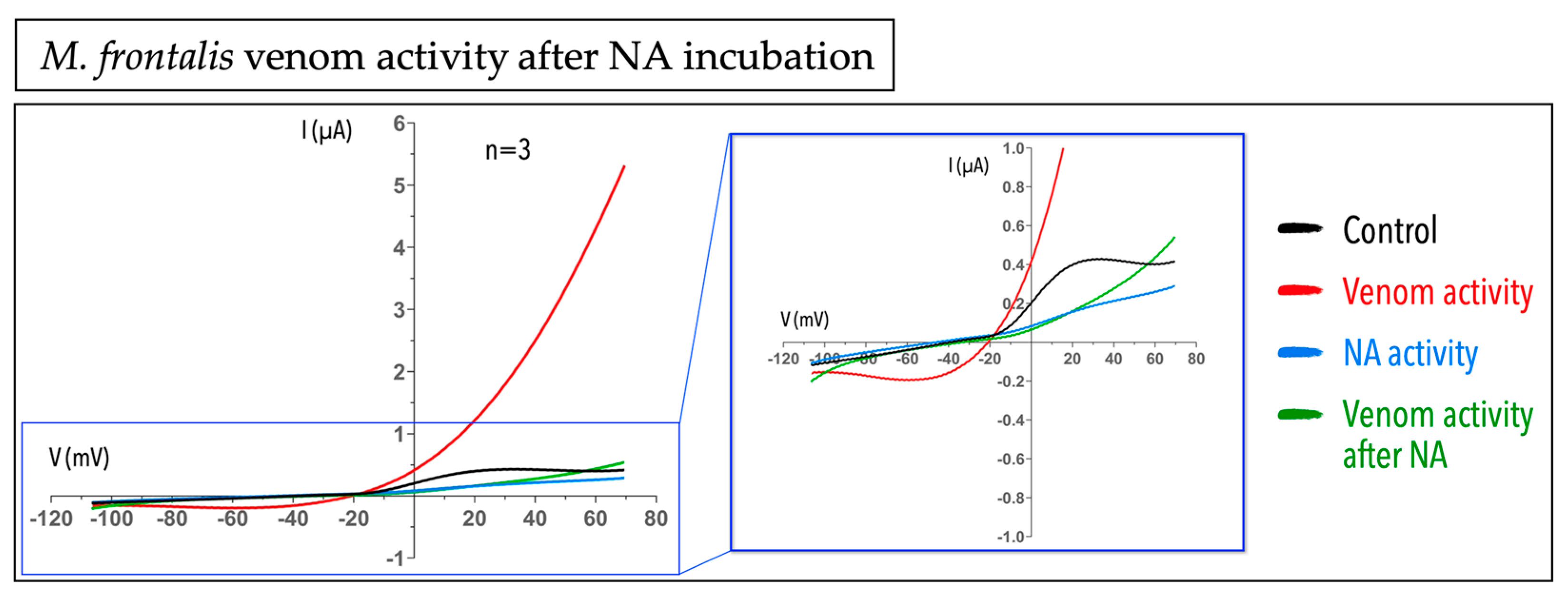
Venoms are complex mixtures of toxins and are designed to cause a variety of damages to many different organisms. One of the well reported Micrurus venom activities is myotoxicity. In this way, as a first step, we decided to test whether these venoms can cause cytotoxicity to X. laevis oocyte, specifically. We used the TEVC technique to measure the electric current in non-injected oocytes after the incubation with Micrurus crude venoms, using a ramp protocol, and compared it with the control current before the exposure to venom. The amount of 50 µg of M. altirostris, M. corallinus, M. frontalis and M. carvalhoi crude venoms did not cause any cytotoxic effect on oocytes (Figure 3A), while 4 µg of Apis mellifera crude venom, used as a positive control, was enough to cause a prominent cytotoxic effect (Figure 3B). A. mellifera venom is already known in the literature to cause cytotoxic/cytolytic effects in different cells [41,42,43,44,45,46]. Interestingly, for the M. frontalis venom, we found an outward current around five times bigger when compared to the control (Figure 3A,C). It means that there is/are molecule(s) in this venom that is/are causing a specific efflux of ions that is not related to a non-selective pore-forming effect in the cell membrane, as we did not find a change in reversal potential when comparing to the control situation (Figure 3D).
Since the four venoms tested did not cause cytotoxic/cytolytic damage to oocytes, we proceeded to the exploration of their activities in oocytes expressing different types of ligand-gated and voltage-gated ion channels (Section 2.4). Because of the limitation in obtaining M. decoratus venom, we did not test the cytotoxic effect in non-injected cells.

2.3. Micrurus Frontalis Venom Evoked Cl Outward Current by Acting Directly or Indirectly on Ca2+-Activated Cl Channels

In the previously cytotoxic experiment with non-injected oocytes, we found an outward current evoked by M. frontalis venom, not observed in any other Micrurus venom tested (Figure 3A,C). We hypothesized that the outward current observed was Cl- ions flowing throughout the cell by direct or indirect activation of Ca2+-activated Cl- channels. To address our hypothesis, we blocked this channel by using niflumic acid (NA), a non-steroidal anti-inflammatory agent that blocks Ca2+-activated Cl channels [47], and evaluated the venom response. In the presence of 100 µM of NA with subsequent incubation of 50 µg of M. frontalis venom, we could not observe the venom activity any longer. It means that in the presence of a chloride channel blocker, the venom cannot act, causing the outward current observed previously, suggesting that the M. frontalis venom may be interfering with either (1) an increase in the intracellular Ca2+ (from external or internal stores), or (2) by directly acting on Ca2+-activated Cl channels (Figure 4).

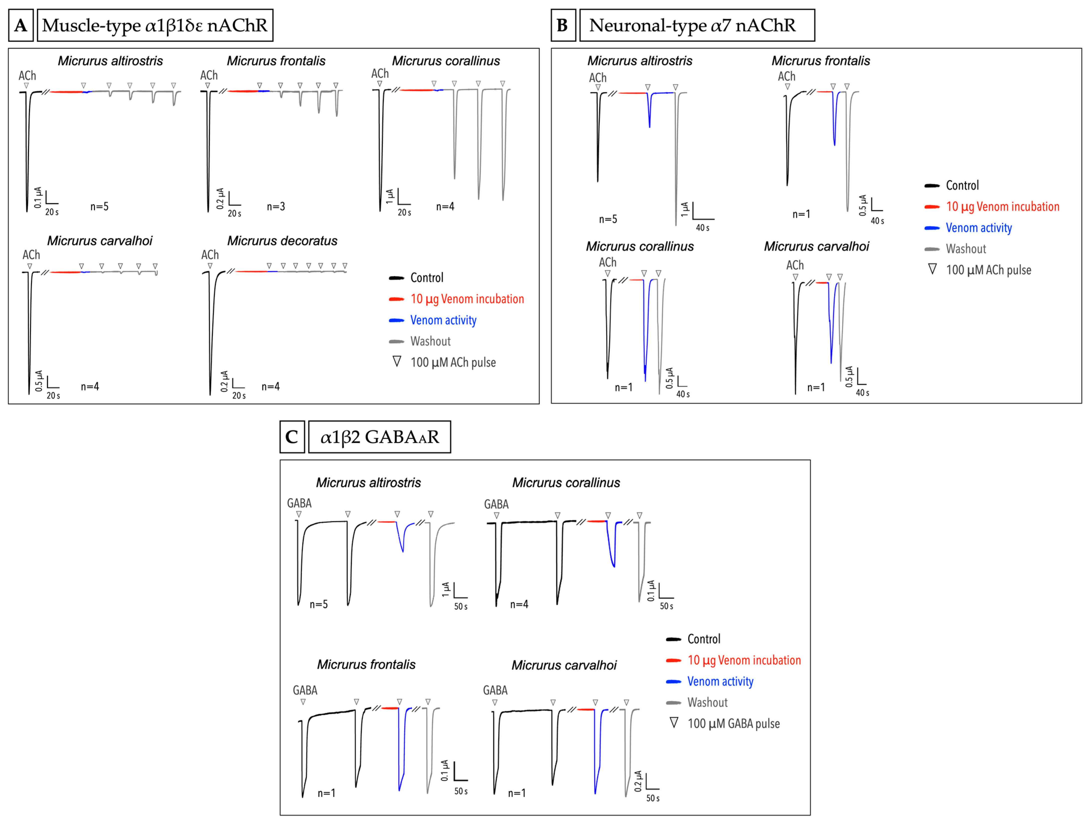
2.4. Micrurus Venoms Act in Different Ion Channels

In order to explore the venoms’ activities on different ligand-gated and voltage-gated ion channels, we expressed different targets in X. laevis oocytes and measured the venom activity using the TEVC. We tested three different ligand-gated ion channels: the muscle-type α1β1δε nicotinic acetylcholine receptor (nAChR), neuronal-type α7 nAChR and α1β2 GABAAR. Briefly, we first applied the agonist (acetylcholine-ACh or γ-aminobutyric acid-GABA) under the flow to obtain controls, then incubated the cell with 10 µg of crude venom under no flow, and subsequently applied the agonist to check the venom activity. Moreover, washout and ACh pulses were given to check whether and how the activities observed would return to the control stage or not. The five Micrurus venoms showed full inhibition of the muscle-type α1β1δε nAChR (Figure 5A). The cell washout followed by agonist pulses application revealed that each venom has different activities and affinities to the receptor. M. altirostris, M. frontalis and M. corallinus have a clear reversible activity, with the M. corallinus venom being almost completely reversible after three washout runs. For M. altirostris and M. frontalis, a prolonged washout had to be carried out to return to the control level. We found M. carvalhoi and M. decoratus venoms to be the most potent and almost irreversible in blocking the α1β1δε nAChR (Figure 5A).
Different from the muscle-type nAChR, the α7 neuronal-type was the target for just two venoms, M. altirostris and M. frontalis. Interestingly, the activity of these two venoms on α7 nAChR was first a partial inhibition and subsequently, after one washout, a potentiation of the current in relation to the control. It means that there are different toxins in the same venom acting differently, as a blocker and as an enhancer, and also with different affinity to the receptor (Figure 5B). For M. corallinus and M. carvalhoi, no significant activity was observed. M. decoratus venom was not included in the experiment with α7 nAChR due to the limited amount of venom available.
For α1β2 GABAAR, we found that M. altirostris and M. corallinus venoms could partially block and modulate the activity of this receptor. Intriguingly, after the venom incubation, we could observe an inversion of the activation and the desensitization behavior of the receptor. In a control condition, the receptor activates quickly until it starts slowly desensitizing under GABA agonist stimulus. On the contrary, after the venom incubation, we could observe a slower activation of the receptor together with an inhibition, and faster desensitization. The mechanism underlying these venoms’ activity must be further explored to understand how the toxins are acting on the inversion of the activation and desensitization processes of the GABAAR. For M. frontalis and M. carvalhoi venoms, no activity was observed on this receptor (Figure 5C). A preliminary study with M. decoratus venom on α1β2 GABAAR showed no effect of this venom on this receptor (data not shown).
Following the screening of Micrurus venom activities on different ligand-gated ion channels, we evaluated their activities on voltage-gated potassium (KV) and sodium (NaV) channels: the Shaker-like KV1.3, and the TTX-sensitive NaV1.2 and NaV1.4. All the five venoms showed inhibition on KV1.3. M. corallinus was the most potent venom in blocking the channel current (48.7% ± 0.1), followed by M. carvalhoi (42.6% ± 1) and M. altirostris (32.8% ± 1.9) venoms. M. frontalis and M. decoratus venoms showed less activity compared to the other venoms (respectively, 28.9% ± 2.2 and 23.3% ± 0.8) (Figure 6A). Furthermore, the activity of all the five venoms was completely reversible after the washout (Figure 6A). Experiments with NaV1.2 (Figure 6B) showed that the five Micrurus venoms have no effect on these channels. Tests on NaV1.4 also demonstrated no effect of the five venoms on this subtype (data not shown).

3. Discussion

The proteomic and transcriptomic analyses of the Micrurus venoms are key contributors to understand the amount, distribution and diversity of the major toxins and other components in these venoms. Based on the venom proteomes available in the literature for the M. altirostris, M. corallinus and M. lemniscatus (=M. carvalhoi), we know that there is a predominance of 3FTxs, which stands for 79.5%, 81.7% and 76.7% in these venoms, respectively [7,9,10]. In contrast, M. frontalis venom has more PLA2 (49.2%) than 3FTx (42.4%) [19]. According to these proteomes [7,9,10] and the chromatographic profiles for the five Micrurus species venoms shown in this work (Figure 2A–E) [7,9,10], we can consider three main toxin distribution regions in the chromatogram. The beginning of the chromatogram is rich in 3FTxs, followed by the middle which is rich in PLA2s, and finally the end of the chromatogram where the toxins with higher molecular weight are found, such as SVMP, LAO and lipases [7,9,10]. The M. altirostris, M. corallinus, M. frontalis and M. carvalhoi RP-HPLC venom profiles reported in this work corroborate the profiles reported in the literature with few differences, which are normally found in different pools of venoms from different animals (Figure 2A–E). The M. decoratus RP-HPLC venom profile (Figure 2E), analyzed for the first time in this study, is considerably less complex in fractions when compared to the M. frontalis venom (Figure 2C), for example. Comparing other Micrurus chromatograms with the venom proteomes described, we can suggest that at least the first two fractions of the M. decoratus venom are significantly composed of 3FTxs (Figure 2E). In addition, we hypothesized that the low complexity of the fractions in this venom may suggest that it is 3FTx-predominant with a low amount and diversity of PLA2 toxins. The M. decoratus venom proteome is under investigation.
Diverse Viperids and Elapid snake venoms are known to be cytotoxic [48,49]. Venoms from Najas are good examples of well-described cytotoxic activity in the Elapid family [50,51,52,53]. In the case of Micrurus venoms, many species were reported as being myotoxic, causing muscle cell death [54,55,56,57]. For example, the isolated Lemnitoxin, a member of the PLA2 protein family and a major component in M. lemniscatus venom, was described as having myotoxic and pro-inflammatory activity [58].
Despite the myotoxicity activity found in several Micrurus species, it is not the front-line activity of these venoms to subdue the preys. Nevertheless, to test the activity of the venom in X. laevis oocytes without masking specific activities or causing cell death, we first checked whether they were cytotoxic for these cells. All the four venoms tested were not cytotoxic after the exposure of the oocytes using a saturating amount of venom, as shown in Figure 3A. In contrast, we used, as a positive control, the venom from the Apis mellifera bee species, well known as having the activity of causing non-ion-selective pores in cell membranes, a cytotoxic/cytolytic effect [41,42,43,44,45,46]. In the electrophysiology experiments using the voltage ramp protocol, when a given venom is forming pores that are non-selective for ions and their conduction, typically two phenomena are observed: (1) a shift of the reversal potential to (wards) 0 mV, and (2) a significant increase in the chord conductance. These two aspects can be clearly seen in the case of the bee venom (Figure 3B,D). We indeed could observe for the bee venom that the voltage at which the potential reverses is situated near 0 mV (slightly positive~3.4 mV), while for Micrurus venoms, it remains negative. An offshoot of the cytotoxic experiments was the activity found in the M. frontalis venom; here, a unique activity was observed by the evocation of an outward current observed in a time-dependent manner (Figure 3A). When blocking the (presumed) Ca2+-activated Cl channels using the blocker NA, we no longer observed the outward current caused by the M. frontalis venom. This is suggestive of the fact that in the presence of the M. frontalis venom, an increase in the intracellular calcium is taking place. Components of M. frontalis venom may be acting directly on this channel (a Ca2+-activated Cl channel) or most likely acting on the cell membrane, causing this effect indirectly, possibly via the action of presynaptic PLA2s.
Micrurus venoms present a preponderant neurotoxic activity, and the main outcome of the envenomation is the blockade of the neuromuscular junction at the peripheral nervous system [21,23]. The action of pre- and postsynaptic neurotoxins, generally PLA2s (also known as β-neurotoxins) and the 3FTxs (or α-neurotoxins), respectively, can act as the main players to the final venom activity. In general, β-neurotoxins can alter the release of acetylcholine, while α-neurotoxins can bind and modulate the cholinergic receptors. Despite this, other mechanisms of action for these α- and β-neurotoxins can be found, and some are still obscure [31,59,60,61]. Furthermore, a diverse range of activities is described for the 3FTx family [32].
Besides the major toxins, the neurotoxicity of Micrurus venoms is complex and other minor molecules can act synergistically, being also important for the envenomation strategy. Based on the classical and better-described activity for the coral snake venoms—the action on cholinergic receptors—in the present work, we tested the five venoms on muscle and neuronal types of nAChR and on GABAAR. The challenge of these venoms against the muscle type α1β1δε nAChR shows that all of them can fully block the receptor, though interestingly differing in the response after the washing of the venom and application of the subsequent pulses of acetylcholine. We can point out two activities found in opposite extremes: M. corallinus venom is almost completely reversible after the third pulse of ACh, while the M. decoratus venom demonstrated no significant reversibility until the sixth pulse (Figure 5A).
All the five venoms showed different reversibility, which is compatible with the diversity of the venom profiles that is leading to different responses. Vital Brazil in 1987, in experiments with isolated rat phrenic nerve-diaphragm, observed that the M. frontalis venom from São Paulo state (Brazil) was reversible for the neuromuscular blockade, while the M. frontalis venom from Mato Grosso (Brazil) was interestingly irreversible [23]. This experiment illustrates that even within the same species, we can find variation in the reversibility, which, in this case, is related to the geographic distribution of the animal. Moreover, pre- and postsynaptic actions can generate different outcomes for the same receptor. Finally, the full blockage of the muscle-type nAChR by the five different Micrurus venoms reinforces the conserved and potent activity of these venoms on cholinergic receptors. Despite the wide range of biological targets, classically, the main 3FTx activity is the disturbance of the cholinergic system, specifically the inhibition of the muscarinic or nicotinic AChRs [31].
The neuronal-type α7 nAChR can also be a target for Micrurus venom toxins. An example is the α-neurotoxin from the venom of the Mexican coral snake M. laticollaris, which blocks not only the muscle-type, but also the α7 neuronal-type of nAChR [62]. In the case of the five Micrurus venoms tested on α7 nAChR in the present work, the most interesting activity was the potentiation of the receptor after the partial blockage caused by the M. altirostris and M. frontalis venoms. At this point, we can only speculate that different toxins with different kinetics for this receptor are taking part in this observation.
The α-neurotoxins can be multifunctional and bind not just to the cholinergic receptors but also to other receptors, such as the GABAAR [35,63]. For example, findings in studies with α-bungarotoxin showed that besides the classical activity reported on the nAChR, this protein can block functional and non-functional GABAAR [34,64]. In Micrurus, two toxins of the 3FTx family from the venom of the Costa Rican coral snake M. mipartitus, named MmTX1 and MmTX2, were reported as strong modulators of GABAAR by allosterically increasing the receptor sensitivity to the agonist [33]. Among the five Micrurus venoms tested here, two showed an interesting modulation of GABAAR.: M. altirostris and M. corallinus venoms, besides partially blocking the receptor, curiously inverted the speed for the activation and desensitization of the receptor when compared to the control. To the best of our knowledge, this modulation was not seen yet for snake toxins and deserves further investigation. Moreover, we decided to use the α1β2 GABAAR subunits, since previous and preliminary tests using α1β2γ2 subunits showed that the α and β were important for the activity while the γ was dispensable (data not shown). This result corroborates with Jean-Pierre Rosso et al. experiments, where two toxins from M. mipartitus venom were tested using the same subunits and it was verified that the γ subunit was not required for the activity [33].
Voltage-gated ion channels are also targets for many venom-derived peptides and proteins [65,66,67,68]. These channels are widely distributed in the human body and are critical in several physiological processes [69] and consequently are involved in many diseases. For example, voltage-gated potassium channels (KVs) [70,71] and voltage-gated sodium channels (NaVs) [70,72] are objects of study in high incidence diseases such as cancer and neurodegenerative disorders [73,74,75,76], and so, are explored as therapeutical targets [77,78,79,80]. Toxins from sea anemone, scorpions, spiders, cone snails, centipedes and snakes are being investigated in studies with KV and NaV [65,67,81,82,83,84,85,86,87,88]. Therefore, in general, animal toxins are of pharmacological interest in the studies using voltage-gated ion channels.
Since Micrurus venoms have been proved to be versatile and often promiscuous in terms of activity and selectivity, as shown in this work, we decided to check whether these venoms can act on voltage-gated ion channels. We tested the Micrurus venoms on KV1.3, noticeably a promising channel for therapeutic drugs [79,89,90] and a known target for toxins [74]. Literature evidence indeed proposes KV1.3 as a key target for multi-therapeutic applications in many human diseases. This channel is mainly found in the immune system and nerve cells, and it can be involved in inflammatory processes, autoimmune disorders, cancer, obesity, etc. [91,92,93]. Specifically, KV1.3 inhibitory molecules are of pharmacological interest since they are considered suppressors for cell proliferation [94,95]. It is worth mentioning that peptides from scorpion venoms are largely found as inhibitors of potassium channels, including the KV1.3 [96,97,98,99]. Perhaps, the natural source with the highest number of peptide modulators of potassium channels discovered to date comes from scorpion venoms [96,98].
In the present work, we demonstrated that the five Micrurus crude venoms tested on KV1.3 can inhibit this channel (Figure 6A), with all of them being reversible after the washout. Notably, an isolated PLA2 toxin called MiDCA1, from Micrurus dumerilii carinicauda, a species from the north of South America, showed reversible inhibition of the KV2.1 [100]. According to our research, only this isolated toxin was reported in Micrurus venoms as a potassium channel inhibitor, to date [100]. This finding, together with our results, led us to hypothesize that this activity may be evolutionarily conserved among this genus and that it might be important for the toxin arsenal to subdue prey. Since we found activity on potassium channels for all the crude venoms tested, we speculate that Micrurus venoms are also a rich source for potassium channel modulators still to be explored.
Finally, we tested the five venoms on NaV1.2 (Figure 6B) and NaV1.4; none of them have demonstrated activity on either channel subtypes. Contrary to the potassium channels, this result shows that sodium channels do not seem to be a target for Micrurus venoms, at least for the five coral snake venoms tested from south and southeast of Brazil on these channel subtypes. In general, sodium channels have not been systematically investigated for coral snake venoms around the world. Nevertheless, an Elapidae species from Indonesia called Long-glanded blue coral snake (Calliophis bivirgatus) was reported as “a snake with the scorpion’s sting”, due to a toxin from the 3FTx family found in its venom which activates the NaV1.4 [101]. Additional investigations of Micrurus venoms are needed for a better understanding of whether or not sodium channels represent a target.

4. Conclusions

In this work, we explored the venom activity of five different Micrurus species from the south and southeast Brazil on six different types of ion channels: three ligand-gated ion channels and three voltage-gated ion channels. We discovered that these venoms can be multifunctional, each of them acting on at least two different types of targets.
Taking into consideration the expected activity on the muscle-type of nAChR, for Micrurus venoms, we demonstrated that in fact all of the five venoms were capable of fully blocking this channel but differed in their reversibility. This result shows that besides the conserved activity, each venom has its particularities. Corroborating with this idea, just some venoms demonstrated activity on GABAAR and the neuronal-type of nAChR. Moreover, we showed that Micrurus venoms may rely on potassium channel inhibitory activity as an important feature of their envenomation strategy since it seems to be conserved among the genus. On the other hand, studies with other species must be carried out to verify if this is indeed a hallmark of the Micrurus genus. Further studies on the specific toxins responsible for the functional effects described in this paper are now ongoing. In conclusion, Micrurus venoms demonstrate to be an invaluable pharmacological and biotechnological source, and so, are noteworthy of deeper investigation.

5. Materials and Methods

5.1. Venoms

Five venoms from different Micrurus species were used in this work: Micrurus altirostris, Micrurus corallinus, Micrurus frontalis, Micrurus carvalhoi (former M. lemniscatus carvalhoi) and Micrurus decoratus (Sisgen code A9F3694). The venoms were extracted from specimens kept in serpentarium and kindly given by the Núcleo de Ofiologia de Porto Alegre-NOPA (M. altirostris) and Instituto Vital Brazil (M. corallinus, M. frontalis, M. carvalhoi and M. decoratus). The venoms were centrifuged to remove debris, lyophilized and stored at −20 to be used further. For all the experiments, pools of adult venoms were used with the exception of M. decoratus venom, which just one specimen was used for the venom extraction. M. decoratus is a rare species and difficult to obtain, being a limiting factor to study its venom.

5.2. RP-HPLC Profile

The crude venom chromatographic profiles were obtained using a reverse-phase high performance liquid chromatography (RP-HPLC). Approximately 500 µg of lyophilized venom was resuspended in 500 µL of 5% acetonitrile and 0.1% trifluoroacetic acid (TFA) solution and centrifuged at 8000× g for 10 min to remove the remaining debris. We used a Teknokroma Europa C18 column (0.4 cm × 25 cm, 5 mm particle size, 300 Å pore size) in a high-performance liquid chromatographic system (HPLC-Shimadzu). The flow rate was 1 mL/min and the experiments were carried out in a linear gradient of 0.1% TFA in water (solution A) and 0.1% TFA in acetonitrile (solution B) in the following condition: 5 min of isocratically 5% of B solution; from 5 to 85 min, 5 to 70% of B solution; 85 to 90 min, 70% of B solution; 90 to 95 min, 5% of B solution. The column was equilibrated with 5% of B solution. The protein detection was monitored at 214 and 280 nm (λ) and the graphs were generated in GraphPad Prism, version 8.0.2 (GraphPad Software, San Diego, CA, USA).

5.3. Electrophysiologic Recordings on Xenopus laevis Oocytes

In order to measure the activity of the venoms in non-injected and injected oocytes with cRNA encoding ion channels, we used the two-electrode voltage clamp (TEVC) technique. The electrophysiologic recordings were carried out on a GeneClamp 500B amplifier equipment (Axon Instruments®, Foster City, CA, USA) controlled by Clampex 10.4 or pCLAMP 10.1 software (Molecular Device, San José, CA, USA) for the data acquisition. Two micropipettes were prepared using capillary glasses (borosilicate WPI 1B120-6) in a microelectrode puller (PUL-1, World Precision Instruments®, Sarasota, FL, USA). Voltage and current electrodes were filled with 3 M KCl and the resistances of both electrodes were kept between 1.0 to 1.5 MΩ. Oocytes were placed in a 0.2 mL recording chamber with a perfusing system. For the whole-cell current recordings, the leak subtraction was carried out with P/4 protocol. The experiments were carried out at room temperature (18 to 22 °C). The data were analyzed in the ClampFit software version 11.0.3 (Molecular Devices, San José, CA, USA) and the final graphs were generated using GraphPad Prism, version 8.0.2 (GraphPad Software, San Diego, CA, USA).
Xenopus laevis frogs used to extract the oocytes were purchased from Nasco (Fort Atkinson, WI, USA) and maintained in the Aquatic Facility of KU Leuven, according to the European Union regulation concerning the welfare of laboratory animals established in the directive 2010/63/EU. The use of X. laevis oocytes was approved by the Animal Ethics Committee of KU Leuven licensed by the number P186/2019. The oocytes in stages V–VI were surgically extracted from mature female animals and anesthetized with 0.1% tricaine solution (aminobenzoic acid ethyl ester, Merck®, Darmstadt, Germany). The oocyte vitellin membranes were defolliculated by incubating with 2.5 mg/mL of collagenase in Ca2+ free ND96 solution (96 mM NaCl; 2 mM KCl; 2 mM MgCl2 and 5 mM HEPES, pH 7.5) at 16 °C, for 1 h 45 min. The defolliculated oocytes were maintained in ND96 (96 mM NaCl; 2 mM KCl; 1.8 mM CaCl2; 2 mM MgCl2 and 5 mM HEPES, pH 7.5) solution supplemented with Theofylline and gentamicin at 16 °C to be used further.

5.3.1. Cytotoxic Experiments with Non-Injected Oocytes

In order to check whether the venoms were cytotoxic to oocytes or not, we used non-injected oocytes, without expressing any particular type of ion channel. For the electrophysiologic measurements, we used a 2-s ramp protocol from −120 to 70 mV, with cells clamped at −20 mV holding potential. Lyophilized venoms were resuspended in ND96 solution for the final concentration of ~0.5 to 10 mg/mL. An amount of 50 µg of crude venom was applied by pipetting directly on the bath, under no flow. The bath perfusion of ND96 solution to wash the oocytes was under the flow (~2 mL/min).

5.3.2. Niflumic Acid Assay

For the electrophysiologic recordings, the ramp protocol was used as described above. In order to verify if outward current after M. frontalis venom application was evoked due to the activation of Ca2+-activated Cl channels, after 20 sweeps of 2-s controls, we blocked Ca2+-activated Cl channels by applying the niflumic acid reagent (Sigma®, St. Louis, MO, USA) directly in the bath by pipetting, for a final concentration of 100 µM. Subsequently, 50 µg of M. frontalis crude venom was applied also by pipetting directly on the bath and the response was evaluated.

5.3.3. Ion Channels Heterologous Expression and Recordings in Oocytes

Six different types of ion channels were used in the present work: (1) human muscle-type of α1β1δε nAChR; (2) human neuronal-type of α7 nAChR; (3) human α1β2 GABAAR; (4) human voltage-gated potassium channel (KV1.3); and (5, 6) rat voltage-gated sodium channels (NaV1.2 and NaV1.4), coexpressed with their rat auxiliary β subunits. For the heterologous expression in oocytes, plasmids for each channel subunit were linearized using appropriate restriction enzymes and transcribed using a T7 or SP6 mMESSAGEmMACHINE transcription kit (Ambion, Austin, TX, USA). For the channel expression, the oocytes were microinjected with cRNA using a micro-injector (Drummond Scientifc®, Broomall, PA, USA) with a final volume ranging from 4 to 60 nL per cell, depending on the channel subtype and the cRNA concentration. For the injection of α1β2 GABAAR and muscle-type α1β1δε nAChR, we used the proportion of 1:1 and 2:1:1:1 of the subunits, respectively. For the injection of both NaV channel types, the proportion of 1:1 was used for the channel and the auxiliary β subunit. The oocytes were incubated in ND96 solution supplemented with 50 mg/L of gentamicin sulfate and 180 mg/L of Theophylline, and kept at 16 °C for 1 to 5 days to express the channels. For the experiments with the ligand-gated ion channels (muscle-type α1β1δε nAChR, neuronal-type α7 nAChR and α1β2 GABAAR), the activation by ligand agonists was also carried out under gravitational flow. In the case of voltage-gated channels (KV1.3, NaV1.2 and NaV1.4), protocols using different voltage steps, depending on the isoform studied, were applied.
For the whole-cell current recordings, different protocols were used depending on the ion channel measured, as follows: For muscle-type α1β1δε nAChR, cells were clamped at −70 mV holding potential. A protocol of 3 pulses of 100 µM acetylcholine (Sigma®, St. Louis, MO, USA) agonist with an interval of ~30 s between each pulse was applied to make controls for each cell experiment. The cell washout was carried out before venom application and ACh pulses were applied to test the venom activity, as in the control. For neuronal-type α7 nAChR, cells were also clamped at −70 mV and 2 or 3 100 µM ACh pulses were applied as the control, followed by cell washout, venom application and ACh pulses to test the activity. For α1β2 GABAAR, cells were clamped at −90 mV. A protocol of 2 pulses of 100 mM of γ-aminobutyric acid (GABA) agonist (Sigma®, St. Louis, MO, USA) for ~30 s and ~3 min of a washout interval between each pulse was performed. Cells were also washed after the controls, to apply the venom subsequently, and 2 pulses of 100 mM GABA were applied to test the activity after the venom incubation. For KV1.3 channels, cells were clamped at a holding potential of −90 mV. The K+ current was evoked by 500 ms depolarizations to 0 mV, followed by 500 ms pulses to −50 mV. Finally, for NaV1.2 and NaV1.4 channels, cells were clamped at −90 mV holding potential and the Na+ current was evoked by 100 ms depolarizations to 0 mV. Lyophilized venoms were resuspended in ND96 solution for the final concentration of 0.5 to 1 mg/mL. For all experiments with ion channels, the amount of ~10 µg of crude venom was applied by pipetting directly on the bath, without flow. For the α1β1δε nAChR and α7 nAChR, the venoms were incubated for ~50 s, and for α1β2 GABAAR, ~1 min. The bath perfusion of ND96 solution to wash the oocytes was under the gravitational flow (~2 mL/min).

Author Contributions

Conceptualization, J.M.K.-F., S.P., R.B.Z. and J.T.; methodology, J.M.K.-F., S.P., R.B.Z. and J.T.; validation, J.M.K.-F., R.B.Z. and J.T.; formal analysis, J.M.K.-F.; investigation, J.M.K.-F., H.B. and E.L.P.-J.; resources, R.B.Z. and J.T.; data curation, J.M.K.-F.; writing—original draft preparation, J.M.K.-F.; writing—review and editing, J.M.K.-F., E.L.P.-J., S.P., R.B.Z. and J.T.; visualization, J.M.K.-F.; supervision, R.B.Z. and J.T.; project administration, J.M.K.-F., R.B.Z. and J.T.; funding acquisition, J.M.K.-F., S.P., R.B.Z. and J.T. All authors have read and agreed to the published version of the manuscript.

Funding

We thank the following financial support: FAPERJ (grants E-26/010.003016/2014; E-26/200.963/2021, E-26-010101030/2018 to R.B.Z.); CNPq (Grant 308376/2020-0 to R.B.Z.); FWO-Vlaanderen (grants GOA4919N, GOE7120N, and GOC2319N to J.T. and 12W7822N to S.P.) and the scholarships PDM/19/164 to S.P., CAPES-PRINT 88887.508234/2020-00 to J.M.K.-F. and CNPq 141949/2018-0 to J.M.K.-F.

Institutional Review Board Statement

Not applicable.

Informed Consent Statement

Not applicable.

Data Availability Statement

Not applicable.

Acknowledgments

We thank Núcleo de Ofiologia de Porto Alegre (NOPA), Instituto Vital Brazil (IVB) and Carlos Correa Netto, to kindly provide the venoms used in this work; and Pedro Rattes to have developed the map of species distribution.

Conflicts of Interest

The authors declare no conflict of interest.

References

  1. Uetz, P.; Koo, M.S.; Aguilar, R.; Catenazzi, A. A Quarter Century of Reptile and Amphibian Databases. Herpetol. Rev. 2021, 52, 246–255. [Google Scholar]
  2. Lomonte, B.; Rangel, J. Snake Venom Lys49 Myotoxins: From Phospholipases A 2 to Non-Enzymatic Membrane Disruptors. Toxicon 2012, 60, 520–530. [Google Scholar] [CrossRef]
  3. Kini, R.M.; Doley, R. Structure, Function and Evolution of Three-Finger Toxins: Mini Proteins with Multiple Targets. Toxicon 2010, 56, 855–867. [Google Scholar] [CrossRef]
  4. Utkin, Y.N. Three-Finger Toxins, a Deadly Weapon of Elapid Venom-Milestones of Discovery. Toxicon 2013, 62, 50–55. [Google Scholar] [CrossRef]
  5. Baudou, F.G.; Rodriguez, J.P.; Fusco, L.; de Roodt, A.R.; de Marzi, M.C.; Leiva, L. South American Snake Venoms with Abundant Neurotoxic Components. Composition and Toxicological Properties. A Literature Review. Acta Trop. 2021, 224, 106119. [Google Scholar] [CrossRef]
  6. Lomonte, B.; Rey-Suárez, P.; Fernández, J.; Sasa, M.; Pla, D.; Vargas, N.; Bénard-Valle, M.; Sanz, L.; Corrêa-Netto, C.; Núñez, V.; et al. Venoms of Micrurus Coral Snakes: Evolutionary Trends in Compositional Patterns Emerging from Proteomic Analyses. Toxicon 2016, 122, 7–25. [Google Scholar] [CrossRef]
  7. Sanz, L.; Quesada-Bernat, S.; Ramos, T.; Casais-e-Silva, L.L.; Corrêa-Netto, C.; Silva-Haad, J.J.; Sasa, M.; Lomonte, B.; Calvete, J.J. New Insights into the Phylogeographic Distribution of the 3FTx/PLA 2 Venom Dichotomy across Genus Micrurus in South America. J. Proteom. 2019, 200, 90–101. [Google Scholar] [CrossRef]
  8. Aird, S.D.; da Silva, N.J.; Qiu, L.; Villar-Briones, A.; Saddi, V.A.; de Telles, M.P.C.; Grau, M.L.; Mikheyev, A.S. Coralsnake Venomics: Analyses of Venom Gland Transcriptomes and Proteomes of Six Brazilian Taxa. Toxins 2017, 9, 187. [Google Scholar] [CrossRef]
  9. Corrêa-Netto, C.; Junqueira-de-Azevedo, I.d.L.M.; Silva, D.A.; Ho, P.L.; Leitão-de-Araújo, M.; Alves, M.L.M.; Sanz, L.; Foguel, D.; Zingali, R.B.; Calvete, J.J. Snake Venomics and Venom Gland Transcriptomic Analysis of Brazilian Coral Snakes, Micrurus Altirostris and M. Corallinus. J. Proteom. 2011, 74, 1795–1809. [Google Scholar] [CrossRef]
  10. Sanz, L.; de Freitas-Lima, L.N.; Quesada-Bernat, S.; Graça-de-Souza, V.K.; Soares, A.M.; Calderón, L.d.A.; Calvete, J.J.; Caldeira, C.A.S. Comparative Venomics of Brazilian Coral Snakes: Micrurus Frontalis, Micrurus Spixii Spixii, and Micrurus Surinamensis. Toxicon 2019, 166, 39–45. [Google Scholar] [CrossRef]
  11. Airi, S.D.; Jorge, N.; Silva, D.A. Comparative Enzymatic Composition Of Brazilian Coral Snake (Micrurus) Venoms. Comp. Biochem. Physiol. Part B Comp. Biochem. 1991, 99, 287–294. [Google Scholar]
  12. Margres, M.J.; Aronow, K.; Loyacano, J.; Rokyta, D.R. The Venom-Gland Transcriptome of the Eastern Coral Snake (Micrurus Fulvius) Reveals High Venom Complexity in the Intragenomic Evolution of Venoms. BMC Genom. 2013, 14, 531. [Google Scholar] [CrossRef] [Green Version]
  13. Aird, S.D. Ophidian Envenomation Strategies and the Role of Purines. Toxicon 2002, 40, 335–393. [Google Scholar] [CrossRef]
  14. da Silva, N.J., Jr.; Aird, S.D. Prey Specificity, Comparative Lethality and Compositional Differences of Coral Snake Venoms. Comp. Biochem. Physiol. Part C Toxicol. Pharmacol. 2001, 128, 425–456. [Google Scholar] [CrossRef]
  15. Rey-Suárez, P.; Núñez, V.; Gutiérrez, J.M.; Lomonte, B. Proteomic and Biological Characterization of the Venom of the Redtail Coral Snake, Micrurus Mipartitus (Elapidae), from Colombia and Costa Rica. J. Proteom. 2011, 75, 655–667. [Google Scholar] [CrossRef]
  16. Kleiz-Ferreira, J.M.; Cirauqui, N.; Trajano, E.A.; Almeida, M.d.S.; Zingali, R.B. Three-Finger Toxins from Brazilian Coral Snakes: From Molecular Framework to Insights in Biological Function. Toxins 2021, 13, 328. [Google Scholar] [CrossRef]
  17. Coelho Da Silva, D.; Araújo De Medeiros, W.A.; de Fátima Correia Batista, I.; Pimenta, D.C.; Lebrun, I.; Francis Abdalla, F.M.; Lopes Sandoval, M.R. Characterization of a New Muscarinic Toxin from the Venom of the Brazilian Coral Snake Micrurus Lemniscatus in Rat Hippocampus. Life Sci. 2011, 89, 931–938. [Google Scholar] [CrossRef] [Green Version]
  18. Silveira De Oliveira, J.; Rossan De Brandão Prieto Da Silva, Á.; Soares, M.B.; Stephano, M.A.; de Oliveira Dias, W.; Raw, I.; Ho, P.L. Cloning and Characterization of an α-Neurotoxin-Type Protein Specific for the Coral Snake Micrurus Corallinus. Biochem. Biophys. Res. Commun. 2000, 267, 887–891. [Google Scholar] [CrossRef]
  19. Moreira, K.G.; Prates, M.V.; Andrade, F.A.C.; Silva, L.P.; Beirão, P.S.L.; Kushmerick, C.; Naves, L.A.; Bloch, C. Frontoxins, Three-Finger Toxins from Micrurus Frontalis Venom, Decrease Miniature Endplate Potential Amplitude at Frog Neuromuscular Junction. Toxicon 2010, 56, 55–63. [Google Scholar] [CrossRef]
  20. Terra, A.L.C.; Moreira-Dill, L.S.; Simões-Silva, R.; Monteiro, J.R.N.; Cavalcante, W.L.G.; Gallacci, M.; Barros, N.B.; Nicolete, R.; Teles, C.B.G.; Medeiros, P.S.M.; et al. Biological Characterization of the Amazon Coral Micrurus Spixii Snake Venom: Isolation of a New Neurotoxic Phospholipase A2. Toxicon 2015, 103, 1–11. [Google Scholar] [CrossRef]
  21. Bucaretchi, F.; de Capitani, E.M.; Vieira, R.J.; Rodrigues, C.K.; Zannin, M.; da Silva, N.J.; Casais-E-Silva, L.L.; Hyslop, S. Coral Snake Bites (Micrurus spp.) in Brazil: A Review of Literature Reports. Clin. Toxicol. 2016, 54, 222–234. [Google Scholar] [CrossRef]
  22. Cecchini, A.L.; Marcussi, S.; Silveira, L.B.; Borja-Oliveira, C.R.; Rodrigues-Simioni, L.; Amara, S.; Stábeli, R.G.; Giglio, J.R.; Arantes, E.C.; Soares, A.M. Biological and Enzymatic Activities of Micrurus Sp. (Coral) Snake Venoms. Comp. Biochem. Physiol. -A Mol. Integr. Physiol. 2005, 140, 125–134. [Google Scholar] [CrossRef]
  23. Vital Brazil, O. Coral Snake Venoms: Mode of Action and Pathophysiology of Experimental Envenomation (1). Rev. Do Inst. De Med. Trop. De Sao Paulo 1987, 29, 119–126. [Google Scholar] [CrossRef] [Green Version]
  24. Moraes, F.V.; Sousa-E-Silva, M.C.C.; Barbaro, K.C.; Leitão, M.A.; Furtado, M.F.D. Biological and Immunochemical Characterization of Micrurus Altirostris Venom and Serum Neutralization of Its Toxic Activities. Toxicon 2002, 41, 71–79. [Google Scholar] [CrossRef]
  25. Bénard-Valle, M.; Carbajal-Saucedo, A.; de Roodt, A.; López-Vera, E.; Alagón, A. Biochemical Characterization of the Venom of the Coral Snake Micrurus Tener and Comparative Biological Activities in the Mouse and a Reptile Model. Toxicon 2014, 77, 6–15. [Google Scholar] [CrossRef]
  26. Francis, B.R.; da Silva, N.J., Jr.; Seebart, C.; Silva, L.L.C.E.; Schmidt, J.J.; Kaiser, I.I. Toxins isolated from the venom of the brazilian coral snake (m1crurus frontalis frontalis) include hemorrhagic type phospholipases a, and postsynaptic neurotoxins. Toxicon 1997, 35, 1193–1203. [Google Scholar] [CrossRef]
  27. Carbajal-Saucedo, A.; Floriano, R.S.; Dal Belo, C.A.; Olvera-Rodríguez, A.; Alagón, A.; Rodrigues-Simioni, L. Neuromuscular Activity of Micrurus Laticollaris (Squamata: Elapidae) Venom In Vitro. Toxins 2013, 6, 359–370. [Google Scholar] [CrossRef] [Green Version]
  28. Dal Belo, C.A.; Toyama, M.H.; Toyama, D.D.O.; Marangoni, S.; Moreno, F.B.; Cavada, B.S.; Fontana, M.D.; Hyslop, S.; Carneiro, E.M.; Boschero, A.C. Determination of the Amino Acid Sequence of a New Phospholipase A 2 (MIDCA1) Isolated from Micrurus Dumerilii Carinicauda Venom. Protein J. 2005, 24, 147–153. [Google Scholar] [CrossRef]
  29. Oliveira, D.A.; Harasawa, C.; Seibert, C.S.; Casais e Silva, L.L.; Pimenta, D.C.; Lebrun, I.; Sandoval, M.R.L. Phospholipases A2 Isolated from Micrurus Lemniscatus Coral Snake Venom: Behavioral, Electroencephalographic, and Neuropathological Aspects. Brain Res. Bull. 2008, 75, 629–639. [Google Scholar] [CrossRef]
  30. dos Santos, R.T.F.; Silva, M.F.P.; Porto, R.M.; Lebrun, I.; de Camargo Gonçalves, L.R.; de Fátima Correia Batista, I.; Sandoval, M.R.L.; Abdalla, F.M.F. Effects of Mlx-8, a Phospholipase A2 from Brazilian Coralsnake Micrurus Lemniscatus Venom, on Muscarinic Acetylcholine Receptors in Rat Hippocampus. J. Venom. Anim. Toxins Incl. Trop. Dis. 2020, 26, e20190041. [Google Scholar] [CrossRef]
  31. Kessler, P.; Marchot, P.; Silva, M.; Servent, D. The Three-Finger Toxin Fold: A Multifunctional Structural Scaffold Able to Modulate Cholinergic Functions. J. Neurochem. 2017, 142, 7–18. [Google Scholar] [CrossRef]
  32. Utkin, Y.N. Last Decade Update for Three-Finger Toxins: Newly Emerging Structures and Biological Activities. World J. Biol. Chem. 2019, 10, 17–27. [Google Scholar] [CrossRef]
  33. Rosso, J.-P.; Schwarz, J.R.; Diaz-Bustamante, M.; Céard, B.; Gutiérrez, J.M.; Kneussel, M.; Pongs, O.; Bosmans, F.; Bougis, P.E. MmTX1 and MmTX2 from Coral Snake Venom Potently Modulate GABAA Receptor Activity. Proc. Natl. Acad. Sci. USA 2015, 112, E891–E900. [Google Scholar] [CrossRef] [Green Version]
  34. Hannan, S.; Mortensen, M.; Smart, T.G. Snake Neurotoxin α-Bungarotoxin Is an Antagonist at Native GABAA Receptors. Neuropharmacology 2015, 93, 28–40. [Google Scholar] [CrossRef] [Green Version]
  35. Kudryavtsev, D.S.; Shelukhina, I.V.; Son, L.V.; Ojomoko, L.O.; Kryukova, E.V.; Lyukmanova, E.N.; Zhmak, M.N.; Dolgikh, D.A.; Ivanov, I.A.; Kasheverov, I.E.; et al. Neurotoxins from Snake Venoms and α-Conotoxin ImI Inhibit Functionally Active Ionotropic γ-Aminobutyric Acid (GABA) Receptors. J. Biol. Chem. 2015, 290, 22747–22758. [Google Scholar] [CrossRef] [Green Version]
  36. Blanchet, G.; Upert, G.; Mourier, G.; Gilquin, B.; Gilles, N.; Servent, D. New α-Adrenergic Property for Synthetic MTβ and CM-3 Three-Finger Fold Toxins from Black Mamba. Toxicon 2013, 75, 160–167. [Google Scholar] [CrossRef]
  37. Maïga, A.; Mourier, G.; Quinton, L.; Rouget, C.; Gales, C.; Denis, C.; Lluel, P.; Sénard, J.-M.; Palea, S.; Servent, D.; et al. G Protein-Coupled Receptors, an Unexploited Animal Toxin Targets: Exploration of Green Mamba Venom for Novel Drug Candidates Active against Adrenoceptors. Toxicon 2012, 59, 487–496. [Google Scholar] [CrossRef]
  38. Rouget, C.; Quinton, L.; Maïga, A.; Gales, C.; Masuyer, G.; Malosse, C.; Chamot-Rooke, J.; Thai, R.; Mourier, G.; de Pauw, E.; et al. Identification of a Novel Snake Peptide Toxin Displaying High Affinity and Antagonist Behaviour for the A2-Adrenoceptors. Br. J. Pharmacol. 2010, 161, 1361–1374. [Google Scholar] [CrossRef] [Green Version]
  39. Rivera-Torres, I.O.; Jin, T.B.; Cadene, M.; Chait, B.T.; Poget, S.F. Discovery and Characterisation of a Novel Toxin from Dendroaspis Angusticeps, Named Tx7335, That Activates the Potassium Channel KcsA. Sci. Rep. 2016, 6, 23904. [Google Scholar] [CrossRef] [Green Version]
  40. Diochot, S.; Baron, A.; Salinas, M.; Douguet, D.; Scarzello, S.; Dabert-Gay, A.-S.; Debayle, D.; Friend, V.; Alloui, A.; Lazdunski, M.; et al. Black Mamba Venom Peptides Target Acid-Sensing Ion Channels to Abolish Pain. Nature 2012, 490, 552–555. [Google Scholar] [CrossRef]
  41. Soliman, C.; Eastwood, S.; Truong, V.K.; Ramsland, P.A.; Elbourne, A. The Membrane Effects of Melittin on Gastric and Colorectal Cancer. PLoS ONE 2019, 14, e0224028. [Google Scholar] [CrossRef] [PubMed] [Green Version]
  42. Borojeni, S.K.; Zolfagharian, H.; Babaie, M.; Javadi, I. Cytotoxic Effect of Bee (A. mellifera) Venom on Cancer Cell Lines. J. Pharmacopunct. 2020, 23. [Google Scholar] [CrossRef] [PubMed]
  43. Adade, C.M.; Chagas, G.S.F.; Souto-Padrón, T. Apis Mellifera Venom Induces Different Cell Death Pathways in Trypanosoma Cruzi. Parasitol. 2012, 139, 1444–1461. [Google Scholar] [CrossRef] [PubMed]
  44. Park, C.; Lee, D.G. Melittin Induces Apoptotic Features in Candida Albicans. Biochem. Biophys. Res. Commun. 2010, 394, 170–172. [Google Scholar] [CrossRef] [PubMed]
  45. Maher, S.; McClean, S. Melittin Exhibits Necrotic Cytotoxicity in Gastrointestinal Cells Which Is Attenuated by Cholesterol. Biochem. Pharmacol. 2008, 75, 1104–1114. [Google Scholar] [CrossRef]
  46. Lee, Y.J.; Kang, S.J.; Kim, B.M.; Kim, Y.J.; Woo, H.D.; Chung, H.W. Cytotoxicity of Honeybee (Apis Mellifera) Venom in Normal Human Lymphocytes and HL-60 Cells. Chem. -Biol. Interact. 2007, 169, 189–197. [Google Scholar] [CrossRef]
  47. White, M.M.; Aylwin, M. Niflumic and Flufenamic Acids Are Potent Reversible Blockers of Ca2(+)-Activated Cl- Channels in Xenopus Oocytes. Mol. Pharmacol. 1990, 37, 720–724. [Google Scholar]
  48. Hiu, J.J.; Yap, M.K.K. Cytotoxicity of Snake Venom Enzymatic Toxins: Phospholipase A2 and L-Amino Acid Oxidase. Biochem. Soc. Trans. 2020, 48, 719–731. [Google Scholar] [CrossRef] [Green Version]
  49. Oliveira, J.C.R.; Montes De Oca, H.; Duarte, M.M.; Diniz, C.R.; Fortes-Dias, C.L. Toxicity of South American Snake Venoms Measured by an in Vitro Cell Culture Assay. Toxicon 2001, 40, 321–325. [Google Scholar] [CrossRef]
  50. Chong, H.P.; Tan, K.Y.; Tan, C.H. Cytotoxicity of Snake Venoms and Cytotoxins From Two Southeast Asian Cobras (Naja Sumatrana, Naja Kaouthia): Exploration of Anticancer Potential, Selectivity, and Cell Death Mechanism. Front. Mol. Biosci. 2020, 7, 583587. [Google Scholar] [CrossRef]
  51. Li, P.-L. Cytotoxicity of Cobra (Naja Naja Kaouthia) Venom on Rabbit Red Blood Cells and S-180 Tumor Cells in the Presence of Tetracaine, Lidocaine and Procaine. Z. Nat. C Biosci. 1980, 35, 268–272. [Google Scholar] [CrossRef]
  52. Khalid, H.; Mukhtar, M.M.; Konstantakopoulos, N. Cytotoxiciy of Naja Nubiae (Serpentes: Elapidae) and Echis Ocellatus (Serpentes: Viperidae) Venoms from Sudan. J. Toxins 2015, 2015, 1–7. [Google Scholar] [CrossRef] [Green Version]
  53. Thangam, R.; Gunasekaran, P.; Kaveri, K.; Sridevi, G.; Sundarraj, S.; Paulpandi, M.; Kannan, S. A Novel Disintegrin Protein from Naja Naja Venom Induces Cytotoxicity and Apoptosis in Human Cancer Cell Lines In Vitro. Process Biochem. 2012, 47, 1243–1249. [Google Scholar] [CrossRef]
  54. Gutiérrez, J.M.; Rojas, G.; da Silva Júnior, N.J.; Núñez, J. Experimental Myonecrosis Induced By The Venoms Of South American Micrurus (Coral Snakes). Toxicon 1992, 30, 1299–1302. [Google Scholar] [CrossRef]
  55. de Roodt, A.R.; Lago, N.R.; Stock, R.P. Myotoxicity and Nephrotoxicity by Micrurus Venoms in Experimental Envenomation. Toxicon 2012, 59, 356–364. [Google Scholar] [CrossRef]
  56. Gutiérrez, J.; Lomonte, B.; Portilla, E.; Cerdas, L.; Rojas, E. Local Effects Induced By Coral Snake Venoms: Evidence Of Myonecrosis After Experimental Inoculations Of Venoms From Five Species. Toxicon 1983, 21, 777–783. [Google Scholar] [CrossRef]
  57. Barros, A.C.; Fernandes, D.P.; Ferreira, L.C.; Dos Santos, M.C. local effects induced by venoms from five species of genus micrurus sp. (coral snakes). Toxicon 1994, 32, 445–452. [Google Scholar] [CrossRef]
  58. Casais-e-Silva, L.L.; Teixeira, C.F.P.; Lebrun, I.; Lomonte, B.; Alape-Girón, A.; Gutiérrez, J.M. Lemnitoxin, the Major Component of Micrurus Lemniscatus Coral Snake Venom, Is a Myotoxic and pro-Inflammatory Phospholipase A2. Toxicol. Lett. 2016, 257, 60–71. [Google Scholar] [CrossRef]
  59. Barber, C.M.; Isbister, G.K.; Hodgson, W.C. Alpha Neurotoxins. Toxicon 2013, 66, 47–58. [Google Scholar] [CrossRef]
  60. Kini, R.M. Excitement Ahead: Structure, Function and Mechanism of Snake Venom Phospholipase A2 Enzymes. Toxicon 2003, 42, 827–840. [Google Scholar] [CrossRef]
  61. Mackessy, S.P. Handbook of Venoms and Toxins of Reptiles; CRC Press: Boca Raton, FL, USA, 2016. [Google Scholar]
  62. Carbajal-Saucedo, A.; López-Vera, E.; Bénard-Valle, M.; Smith, E.N.; Zamudio, F.; de Roodt, A.R.; Olvera-Rodríguez, A. Isolation, Characterization, Cloning and Expression of an Alpha-Neurotoxin from the Venom of the Mexican Coral Snake Micrurus Laticollaris (Squamata: Elapidae). Toxicon 2013, 66, 64–74. [Google Scholar] [CrossRef] [PubMed]
  63. Son, L.; Kryukova, E.; Ziganshin, R.; Andreeva, T.; Kudryavtsev, D.; Kasheverov, I.; Tsetlin, V.; Utkin, Y. Novel Three-Finger Neurotoxins from Naja Melanoleuca Cobra Venom Interact with GABAA and Nicotinic Acetylcholine Receptors. Toxins 2021, 13, 164. [Google Scholar] [CrossRef] [PubMed]
  64. Mccann, C.M.; Bracamontes, J.; Steinbach, J.H.; Sanes, J.R. The Cholinergic Antagonist-Bungarotoxin Also Binds and Blocks a Subset of GABA Receptors. Proc. Natl. Acad. Sci. USA 2006, 103, 5149–5154. [Google Scholar] [CrossRef] [PubMed] [Green Version]
  65. Wu, Y.; Ma, H.; Zhang, F.; Zhang, C.; Zou, X.; Cao, Z. Selective Voltage-Gated Sodium Channel Peptide Toxins from Animal Venom: Pharmacological Probes and Analgesic Drug Development. ACS Chem. Neurosci. 2018, 9, 187–197. [Google Scholar] [CrossRef]
  66. Kalia, J.; Milescu, M.; Salvatierra, J.; Wagner, J.; Klint, J.K.; King, G.F.; Olivera, B.M.; Bosmans, F. From Foe to Friend: Using Animal Toxins to Investigate Ion Channel Function. J. Mol. Biol. 2015, 427, 158–175. [Google Scholar] [CrossRef] [Green Version]
  67. Norton, R.S.; Chandy, K.G. Venom-Derived Peptide Inhibitors of Voltage-Gated Potassium Channels. Neuropharmacology 2017, 127, 124–138. [Google Scholar] [CrossRef]
  68. Bordon, K.d.C.F.; Cologna, C.T.; Fornari-Baldo, E.C.; Pinheiro-Júnior, E.L.; Cerni, F.A.; Amorim, F.G.; Anjolette, F.A.P.; Cordeiro, F.A.; Wiezel, G.A.; Cardoso, I.A.; et al. From Animal Poisons and Venoms to Medicines: Achievements, Challenges and Perspectives in Drug Discovery. Front. Pharmacol. 2020, 11, 1132. [Google Scholar] [CrossRef]
  69. Lai, H.C.; Jan, L.Y. The Distribution and Targeting of Neuronal Voltage-Gated Ion Channels. Nat. Rev. Neurosci. 2006, 7, 548–562. [Google Scholar] [CrossRef]
  70. Terlau, H.; Stühmer, W. The S Tructure And Function Of Voltage-Gated Na + And K + Channels. Naturwissenschaften 1998, 85, 437–444. [Google Scholar] [CrossRef]
  71. Yellen, G. The Voltage-Gated Potassium Channels and Their Relatives. Nature 2002, 419, 35–42. [Google Scholar] [CrossRef]
  72. Kwong, K.; Carr, M.J. Voltage-Gated Sodium Channels. Curr. Opin. Pharmacol. 2015, 22, 131–139. [Google Scholar] [CrossRef] [PubMed]
  73. Pardo, L.A.; Stühmer, W. The Roles of K + Channels in Cancer. Nat. Rev. Cancer 2014, 14, 39–48. [Google Scholar] [CrossRef] [PubMed]
  74. Waters, M.F.; Minassian, N.A.; Stevanin, G.; Figueroa, K.P.; Bannister, J.P.A.; Nolte, D.; Mock, A.F.; Evidente, V.G.H.; Fee, D.B.; Müller, U.; et al. Mutations in Voltage-Gated Potassium Channel KCNC3 Cause Degenerative and Developmental Central Nervous System Phenotypes. Nat. Genet. 2006, 38, 447–451. [Google Scholar] [CrossRef]
  75. Mao, W.; Zhang, J.; Körner, H.; Jiang, Y.; Ying, S. The Emerging Role of Voltage-Gated Sodium Channels in Tumor Biology. Front. Oncol. 2019, 9, 124. [Google Scholar] [CrossRef] [Green Version]
  76. Eijkelkamp, N.; Linley, J.E.; Baker, M.D.; Minett, M.S.; Cregg, R.; Werdehausen, R.; Rugiero, F.; Wood, J.N. Neurological Perspectives on Voltage-Gated Sodium Channels. Brain 2012, 135, 2585–2612. [Google Scholar] [CrossRef]
  77. Lawson, K.; Mckay, N.G. Modulation of Potassium Channels as a Therapeutic Approach. Curr. Pharm. Des. 2006, 12, 459–470. [Google Scholar] [CrossRef]
  78. Wickenden, A.D. K + Channels as Therapeutic Drug Targets. Pharmacol. Ther. 2002, 94, 157–182. [Google Scholar] [CrossRef]
  79. Wulff, H.; Castle, N.A.; Pardo, L.A. Voltage-Gated Potassium Channels as Therapeutic Targets. Nat. Rev. Drug Discov. 2009, 8, 982–1001. [Google Scholar] [CrossRef] [Green Version]
  80. Mantegazza, M.; Curia, G.; Biagini, G.; Ragsdale, D.S.; Avoli, M. Voltage-Gated Sodium Channels as Therapeutic Targets in Epilepsy and Other Neurological Disorders. Lancet Neurol. 2010, 9, 413–424. [Google Scholar] [CrossRef]
  81. Tajti, G.; Wai, D.C.C.; Panyi, G.; Norton, R.S. The Voltage-Gated Potassium Channel KV1.3 as a Therapeutic Target for Venom-Derived Peptides. Biochem. Pharmacol. 2020, 181, 114146. [Google Scholar] [CrossRef]
  82. Jouirou, B.; Mouhat, S.; Andreotti, N.; de Waard, M.; Sabatier, J.M. Toxin Determinants Required for Interaction with Voltage-Gated K + Channels. Toxicon 2004, 43, 909–914. [Google Scholar] [CrossRef] [PubMed]
  83. Mouhat, S.; Andreotti, N.; Jouirou, B.; Sabatier, J.-M. Animal Toxins Acting on Voltage-Gated Potassium Channels. Curr. Pharm. Des. 2008, 14, 2503–2518. [Google Scholar] [CrossRef] [PubMed]
  84. Mouhat, S.; Jouirou, B.; Mosbah, A.; de Waard, M.; Sabatier, J.-M. Diversity of Folds in Animal Toxins Acting on Ion Channels. Biochem. J. 2004, 378, 716–726. [Google Scholar] [CrossRef]
  85. Boldrini-França, J.; Pinheiro-Junior, E.L.; Peigneur, S.; Pucca, M.B.; Cerni, F.A.; Borges, R.J.; Costa, T.R.; Carone, S.E.I.; Fontes, M.R.d.M.; Sampaio, S.V.; et al. Beyond Hemostasis: A Snake Venom Serine Protease with Potassium Channel Blocking and Potential Antitumor Activities. Sci. Rep. 2020, 10, 4476. [Google Scholar] [CrossRef] [Green Version]
  86. An, D.; Pinheiro-Junior, E.L.; Béress, L.; Gladkikh, I.; Leychenko, E.; Undheim, E.A.B.; Peigneur, S.; Tytgat, J. AsKC11, a Kunitz Peptide from Anemonia Sulcata, Is a Novel Activator of G Protein-Coupled Inward-Rectifier Potassium Channels. Mar. Drugs 2022, 20, 140. [Google Scholar] [CrossRef] [PubMed]
  87. Gladkikh, I.; Peigneur, S.; Sintsova, O.; Lopes Pinheiro-Junior, E.; Klimovich, A.; Menshov, A.; Kalinovsky, A.; Isaeva, M.; Monastyrnaya, M.; Kozlovskaya, E.; et al. Kunitz-Type Peptides from the Sea Anemone Heteractis Crispa Demonstrate Potassium Channel Blocking and Anti-Inflammatory Activities. Biomedicines 2020, 8, 473. [Google Scholar] [CrossRef] [PubMed]
  88. Pinheiro-Junior, E.L.; Kalina, R.; Gladkikh, I.; Leychenko, E.; Tytgat, J.; Peigneur, S. A Tale of Toxin Promiscuity: The Versatile Pharmacological Effects of Hcr 1b-2 Sea Anemone Peptide on Voltage-Gated Ion Channels. Mar. Drugs 2022, 20, 147. [Google Scholar] [CrossRef]
  89. Teisseyre, A.; Palko-Labuz, A.; Sroda-Pomianek, K.; Michalak, K. Voltage-Gated Potassium Channel Kv1.3 as a Target in Therapy of Cancer. Front. Oncol. 2019, 9, 933. [Google Scholar] [CrossRef]
  90. Schmalhofer, W.A.; Bao, J.; McManus, O.B.; Green, B.; Matyskiela, M.; Wunderler, D.; Bugianesi, R.M.; Felix, J.P.; Hanner, M.; Linde-Arias, A.R.; et al. Identification of a New Class of Inhibitors of the Voltage-Gated Potassium Channel, Kv1.3, with Immunosuppressant Properties. Biochemistry 2002, 41, 7781–7794. [Google Scholar] [CrossRef]
  91. Pérez-Verdaguer, M.; Capera, J.; Serrano-Novillo, C.; Estadella, I.; Sastre, D.; Felipe, A. The Voltage-Gated Potassium Channel Kv1.3 Is a Promising Multitherapeutic Target against Human Pathologies. Expert Opin. Ther. Targets 2016, 20, 577–591. [Google Scholar] [CrossRef]
  92. Beeton, C.; Wulff, H.; Standifer, N.E.; Azam, P.; Mullen, K.M.; Pennington, M.W.; Kolski-Andreaco, A.; Wei, E.; Grino, A.; Counts, D.R.; et al. Kv1.3 Channels Are a Therapeutic Target for T Cell-Mediated Autoimmune Diseases. Proc. Natl. Acad. Sci. USA 2006, 103, 17414–17419. [Google Scholar] [CrossRef] [PubMed] [Green Version]
  93. Rangaraju, S.; Chi, V.; Pennington, M.W.; Chandy, K.G. Kv1.3 Potassium Channels as a Therapeutic Target in Multiple Sclerosis. Expert Opin. Ther. Targets 2009, 13, 909–924. [Google Scholar] [CrossRef]
  94. Gubič, Š.; Hendrickx, L.A.; Toplak, Ž.; Sterle, M.; Peigneur, S.; Tomašič, T.; Pardo, L.A.; Tytgat, J.; Zega, A.; Mašič, L.P. Discovery of KV1.3 Ion Channel Inhibitors: Medicinal Chemistry Approaches and Challenges. Med. Res. Rev. 2021, 41, 2423–2473. [Google Scholar] [CrossRef]
  95. Chandy, K.G.; Norton, R.S. Peptide Blockers of Kv1.3 Channels in T Cells as Therapeutics for Autoimmune Disease. Curr. Opin. Chem. Biol. 2017, 38, 97–107. [Google Scholar] [CrossRef]
  96. Kuzmenkov, A.I.; Grishin, E.V.; Vassilevski, A.A. Diversity of Potassium Channel Ligands: Focus on Scorpion Toxins. Biochemistry 2015, 80, 1764–1799. [Google Scholar] [CrossRef]
  97. Quintero-Hernández, V.; Jiménez-Vargas, J.M.; Gurrola, G.B.; Valdivia, H.H.; Possani, L.D. Scorpion Venom Components That Affect Ion-Channels Function. Toxicon 2013, 76, 328–342. [Google Scholar] [CrossRef] [PubMed] [Green Version]
  98. Kuzmenkov, A.I.; Krylov, N.A.; Chugunov, A.O.; Grishin, E.V.; Vassilevski, A.A. Kalium: A Database of Potassium Channel Toxins from Scorpion Venom. Database 2016, 2016, baw056. [Google Scholar] [CrossRef] [PubMed] [Green Version]
  99. Bergeron, Z.L.; Bingham, J.P. Scorpion Toxins Specific for Potassium (K+) Channels: A Historical Overview of Peptide Bioengineering. Toxins 2012, 4, 1082–1119. [Google Scholar] [CrossRef] [Green Version]
  100. Schütter, N.; Barreto, Y.C.; Vardanyan, V.; Hornig, S.; Hyslop, S.; Marangoni, S.; Rodrigues-Simioni, L.; Pongs, O.; Dal Belo, C.A. Inhibition of Kv2.1 Potassium Channels by MiDCA1, a Pre-Synaptically Active PLA2-Type Toxin from Micrurus Dumerilii Carinicauda Coral Snake Venom. Toxins 2019, 11, 335. [Google Scholar] [CrossRef] [Green Version]
  101. Yang, D.C.; Deuis, J.R.; Dashevsky, D.; Dobson, J.; Jackson, T.N.W.; Brust, A.; Xie, B.; Koludarov, I.; Debono, J.; Hendrikx, I.; et al. The Snake with the Scorpion’s Sting: Novel Three-Finger Toxin Sodium Channel Activators from the Venom of the Long-Glanded Blue Coral Snake (Calliophis Bivirgatus). Toxins 2016, 8, 303. [Google Scholar] [CrossRef]
Figure 1. Geographic distribution of the five Micrurus species. The colored areas represent the current consolidated geographic distribution of the Micrurus species, and the dotted colored lines represent the distribution areas under scientific revision. The areas where the specimens used in this work were collected are indicated in the map with the names of the species.
Ijms 23 07714 g001
Figure 2. Crude venom chromatographic profiles. (A) Micrurus altirostris, (B) Micrurus corallinus, (C) Micrurus frontalis, (D) Micrurus carvalhoi and (E) Micrurus decoratus venoms. A total of 500 µg of crude venom was used to obtain the RP-HPLC profile. Based on the published proteome information of Micrurus species venoms [8,9,10], we show in the figure the approximate elution regions of 3FTxs and PLA2s, marked by dotted lines in red and green, respectively. The right superior panel in each figure shows the full area of the chromatogram, where the blue box represents the zoomed area and the purple line represents the acetonitrile (ACN) gradient.
Ijms 23 07714 g002
Figure 3. Cytotoxic evaluation of crude venoms on X. laevis non-injected oocytes. (A) Micrurus altirostris, Micrurus corallinus, Micrurus carvalhoi and Micrurus frontalis venom activity. (B) Positive control-Apis mellifera venom activity. The black lines show the control, and the red lines show the venom activity. In panel (A), the dotted dark gray and light gray lines show 100 s and 200 s of past time from the venom activity, respectively. The blue box shows a zoom of the graph on panel (B). The graphs show the trace of one experiment from a series of independent experiments. (C) Inward and outward current graph. (D) Reversal potential graph.
Ijms 23 07714 g003
Figure 4. Micrurus frontalis venom causes a Cl outward current by acting directly or indirectly on Ca2+-activated Cl channels in X. laevis oocytes. The black line shows the control (the oocyte perfused with ND96); the red line shows the activity of 50 µg of M. frontalis venom on oocytes; the blue line shows the oocyte current in the presence of 100 µM of NA; and the green line shows the activity of 50 µg of M. frontalis venom after cell incubation with 100 µM of NA, where the outward current evoked by the venom is no longer observed. The blue box on the right of the panel shows a zoom of the graph. The graphs show the trace of one experiment from a series of independent experiments.
Ijms 23 07714 g004
Figure 5. Screening of crude venoms from different Micrurus species in different ligand-gated ion channels. (A) Muscle-type α1β1δε nAChR, (B) Neuronal-type α7 nAChR and (C) α1β2 GABAAR. The black lines represent the control pulses, the red lines represent the venom incubation, the blue lines represent the receptor activity after the incubation with 10 µg of crude venom, and the gray lines represent the agonist pulse after the cell washout. The triangles mark the point where the application of the agonist was started. The n from each experiment is shown as well as the current/time reference bar. The graphs show the trace of one experiment from a series of independent experiments, with the exception of tests with n = 1.
Ijms 23 07714 g005
Figure 6. Screening of crude venoms from different Micrurus species in different voltage-gated ion channels. (A) KV1.3. The inhibition percentage and the SEM are shown in blue for each graph. (B) NaV1.2. The black lines represent the control and the blue lines represent the channel response after incubation with 10 µg of crude venom. The dotted lines represent the zero current level. The n from each experiment is shown as well as the current/time reference bar. The graphs show the trace of one experiment from a series of independent experiments, with the exception of tests with n = 1.
Ijms 23 07714 g006
Publisher’s Note: MDPI stays neutral with regard to jurisdictional claims in published maps and institutional affiliations.

