Review

Vitamin D and the Kidney: Two Players, One Console

Nephrology, Dialysis and Renal Transplant Unit, IRCCS-Azienda Ospedaliero-Universitaria di Bologna, Alma Mater Studiorum University of Bologna, 40138 Bologna, Italy
*
Author to whom correspondence should be addressed.
Int. J. Mol. Sci. 2022, 23(16), 9135; https://0-doi-org.brum.beds.ac.uk/10.3390/ijms23169135
Submission received: 29 June 2022 / Revised: 8 August 2022 / Accepted: 11 August 2022 / Published: 15 August 2022
(This article belongs to the Special Issue The Role of Vitamin D in Human Health and Diseases 2.0)

Abstract

:
Vitamin D belongs to the group of liposoluble steroids mainly involved in bone metabolism by modulating calcium and phosphorus absorption or reabsorption at various levels, as well as parathyroid hormone production. Recent evidence has shown the extra-bone effects of vitamin D, including glucose homeostasis, cardiovascular protection, and anti-inflammatory and antiproliferative effects. This narrative review provides an overall view of vitamin D’s role in different settings, with a special focus on chronic kidney disease and kidney transplant.

1. Introduction

The denomination “vitamin D” refers to a group of liposoluble, steroidal compounds crucial for intestinal absorption and for metabolism regulation of calcium and phosphates [1]. The most important isoforms in human physiology are ergocalciferol (vitamin D2) and cholecalciferol (vitamin D3), also known as calciols; while the first one is only synthesized in plants and fungi (dietary intake), the second one is both exogenous and produced endogenously from the photolysis of 7-dehydrocholesterol by UVB radiation in the skin [2]. Calciols undergo a two-step hydroxylation to turn into the biologically active form, calcitriol. First, vitamin D 25-hydroxylase in the liver mediates D2/D3 to change into 25(OH)D (calcidiol), a quantifiable form mostly used to determine vitamin D levels in serum, and it is defined as a native form. The next step is the hydroxylation on carbon 1 in the kidney’s proximal tubule to form calcitriol, also referred as 1,25-dihydroxyvitamin D [1,25(OH)2D]. Serum 1,25(OH)2D provides little information about vitamin D status, and it is usually normal or even elevated when hyperparathyroidism associates with vitamin D deficiency [3].
1,25(OH)2D reaches the target organs via a vitamin D-binding protein (VDBP) in systemic circulation, then binds to the local vitamin D receptor (VDR). It is known that the VDR belongs to a wide group of ligand-activated nuclear transcription factors, and it can boast an almost ubiquitous and tissue-dependent expression in nucleated cells [4]. Besides triggering absorption, output, and mobilization of both calcium and phosphorus, vitamin D also exerts several non-osteogenic and non-calcemic functions, thus representing a key player in extraskeletal health [3].
To avoid intoxication, calcidiol and calcitriol are strictly regulated by 25(OH)D 24-hydroxylase (CYP24A1), which is the primary vitamin D-inactivating enzyme for both compounds [5]. Moreover, the parathyroid hormone (PTH) and fibroblast growth factor 23 (FGF23) also regulate vitamin D metabolism. PTH is produced by the parathyroid glands secondarily to low serum calcium levels; it both stimulates bone turnover and upregulates 1,25(OH)2D levels due to the induction of renal expression of the involved cytochrome (CYP27B1). FGF23 instead is produced by osteoblasts and osteoclasts in response to high phosphate and calcitriol serum levels and downregulates calcitriol production by inhibiting CYP27B1 in the kidney [6,7]. In Figure 1, the main systemic effect of 1,25(OH)2D are exposed.

2. Vitamin D in Bone Homeostasis

Vitamin D has direct and indirect control of bone-matrix formation, as its main physiologic function is the modulation of calcium and phosphorus absorption or reabsorption at various levels. In this frame, the kidney has a major involvement: once calcium and inorganic phosphorus are filtered to preurine, 1,25(OH)2D, together with PTH, regulates their reabsorption through various channels and transporters in distal, tubular segments [8]. In conditions of normal renal function, about 98% of the filtered calcium is reabsorbed in the kidney; in proximal tubules, where thiazide diuretics, 1,25(OH)2D, and PTH have no influence, Na-dependent, paracellular mechanisms mediate the uptake of 50–60% of the whole load of calcium. The descending loop and the thin, ascending limb of the loop of Henle play only a minor role in calcium homeostasis. On the other hand, important percentages of the reuptake of the filtered mineral occur in the thick, ascending limb (20%), distal tubule (10–15%), and collecting duct (5%), where calcium reabsorption is ATP dependent and mediated by epithelial calcium channels, calbindin, and the plasma membrane Ca2+ ATPase (ATP2B1) [9,10,11].
Another important function of vitamin D is the enhancement of intestinal calcium and phosphorus reabsorption. This is indeed demonstrated by the great vitamin D influence on the amount of enteric calcium uptake: with 25(OH)D insufficiency, only 10–20% of dietary calcium intake eventually enters into the bloodstream, while adequate levels of the prohormone improve the absorption to 30–40% [12,13]. Many of the direct effects of vitamin D on the skeletal tissue are not completely known. However, there is a large amount of evidence to suggest that vitamin D involvement in bone-tissue deposition and remodeling is represented not only through the regulation of Ca/P serum levels with the close coordination of PTH, but also via the direct effect on bone cells expressing VDR, osteoblasts, and osteoclasts [14]. Despite the fact that the 1α-hydroxylation of 25(OH)D to 1,25(OH)2D in bone cells was described many years ago, the discovery of its autocrine/paracrine activity for osteoblast and osteoclast maturation and proliferation is relatively recent [15]. It has been proven that 1,25(OH)2D promotes the expression of RANKL, osteocalcin, and osteopontin, associated with osteoblast maturation and mineralization. Moreover, 1,25(OH)2D also controls hyperactive osteoclastic resorptive activity and upregulates the expression of FGF23 and sclerostin via the VDR [16].

3. Vitamin D in Chronic Kidney Disease and End Stage Renal Disease

Patients with chronic kidney disease (CKD) and end-stage renal disease (ESRD) present more severe vitamin D deficiency and insufficiency compared to the healthy population. Different definitions of vitamin D deficiency and insufficiency have been provided over the last years, resulting in heterogeneous guidelines, ranges, and cut-offs. However, most clinicians refer to the Endocrine Society’s recommendations, where 25(OH)D concentrations < 20 ng/mL are defined as deficiency, concentrations between 21 and 29 ng/mL as insufficiency, and serum levels > 30 ng/mL as normal/sufficiency [16]. Given the serious dietary restrictions in subjects with impaired renal function and the presence of comorbidities that may influence hospitalization and mobility (leading to lower sun exposure), CKD patients commonly require vitamin D supplementation, mainly cholecalciferol and calcifediol-based supplements [17]. Moreover, the 1α-hydroxylation of 25(OH)D is impaired due to damaged kidney tissue. The resulting hypocalcemia and hyperphosphoremia, secondary to kidney failure, lead to secondary hyperparathyroidism and increased serum levels of the hyperphosphaturic, osteocyte-derived fibroblast growth factor 23 (FGF23) [18]. PTH and FGF23 have opposite effects on the regulation of 1α-hydroxylase: while PTH enhances its expression in order to invert the trend of calcium loss, FGF23, which is triggered by phosphate retention, inhibits renal 1α-hydroxylase expression [7]. Long-term 25(OH)D and 1,25(OH)2D insufficiency and secondary hyperparathyroidism result in a broad spectrum of bone damage, commonly found in the CKD/ESRD population known as chronic kidney disease–mineral and bone disorder (CKD-MBD) [19].

4. Vitamin D and CKD-MBD

Protracted 25(OH)D and 1,25(OH)2D deficiency causes a drop in bone mineral density and progressive bone loss, thus burdening the patient with a wide range of bone disorders, a higher risk of pathological fractures, significant morbidity and mortality, and ultimately, increased healthcare costs [20,21].
In clinical practice, multiple designations are used to indicate CKD-related bone diseases, and they can be summed up in three fundamental, pathological entities: osteoporosis, CKD-MBD, and renal osteodystrophy [22].
Osteoporosis is defined as a systemic, skeletal disorder, where bone strength and resistance are compromised, and thus, affected patients have an elevated risk of fracture due to reduction in bone mass density (BMD, mineral quantity per square centimeter, expressed as g/cm2) and bone quality (BQ, comprehensive of microarchitecture, mineralization, turnover, and microcrack accumulation) [23,24,25,26,27,28]. According to the World Health Organization (WHO), “osteoporosis is defined as a BMD that lies 2.5 standard deviations or more below the average value for young healthy women (a T-score of < −2.5 SD)”. A second, higher threshold that lies between −1 and −2.5 SD describes “low bone mass” or osteopenia [23].
CKD-MBD is a systemic disorder of mineral metabolism, initiated by phosphorus retention and elevated levels of FGF23 and PTH, resulting in a detrimental rebound on skeletal integrity. The disease is characterized by alterations of the principal CKD-MBD biomarkers (calcium, phosphorus, vitamin D, and PTH) associated with anomalies in bone turnover, mineralization, and volume (TMV); extraskeletal calcifications; and atherosclerosis [29]. In Figure 2, pathogenesis of CKD-MBD is schematized.
Lastly, the denomination “renal osteodystrophy” describes the different morphological pictures of bone disease that can be diagnosed in CKD through bone biopsy [30], according to TMV classification. In this case, the cortical bone is of predominant interest [22]. Osteitis fibrosa cystica is the main one among these skeletal disorders and is characterized by high bone turnover that triggers the production of fibrous bone instead of resistant, lamellar bone, resulting from high serum PTH levels [22]. Conversely, in adynamic bone disease, low bone turnover is common, due to reduced osteoblasts and osteoclasts activity. The ability of bone to release or store calcium is consequently compromised, resulting in broad oscillation of calcium levels [24,25].
The physiological concentration of 25(OH)D has inhibitory effects on PTH transcription [28]. In secondary hyperparathyroidism, 25(OH)D has a synergistic effect with 1,25(OH)2D on PTH production [28].
Vitamin D deficiency (both 25(OH)D and 1,25(OH)2D) is highly prevalent in the CKD population. Previously, a cross-sectional analysis of 825 HD patients showed that 78% of the cohort had vitamin D (25(OH)D) deficiency (<30 ng/mL) and 18% had severe deficiency (<10 ng/mL). Moreover, they demonstrated that 25(OH)D deficiency was associated with increased early mortality [28]. This phenomenon contributes to the development of high PTH levels and the worsening of secondary hyperparathyroidism.
Some studies have reported the association between free 25(OH)D and serum PTH decline [31]. Nevertheless, some others have not reached such conclusions [32]. In fact, it is still uncertain if levels of 25(OH)D may represent the total, biologically active vitamin D. In fact, supplements of both cholecalciferol and calcifediol are effective in increasing the total and free 25(OH)D level and are associated with a serum PTH-level decline [33]. In CKD patients, supplementation with cholecalciferol showed a significant increase in serum 25(OH)D concentration and a decrease in PTH levels when compared with the placebo [34]. More recently, Westerberg reported that high-dose cholecalciferol (8000 IU/day) in patients with CKD stages 3–4 prevents the development of secondary hyperparathyroidism, with no increase in the risk of hypercalcemia and hyperphosphatemia [35].
The 2017 KDIGO CKD-MBD Guideline suggests that vitamin D deficiency should be corrected if CKD stages 3 to 5a, not-yet-dialyzed-patients have a progressive or persistently high PTH level [19]. Vitamin D administration can be considered the adjuvant therapy for secondary hyperparathyroidism prevention because of the high prevalence of vitamin D deficiency in the general population and in CKD patients. Moreover, vitamin D has multiple pleiotropic and systemic effects, as described above. Although more evidences support the benefit of initiating vitamin D supplementation to lower the development of secondary hyperparathyroidism, the efficiency of vitamin D administration for this purpose still needs more randomized, controlled trials to prove.

5. Effect of Vitamin D Therapy

Due to the long life of complex 25(OH)D and the vitamin D-binding protein (15 days), daily, weekly, or monthly administration regimens can be efficient for restoring 25(OH)D levels [18,36,37].
At present, there is no current evidence to prefer one formulation of nutritional vitamin D over another in CKD, and no evidence has been found analyzing the benefit that derives from combining nutritional (ergocalciferol, cholecalciferol, and calcifediol) and activated vitamin D (VDRAs, calcitriol, and paricalcitol) [14]. The latest reports indicate that in patients with CKD, nutritional forms of vitamin D have poor PTH-lowering efficacy and vitamin D supplementation is inferior to VDRAs for hyperparathyroidism treatment, particularly in dialysis patients [14,27]. However, cholecalciferol supplementation in dialysis patients causes an increase in both 25(OH)D and 1,25(OH)2D levels, suggesting that extra-renal activity may be significant in these patients [14]. These effects depend on the vitamin D dosage, the type of vitamin D compounds, the duration of the study, and the examined population.
Kandula et al. reported that nutritional vitamin D leads to increased 25(OH)D levels without influencing calcium and phosphorus levels but causes a reduction in the serum PTH level (41% decrease), mostly in dialysis patients [38,39,40]. Jean et al. described a positive effect of systematic 25(OH)D supplementation during the pre-dialysis period to prevent secondary hyperparathyroidism (SHPT) [41].
Concerning the mineral metabolism, vitamin D has shown multiple effects that involve renal failure progression and cardiovascular disease. High-dose cholecalciferol administration seems to ameliorate cardiovascular and endothelial parameters in children with CKD, measured through flow-mediated dilatation, arterial stiffness, and plasmatic dosage of homocysteine and von Willebrand [42]. Nonetheless, Karakas et al. confirmed that the administration of cholecalciferol improved the percentage of flow-mediated dilatation in patients under chronic dialysis treatment [43].
In diabetic CKD patients using angiotensin-converting enzyme inhibitors, a decrease in proteinuria by adding native vitamin D was described [44]. A RCT by Meireless et al. revealed that cholecalciferol promoted the upregulation of CYP27B1 and VDR expression in monocytes and decreased serum IL-6 and C-reactive protein levels [45]. In a recent meta-analysis, Mann et al. lacked finding significant effects of vitamin D supplementation on mortality [46].
In 2014, a Cochrane analysis showed some evidences that vitamin D may decrease all-cause mortality and cancer mortality in elderly participants. Elevated urinary calcium excretion, renal insufficiency, cancer, and cardiovascular, gastrointestinal, psychiatric, or skin disorders were not statistically significantly influenced by vitamin D supplementation [47].

6. Vitamin D and Kidney Transplantation

In kidney transplant recipients, the underlying causes of the altered metabolism of vitamin D, referred to as both 25(OH)D deficiency and reduced levels of 1,25(OH)2D, are still unclear. Although many uremic alterations are recovered by the restored kidney function, vitamin D metabolism usually remains imbalanced and suboptimal [48].
As observed in CKD/ESRD patients, vitamin D deficiency represents a trigger of CKD-MBD, and it has been associated with worse clinical outcomes due to the impairment of its pleiotropic effects, especially those involving the renal and cardiovascular systems [16,37,43]. Vitamin D deficiency is associated with deteriorated kidney function and worse long-term clinical outcomes [49] that can be due to the higher rates of rejection episodes and proteinuria onset [50]. Filipov et al. demonstrated that poor vitamin D status results in higher proteinuria after kidney transplantation [51]. The possible antiproteinuric mechanisms of vitamin D are the inhibition of the renin–angiotensin–aldosterone system (RAAS), nuclear factor κΒ (NFKB1) inactivation, Wnt/β catenin (WNT1/CTNNB1) pathway suppression, and upregulation of slit-diaphragm proteins. However, up to now, there is not strong evidence of a favorable effect of vitamin D therapy as a disease-modifying factor in terms of proteinuria, interstitial fibrosis/tubular atrophy (IF/TA), or graft function [48,52].
Lifelong immunosuppressive therapy is mandatory in kidney transplants to prevent allograft rejection, and it might be one of the culprits of CKD-MBD: many studies have demonstrated how calcineurin inhibitors and steroids have a negative effect on the vitamin D system and bone metabolism [53], while sirolimus has been described as a bone-sparing drug, with no skeletal side effects [54].
Table 1 summarizes the main studies on the effects of 25(OH)D supplementation in renal patients.

7. Immunomodulatory Effects of Vitamin D

The classic functions of vitamin D are the regulation of calcium in bone and mineral homeostasis [55]. In addition, VDR is expressed in immune cells, such as macrophages, dendritic cells, B and T lymphocytes, and neutrophils. This suggests that vitamin D may play an important role in the regulation of the immune system [56,57].
Recently, some studies have shown that 1,25(OH)2D regulates both adaptive and innate immunity but in opposite directions. In fact, 1,25(OH)2D inhibits the adaptive immune response and enhances the innate immune response [58].
Previously, some studies have demonstrated vitamin D-dependent, antimicrobial activity [59]. In particular, calcitriol can reduce the expression of MHC class II molecules, as well as co-stimulatory molecules (CD80, CD86), which also results in a decline of IL-12 secretion [60].
Chen et al. studied the effect of 25(OH)D administration on innate immune cells. They found an enhanced production of IL-1beta and IL-8 by both neutrophils and macrophages, while the phagocytic capacity was suppressed in these cells [61].
Furthermore, the immune-modulating effects of vitamin D and its analogs have been well-characterized in dendritic cells: these cells are antigen-presenting cells that stimulate lymphocytes through antigen presentation. Griffin et al., have shown a robust vitamin D-dependent inhibition of the maturation, differentiation, and survival of dentritics cells [62]. Moreover, in the course of the inflammatory process, vitamin D strongly inhibits the migration and maturation of dentritics cells, causing a reduction in antigen presentation and an activation of T cells. Furthermore, Il-2 production decreases while IL-10 expression increases, leading to the suppression of the T helper 1 (Th1) phenotype. Therefore, by maintaining dentritic cells in an immature phenotype, vitamin D and its analogs contribute to an induction of a tolerogenic state [63,64]. In addition, vitamin D suppresses the proliferation of B cells and immunoglobulin production. It also suppresses the differentiation of B cells into plasma cells [65,66]. Naïve B cells express very low levels of VDR. However, the activation of B cells induces VDR expression. Moreover, vitamin D signaling potentiates apoptosis of activated B cells and inhibits memory B-cell formation and the secretion of immunoglobulins IgG and IgM in activated B cells [67].

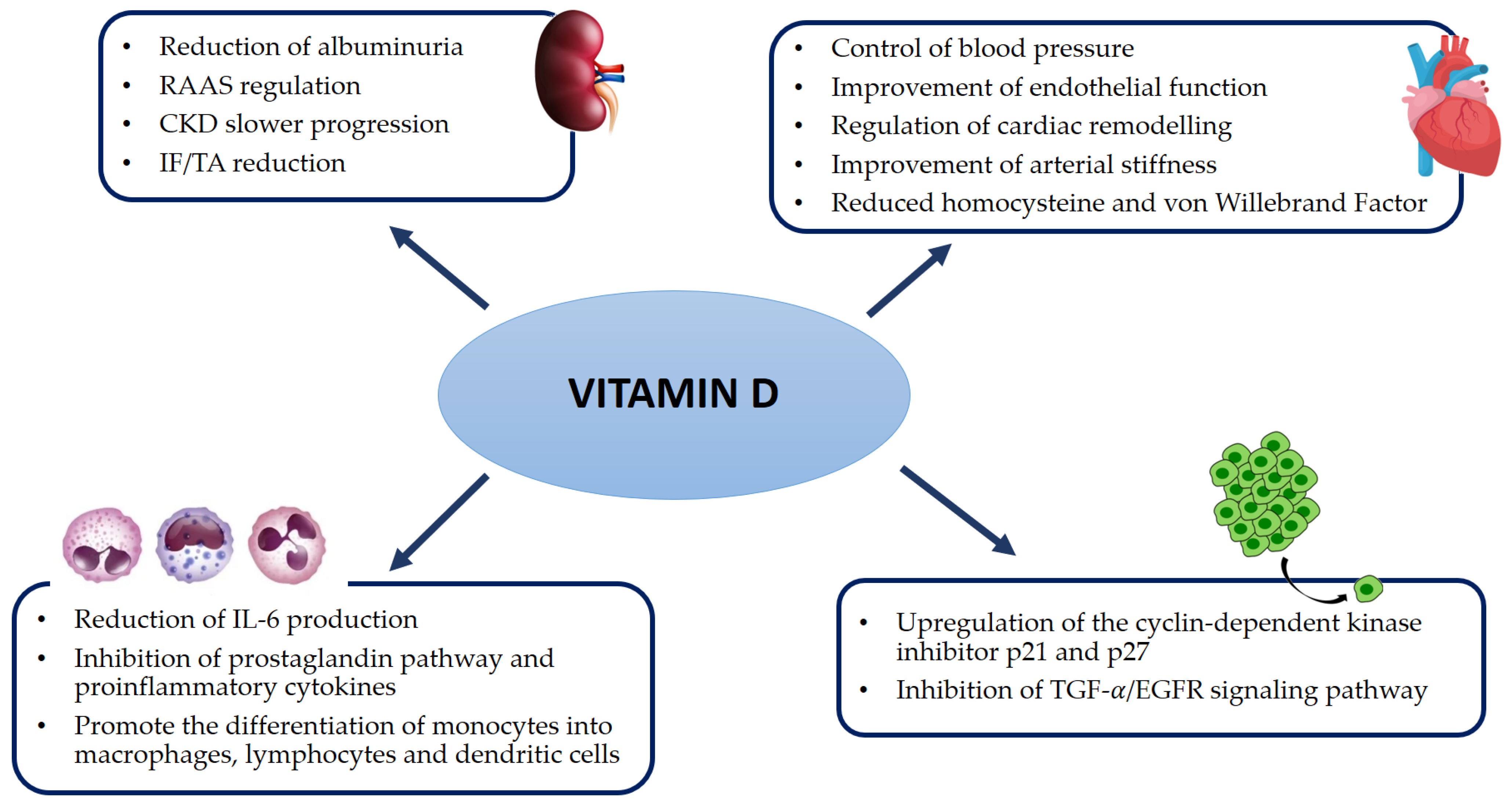
8. Pleiotropic Effects of Vitamin D

Over the last few years, increasing evidence has been revealed about the impact of vitamin D on cardiovascular health, inflammatory status, cancer, and progression of CKD. The discovery of the VDR enabled multiple investigations on the association of vitamin D deficiency with acute and chronic diseases. Due to the wider distribution of the VDR, vitamin D is associated with several pleiotropic effects: renal-function preservation, regulation of blood pressure, glycemic control, regulation of cellular proliferation, regulation of the renin-angiotensin-aldosterone system (RAAS), and immunomodulation properties [68,69].
Vitamin D plays a central role in cardiovascular health, as shown by the expression of the dedicated signaling apparatus at almost all levels of the cardiovascular system, i.e., endothelial cells, cardiomyocytes, and smooth muscle cells of vessels [70,71,72,73]. Experimental studies conducted on VDR-knockout mice highlighted a dramatic increase in cardiovascular dysfunction in affected animals that developed ventricular hypertrophy, heart failure, hypertension, and upregulation of RAAS. Evidence suggests that such comorbidities improve following vitamin D supplementation [4].
It has been found that 25(OH)D deficiency is associated with accelerated arteriosclerosis and endothelial dysfunction in ESRD patients, with a subsequent increase in cardiovascular risk. Moreover, a suppression of cardiomyocytes proliferation in case of vitamin D deficiency has been hypothesized [74].
Several prospective observational studies investigated 25(OH)D levels and the risk of CVD, and the clinical endpoints were various myocardial infarction, combined cardiovascular disease, stroke, and cardiovascular mortality [75]. The Framingham Offspring Study recruited 1739 participants free of CVD at the baseline. Over an average follow-up time of 5 years, lower 25(OH)D levels were associated with a risk of cardiovascular events that was 1.62 times higher [72]. Similarly, the Health Professionals Follow-up Study revealed that the incidence of acute myocardial infarction was 2.42 times higher in men with 25(OH)D levels < 15 ng/mL, compared to those with levels above 30 ng/mL [76]. On the other hand, the NHANES III study, which included data from more than 13,300 participants followed for 8.7 years, showed only a trend towards an increased risk in the lowest (<17.8 ng/mL) compared with the highest 1,25(OH)2D [77]. In a prospective cohort study, as the subset of the MrOS study, no significant association was found between 25(OH)D deficiency (<15 ng/mL) and cardiovascular incidence (coronary heart disease and cerebrovascular attack) compared with vitamin D sufficiency (>30 ng/mL) [78].
Several studies evaluated not only have changes in cardiovascular risk with low 25(OH)D levels, but also with the contribution of higher levels. Most of these suggest that risk does not decrease with levels >30 ng/mL [79,80]. Some others even suggested a possible U-shaped relation, with a possible increase in cardiovascular disease risk at high 25(OH)D D levels (>60 ng/mL) [81]. Finally, if the observational data provided evidence of the association between low 25(OH)D levels and increased cardiovascular risk, evidences are still limited to support the view that higher levels of 25(OH)D are linked with a similar decrease in risk.
Regarding the control of the inflammatory status, accumulating data indicate that vitamin D exerts anti-inflammatory effects through many ways, namely by inhibition of the prostaglandin pathway, proinflammatory cytokines, and NFKB. Moreover, it provides antioxidant defense against ROS, thus avoiding the perpetuation of pro-inflammatory responses and DNA damage [82].
Another function attributed to vitamin D is the ability to promote the differentiation of monocytes into macrophages, lymphocytes, and dendritic cells, which are the first line of defense of the innate immune system and infection control [83].
Several studies have also highlighted an association between sufficient vitamin D status and cancer prevention in several malignancies, namely prostate, breast, and colon cancer. This protective role can be explained by vitamin D-mediated upregulation of the cyclin-dependent kinase inhibitors p21 and p27 and inhibition of the TGF-α/EGFR growth pathway [84].
Furthermore, many studies focused on nephropathies reported that active vitamin D protects the kidneys through its anti-inflammatory and antifibrotic effects. Calcitriol has proven to have inhibitory effects on renal interstitial myofibroblasts, thus decelerating the progression to renal interstitial fibrosis. Experimental studies involving knockout mice lacking active vitamin D receptors revealed elevated levels of renin and angiotensin II in the mice’s blood, which caused a significant rise in blood pressure and subsequent cardiac hypertrophy [85,86,87,88]. Figure 3 is a schematic representation of the main pleiotropic systemic effects of vitamin D.

9. Conclusions

Recently, the function of vitamin D has been extensively investigated. The discovery of the VDR can lead to a better understanding of the relationship of acute and chronic diseases with vitamin D deficiency. Results of vitamin D trials vary for the general population and renal patients. The discrepancies may be due to differences in the baseline serum 25(OH)D levels, vitamin D doses and treatment periods, adherence to supplementation, and VDR genetic polymorphisms [89].
Therefore, the application of vitamin D in disease treatment and prevention is far from been achieved. Further investigation is required to pursue this aim. Regarding vitamin D reference values, there is so far still no univocal consensus on the reference values of vitamin D’s status. The optimal serum concentration of 25(OH)D has been considered to not lead to a PTH elevation [90]. Such a view seems to be obsolete, and it is the result of partial knowledge of the biological activity of vitamin D. Moreover, the bioaccessibility of vitamin D in foods must be considered. There is, however, a lack of kinetic data that allows for the prediction of vitamin D’s stability under industrial processing conditions [91].

Author Contributions

Conceptualization, F.Z. and A.C.; methodology, F.Z. and M.C.; software, M.C.; validation, C.D., M.C. and G.L.M.; formal analysis, F.Z.; investigation, A.C. and M.N.; resources, M.D.N.; data curation, F.T.; writing—original draft preparation, A.C. and F.Z.; writing—review and editing, A.C., F.Z. and M.C.; visualization, A.S. and A.L.C.C.; supervision, C.D. and G.C. All authors have read and agreed to the published version of the manuscript.

Funding

This research received no external funding.

Institutional Review Board Statement

Not applicable.

Informed Consent Statement

Not applicable.

Data Availability Statement

Not applicable.

Conflicts of Interest

The authors declare no conflict of interest.

References

  1. Heaney, R.P. Vitamin D in Health and Disease. Clin. J. Am. Soc. Nephrol. 2008, 3, 1535–1541. [Google Scholar] [CrossRef]
  2. Holick, M.F. Vitamin D Status: Measurement, Interpretation, and Clinical Application. Ann. Epidemiol. 2009, 19, 73–78. [Google Scholar] [CrossRef] [PubMed]
  3. Holick, M.F. High Prevalence of Vitamin D Inadequacy and Implications for Health. Mayo Clin. Proc. 2006, 81, 353–373. [Google Scholar] [CrossRef] [PubMed]
  4. Bouillon, R.; Carmeliet, G.; Verlinden, L.; van Etten, E.; Verstuyf, A.; Luderer, H.F.; Lieben, L.; Mathieu, C.; DeMay, M. Vitamin D and Human Health: Lessons from Vitamin D Receptor Null Mice. Endocr. Rev. 2008, 29, 726–776. [Google Scholar] [CrossRef] [PubMed]
  5. Jones, G.; Prosser, D.E.; Kaufmann, M. Cytochrome P450-mediated metabolism of vitamin D. J. Lipid Res. 2014, 55, 13–31. [Google Scholar] [CrossRef]
  6. Zierold, C.; Nehring, J.A.; Deluca, H.F. Nuclear receptor 4A2 and C/EBPβ regulate the parathyroid hormone-mediated transcriptional regulation of the 25-hydroxyvitamin D3-1α-hydroxylase. Arch. Biochem. Biophys. 2007, 460, 233–239. [Google Scholar] [CrossRef] [PubMed]
  7. Perwad, F.; Azam, N.; Zhang, M.Y.; Yamashita, T.; Tenenhouse, H.S.; Portale, A.A. Dietary and Serum Phosphorus Regulate Fibroblast Growth Factor 23 Expression and 1,25-Dihydroxyvitamin D Metabolism in Mice. Endocrinology 2005, 146, 5358–5364. [Google Scholar] [CrossRef]
  8. Kumar, R.; Tebben, P.J.; Thompson, J.R. Vitamin D and the kidney. Arch. Biochem. Biophys. 2012, 523, 77–86. [Google Scholar] [CrossRef]
  9. Caudarella, R.; Vescini, F.; Buffa, A.; Sinicropi, G.; Rizzoli, E.; La Manna, G.; Stefoni, S. Bone mass loss in calcium stone disease: Focus on hypercalciuria and metabolic factors. J. Nephrol. 2003, 16, 260–266. [Google Scholar] [PubMed]
  10. Friedman, P.A.; Gesek, F.A. Cellular calcium transport in renal epithelia: Measurement, mechanisms, and regulation. Physiol. Rev. 1995, 75, 429–471. [Google Scholar] [CrossRef]
  11. Borke, J.L.; Penniston, J.T.; Kumar, R. Recent advances in calcium transport by the kidney. Semin. Nephrol. 1990, 10, 15–23. [Google Scholar] [PubMed]
  12. Khazai, N.; Judd, S.E.; Tangpricha, V. Calcium and vitamin D: Skeletal and extraskeletal health. Curr. Rheumatol. Rep. 2008, 10, 110–117. [Google Scholar] [CrossRef]
  13. Holick, M.F. The vitamin D deficiency pandemic: Approaches for diagnosis, treatment and prevention. Rev. Endocr. Metab. Disord. 2017, 18, 153–165. [Google Scholar] [CrossRef] [PubMed]
  14. Capelli, I.; Cianciolo, G.; Gasperoni, L.; Galassi, A.; Ciceri, P.; Cozzolino, M. Nutritional vitamin D in CKD: Should we measure? Should we treat? Clin. Chim. Acta 2020, 501, 186–197. [Google Scholar] [CrossRef] [PubMed]
  15. Morrone, L.F.; Bolasco, P.; Camerini, C.; Cianciolo, G.; Cupisti, A.; Galassi, A.; Mazzaferro, S.; Russo, D.; Russo, L.; Cozzolino, M. Vitamin D in patients with chronic kidney disease: A position statement of the Working Group “Trace Elements and Mineral Metabolism” of the Italian Society of Nephrology. J. Nephrol. 2016, 29, 305–328. [Google Scholar] [CrossRef]
  16. Kogawa, M.; Findlay, D.M.; Anderson, P.H.; Ormsby, R.; Vincent, C.; Morris, H.A.; Atkins, G.J. Osteoclastic Metabolism of 25(OH)-Vitamin D3: A Potential Mechanism for Optimization of Bone Resorption. Endocrinology 2010, 151, 4613–4625. [Google Scholar] [CrossRef] [PubMed]
  17. Anderson, P.H.; Atkins, G.J.; Turner, A.G.; Kogawa, M.; Findlay, D.M.; Morris, H.A. Vitamin D metabolism within bone cells: Effects on bone structure and strength. Mol. Cell. Endocrinol. 2011, 347, 42–47. [Google Scholar] [CrossRef]
  18. Cianciolo, G.; Cappuccilli, M.; Tondolo, F.; Gasperoni, L.; Zappulo, F.; Barbuto, S.; Iacovella, F.; Conte, D.; Capelli, I.; La Manna, G. Vitamin D Effects on Bone Homeostasis and Cardiovascular System in Patients with Chronic Kidney Disease and Renal Transplant Recipients. Nutrients 2021, 13, 1453. [Google Scholar] [CrossRef] [PubMed]
  19. Kidney Disease: Improving Global Outcomes (KDIGO) CKD-MBD Update Work Group. KDIGO 2017 Clinical Practice Guideline Update for the Diagnosis, Evaluation, Prevention, and Treatment of Chronic Kidney Disease–Mineral and Bone Disorder (CKD-MBD). Kidney Int. Suppl. 2017, 7, 1–59. [Google Scholar] [CrossRef]
  20. Lips, P.; van Schoor, N.M. The effect of vitamin D on bone and osteoporosis. Best Pract. Res. Clin. Endocrinol. Metab. 2011, 25, 585–591. [Google Scholar] [CrossRef] [PubMed]
  21. Fusaro, M.; Aghi, A.; Mereu, M.C.; Giusti, A. Fratture da fragilità nella Malattia Renale Cronica (MRC) [Fragility fracture in the Chronic Kidney Disease (CKD)]. G. Ital. Nefrol. 2017, 6, 34. [Google Scholar]
  22. Pazianas, M.; Miller, P.D. Osteoporosis and Chronic Kidney Disease–Mineral and Bone Disorder (CKD-MBD): Back to Basics. Am. J. Kidney Dis. 2021, 78, 582–589. [Google Scholar] [CrossRef] [PubMed]
  23. Felsenberg, D.; Boonen, S. The bone quality framework: Determinants of bone strength and their interrelationships, and implications for osteoporosis management. Clin. Ther. 2005, 27, 1–11. [Google Scholar] [CrossRef] [PubMed]
  24. Hampson, G.; Elder, G.J.; Cohen-Solal, M.; Abrahamsen, B. A review and perspective on the assessment, management and prevention of fragility fractures in patients with osteoporosis and chronic kidney disease. Endocrine 2021, 73, 509–529. [Google Scholar] [CrossRef] [PubMed]
  25. Peugh, J.; Khalil, A.; Chan, M.R.; Hansen, K.E. Teriparatide Treatment for Hypercalcemia Associated With Adynamic Bone Disease. JBMR Plus 2019, 3, e10176. [Google Scholar] [CrossRef]
  26. Kawahara, M.; Iwasaki, Y.; Sakaguchi, K.; Taguchi, T.; Nishiyama, M.; Nigawara, T.; Tsugita, M.; Kambayashi, M.; Suda, T.; Hashimoto, K. Predominant role of 25OHD in the negative regulation of PTH expression: Clinical relevance for hypovitaminosis D. Life Sci. 2008, 82, 677–683. [Google Scholar] [CrossRef] [PubMed]
  27. Arcidiacono, M.V.; Yang, J.; Fernandez, E.; Dusso, A. The induction of C/EBP contributes to vitamin D inhibition of ADAM17 expression and parathyroid hyperplasia in kidney disease. Nephrol. Dial. Transplant. 2015, 30, 423–433. [Google Scholar] [CrossRef]
  28. Wolf, M.; Shah, A.; Gutierrez, O.; Ankers, E.; Monroy, M.; Tamez, H.; Steele, D.; Chang, Y.; Camargo, C.A., Jr.; Tonelli, M.; et al. Vitamin D levels and early mortality among incident hemodialysis patients. Kidney Int. 2007, 72, 1004–1013. [Google Scholar] [CrossRef]
  29. Sözen, T.; Özışık, L.; Başaran, N.Ç. An overview and management of osteoporosis. Eur. J. Rheumatol. 2017, 4, 46–56. [Google Scholar] [CrossRef]
  30. Moe, S.; Drüeke, T.; Cunningham, J.; Goodman, W.; Martin, K.; Olgaard, K.; Ott, S.; Sprague, S.; Lameire, N.; Eknoyan, G.; et al. Definition, evaluation, and classification of renal osteodystrophy: A position statement from Kidney Disease: Improving Global Outcomes (KDIGO). Kidney Int. 2006, 69, 1945–1953. [Google Scholar] [CrossRef] [PubMed]
  31. Mizobuchi, M.; Ogata, H.; Hatamura, I.; Saji, F.; Koiwa, F.; Kinugasa, E.; Koshikawa, S.; Akizawa, T. Activation of calcium-sensing receptor accelerates apoptosis in hyperplastic parathyroid cells. Biochem. Biophys. Res. Commun. 2007, 362, 11–16. [Google Scholar] [CrossRef]
  32. Aloia, J.; Dhaliwal, R.; Mikhail, M.; Shieh, A.; Stolberg, A.; Ragolia, L.; Fazzari, M.; Abrams, S.A. Free 25(OH)D and Calcium Absorption, PTH, and Markers of Bone Turnover. J. Clin. Endocrinol. Metab. 2015, 100, 4140–4145. [Google Scholar] [CrossRef]
  33. Shieh, A.; Ma, C.; Chun, R.F.; Wittwer-Schegg, J.; Swinkels, L.; Huijs, T.; Wang, J.; Donangelo, I.; Hewison, M.; Adams, J.S. Associations Between Change in Total and Free 25-Hydroxyvitamin D With 24,25-Dihydroxyvitamin D and Parathyroid Hormone. J. Clin. Endocrinol. Metab. 2018, 103, 3368–3375. [Google Scholar] [CrossRef] [PubMed]
  34. Chandra, P.; Binongo, J.N.; Ziegler, T.R.; Schlanger, L.E.; Wang, W.; Someren, J.T.; Tangpricha, V. Cholecalciferol (Vitamin D3) Therapy and Vitamin D Insufficiency in Patients with Chronic Kidney Disease: A Randomized Controlled Pilot Study. Endocr. Pract. 2008, 14, 10–17. [Google Scholar] [CrossRef] [PubMed]
  35. Westerberg, P.A.; Sterner, G.; Ljunggren, O.; Isaksson, E.; Elvarson, F.; Dezfoolian, H.; Linde, T. High doses of cholecalciferol alleviate the progression of hyperparathyroidism in patients with CKD Stages 3–4: Results of a 12-week double-blind, randomized, controlled study. Nephrol. Dial. Transplant. 2018, 33, 466–471. [Google Scholar] [CrossRef]
  36. Massart, A.; Debelle, F.D.; Racapé, J.; Gervy, C.; Husson, C.; Dhaene, M.; Wissing, K.M.; Nortier, J.L. Biochemical Parameters After Cholecalciferol Repletion in Hemodialysis: Results From the VitaDial Randomized Trial. Am. J. Kidney Dis. 2014, 64, 696–705. [Google Scholar] [CrossRef]
  37. Ish-Shalom, S.; Segal, E.; Salganik, T.; Raz, B.; Bromberg, I.L.; Vieth, R. Comparison of Daily, Weekly, and Monthly Vitamin D3 in Ethanol Dosing Protocols for Two Months in Elderly Hip Fracture Patients. J. Clin. Endocrinol. Metab. 2008, 93, 3430–3435. [Google Scholar] [CrossRef]
  38. Kandula, P.; Dobre, M.; Schold, J.D.; Schreiber, M.J., Jr.; Mehrotra, R.; Navaneethan, S.D. Vitamin D Supplementation in Chronic Kidney Disease: A Systematic Review and Meta-Analysis of Observational Studies and Randomized Controlled Trials. Clin. J. Am. Soc. Nephrol. 2011, 6, 50–62. [Google Scholar] [CrossRef]
  39. Alvarez, J.A.; Law, J.; Coakley, K.E.; Zughaier, S.M.; Hao, L.; Shahid Salles, K.; Wasse, H.; Gutiérrez, O.M.; Ziegler, T.R.; Tangpricha, V. High-dose cholecalciferol reduces parathyroid hormone in patients with early chronic kidney disease: A pilot, randomized, double-blind, placebo-controlled trial. Am. J. Clin. Nutr. 2012, 96, 672–679. [Google Scholar] [CrossRef]
  40. Cupisti, A.; Egidi, M.F.; Vigo, V.; Baronti, M.E.; D’Alessandro, C.; Ghiadoni, L. Vitamin D status and cholecalciferol supplementation in chronic kidney disease patients: An Italian cohort report. Int. J. Nephrol. Renovacs. Dis. 2015, 8, 151–157. [Google Scholar] [CrossRef]
  41. Jean, G.; Vanel, T.; Terrat, J.C.; Chazot, C. Prevention of secondary hyperparathyroidism in hemodialysis patients: The key role of native vitamin D supplementation. Hemodial. Int. 2010, 14, 486–491. [Google Scholar] [CrossRef] [PubMed]
  42. Aytac, M.B.; Deveci, M.; Bek, K.; Kayabey, O.; Ekinci, Z. Effect of cholecalciferol on local arterial stiffness and endothelial dysfunction in children with chronic kidney disease. Pediatr. Nephrol. 2016, 31, 267–277. [Google Scholar] [CrossRef] [PubMed]
  43. Karakas, Y.; Sahin, G.; Urfali, F.E.; Bal, C.; Degirmenci, N.A.; Sirmagul, B. Effect of vitamin D supplementation on endothelial dysfunction in hemodialysis patients. Hemodial. Int. 2017, 21, 97–106. [Google Scholar] [CrossRef]
  44. Kim, M.J.; Frankel, A.H.; Donaldson, M.; Darch, S.J.; Pusey, C.D.; Hill, P.D.; Mayr, M.; Tam, F.W. Oral cholecalciferol decreases albuminuria and urinary TGF-β1 in patients with type 2 diabetic nephropathy on established renin–angiotensin–aldosterone system inhibition. Kidney Int. 2011, 80, 851–860. [Google Scholar] [CrossRef]
  45. Meireles, M.S.; Kamimura, M.A.; Dalboni, M.A.; Giffoni de Carvalho, J.T.; Aoike, D.T.; Cuppari, L. Effect of cholecalciferol on vitamin D-regulatory proteins in monocytes and on inflammatory markers in dialysis patients: A randomized controlled trial. Clin. Nutr. 2016, 35, 1251–1258. [Google Scholar] [CrossRef]
  46. Mann, M.C.; Hobbs, A.J.; Hemmelgarn, B.R.; Roberts, D.J.; Ahmed, S.B.; Rabi, D.M. Effect of oral vitamin D analogs on mortality and cardiovascular outcomes among adults with chronic kidney disease: A meta-analysis. Clin. Kidney J. 2015, 8, 41–48. [Google Scholar] [CrossRef]
  47. Bjelakovic, G.; Gluud, L.L.; Nikolova, D.; Whitfield, K.; Wetterslev, J.; Simonetti, R.G.; Bjelakovic, M.; Gluud, C. Vitamin D supplementation for prevention of mortality in adults. Cochrane Database Syst. Rev. 2014, CD007470. [Google Scholar] [CrossRef]
  48. Cianciolo, G.; Galassi, A.; Capelli, I.; Angelini, M.L.; La Manna, G.; Cozzolino, M. Vitamin D in Kidney Transplant Recipients: Mechanisms and Therapy. Am. J. Nephrol. 2016, 43, 397–407. [Google Scholar] [CrossRef]
  49. Obi, Y.; Hamano, T.; Ichimaro, N.; Tomida, K.; Matsui, I.; Fujii, N.; Okumi, M.; Kaimori, J.-Y.; Yazawa, K.; Kokado, Y. Vitamin D deficiency predicts decline in kidney allograft function: A prospective cohort study. J. Clin. Endocrinol. Metab. 2014, 99, 527–535. [Google Scholar] [CrossRef]
  50. Lee, J.R.; Dadhania, D.; August, P.; Lee, J.B.; Suthanthiran, M.; Muthukumar, T. Circulating levels of 25-hydroxyvitamin D and acute cellular rejection in kidney allograft recipients. Transplantation 2014, 98, 292–299. [Google Scholar] [CrossRef]
  51. Filipov, J.J.; Zlatkov, B.K.; Dimitrov, E.P.; Svinarov, D.A. Higher 25-Hydroxyvitamin D Levels Are Associated With Lower Proteinuria in Kidney Transplant Recipients. Exp. Clin. Transplant. 2016, 14, 629–633. [Google Scholar] [PubMed]
  52. Courbebaisse, M.; Souberbielle, J.-C.; Thervet, E. Potential Nonclassical Effects of Vitamin D in Transplant Recipients. Transplantation 2010, 89, 131–137. [Google Scholar] [CrossRef] [PubMed]
  53. Courbebaisse, M.; Xu-Dubois, Y.-C.; Thervet, E.; Prié, D.; Zuber, J.; Kreis, H.; Legendre, C.; Rondeau, E.; Pallet, N. Cholecalciferol Supplementation Does Not Protect Against Renal Allograft Structural and Functional Deterioration: A Retrospective Study. Transplantation 2011, 91, 207–212. [Google Scholar] [CrossRef]
  54. Westenfeld, R.; Schlieper, G.; Wöltje, M.; Gawlik, A.; Brandenburg, V.; Rutkowski, P.; Floege, J.; Jahnen-Dechent, W.; Ketteler, M. Impact of sirolimus, tacrolimus and mycophenolate mofetil on osteoclastogenesis--implications for post-transplantation bone disease. Nephrol. Dial. Transplant. 2011, 26, 4115–4123. [Google Scholar] [CrossRef]
  55. Christakos, S.; Dhawan, P.; Verstuyf, A.; Verlinden, L.; Carmeliet, G. Vitamin D: Metabolism, Molecular Mechanism of Action, and Pleiotropic Effects. Physiol. Rev. 2016, 96, 365–408. [Google Scholar] [CrossRef]
  56. Cantorna, M.T.; Snyder, L.; Lin, Y.-D.; Yang, L. Vitamin D and 1,25(OH)2D Regulation of T cells. Nutrients 2015, 7, 3011–3021. [Google Scholar] [CrossRef]
  57. Bhalla, A.K.; Amento, E.P.; Clemens, T.L.; Holick, M.F.; Krane, S.M. Specific high-affinity receptors for 1,25-dihydroxyvitamin D3 in human peripheral blood mononuclear cells: Presence in monocytes and induction in T lymphocytes following activation. J. Clin. Endocrinol. Metab. 1983, 57, 1308–1310. [Google Scholar] [CrossRef]
  58. Baeke, F.; Takiishi, T.; Korf, H.; Gysemans, C.; Mathieu, C. Vitamin D: Modulator of the immune system. Curr. Opin. Pharmacol. 2010, 10, 482–496. [Google Scholar] [CrossRef]
  59. Kim, E.W.; Teles, R.M.B.; Haile, S.; Liu, P.T.; Modlin, R.L. Vitamin D status contributes to the antimicrobial activity of macrophages against Mycobacterium leprae. PLOS Neglected Trop. Dis. 2018, 12, e0006608. [Google Scholar] [CrossRef]
  60. Piemonti, L.; Monti, P.; Sironi, M.; Fraticelli, P.; Leone, B.E.; Dal Cin, E.; Allavena, P.; Di Carlo, V. Vitamin D3 Affects Differentiation, Maturation, and Function of Human Monocyte-Derived Dendritic Cells. J. Immunol. 2000, 164, 4443–4451. [Google Scholar] [CrossRef]
  61. Chen, L.; Eapen, M.S.; Zosky, G.R. Vitamin D both facilitates and attenuates the cellular response to lipopolysaccharide. Sci. Rep. 2017, 7, 45172. [Google Scholar] [CrossRef] [PubMed]
  62. Griffin, M.D.; Lutz, W.H.; Phan, V.A.; Bachman, L.A.; McKean, D.J.; Kumar, R. Potent Inhibition of Dendritic Cell Differentiation and Maturation by Vitamin D Analogs. Biochem. Biophys. Res. Commun. 2000, 270, 701–708. [Google Scholar] [CrossRef] [PubMed]
  63. Bartels, L.E.; Hvas, C.L.; Agnholt, J.; Dahlerup, J.F.; Agger, R. Human dendritic cell antigen presentation and chemotaxis are inhibited by intrinsic 25-hydroxy vitamin D activation. Int. Immunopharmacol. 2010, 10, 922–928. [Google Scholar] [CrossRef] [PubMed]
  64. Dam, T.N.; Møller, B.; Hindkjaer, J.; Kragballe, K. The vitamin D3 analog calcipotriol suppresses the number and antigen-presenting function of Langerhans cells in normal human skin. J. Investig. Dermatol. Symp. Proc. 1996, 1, 72–77. [Google Scholar]
  65. Rolf, L.; Muris, A.-H.; Hupperts, R.; Damoiseaux, J. Vitamin D effects on B cell function in autoimmunity. Ann. N. Y. Acad. Sci. 2014, 1317, 84–91. [Google Scholar] [CrossRef]
  66. Chen, S.; Sims, G.P.; Chen, X.X.; Gu, Y.Y.; Chen, S.; Lipsky, P.E. Modulatory Effects of 1,25-Dihydroxyvitamin D3on Human B Cell Differentiation. J. Immunol. 2007, 179, 1634–1647. [Google Scholar] [CrossRef]
  67. Morgan, J.W.; Kouttab, N.; Ford, D.; Maizel, A.L. Vitamin D-Mediated Gene Regulation in Phenotypically Defined Human B Cell Subpopulations. Endocrinology 2000, 141, 3225–3234. [Google Scholar] [CrossRef]
  68. Levin, G.P.; Robinson-Cohen, C.; de Boer, I.H.; Houston, D.K.; Lohman, K.; Liu, Y.; Kritchevsky, S.B.; Cauley, J.A.; Tanaka, T.; Ferrucci, L.; et al. Genetic Variants and Associations of 25-Hydroxyvitamin D Concentrations With Major Clinical Outcomes. JAMA 2012, 308, 1898–1905. [Google Scholar] [CrossRef]
  69. Galassi, A.; Bellasi, A.; Auricchio, S.; Papagni, S.; Cozzolino, M. Which Vitamin D in CKD-MBD? The Time of Burning Questions. BioMed Res. Int. 2013, 2013, 864012. [Google Scholar] [CrossRef]
  70. Mozos, I.; Marginean, O. Links between Vitamin D Deficiency and Cardiovascular Diseases. BioMed Res. Int. 2015, 2015, 109275. [Google Scholar] [CrossRef]
  71. Pilz, S.; Gaksch, M.; O’Hartaigh, B.; Tomaschitz, A.; Marz, W. The role of vitamin D deficiency in cardiovascular disease: Where do we stand in 2013? Arch. Toxicol. 2013, 87, 2083–2103. [Google Scholar] [CrossRef] [PubMed]
  72. Wang, T.J.; Pencina, M.J.; Booth, S.L.; Jacques, P.F.; Ingelsson, E.; Lanier, K.; Benjamin, E.J.; D’Agostino, R.B.; Wolf, M.; Vasan, R.S. Vitamin D Deficiency and Risk of Cardiovascular Disease. Circulation 2008, 117, 503–511. [Google Scholar] [CrossRef]
  73. Lee, J.H.; O’Keefe, J.H.; Bell, D.; Hensrud, D.D.; Holick, M.F. Vitamin D deficiency an important, common, and easily treatable cardiovascular risk factor? J. Am. Coll. Cardiol. 2008, 52, 1949–1956. [Google Scholar] [CrossRef] [PubMed]
  74. Weishaar, R.E.; Simpson, R.U. Vitamin D3 and cardiovascular function in rats. J. Clin. Investig. 1987, 79, 1706–1712. [Google Scholar] [CrossRef] [PubMed]
  75. Wang, L.; Song, Y.; Manson, J.E.; Pilz, S.; Marz, W.; Michaelsson, K.; Lundqvist, A.; Jassal, S.K.; Barrett-Connor, E.; Zhang, C.; et al. Circulating 25-hydroxy-vitamin D and risk of cardiovascular disease: A meta-analysis of prospective studies. Circ. Cardiovasc. Qual. Outcomes 2012, 5, 819–829. [Google Scholar] [CrossRef]
  76. Giovannucci, E.; Liu, Y.; Hollis, B.W.; Rimm, E.B. 25-Hydroxyvitamin D and risk of myocardial infarction in men: A prospective study. Arch. Internal Med. 2008, 168, 1174–1180. [Google Scholar] [CrossRef] [PubMed]
  77. Melamed, M.L.; Michos, E.D.; Post, W.; Astor, B. 25-Hydroxyvitamin D Levels and the Risk of Mortality in the General Population. Arch. Int. Med. 2008, 168, 1629–1637. [Google Scholar] [CrossRef] [PubMed]
  78. Messenger, W.; Nielson, C.M.; Li, H.; Beer, T.; Barrett-Connor, E.; Stone, K.; Shannon, J. Serum and dietary vitamin D and cardiovascular disease risk in elderly men: A prospective cohort study. Nutr. Metab. Cardiovasc. Dis. 2012, 22, 856–863. [Google Scholar] [CrossRef]
  79. Kendrick, J.; Targher, G.; Smits, G.; Chonchol, M. 25-Hydroxyvitamin D deficiency is independently associated with cardiovascular disease in the Third National Health and Nutrition Examination Survey. Atherosclerosis 2009, 205, 255–260. [Google Scholar] [CrossRef]
  80. Chang, Q.; Hoefs, S.; van der Kemp, A.W.; Topala, C.N.; Bindels, R.J.; Hoenderop, J.G. The beta-glucuronidase klotho hydrolyzes and activates the TRPV5 channel. Science 2005, 310, 490–493. [Google Scholar] [CrossRef]
  81. Durup, D.; Jørgensen, H.L.; Christensen, J.; Schwarz, P.; Heegaard, A.-M.; Lind, B. A Reverse J-Shaped Association of All-Cause Mortality with Serum 25-Hydroxyvitamin D in General Practice: The CopD Study. J. Clin. Endocrinol. Metab. 2012, 97, 2644–2652. [Google Scholar] [CrossRef]
  82. Jeon, S.-M.; Shin, E.-A. Exploring vitamin D metabolism and function in cancer. Exp. Mol. Med. 2018, 50, 1–14. [Google Scholar] [CrossRef] [PubMed]
  83. Tan, X.; Wen, X.; Liu, Y. Paricalcitol Inhibits Renal Inflammation by Promoting Vitamin D Receptor–Mediated Sequestration of NF-κB Signaling. J. Am. Soc. Nephrol. 2008, 19, 1741–1752. [Google Scholar] [CrossRef] [PubMed]
  84. Foltyn, V.N.; Bendikov, I.; De Miranda, J.; Panizzutti, R.; Dumin, E.; Shleper, M.; Li, P.; Toney, M.D.; Kartvelishvily, E.; Wolosker, H. Serine Racemase Modulates Intracellular D-Serine Levels through an α,β-Elimination Activity. J. Biol. Chem. 2005, 280, 1754–1763. [Google Scholar] [CrossRef] [PubMed]
  85. Chen, S.; Law, C.S.; Grigsby, C.L.; Olsen, K.; Hong, T.-T.; Zhang, Y.; Yeghiazarians, Y.; Gardner, D.G. Cardiomyocyte-Specific Deletion of the Vitamin D Receptor Gene Results in Cardiac Hypertrophy. Circulation 2011, 124, 1838–1847. [Google Scholar] [CrossRef] [PubMed]
  86. Lai, Y.-H.; Fang, T.-C. The Pleiotropic Effect of Vitamin D. ISRN Nephrol. 2013, 2013, 898125. [Google Scholar] [CrossRef]
  87. Zittermann, A. Vitamin D in preventive medicine: Are we ignoring the evidence? Br. J. Nutr. 2003, 89, 552–572. [Google Scholar] [CrossRef] [PubMed]
  88. Li, Y.C.; Kong, J.; Wei, M.; Chen, Z.-F.; Liu, S.Q.; Cao, L.-P. 1,25-Dihydroxyvitamin D3 is a negative endocrine regulator of the renin-angiotensin system. J. Clin. Investig. 2002, 110, 229–238. [Google Scholar] [CrossRef] [PubMed]
  89. Mohseni, H.; Hosseini, S.A.; Amani, R.; Ekrami, A.; Ahmadzadeh, A.; Latifi, S.M. Circulating 25-Hydroxy Vitamin D Relative to Vitamin D Receptor Polymorphism after Vitamin D3 Supplementation in Breast Cancer Women: A Randomized, Double-Blind Controlled Clinical Trial. Asian Pac. J. Cancer Prev. 2017, 18, 1953–1959. [Google Scholar] [CrossRef] [PubMed]
  90. Cantorna, M.T.; Mahon, B. Mounting Evidence for Vitamin D as an Environmental Factor Affecting Autoimmune Disease Prevalence. Exp. Biol. Med. 2004, 229, 1136–1142. [Google Scholar] [CrossRef] [PubMed]
  91. Lavelli, V.; D’Incecco, P.; Pellegrino, L. Vitamin D Incorporation in Foods: Formulation Strategies, Stability, and Bioaccessibility as Affected by the Food Matrix. Foods 2021, 10, 1989. [Google Scholar] [CrossRef] [PubMed]
Figure 1. Systemic effect of vitamin D. Ca, calcium; P, phosphorus; PTH, parathyroid hormone.
Ijms 23 09135 g001
Figure 2. CKD-MBD pathogenesis and its main systemic effects. FGF23, fibroblast growth factor 23; P, phosphorus; PTH, parathyroid hormone; Ca, calcium.
Ijms 23 09135 g002
Figure 3. Pleiotropic effect of vitamin D. CKD, chronic kidney disease; EGFR, epidermal growth factor receptor; ESRD, end-stage renal disease; IF/TA, interstitial fibrosis/tubular atrophy; IL-6, interleukin 6; RAAS, renin-angiotensin-aldosterone system; TGF-α, transforming growth factor alpha.
Ijms 23 09135 g003
Table 1. Most representative studies on the effects of native vitamin D supplementation in the nephrology clinical setting.
AuthorsVitamin D FormulationDosageStudy DesignPatientsStudy LengthResults
Kandula et al. [38]Ergocalciferol or cholecalciferolObservational study 4000 to 50,000 IU daily. RCTs rom 20,000 IU weekly to 25,000 IU monthly Systematic review and meta-analysisCKD: pre-dialysis, hemodialysis, peritoneal dialysis and KTRs1966 to September 2009No influence on Ca and P levels
Reduction of PTH
Alvarez et al. [39]Cholecalciferol50,000 IU/week for 12 weeks followed by 50,000 IU every other week for 40 weeksProspective46 early CKD (stages 2–3)1 yearPrevent vitamin D insufficiency
Improvement of serum PTH
Cupisti et al. [40]Cholecalciferol10,000 IU once-a-week Cohort study 405 CKD patients (stages 2–4)12 monthsReduction of PTH
Jean et al. [41]Cholecalciferol and calcifediol cholecalciferol 100,000 U/month calcifediol 10–50 μg/dProspective All incident and prevalent hemodialysis patients in a single centerThree observation periods of 1-yr eachReduction of the incidence of SHPT
Aytac et al. [42]Cholecalciferolsingle dose of 300,000 IU of oral cholecalciferolProspective41 CKD children and 24 healthy subjects free of any underlying cardiac or renal disease12 weeksIncrease in flow mediated dilatation, reduction in arterial stiffness
Reduction of plasmatic Hcy and von Willebrand factor
Karakas et al. [43]Cholecalciferol50,000 units weekly Prospective44 hemodialysis patients and 24 healthy8 weeksIncrease in flow-mediated dilatation
Kim et al. [44]Cholecalciferol40,000 units weekly for 8 weeks and then monthlyProspective63 patients with diabetic nephropathy 4 MonthsDecrease in proteinuria in addition to ACE-i
Meireless et al. [45]Cholecalciferol50,000 IU of cholecalciferol twice weeklyProspective38 dialysis patients 12 weeksUpregulation of CYP27B1 and VDR expression in monocytes
Lower serum IL-6 and CRP levels
Mann et al. [46]Cholecalciferol, doxecalciferol, paracalcitol or alfacalcidol 0.25 ug per day to 200,000 IU per weekSystematic reviewAdults with CKD (≤60 mL/min/1.73 m2), including dialysis-dependent ESRD3–104 weeksLack of significant effects of vitamin D supplementation on mortality
ACE-I, angiotensin-converting enzyme inhibitors; CKD, chronic kidney disease; CRP, C-reactive protein; ESRD, end stage renal disease; IL-6, interleukin 6; Hcy, homocysteine; PTH, parathyroid hormone; SHPT, secondary hyperparathyroidism; VDR, vitamin D receptor.
Publisher’s Note: MDPI stays neutral with regard to jurisdictional claims in published maps and institutional affiliations.

