Article

Anthocyanin Biosynthesis Associated with Natural Variation in Autumn Leaf Coloration in Quercus aliena Accessions

1
Key Laboratory of Silviculture and Conservation of the Ministry of Education, College of Forestry, Beijing Forestry University, Beijing 100083, China
2
Research Center of Deciduous Oaks, Beijing Forestry University, Beijing 100083, China
*
Author to whom correspondence should be addressed.
Int. J. Mol. Sci. 2022, 23(20), 12179; https://0-doi-org.brum.beds.ac.uk/10.3390/ijms232012179
Submission received: 13 September 2022 / Revised: 29 September 2022 / Accepted: 9 October 2022 / Published: 12 October 2022
(This article belongs to the Section Molecular Plant Sciences)

Abstract

:
Quercus aliena is an economically important tree species and one of the dominant native oak species in China. Although its leaves typically turn yellow in autumn, we observed natural variants with red leaves. It is important to understand the mechanisms involved in leaf color variation in this species. Therefore, we compared a Q. aliena tree with yellow leaves and three variants with red leaves at different stages of senescence in order to determine the causes of natural variation. We found that the accumulation of anthocyanins such as cyanidin 3-O-glucoside and cyanidin 3-O-sambubiglycoside had a significant effect on leaf coloration. Gene expression analysis showed upregulation of almost all genes encoding enzymes involved in anthocyanin synthesis in the red-leaved variants during the early and main discoloration stages of senescence. These findings are consistent with the accumulation of anthocyanin in red variants. Furthermore, the variants showed significantly higher expression of transcription factors associated with anthocyanin synthesis, such as those encoded by genes QaMYB1 and QaMYB3. Our findings provide new insights into the physiological and molecular mechanisms involved in autumn leaf coloration in Q. aliena, as well as provide genetic resources for further development and cultivation of valuable ornamental variants of this species.

1. Introduction

Anthocyanins, the glycosides of anthocyanidins, are water-soluble flavonoids that are produced in the cytoplasm of plants and that accumulate in vacuoles. They are one of the main pigments responsible for the coloration of flowers, fruits, and leaves [1]. Additionally, they contribute to important physiological functions such as plant defense, photoprotection, free radical scavenging, and osmotic regulation [2,3]. There are six types of anthocyanins commonly found in plants: pelargonidin 3-O-glucosides, cyanidin 3-O-glucosides, delphinidin 3-O-glucosides, peonidin 3-O-glucosides, petunidin 3-O-glucosides, and malvidin 3-O-glucosides [4]. Biosynthesis of anthocyanins is an important branch of flavonoid metabolism. Their precursor, phenylalanine, is converted into anthocyanins through a step-by-step process involving phenylalanine ammonia lyase (PAL), cinnamate-4-hydroxylase (C4H), 4-coumaryl (4CL), chalcone synthase (CHS), chalcone isomerase (CHI), flavanone 3-hydroxylase (F3H), flavanone-3′-hydroxylase (F3′H), flavanone-3′,5′-hydroxylase (F3′5′H), dihydroflavonol-4-reductase (DFR), anthocyanidin synthase (ANS), and UDP 3-O-glucosyltransferases (UFGT) [5,6]. Anthocyanin biosynthesis is regulated by enzyme-encoding genes in the anthocyanin biosynthetic pathway, and it can be influenced by transcription factors of the R2R3-MYB and bHLH families, which interact with WD40 protein to form an MYB–bHLH–WD40 complex that regulates anthocyanin biosynthesis [1].
Genes regulating anthocyanin biosynthesis have been studied extensively in many plants, including the model plant Arabidopsis thaliana [7], and other plants, such as Populus (poplar) [8], apple [9], grape [10], strawberry [11], tomato [12], and Dendrobium officinale [13]. Previous studies have also examined anthocyanin biosynthesis during the process of leaf senescence. In Liquidambar formosana, the gene LfMYB113 regulates color change in autumn leaves: its protein product activates the promoter regions of LfDFR1 and LfDFR2 [14], while the red leaves of Pistacia chinensis strongly express PcMYB113 in autumn, which is significantly associated with anthocyanin content [15]. The protein encoded by MdbHLH3, whose expression is induced by the protein encoded by MdABI5, can interact with the protein encoded by MdMYB1 to regulate anthocyanin synthesis in apple leaves [9]. The protein encoded by MdbHLH3 also modulates leaf senescence by regulating dehydratase-enolase-phosphatase complex 1, encoded by MdDEP1 [16]. Transcriptome analysis has also shown that the key regulators of leaf senescence in L. formosana-LfWRKY75, LfNAC1, and LfMYB113 are associated with the regulation of chlorophyll degradation and anthocyanin biosynthesis [17]. Here, this is the first study addressing autumn leaf color variation in Q. aliena.
Quercus aliena is a deciduous white oak tree species belonging to the Fagaceae family. It is native to China, Japan, and South Korea, and it is distributed at an altitude of 100–2000 m. Different parts of this economically important tree species have been used as raw materials for furniture, buildings, animal feed, starch, alcohol, and medicines [18,19,20,21]. Additionally, this species has an abundance of branches and leaves, and is therefore often used for urban and rural greening [22]. During cultivation, we found three natural variants of Q. aliena in Beijing, China that were significantly different in terms of their autumn leaf colors: the autumn leaves of Q. aliena are typically yellow, but the variants we observed had red leaves. Therefore, we aimed to examine the mechanisms involved in the natural variation of autumn leaves in Q. aliena in order to provide new insights into the influence of anthocyanin biosynthesis on leaf coloration.
In this study, variation in autumn leaf coloration was examined by comparing contents of total anthocyanins, total carotenoids, chlorophyll a, and chlorophyll b in the leaves of a typical Q. aliena plant and three natural variants. We found that anthocyanin played a major role in determining autumn leaf color variation and explored the factors contributing to anthocyanin accumulation in Q. aliena. Our findings may provide a basis for the development and cultivation of valuable ornamental varieties in Q. aliena, as well as new insights into the molecular mechanisms of anthocyanin biosynthesis during autumn leaf senescence.

2. Results

2.1. Changes of Autumn Leaves in Q. aliena Accessions

In our previous study, specific red-leaf variants were obtained through selective breeding of seedlings. As the serious introgression in Quercus species, molecular marker analysis was performed to explore the relationships of the four trees. The three red-leaved variants belonged to Q. aliena, and had a close relationship with QAC (Figure S1a). When compared with the populations of Q. aliena and its related species, the three variants and QAC all belonged to the Q. aliena populations, and had divergent relationships with other species in Quercus, such as Q. mongolica, Q. dentata, and Q. variabilis. There was no significant difference in autumn leaves length, width, and area (Figure S1b,c,d), and only special leaf colors were found in the four trees, which indicated that leaf color variations do not cause or accompany any other growth changes in leaf. Based on the RHS color chart, the leaf color of the red-leaved variants corresponded to the greyed-purple group, while the leaf color of the yellow-leaved tree corresponded to the greyed-orange group (Figure 1). In order to examine whether the reddening of leaves had an effect on photosynthetic capacity, we calculated Fv/Fm values for all four trees at three different stages of senescence (S1–S3), and found that the values tended to decrease as senescence progressed. Fv/Fm values for all four trees were significantly lower in the S3 stage than in the S1 stage, indicating higher potential maximum maximal quantum yield of PS II in the green stage (S1) than in the main discoloration stage (S3, Figure 2). At the S2 stage, QAC had a higher Fv/Fm value than the red-leaved variants (QA1, QA2, and QA3), but there were no significant differences in Fv/Fm among the four trees at S1 or S3. These results indicated that photosynthetic capacity is only affected by leaf color variation in the early discoloration stage.

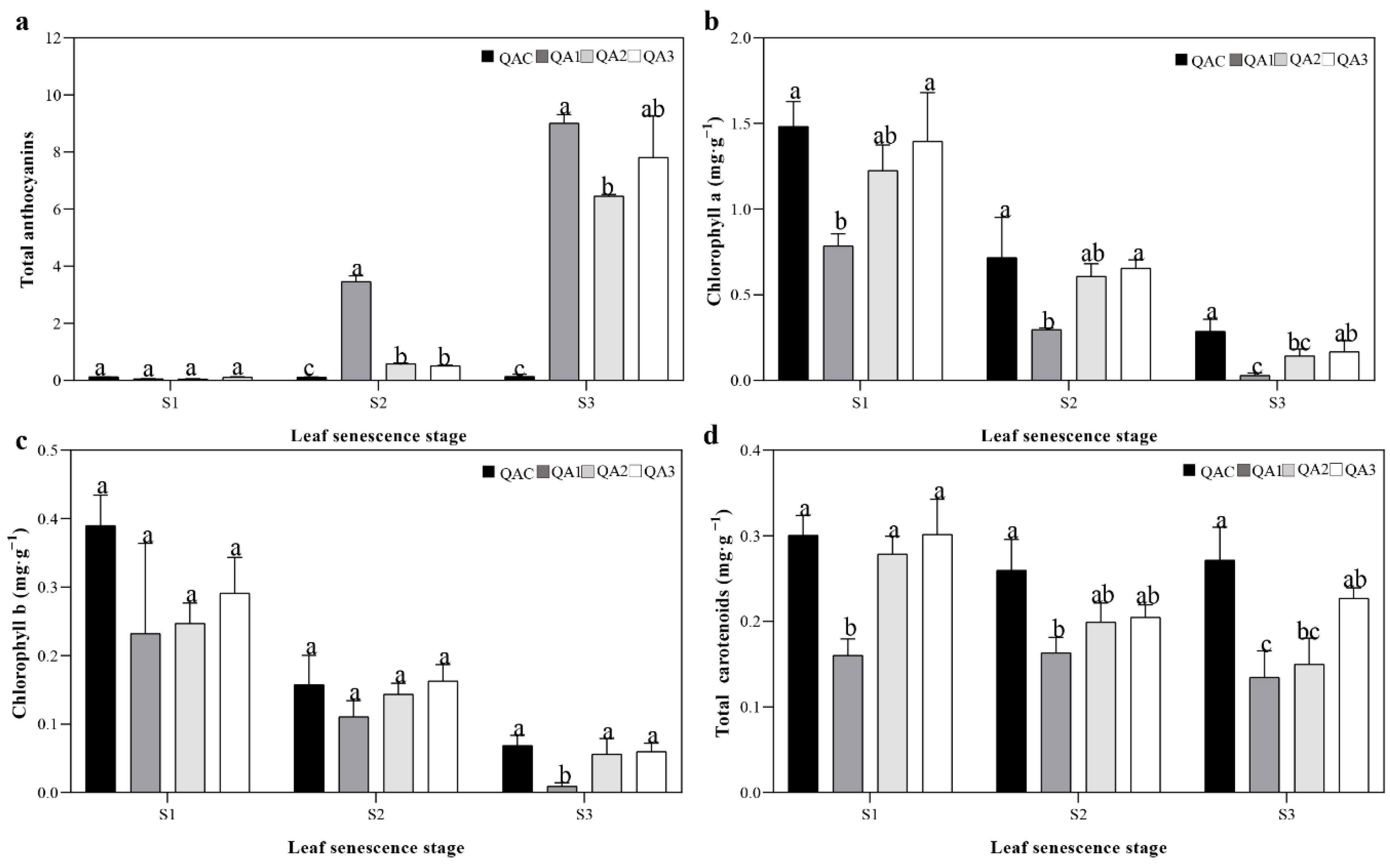
2.2. Contents of Total Anthocyanins, Total Carotenoids, and Chlorophyll in Q. aliena Accessions

In order to explore the cause of reddening of leaves in Q. aliena, we measured the content of total anthocyanins, total carotenoids, chlorophyll a, and chlorophyll b at S1, S2, and S3 in all four trees. Compared to QAC, we observed an upward trend in the total anthocyanin content of the red-leaved variants as senescence progressed. At S3, the QA1, QA2, and QA3 leaves had significantly higher anthocyanin content than the QAC leaves, while at S1, there were no significant differences in anthocyanin content among the four accessions (Figure 3a). In fact, at S3, the anthocyanin content in QA1, QA2, and QA3 was 72–163 times higher than that measured at S1 (Figure 3a). On the other hand, we observed a downward trend in chlorophyll a and chlorophyll b content with the progression of senescence in all four trees. At S1, the content of chlorophyll a was 5–28 times higher than that measured at S3, while the content of chlorophyll b was 5–24 times higher than that measured at S3 (Figure 3b,c). In addition, at S3, the content of chlorophyll a and chlorophyll b in QAC was slightly higher than that in red-leaved variants, especially than QA1 (Figure 3b,c). In terms of total carotenoids, QA1, QA2, and QA3 leaves had slightly lower levels of carotenoids than QAC at S2 and S3 (Figure 3d). Based on the above, we believed that the leaf color variation in Q. aliena is mainly caused by the of anthocyanin accumulation.
Based on our analysis of individual anthocyanin components in the autumn leaves of the four Q. aliena accessions, we found substantially higher levels of cyanidin 3-O-glucoside and cyanidin 3-O-sambubioside in the QA1, QA2, and QA3 samples than in QAC (Table 1). This indicates that high cyanidin concentrations may contribute to the red color observed in the leaves of these natural variants. Additionally, procyanidin B1 and procyanidin B3 levels were 2–3 times higher in QA1, QA2, and QA3 than in QAC, while high content of delphinidin 3-O-glucoside was observed only in QA3.

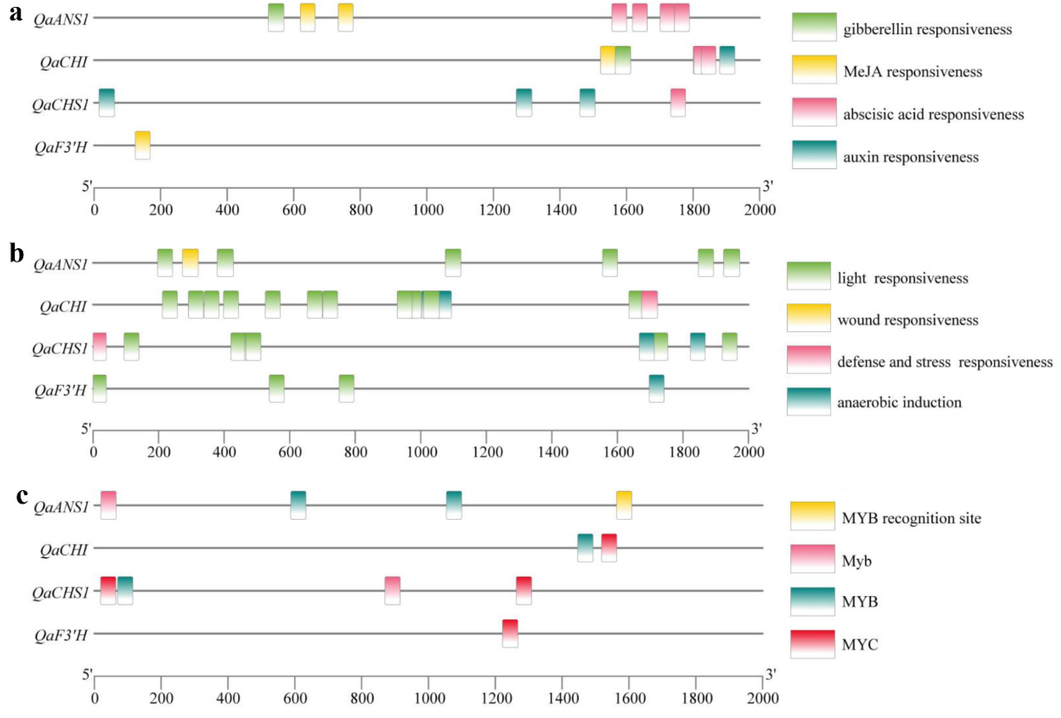
2.3. Expression of Genes Associated with Anthocyanin Biosynthesis in Q. aliena

In order to identify genes that may regulate red coloration in the leaves of Q. aliena variants, we cloned and sequenced 25 Q. aliena genes homologous to genes previously shown in other species to be involved in anthocyanin biosynthesis (Table S1). Additionally, semi-quantitative and real-time quantitative PCR were performed to sequence genes associated with each of the three senescence stages (S1–S3). Among the 25 genes associated with anthocyanin biosynthesis, we found that only 18 were expressed during leaf color variation in autumn. As senescence progressed, the expression of these 18 genes in QAC changed by 0–2 fold, while significant changes were found in the relative expression of anthocyanin synthesis-related genes in QA1, QA2, and QA3 (Figure S3). At S2, we observed high expression of QaMYB1 in all three red-leaved variants, high expression of QaDFR1 and QaANS1 in QA1 and QA3, high expression of QaCHS2 and QaMYB3 in QA1, as well as high levels of QaMYB2 and QaF3′H in QA3 (Figure S3b–d). When we compared relative expression of anthocyanin synthesis-related genes between the red-leaved variants and the yellow-leaved tree, we found that all the genes except for Qa4CL1 were expressed at significantly higher levels in QA1 than in QAC at S2 and S3 (Figure S4). However, different gene expression patterns were found in QA2 and QA3, and only QaCHS1, QaCHI, QaF3′H, QaANS1, and QaMYB1 were expressed at significantly higher levels in all three red-leaved variants than in QAC. Based on quantitative real-time PCR, expression of QaCHS2, QaDFR1, QaANS1, QaMYB1, and QaMYB3 in QA1 were higher at S2 and S3 (Figure 4a) than at S1, which was consistent with the semi-quantitative PCR results. Significant changes in gene expression levels in QA2 and QA3 were also verified using quantitative PCR. Once again, when we compared the red-leaved variants and the yellow-leaved control, we found higher expression of Qa4CL2, QaCHS2, QaCHI, QaF3H, QaF3′H, QaDFR1, QaANS1, QaMYB1, and QaMYB3 in QA1 than in QAC (Figure 4b). Although the expression patterns of anthocyanin synthesis-related genes in the three variants were not exactly consistent, the increased expression of these genes is obvious as compared to QAC. In addition, the gene sequences were also compared between the three variants and QAC. However, there were no difference in amino acid sequences in all genes, such as QaANS1, QaMYB1 and QaMYB3 (Figure S5), which indicated that the leaf color variation in Q. aliena is not caused by the gene body mutation in anthocyanins synthesis.
Based on the genome in our laboratory (data not shown), the promoter regions of the four key genes were analyzed. Thereinto, light-responsive, auxin and ABA-responsive elements were the common elements (Figure 5a,b). The potential MYB and bHLH binding elements were also analyzed, Myb motif (TAACTG), MYB recognition site (CCGTTG) and MYB motif ([T/C]AACCA) were found in QaCHS1, QaCHI, and QaANS1; and MYC motif (CAT[T/G]TG) was found in QaCHI, QaCHS1 and QaF3′H, which suggested that these genes may be regulated by the MYB and bHLH transcription factors (Figure 5c). In order to rule out the influence of leaf senescence, we performed a correlation analysis to compare anthocyanin content and gene expression (based on semi-quantitative data) in all four trees at S2 and S3 (Table 2). At S2, most of the anthocyanin synthesis-related genes significantly correlated with QaMYB1 expression and anthocyanin content, except for QaPAL2, Qa4CL1, Qa4CL2, QaCHS1, QaF3′H, and QaMYB2. However, at S3, expression of only QaCHS1 and QaF3′H significantly correlated with anthocyanin content. Therefore, we speculated that the regulation role of QaMYB1 in anthocyanin synthesis maybe time-specific, which needs to be further studied.

2.4. Homology Alignment and Phylogenetic Analysis of MYB Transcription Factors

Since QaMYB1 and QaMYB3 expression changed significantly during senescence, we cloned and analyzed the coding DNA sequences of these two genes from the four accessions and found no significant amino acid differences among the four encoded proteins. In addition, we constructed a phylogenetic tree with other MYB transcription factors such as DcMYB113 [23], PtrMYB119 [8], FaMYB10 [24] and MdMYB9 [25], which have been studied extensively in other species. The phylogenetic analysis showed that QaMYB1 and QaMYB3 clustered together in the same branch with MdMYB1, FaMYB10, AtMYB90, and VvMYBA1, all of which are involved in anthocyanin biosynthesis (Figure 6a). In addition, QaMYB2 clustered together with MdMYB9, FaMYB9, FaMYB11, and VvMYBPA1, which are involved in proanthocyanin synthesis in plants (Figure 6a). In fact, MYB genes related to anthocyanin synthesis in different species formed independent branches, indicating that the MYB gene family is conserved among all plant species. The results of the homology alignment analysis showed that the three MYB transcription factors of Q. aliena are relatively conserved at the N-terminus but substantially different at the C-terminus. All three members contain two MYB gene family-specific domains at the N-terminus of the sequence, the R2 and R3 domains, indicating that they all belong to the R2R3-MYB gene family. However, only the proteins encoded by QaMYB2 and QaMYB3 contain a unique bHLH transcription factor-binding domain, [D/E]Lx2[R/K]x3Lx6Lx3R, in the R3 domain (Figure 6b), which suggests that these two proteins may bind to bHLH transcription factors to form the MBW complex.

3. Discussion

3.1. Anthocyanin Accumulation Causes Reddening of Leaves in Q. aliena Variants

The immediate cause of color change in plants can be attributed to a change in the content of three main pigments: chlorophyll, carotenoids, and flavonoids [26]. Anthocyanins are major flavonoids that appear red, blue, or purple depending on the conditions [27]. In the present study, the contents of chlorophyll, carotenoids, and anthocyanins were all analyzed, and only significant differences were found in anthocyanins, which indicate the accumulation of anthocyanins, especially cyanidin 3-O-glucoside (the glycosylation product of cyanidin [28]), contributed to the autumn leaf coloration of the three natural variants of Q. aliena. We also found that anthocyanin accumulation and changes in leaf color were strongly associated with leaf senescence, consistent with previous work [29,30,31]. For example, a study of 89 deciduous tree species reported that 62 contained anthocyanins during leaf senescence [30], indicating that anthocyanin accumulation is a common phenomenon during senescence. During the process of leaf senescence in the red-leaved variants of Q. aliena, we found that the increase in anthocyanin content was accompanied by a decrease in chlorophyll content. However, there was no significant difference in chlorophyll content among QAC, QA2, and QA3, indicating that anthocyanin accumulation had no effect on the degradation of chlorophyll. During the early discoloration stage (S2), Fa/Fv values were lower in the red-leaved variants than in the yellow-leaved control, and we speculate that this may be due to the higher anthocyanin content. An increase in anthocyanin content can reduce the amount of light energy that can be absorbed and utilized by photosynthesis, thus slowing the rate of photosynthesis [32]. Additionally, a study of red maples showed that high anthocyanin content during leaf senescence can protect the plants from extreme environments such as low temperature and high solar radiation [33]. In the present study, leaf color in the Q. aliena accessions changed between mid-October and mid-November. During this period, we observed that the daily average temperature dropped substantially from 19.1 °C at the green stage (S1, late September) to 9.5 °C at the main discoloration stage (S3, early November; Figure S2). Previous studies have shown that low temperatures can promote anthocyanin accumulation and synthesis in plants such as Brassica rapa, apple, and red grapes [34,35,36]. Further research is required to understand whether and how low temperatures can cause changes in leaf coloration, and these insights will be useful for the cultivation and development of ornamental varieties of Q. aliena.

3.2. Changes in Anthocyanin Biosynthesis Alter Leaf Color in Q. aliena Variants

Anthocyanin biosynthesis is regulated by a series of enzymatic reactions, where an increase in the expression of key genes causes a corresponding increase in the expression of enzymes associated with the synthesis and accumulation of anthocyanins [37]. This accumulation of anthocyanin can alter plant color. Previous studies on natural variants have shown that high anthocyanin content was caused by the entire anthocyanin biosynthetic pathway rather than specific genes. Red variants of radish were found to express more PAL, C4H, 4CL, CHS, CHI, F3H, DFR, LDOX, and UGT enzymes than white variants, and CHS plays an important role in root coloration [38]. When four variants of radish were compared, anthocyanin content was found to be associated with levels of expression of RsUFGT, RsF3H, RsANS, RsCHS3, and RsF3′H1; and RsUFGT was found to be the key control point involved in leaf coloration [39]. Similarly, expression levels of F3′5′H, DFR, and UFGT correlated significantly with anthocyanin accumulation in pepper [40]. Our results show a significant upward trend in most of the anthocyanin synthesis-related genes in the red-leaved variants during the early and main discoloration stages (S2 and S3), indicating that the accumulation of anthocyanins in oak leaves was caused by upregulation of the entire anthocyanin biosynthesis pathway, consistent with the abovementioned studies. However, we observed differential expression of anthocyanin synthesis-related genes across the three red-leaved variants, which may be caused by the different coloration patterns. Additionally, similar results were also found in natural variants of apple [41] and poplar [42]. In the present study, the expression patterns of QaCHS1, QaCHI, QaF3′H, and QaANS1 were significantly different across all three variants at both S2 and S3, suggesting that these genes may be the key regulators of anthocyanin synthesis in Q. aliena. Since a study of Pistacia chinensis showed that anthocyanin biosynthesis-related genes such as CHS and CHI were regulated by MYB transcription factors during autumn leaf discoloration [43]. Thus, based on the Q. aliena genome in our laboratory (data not shown), promoter sequences of the key genes, QaCHS1, QaCHI, QaF3′H, and QaANS1, were analyzed. Thus, potential MYB binding sites were found in QaCHS1, QaCHI, and QaANS1 (Figure 4). The Myb motif and MYB recognition site are proven to play roles in dehydration stress and abscisic acid signaling though AtMYB2 [44,45]. Additionally, the MYB motif has dual roles in water stress and flavonoid biosynthesis [44,46]. Additionally, a bHLH motif, MYC motif was found in QaCHI, QaCHS1 and QaF3′H, which can bind to AtMYC to play roles in abiotic stress [47]. Future studies should investigate whether MYB or bHLH transcription factors are involved in leaf coloration in Q. aliena.

3.3. QaMYB1 Probably Regulates Anthocyanin Biosynthesis in Q. aliena

Studies based on several different species such as grape, apple, strawberry and petunia have shown that the synthesis of plant anthocyanins is closely related to MYB transcription factors [48]. In grapes, VvMYBAs expression is closely related to fruit coloration: the stronger the VvMYBAs expression, the darker the fruit [49,50]. MdMYB10 expression correlates with anthocyanin accumulation in apples, and expression is higher in cultivars with red fruits than in those with green fruits [51]. In the present study, we used VvMYBA1 in grape and MdMYB10 in apple as indices and the Q. robur genome sequence as a reference in order to clone and sequence the QaMYB1, QaMYB2, and QaMYB3 genes in Q. aliena. QaMYB1 was strongly expressed in the leaves of all three red-leaved variants during the early (S2) and main discoloration (S3) stages. In addition, we observed that the expression of QaMYB1 was higher in all three variants than in the yellow-leaved control (QAC). Further phylogenetic tree analysis showed that QaMYB1 and QaMYB3 were in the same clade as other MYB gene family members related to anthocyanin synthesis, while QaMYB2 was in the same clade as the MYB gene family members related to proanthocyanin synthesis. These findings suggest that QaMYB1 may play a role in autumn leaf coloration in Q. aliena.
Previous studies have also shown that MYB transcription factors can affect anthocyanin accumulation by binding the promoters of anthocyanin biosynthesis-related genes directly or through the MYB-bHLH-WD40 complex [52,53]. In strawberry, FvMYB10 binds to the promoter regions of genes encoding key enzymes such as FvCHS2 and FvDFR1, and it activates their expression, promoting the accumulation of anthocyanins [54]. In Triadica sebifera, the protein encoded by SsMYB1 activates anthocyanin biosynthesis by binding to the promoters of SsDFR1 and SsANS [55]. In kiwi fruit, the protein encoded by AcMYBF110 plays an important role in the regulation of anthocyanin accumulation by forming the MYB-bHLH-WD40 complex [56]. In the present study, QaMYB1 and QaMYB3 significantly correlated with the expression levels of other anthocyanin synthesis-related genes, including QaPAL1, QaCHS2, QaCHI, QaF3H, QaDFR1, QaDFR2, QaDFR3, QaUFGT1, and QaANS1 at the S2 stage, indicating that transcription factors may regulate the expression of anthocyanin synthesis-related genes and subsequent anthocyanin accumulation in Q. aliena. However, the homology alignment identified mutations in the sequence motif within the protein encoded by QaMYB1 that may affect binding to bHLH transcription factors. Notably, we were unable to identify specific bHLH transcription factors in Q. aliena. Although the homologue of MdbHLH33 could be found in Q. robur genome, there was no product in Q. aliena. Further research is required to understand how QaMYB1 may regulate anthocyanin synthesis in Q. aliena. A study of FvMYB10 in strawberry showed that mutations in MYB10 can cause differences in anthocyanin content [57]. Although we observed no significant differences in the sequences of proteins encoded by QaMYB1 between the yellow-leaved control and the red-leaved variants, we cannot exclude the possibility that differences in gene expression may contribute to leaf color variation. Additionally, and notably, the promoter regions of QaMYB1 in the four trees were analyzed, and an important base mutation was found in the cis-regulatory element, aaaAaaC(G/C)GTTA (MYB binding motif involved in flavonoid biosynthetic genes regulation), which may influence the expression of QaMYB1 (Figure S6). This possibility should be studied further. In addition, glutathione S-transferases (GSTs), anthocyanin transporters, have been shown to play important roles in anthocyanin accumulation [58]. It can also interact with the MYB transcription factor, such as PpMYB10.1 [59] and AcMYBF110 [60], to regulate the accumulation of anthocyanins. The loss of GSTs’ function can also cause the flower and fruit color variation in peach [61], or fruit color variation in apple [62]. Therefore, further exploring the mechanism of anthocyanin transport in Q. aliena variants has the potential roles to explain leaf color variation in autumn.

4. Materials and Methods

4.1. Plant Material

We collected plant data from four 5-year-old Q. aliena trees grown in the Beijing Dadongliu Nursery in Beijing, China. Three of the trees (QA1, QA2 and QA3) had red leaves and were natural variants; the fourth tree (QAC) had typical yellow leaves and was considered the control (Figure 1). All the four trees were planted together under the same conditions. Senescence in plants is typically stratified into three stages based on the degree of change in leaf color: the green stage (28 September 2020; S1), early discoloration stage (21 October 2020; S2), and main discoloration stage (27 October 2020; S3) (Figure 1). Matured leaves (fourth to sixth from the top of the branch) were collected at each of these stages from all four trees, frozen in liquid nitrogen, and stored at −80 °C until subsequent experiments.

4.2. Leaves Phenotypic Analysis

The color of mature leaves was determined by comparing them to the RHS Large Color Chart (MARK0011, Royal Horticultural Society, London, UK). The length, width and area of leaves were analyzed at the main discoloration stage by using CI-3000 Portable Leaf Area Meter (LI–COR, Lincoln, NE, USA). Fifteen leaves were selected for each tree.

4.3. Analysis of Simple Sequence Repeat Markers

Based on the previous research about Quercus [63], nine pairs of primers (Table S2) were selected to analyze phylogenetic relationships among the four samples (QA1, QA2, QA3, and QAC), as well as their relationships to additional Q. aliena individuals, Q. mongolica, Q. dentata, and Q. variabilis. The simple sequence repeat procedure was performed as described [64], and the reaction products were sent to Beijing Ruibo Xingke Biotechnology Co., Ltd., Beijing, China for capillary electrophoresis analysis. Preliminary analysis of the electrophoresis results was performed using the GeneMarker v2.2.0 (SoftGenetics, LLC., State College, PA, USA), and clustering analysis was performed using the UPGMA clustering method in SAHN mode in NTSYS 2.10e [65].

4.4. Measurement of Chlorophyll Fluorescence

The chlorophyll fluorescence of Q. aliena leaves was measured using the MINI-PAM-II ultra-portable modulated chlorophyll fluorometer (Heinz Walz, Effeltrich, Germany). We calculated the ratio of variable fluorescence to maximal fluorescence (Fv/Fm) to compare the potential maximum maximal quantum yield of PS II of the leaves at S1, S2, and S3. Four leaves were selected to do analysis after adequate pre-darkening (>30 min) at all three stages.

4.5. Quantification of Total Anthocyanins, Total Carotenoids, Chlorophyll a, and Chlorophyll b

The extraction and quantification of total anthocyanin content from Q. aliena leaves were performed as described [66], with some modifications. After removing veins, leaf samples (0.4 g) were added to test tubes containing 2 mL of 1% (v/v) hydrochloric acid-methanol solution. After mixing, the samples were placed in the dark at 4 °C for 24 h to allow further extraction. Next, the mixture was centrifuged at 4 °C (12,000 rpm, 30 min), and the absorbance of the supernatant was measured at 530 nm and 657 nm using a Cary 300 UV-Vis spectrophotometer (Agilent Technologies, Santa Clara, CA, USA). Total anthocyanin content (Qta) was calculated as the total anthocyanin content per gram of fresh weight (FW) using the following formula Equation (1). Individual anthocyanin components in each sample were identified and analyzed by technicians at the Wuhan Medwell Biotechnology Co. Ltd., Wuhan, China using an ultra-high performance liquid chromatography system (ExionLC™ AD, https://sciex.com.cn/, accessed on 19 November 2021) equipped with an ACQUITY BEH C18 column (1.7 µm, 2.1 × 100 mm2) and coupled to a mass spectrometry system (QTRAP® 6500+, https://sciex.com.cn/, accessed on 19 November 2021).
Chlorophyll a, chlorophyll b, and total carotenoids were extracted and quantification from Q. aliena leaves as described [67], with some modifications. After removing veins, leaf samples (0.2 g) were extracted using 80% (v/v) acetone for 5 min with grinding. The mixture was then filtered into a 25-mL brown volumetric flask, diluted with 80% acetone to volume, and mixed. The absorbance of the extracts was measured at 663 nm and 646 nm using the Cary 300 UV-Vis spectrophotometer. Contents of chlorophyll a (Qca), chlorophyll b (Qcb), and total carotenoids (Qtc) were calculated per gram of FW using the following formulas Equations (2)–(7). Contents of chlorophyll a, chlorophyll b, and total carotenoids were calculated per gram of FW.
Q t a = ( A 530 0.25 × A 657 ) / F W
C c a   mg   L 1 = 12.21 × A 663 2.81 × A 646
C c b mg   L 1 = 20.13 × A 646 5.03 × A 663
C t c mg   L 1 = 1000 × A 470 3.27 × C c a 104 × C c b / 229
Q c a   mg   g 1 = C c a × V × N / F W
Q c b   mg   g 1 = C c b × V × N / F W
Q t c   mg   g 1 = C t c × V × N / F W
Thereinto, Cca is the concentration of chlorophyll a; Ccb is the concentration of chlorophyll b; Ctc is the concentration of total carotenoids; V is the extract volume and N is the dilution factor.

4.6. Cloning of Genes Associated with Anthocyanin Biosynthesis

Total RNA extraction was performed based on the modified CTAB method [68], and the cDNA strand was synthesized using the Reverse Transcription System I (Promega, Madison, WI, USA). Genes involved in anthocyanin biosynthesis were identified based on previous studies (Table S1) and the genome of Q. robur [69], which is closely related to Q. aliena. Sequences beginning 2000 bp upstream of the transcription start site were used for identification of potential cis-acting elements using the PlantCARE tool (http://bioinformatics.psb.ugent.be/webtools/plantcare/html/, accessed on 8 June 2022), and the results were visualized using TBtools (v1.098721). A phylogenetic tree was constructed using Bioedit and MEGA11.0 (https://www.megasoftware.net/home, accessed on 5 March 2022). We used the Poisson model in the Neighbor-Joining algorithm, the “complete deletion” mode, and a bootstrap value of 1000 to construct the tree [70]. The homologous sequence alignment was analyzed using DNAMAN 9.0.1.116 (Lynnon Biosoft, San Ramon, CA, USA).

4.7. Semi-Quantitative and Quantitative Gene Expression Analyses

According to the conserved partial sequences, primer sequences for semi-quantitative PCR and real-time quantitative PCR were designed and shown in Table S2, and the amplicons of each primer were verified by sanger sequencing. Data collected from the semi-qPCR were photographed with a gel imager (Gel DocTM XR+, Bio-Rad, Hercules, CA, USA) and analyzed using Image Lab (version 6.0.1, Bio-Rad) in order to quantify the brightness of the product bands. The QaACTIN gene of Q. aliena was used as the internal reference [71,72]. The S1 stage samples were used as control groups when comparing the difference between leaf senescence process, and the QAC samples were used as control groups when comparing the difference between the four accessions. The gene relative expression was calculated based on the luminance ratio method. Quantitative real-time PCR was performed using the StepOne PlusTM real-time PCR system with Tower detection system (ABI, Worcester, MA, USA) and the ChamQ Universal SYBR qPCR Master Mix (Vazyme Biotech Co., Ltd., Nanjing, China). Here, again, the QaACTIN gene was used as an internal reference, and the S1 stage and QAC samples were considered as controls. Relative gene expression was calculated using the 2∆∆Ct method.

4.8. Statistical Analyses

All data were subjected to one-way analysis of variance (ANOVA) using SPSS 22.0 (IBM, Armonk, NY, USA). Relationships between genes relative expression and anthocyanin content were analyzed between different individuals at the same stage based on Pearson’s correlation coefficients. Significance levels for two-tailed tests were defined as p < 0.05 and p < 0.01.

5. Conclusions

The natural variants of Q. aliena are a good model system to understand the mechanism involved in anthocyanin biosynthesis and their subsequent effects on autumn leaf coloration (Figure 7). Our findings indicates that the accumulation of anthocyanins, especially cyanidin 3-O-glucoside, is the main cause of the reddening of autumn leaves in the natural variants of Q. aliena. Differential expression analysis of genes related to anthocyanin biosynthesis in the four accessions indicate that the leaf color variation may be regulated by several genes, including QaCHS1, QaCHI, QaF3′H, and QaANS1. Additionally, QaMYB1 expression is significantly associated with anthocyanin content and with expression levels of most of the anthocyanin synthesis-related genes at the early discoloration stage. The results of this study not only provide insights into the molecular and physiological causes of leaf color variation in Q. aliena, but also a reference for studying leaf color changes during leaf senescence.

Supplementary Materials

The following supporting information can be downloaded at: https://0-www-mdpi-com.brum.beds.ac.uk/article/10.3390/ijms232012179/s1.

Author Contributions

X.Y., N.Y. and G.L. designed the work; X.Y. and N.Y. performed experiments and analyzed data; Q.Z. and M.C. participated in the gene cloning experiment; Z.P. performed the cloning and analysis of promoter regions. The collection of experimental materials and physiological measurements were performed with the help of H.Z. and Y.G.; X.Y. and N.Y. drafted the manuscript; and Q.Y. and G.L. revised the manuscript. All authors have read and agreed to the published version of the manuscript.

Funding

This work was funded by National Key R&D Program of China 2021YFD2200302.

Institutional Review Board Statement

Not applicable.

Informed Consent Statement

Not applicable.

Data Availability Statement

The data that support the findings of this study are available in insert article and supplementary material here.

Acknowledgments

We gratefully acknowledge the families who participated by providing research assistance. We gratefully acknowledge the infrastructure support of the Beijing Advanced Innovation Center for Tree Breeding by Molecular Design, Beijing Forestry University. We also thank the families who participated by providing research assistance.

Conflicts of Interest

The authors declare that they have no conflict of interest.

References

  1. Shi, Q.; Zhou, L.; Li, K.; Wang, Y. Advances in transcriptional regulation of anthocyanin synthesis in plants. For. Sci. Res. 2015, 28, 570–576. [Google Scholar] [CrossRef]
  2. Boldt, J.; Erwin, J.E.; Meyer, M.H. Foliar Anthocyanins: A Horticultural Review. Hortic. Rev. 2014, 42, 209–252. [Google Scholar] [CrossRef]
  3. Karageorgou, P.; Manetas, Y. The importance of being red when young: Anthocyanins and the protection of young leaves of Quercus coccifera from insect herbivory and excess light. Tree Physiol. 2006, 26, 613–621. [Google Scholar] [CrossRef] [PubMed] [Green Version]
  4. Cabrita, L.; Fossen, T.; MAndersen, Ø. Colour and stability of the six common anthocyanidin 3-glucosides in aqueous solutions. Food Chem. 2000, 68, 101–107. [Google Scholar] [CrossRef]
  5. Grotewold, E. The genetics and biochemistry of floral pigments. Annu. Rev. Plant Biol. 2006, 57, 761–780. [Google Scholar] [CrossRef]
  6. Holton, T.A.; Cornish, E.C. Genetics and Biochemistry of Anthocyanin Biosynthesis. Plant Cell 1995, 7, 1071–1083. [Google Scholar] [CrossRef]
  7. Shi, M.-Z.; Xie, D.-Y. Biosynthesis and Metabolic Engineering of Anthocyanins in Arabidopsis thaliana. Recent Pat. Biotechnol. 2014, 8, 47–60. [Google Scholar] [CrossRef] [Green Version]
  8. Cho, J.-S.; Nguyen, V.P.; Jeon, H.-W.; Kim, M.-H.; Eom, S.H.; Lim, Y.J.; Kim, W.-C.; Park, E.-J.; Choi, Y.-I.; Ko, J.-H. Overexpression of PtrMYB119, a R2R3-MYB transcription factor from Populus trichocarpa, promotes anthocyanin production in hybrid poplar. Tree Physiol. 2016, 36, 1162–1176. [Google Scholar] [CrossRef] [Green Version]
  9. An, J.; Zhang, X.; Liu, Y.; Wang, X.; You, C.; Hao, Y. ABI5 regulates ABA-induced anthocyanin biosynthesis by modulating the MYB1-bHLH3 complex in apple. J. Exp. Bot. 2020, 72, 1460–1472. [Google Scholar] [CrossRef]
  10. Azuma, A.; Yakushiji, H.; Koshita, Y.; Kobayashi, S. Flavonoid biosynthesis-related genes in grape skin are differentially regulated by temperature and light conditions. Planta 2012, 236, 1067–1080. [Google Scholar] [CrossRef]
  11. Schaart, J.G.; Dubos, C.; De La Fuente, I.R.; van Houwelingen, A.M.M.L.; de Vos, R.C.H.; Jonker, H.H.; Xu, W.; Routaboul, J.-M.; Lepiniec, L.; Bovy, A.G. Identification and characterization of MYB-bHLH-WD40 regulatory complexes controlling proanthocyanidin biosynthesis in strawberry (Fragaria × ananassa) fruits. New Phytol. 2013, 197, 454–467. [Google Scholar] [CrossRef] [PubMed]
  12. Jian, W.; Cao, H.; Yuan, S.; Liu, Y.; Lu, J.; Lu, W.; Li, N.; Wang, J.; Zou, J.; Tang, N.; et al. SlMYB75, an MYB-type transcription factor, promotes anthocyanin accumulation and enhances volatile aroma production in tomato fruits. Hortic. Res. 2019, 6, 15. [Google Scholar] [CrossRef] [Green Version]
  13. Jia, N.; Wang, J.; Wang, Y.; Ye, W.; Liu, J.; Jiang, J.; Sun, J.; Yan, P.; Wang, P.; Wang, F.; et al. The Light-Induced WD40-Repeat Transcription Factor DcTTG1 Regulates Anthocyanin Biosynthesis in Dendrobium candidum. Front. Plant Sci. 2021, 12, 633333. [Google Scholar] [CrossRef] [PubMed]
  14. Wen, C.-H.; Chu, F.-H. A R2R3-MYB Gene LfMYB113 is Responsible for Autumn Leaf Coloration in Formosan sweet gum (Liquidambar formosana Hance). Plant Cell Physiol. 2017, 58, 508–521. [Google Scholar] [CrossRef] [PubMed]
  15. Zhou, W.; Zhang, Q.; Sun, Y.; Yang, L.; Wang, Z. Genome-wide identification and characterization of R2R3-MYB family in Hypericum perforatum under diverse abiotic stresses. Int. J. Biol. Macromol. 2019, 145, 341–354. [Google Scholar] [CrossRef]
  16. Hu, D.-G.; Sun, C.-H.; Zhang, Q.-Y.; Gu, K.-D.; Hao, Y.-J. The basic helix-loop-helix transcription factor MdbHLH3 modulates leaf senescence in apple via the regulation of dehydratase-enolase-phosphatase complex 1. Hortic. Res. 2020, 7, 50. [Google Scholar] [CrossRef] [Green Version]
  17. Wen, C.-H.; Lin, S.-S.; Chu, F.-H. Transcriptome Analysis of a Subtropical Deciduous Tree: Autumn Leaf Senescence Gene Expression Profile of Formosan Gum. Plant Cell Physiol. 2015, 56, 163–174. [Google Scholar] [CrossRef] [Green Version]
  18. Li, Q.; Gu, M.; Deng, M. In Vitro Propagation of Oriental White Oak Quercus aliena Blume. Forests 2019, 10, 463. [Google Scholar] [CrossRef] [Green Version]
  19. Liu, Y.; Li, Y.; Song, J.; Zhang, R.; Yan, Y.; Wang, Y.; Du, F.K. Geometric morphometric analyses of leaf shapes in two sympatric Chinese oaks: Quercus dentata Thunberg and Quercus aliena Blume (Fagaceae). Ann. For. Sci. 2018, 75, 90. [Google Scholar] [CrossRef] [Green Version]
  20. Lu, S.; Hou, M.; Du, F.K.; Li, J.; Yin, K. Complete chloroplast genome of the Oriental white oak: Quercus aliena Blume. Mitochondrial Dna 2015, 27, 2802–2804. [Google Scholar] [CrossRef]
  21. Jin, Y.-S.; Heo, S.-I.; Lee, M.-J.; Rhee, H.-I.; Wang, M.-H. Free radical scavenging and hepatoprotective actions of Quercus aliena acorn extract against CCl4-induced liver. Free Radic. Res. 2005, 39, 1351–1358. [Google Scholar] [CrossRef] [PubMed]
  22. Sun, Q. Quercus aliena Bl. Seedling and silviculture techniques. Mod. Hortic. 2010, 11, 45. [Google Scholar] [CrossRef]
  23. Xu, Z.-S.; Yang, Q.-Q.; Kai, F.; Yu, X.; Xiong, A.-S. DcMYB113, a root-specific R2R3-MYB, conditions anthocyanin biosynthesis and modification in carrot. Wiley-Blackwell Online Open 2020, 18, 1585–1597. [Google Scholar] [CrossRef] [PubMed] [Green Version]
  24. Laura, M.-P.; Guadalupe, C.-L.; Francisco, A.-R.; Thomas, H.; Ludwig, R.; Antonio, R.-F.; Luis, C.J.; Wilfried, S.; Juan, M.-B.; Rosario, B.-P. MYB10 plays a major role in the regulation of flavonoid/phenylpropanoid metabolism during ripening of Fragaria × ananassa fruits. J. Exp. Bot. 2013, 65, 401. [Google Scholar] [CrossRef] [Green Version]
  25. An, J.; Xu, R.; Liu, X.; Zhang, J.; Wang, X.; You, C.; Hao, Y. Jasmonate induces biosynthesis of anthocyanin and proanthocyanidin in apple by mediating the JAZ1–TRB1–MYB9 complex. Plant J. 2021, 106, 1414–1430. [Google Scholar] [CrossRef]
  26. Iwashina, T. Contribution to flower colors of flavonoids including anthocyanins: A review. Nat. Prod. Commun. 2015, 10, 529–544. [Google Scholar] [CrossRef] [Green Version]
  27. Winkel-Shirley, B. Flavonoid Biosynthesis. A Colorful Model for Genetics, Biochemistry, Cell Biology, and Biotechnology. Plant Physiol. 2001, 126, 485–493. [Google Scholar] [CrossRef] [Green Version]
  28. Hu, R.; He, Y.; He, Z.; Wu, S. Progress in studies on absorption, metabolism and physiological functions of centaurea -3- glucoside. Chin. J. Anim. Nutr. 2019, 31, 2052–2062. [Google Scholar] [CrossRef]
  29. Tallis, M.J.; Lin, Y.; Rogers, A.; Zhang, J.; Street, N.R.; Miglietta, F.; Karnosky, D.F.; Angelis, P.D.; Calfapietra, C.; Taylor, G. The transcriptome of Populus in elevated CO2 reveals increased anthocyanin biosynthesis during delayed autumnal senescence. New Phytol. 2010, 186, 415–428. [Google Scholar] [CrossRef] [Green Version]
  30. Lee, D.; O’Keefe, J.; MicheleHolbrook, N.; Feild, T. Pigment dynamics and autumn leaf senescence in a New England deciduous forest, eastern USA. Ecol. Res. 2003, 18, 677–694. [Google Scholar] [CrossRef]
  31. Hoch, W.A.; Singsaas, E.L.; McCown, B.H. Resorption Protection. Anthocyanins Facilitate Nutrient Recovery in Autumn by Shielding Leaves from Potentially Damaging Light Levels. Plant Physiol. 2003, 133, 1296–1305. [Google Scholar] [CrossRef] [PubMed] [Green Version]
  32. Zhu, X.-G.; Long, S.P.; Ort, D.R. What is the maximum efficiency with which photosynthesis can convert solar energy into biomass? Curr. Opin. Biotechnol. 2008, 19, 153–159. [Google Scholar] [CrossRef] [PubMed]
  33. Rachel, A.; Peter, R. Early Autumn Senescence in Red Maple (Acer rubrum L.) Is Associated with High Leaf Anthocyanin Content. Plants 2015, 4, 505–522. [Google Scholar] [CrossRef] [Green Version]
  34. He, Q.; Ren, Y.; Zhao, W.; Li, R.; Zhang, L. Low Temperature Promotes Anthocyanin Biosynthesis and Related Gene Expression in the Seedlings of Purple Head Chinese Cabbage (Brassica rapa L.). Genes 2020, 11, 81. [Google Scholar] [CrossRef] [PubMed] [Green Version]
  35. Song, T.; Li, K.; Wu, T.; Wang, Y.; Zhang, X.; Xu, X.; Yao, Y.; Han, Z. Identification of new regulators through transcriptome analysis that regulate anthocyanin biosynthesis in apple leaves at low temperatures. PLoS ONE 2019, 14, e0210672. [Google Scholar] [CrossRef] [PubMed]
  36. Gao-Takaia, M.; Katayama-Ikegamia, A.; Matsudab, K.; Shindoa, H.; Uemaea, S.; Oyaizua, M. A low temperature promotes anthocyanin biosynthesis but does not accelerate endogenous abscisic acid accumulation in red-skinned grapes. Plant Sci. 2019, 283, 165–176. [Google Scholar] [CrossRef] [PubMed]
  37. Liu, H.; Liu, Z.; Wu, Y.; Zheng, L.; Zhang, G. Regulatory Mechanisms of Anthocyanin Biosynthesis in Apple and Pear. Int. J. Mol. Sci. 2021, 22, 8441. [Google Scholar] [CrossRef]
  38. Yu, R.; Du, X.; Li, J.; Liu, L.; Hu, C.; Yan, X.; Xia, Y.; Xu, H. Identification and differential expression analysis of anthocyanin biosynthetic genes in root-skin color variants of radish (Raphanus sativus L.). Genes Genom. 2020, 42, 413–424. [Google Scholar] [CrossRef]
  39. Muleke, E.M.M.; Fan, L.; Wang, Y.; Xu, L.; Zhu, X.; Zhang, W.; Cao, Y.; Karanja, K.B.; Liu, L. Coordinated Regulation of Anthocyanin Biosynthesis Genes Confers Varied Phenotypic and Spatial-Temporal Anthocyanin Accumulation in Radish (Raphanus sativus L.). Front. Plant Sci. 2017, 8, 1243. [Google Scholar] [CrossRef] [Green Version]
  40. Aza-González, C.; Herrera-Isidrón, L.; Núez-Palenius, H.G.; Vega, O.M.D.L.; Ochoa-Alejo, N. Anthocyanin accumulation and expression analysis of biosynthesis-related genes during chili pepper fruit development. Biol. Plant. 2012, 57, 49–55. [Google Scholar] [CrossRef]
  41. Ben-Yehudah, G.; Korchinsky, R.; Redel, G.; Ovadya, R.; Cohen, Y. Colour accumulation patterns and the anthocyanin biosynthetic pathway in ‘Red Delicious’ apple variants. J. Pomol. Hortic. Sci. 2005, 80, 187–192. [Google Scholar] [CrossRef]
  42. Chen, X.; Liu, H.; Wang, S.; Zhang, C.; Zhang, J. Combined Transcriptome and Proteome Analysis Provides Insights into Anthocyanin Accumulation in the Leaves of Red-Leaved Poplars. Plant Mol. Biol. 2021, 106, 491–503. [Google Scholar] [CrossRef]
  43. Song, X.; Yang, Q.; Liu, Y.; Li, J.; Chang, X.; Xian, L.; Zhang, J. Genome-wide identification of Pistacia R2R3-MYB gene family and function characterization of PcMYB113 during autumn leaf coloration in Pistacia chinensis. Int. J. Biol. Macromol. 2021, 192, 16–27. [Google Scholar] [CrossRef]
  44. Urao, T.; Yamaguchi-Shinozaki, K.; Urao, S.; Shinozaki, K. An Arabidopsis myb homolog is induced by dehydration stress and its gene product binds to the conserved MYB recognition sequence. Plant Cell 1993, 5, 1529–1539. [Google Scholar] [CrossRef] [PubMed] [Green Version]
  45. Abe, H.; Urao, T.; Ito, T.; Seki, M.; Shinozaki, K.; Yamaguchi-Shinozaki, K. Arabidopsis AtMYC2 (bHLH) and AtMYB2 (MYB) function as transcriptional activators in abscisic acid signaling. Plant Cell 2003, 15, 63–78. [Google Scholar] [CrossRef] [PubMed] [Green Version]
  46. Solano, R.; Nieto, C.; Avila, J.; Cañas, L.; Diaz, I.; Paz-Ares, J. Dual DNA binding specificity of a petal epidermis-specific MYB transcription factor (MYB.Ph3) from Petunia hybrida. EMBO J. 1995, 14, 1773–1784. [Google Scholar] [CrossRef]
  47. Chinnusamy, V.; Schumaker, K.; Zhu, J.K. Molecular genetic perspectives on cross-talk and specificity in abiotic stress signalling in plants. J. Exp. Bot. 2004, 55, 225–236. [Google Scholar] [CrossRef] [PubMed]
  48. Naing, A.H.; Kim, C.K. Roles of R2R3-MYB transcription factors in transcriptional regulation of anthocyanin biosynthesis in horticultural plants. Plant Mol. Biol. 2018, 98, 1–18. [Google Scholar] [CrossRef] [PubMed]
  49. Poudel, P.; Azuma, A.; Kobayashi, S.; Koyama, K.; Goto-Yamamoto, N. VvMYBAs induce expression of a series of anthocyanin biosynthetic pathway genes in red grapes (Vitis vinifera L.). Sci. Hortic. 2021, 283, 110304. [Google Scholar] [CrossRef]
  50. Kobayashi, S.; Ishimaru, M.; Hiraoka, K.; Honda, C. Myb-related genes of the Kyoho grape (Vitis labruscana) regulate anthocyanin biosynthesis. Planta 2002, 215, 924–933. [Google Scholar] [CrossRef] [PubMed]
  51. Telias, A.; Lin-Wang, K.; Stevenson, D.E.; Cooney, J.M.; Bradeen, J.M. Apple skin patterning is associated with differential expression of MYB10. BMC Plant Biol. 2011, 11, 93. [Google Scholar] [CrossRef] [PubMed] [Green Version]
  52. Xu, W.; Grain, D.; Bobet, S.; Gourrierec, J.L.; Thévenin, J.; Kelemen, Z.; Lepiniec, L.; Dubos, C. Complexity and robustness of the flavonoid transcriptional regulatory network revealed by comprehensive analyses of MYB-bHLH-WDR complexes and their targets in Arabidopsis seed. New Phytol. 2014, 202, 15–18. [Google Scholar] [CrossRef] [PubMed]
  53. Gonzalez, A.; Zhao, M.; Leavitt, J.M.; Lloyd, A.M. Regulation of the anthocyanin biosynthetic pathway by the TTG1/bHLH/Myb transcriptional complex in Arabidopsis seedlings. Plant J. 2010, 53, 814–827. [Google Scholar] [CrossRef]
  54. Chen, G.; Pengbo, X.; Pan, J.; Li, Y.; Zhou, J.; Kuang, H.; Lian, H. Inhibition of FvMYB10 transcriptional activity promotes color loss in strawberry fruit. Plant Sci. 2020, 298, 110578. [Google Scholar] [CrossRef] [PubMed]
  55. Chen, X.; Li, M.; Jun, N.; Hou, J.; Shu, X.; Zhao, W.; Su, P.; Wang, D.; Shah, F.A.; Huang, S.; et al. The R2R3-MYB transcription factor SsMYB1 positively regulates anthocyanin biosynthesis and determines leaf color in Chinese tallow (Sapium sebiferum Roxb.). Ind. Crop. Prod. 2021, 164, 113335. [Google Scholar] [CrossRef]
  56. Liu, Y.; Ma, K.; Qi, Y.; Lv, G.; Ren, X.; Liu, Z.; Ma, F. Transcriptional Regulation of Anthocyanin Synthesis by MYB-bHLH-WDR Complexes in Kiwifruit (Actinidia chinensis). J. Agric. Food Chem. 2021, 69, 3677–3691. [Google Scholar] [CrossRef]
  57. Castillejo, C.; Waurich, V.; Wagner, H.; Ramos, R.; Oiza, N.; Muñoz, P.; Triviño, J.C.; Caruana, J.; Liu, Z.; Cobo, N.; et al. Allelic Variation of MYB10 Is the Major Force Controlling Natural Variation in Skin and Flesh Color in Strawberry (Fragaria spp.) Fruit. Plant Cell 2020, 12. [Google Scholar] [CrossRef] [PubMed]
  58. Li, H.; Yang, Y.; Li, H.; Wang, W.; Zheng, H.; Tao, J. Genome-Wide Identification of Glutathione S-Transferase and Expression Analysis in Response to Anthocyanin Transport in the Flesh of the New Teinturier Grape Germplasm ‘Zhongshan-HongYu’. Int. J. Mol. Sci. 2022, 32, 7717. [Google Scholar] [CrossRef] [PubMed]
  59. Zhao, Y.; Dong, W.; Zhu, Y.; Allan, A.C.; Lin-Wang, K.; Xu, C. PpGST1, an anthocyanin-related glutathione S-transferase gene, is essential for fruit coloration in peach. Plant Biotechnol. J. 2020, 18, 1284–1295. [Google Scholar] [CrossRef] [PubMed] [Green Version]
  60. Liu, Y.; Qi, Y.; Zhang, A.; Wu, H.; Liu, Z.; Ren, X. Molecular cloning and functional characterization of AcGST1, an anthocyanin-related glutathione S-transferase gene in kiwifruit (Actinidia chinensis). Plant Mol. Biol. 2019, 100, 451–465. [Google Scholar] [CrossRef] [PubMed]
  61. Lu, Z.; Cao, H.; Pan, L.; Niu, L.; Wei, B.; Cui, G.; Wang, L.; Yao, J.L.; Zeng, W.; Wang, Z. Two loss-of-function alleles of the glutathione S-transferase (GST) gene cause anthocyanin deficiency in flower and fruit skin of peach (Prunus persica). Plant J. Cell Mol. Biol. 2021, 107, 1320–1331. [Google Scholar] [CrossRef] [PubMed]
  62. El-Sharkawy, I.; Liang, D.; Xu, K. Transcriptome analysis of an apple (Malus × domestica) yellow fruit somatic mutation identifies a gene network module highly associated with anthocyanin and epigenetic regulation. J. Exp. Bot. 2015, 66, 7359–7376. [Google Scholar] [CrossRef] [PubMed] [Green Version]
  63. Wang, Y. Natural Hybridization between Quercus dentata and Q. mongolica-liaotungensis Revealed by Microsatellite Markers. Master’s Thesis, Shandong University, Shandong, China, 2012. [Google Scholar]
  64. Yang, X.; Gao, K.; Chen, Z.; Yang, X.; Rao, P.; Zhao, T.; An, X. Development and application of EST-SSR markers in Koelreuteria paniculata Laxm. using a transcriptomic approach. Biotechnology 2017, 16, 45–56. [Google Scholar] [CrossRef]
  65. Rohlf, F. NTSYS-pc: Numerical Taxonomy System Ver. 2.1; Exeter Publishing Ltd.: Setauket, NY, USA, 2002. [Google Scholar]
  66. Zhang, K.M.; Wang, J.W.; Guo, M.L.; Du, W.L.; Wang, X. Short-day signals are crucial for the induction of anthocyanin biosynthesis in Begonia semperflorens under low temperature condition. J. Plant Physiol. 2016, 204, 1–7. [Google Scholar] [CrossRef] [PubMed]
  67. Lichtenthaler, H.K.; Wellburn, A.R. Determinations of total carotenoids and chlorophylls a and b of leaf extracts in different solvents. Analysis 1983, 11, 591–592. [Google Scholar] [CrossRef] [Green Version]
  68. Gambino, G.; Perrone, I.; Gribaudo, I. A Rapid and effective method for RNA extraction from different tissues of grapevine and other woody plants. Phytochem. Anal. 2008, 19, 520–525. [Google Scholar] [CrossRef] [PubMed]
  69. Plomion, C.; Aury, J.-M.; Amselem, J.; Leroy, T.; Murat, F.; Duplessis, S.; Faye, S.; Francillonne, N.; Labadie, K.; Le Provost, G.; et al. Oak genome reveals facets of long lifespan. Nat. Plants 2018, 4, 440–452. [Google Scholar] [CrossRef] [PubMed] [Green Version]
  70. Rogers, E.L.; Ruskin, P.E. MEGA: A Computer Assisted Educational Tool for Multidisciplinary Education in Geriatrics and Aging. Gerontol. Geriatr. Educ. 1997, 18, 23–31. [Google Scholar] [CrossRef]
  71. Pérez, M.; Cañal, M.J.; Toorop, P.E. Expression analysis of epigenetic and abscisic acid-related genes during maturation of Quercus suber somatic embryos. Plant Cell Tissue Organ Cult. (PCTOC) 2015, 121, 353–366. [Google Scholar] [CrossRef]
  72. Mallón, R.; Valladares, S.; Corredoira, E.; Vieitez, A.; Vidal, N. Overexpression of the chestnut CsTL1 gene coding for a thaumatin-like protein in somatic embryos of Quercus robur. Plant Cell Tissue Organ Cult. (PCTOC) 2014, 116, 141–151. [Google Scholar] [CrossRef]
Figure 1. Representative images of whole plant and autumn leaf coloration in four Quercus aliena accessions. Whole plant images of QAC (a), QA1 (e), QA2 (i), and QA3 (m) at S3; green leaves of QAC (b), QA1 (f), QA2 (j), and QA3 (n) at S1; green-yellow leaves of QAC (c) at S2; green-red leaves of QA1 (g), QA2 (k), and QA3 (o) at S2; green-yellow leaves of QAC (d) at S3; and red leaves of QA1 (h), QA2 (l), and QA3 (p) at S3.
Ijms 23 12179 g001
Figure 2. Changes in potential maximum maximal quantum yield of PS II (Fv/Fm) during leaf senescence. Bars represent the standard deviation (SD, n = 3), and lowercase letters represent significant differences at p < 0.05.
Ijms 23 12179 g002
Figure 3. Content of (a) total anthocyanins, (b) chlorophyll a, (c) chlorophyll b, and (d) total carotenoids in Q. aliena accessions during leaf senescence. Bars represent standard deviation (SD), and lowercase letters represent significant differences at p < 0.05.
Ijms 23 12179 g003
Figure 4. Expression of selected genes based on quantitative PCR. (a) Comparison of genes expressed in QA1, QA2, and QA3 during senescence (S1–S3); (b) comparison between QAC and QA1 at S2 and S3. Bars represent standard deviation (SD, n = 3).
Ijms 23 12179 g004
Figure 5. Analysis of cis-acting elements for anthocyanin synthesis-related genes in Q. aliena. (a) Plant hormone responsive elements; (b) environment responsive elements; (c) MYB and bHLH transcription factors binding elements.
Ijms 23 12179 g005
Figure 6. (a) Systematic evolution and (b) homology analysis of three MYB transcription factors in Q. aliena.
Ijms 23 12179 g006
Figure 7. Regulatory network of anthocyanin synthesis in the leaves of Q. aliena. Heat maps represent the gene expression in QAC, QA1, QA2 and QA3 (left to right) at early discoloration stage (S2, the top) and main discoloration stage (S3, the bottom).
Ijms 23 12179 g007
Table 1. Analysis of anthocyanin components in four Q. aliena accessions.
Compound ClassCompoundMolecular Weight (g/mol)QACQA1QA2QA3
CyanidinCyanidin 3-O-glucoside449.1111.03777.96662.40583.98
CyanidinCyanidin 3-O-sambubioside581.150.0376.4957.0310.56
DelphinidinDelphinidin 3-O-glucoside465.100.353.863.0336.74
ProcyanidinProcyanidin B1578.1449.52191.28195.87130.92
ProcyanidinProcyanidin B3578.14133.55521.95481.77229.01
FlavonoidQuercetin 3-O-glucoside464.10543.53796.13418.63439.02
Values are μg/g, unless otherwise mentioned. QA1-3, red-leaved variants; QAC, typical yellow-leaved control.
Table 2. Correlation analysis comparing anthocyanin content and anthocyanin synthesis-related gene expression in Q. aliena accessions.
GeneS2 StageS3 Stage
ACsQaMYB1QaMYB2QaMYB3QaWD40ACsQaMYB1QaMYB2QaMYB3QaWD40
QaPAL10.992 **1.000 **0.7211.000 **1.000 **0.7070.952 *0.8650.980 *0.981 *
QaPAL20.8940.8720.5090.8630.8610.7990.953 *0.7290.8710.908
Qa4CL1−0.556−0.511−0.959 *−0.509−0.511−0.882−0.990 **−0.657−0.911−0.852
Qa4CL20.9400.9410.5180.9360.9350.7820.969 *0.7850.9260.944
QaCHS10.8800.8520.5160.8410.8390.998 **0.9050.2930.6510.585
QaCHS20.992 **1.000 **0.7111.000 **1.000 **0.7450.958 *0.8090.982 *0.935
QaCHI0.970 *0.952 *0.6660.9450.9440.8460.964 *0.6740.8500.876
QaF3H0.992 **0.999 **0.7020.999 **0.999 **0.7190.9340.8180.9140.955 *
QaF3′H0.9460.9350.5690.9290.9270.961 *0.8220.1690.5700.455
QaDFR10.993 **1.000 **0.7141.000 **1.000 **0.7210.9430.8120.983 *0.927
QaDFR20.993 **1.000 **0.7221.000 **1.000 **0.6010.9000.9240.996 **0.991 **
QaDFR30.993 **1.000 **0.7291.000 **1.000 **0.5490.8530.8990.988 *0.941
QaANS10.971 *0.964 *0.6190.959 *0.958 *0.9460.988 *0.5510.8380.789
QaUFGT10.992 **0.998 **0.6880.997 **0.997 **0.4970.8260.9280.986 *0.950 *
QaWD400.991 **1.000 **0.7231.000 ** 0.5570.8740.9440.976 *
QaMYB30.992 **1.000 **0.722 0.6170.9050.905
QaMYB20.7660.725 0.2570.671
QaMYB10.994 ** 0.889
* p < 0.05; ** p < 0.01. ACs, total anthocyanin content.
Publisher’s Note: MDPI stays neutral with regard to jurisdictional claims in published maps and institutional affiliations.

