
Leveraging Blood-Based Diagnostics to Predict Tumor Biology and Extend the Application and Personalization of Radiotherapy in Liver Cancers


Abstract
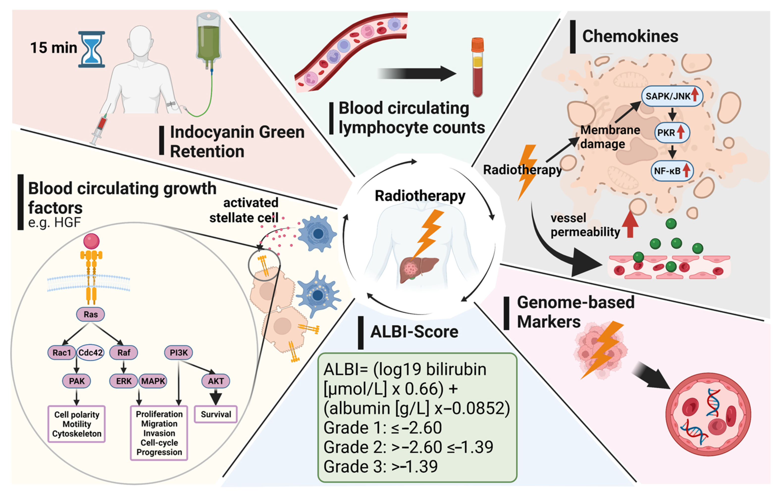
:1. Introduction
2. Clinical Scores: Child–Pugh, Barcelona Clinic Liver Cancer (BCLC), and Albumin–Bilirubin (ALBI) Grades
3. Indocyanine Green (ICG) Test
4. Hepatocyte Growth Factor (HGF)
5. Cytokines
6. Circulating Blood Cells
7. Genomic Biomarkers
8. Other Soluble Factors
9. Summary and Future Directions
10. Conclusions
Author Contributions
Funding
Institutional Review Board Statement
Informed Consent Statement
Data Availability Statement
Acknowledgments
Conflicts of Interest
References
- Llovet, J.M.; Kelley, R.K.; Villanueva, A.; Singal, A.G.; Pikarsky, E.; Roayaie, S.; Lencioni, R.; Koike, K.; Rossi-Zucman, J.; Fin, R.S. Hepatocellular carcinoma. Nat. Rev. Dis. Primers 2021, 7, 6. [Google Scholar] [CrossRef] [PubMed]
- Singal, A.G.; Lampertico, P.; Nahon, P. Epidemiology and surveillance for hepatocellular carcinoma: New trends. J. Hepatol. 2020, 72, 250–261. [Google Scholar] [CrossRef] [PubMed] [Green Version]
- van Dams, R.; Wu, T.C.; Kishan, A.U.; Raldow, A.C.; Chu, F.-I.; Hernandez, J.; Lamb, J.M.; Mikaeilian, A.; Low, D.A.; Steinberg, M.L. Ablative Radiotherapy for Liver Tumors Using Stereotactic MRI-Guidance: A Prospective Phase I Trial. Radiother. Oncol. 2021, 21, S0167-8140. [Google Scholar] [CrossRef] [PubMed]
- Gani, C.; Boeke, S.; McNair, H.; Ehlers, J.; Nachbar, M.; Mönnich, D.; Stolte, A.; Boldt, J.; Marks, C.; Winter, J.; et al. Marker-less online MR-guided stereotactic body radiotherapy of liver metastases at a 1.5 T MR-Linac—Feasibility, workflow data and patient acceptance. Clin. Transl. Radiat. Oncol. 2020, 26, 55–61. [Google Scholar] [CrossRef]
- Romesser, P.B.; Tyagi, N.; Crane, C.H. Magnetic Resonance Imaging-Guided Adaptive Radiotherapy for Colorectal Liver Metastases. Cancers 2021, 13, 1636. [Google Scholar] [CrossRef]
- Mathew, A.S.; Dawson, L.A. Current Understanding of Ablative Radiation Therapy in Hepatocellular Carcinoma. J. Hepatocell. Carcinoma 2021, 8, 575–586. [Google Scholar] [CrossRef]
- Kim, N.; Cheng, J.; Huang, W.-Y.; Kimura, T.; Zeng, Z.C.; Lee, V.H.; Kay, C.S.; Seong, J. Dose-Response Relationship in Stereotactic Body Radiation Therapy for Hepatocellular Carcinoma: A Pooled Analysis of an Asian Liver Radiation Therapy Group Study. Int. J. Radiat. Oncol. Biol. Phys. 2021, 109, 464–473. [Google Scholar] [CrossRef]
- Park, H.C.; Seong, J.; Han, K.H.; Chon, C.Y.; Moon, Y.M.; Suh, C.-O. Dose-response relationship in local radiotherapy for hepatocellular carcinoma. Int. J. Radiat. Oncol. Biol. Phys. 2002, 54, 150–155. [Google Scholar] [CrossRef]
- Romero, A.M.; Wunderink, W.; Hussain, S.M.; de Pooter, J.; Heijmen, B.J.M.; Nowak, P.C.J.M.; Nuyttens, J.J.; Brandwijk, R.P.; Verhoef, C.; Ijzermans, J.N.M.; et al. Stereotactic body radiation therapy for primary and metastatic liver tumors: A single institution phase i-ii study. Acta Oncol. 2006, 45, 831–837. [Google Scholar] [CrossRef]
- Hong, T.S.; DeLaney, T.F.; Mamon, H.J.; Willett, C.G.; Yeap, B.Y.; Niemierko, A.; Wolfgang, J.A.; Lu, H.-M.; Adams, J.; Weyman, E.A. A prospective feasibility study of respiratory-gated proton beam therapy for liver tumors. Pr. Radiat. Oncol. 2014, 4, 316–322. [Google Scholar] [CrossRef] [Green Version]
- Guha, C.; Kavanagh, B.D. Hepatic radiation toxicity: Avoidance and amelioration. Semin. Radiat. Oncol. 2011, 21, 256–263. [Google Scholar] [CrossRef] [Green Version]
- Dawson, L.A.; Normolle, D.; Balter, J.M.; McGinn, C.J.; Lawrence, T.S.; Haken, R.T. Analysis of radiation-induced liver disease using the Lyman NTCP model. Int. J. Radiat. Oncol. Biol. Phys. 2002, 53, 810–821. [Google Scholar] [CrossRef]
- Fitch, M.I.; Sharp, L.; Hanly, P.; Longo, C.J. Experiencing financial toxicity associated with cancer in publicly funded healthcare systems: A systematic review of qualitative studies. J. Cancer Surviv. 2021, 1–15. [Google Scholar] [CrossRef]
- Di Tommaso, L.; Spadaccini, M.; Donadon, M.; Personeni, N.; Elamin, A.; Aghemo, A.; Lleo, A. Role of liver biopsy in hepatocellular carcinoma. World J. Gastroenterol. 2019, 25, 6041–6052. [Google Scholar] [CrossRef]
- Llovet, J.M.; Bruix, J. Prognosis of hepatocellular carcinoma: The BCLC staging classification. Semin. Liver Dis. 1999, 19, 329–338. [Google Scholar] [CrossRef]
- Knox, J.J. Addressing the interplay of liver disease and hepatocellular carcinoma on patient survival: The ALBI scoring model. J. Clin. Oncol. 2015, 33, 529–531. [Google Scholar] [CrossRef]
- Na, S.K.; Yim, S.Y.; Suh, S.J.; Jung, Y.K.; Kim, J.H.; Seo, Y.S.; Yim, H.J.; Yeon, J.E.; Byun, K.S.; Um, S.H. ALBI versus Child-Pugh grading systems for liver function in patients with hepatocellular carcinoma. J. Surg. Oncol. 2018, 117, 912–921. [Google Scholar] [CrossRef]
- Wang, Y.-Y.; Zhong, J.-H.; Su, Z.-Y.; Huang, J.-F.; Lu, S.-D.; Xiang, B.-D.; Ma, L.; Qi, L.-N.; Ou, B.-N.; Li, L.-Q. Albumin-bilirubin versus Child-Pugh score as a predictor of outcome after liver resection for hepatocellular carcinoma. Br. J. Surg. 2016, 103, 725–734. [Google Scholar] [CrossRef]
- Johnson, P.J.; Berhane, S.; Kagebayashi, C.; Satomura, S.; Teng, M.; Reeves, H.L.; O’Beirne, J.; Fox, R.; Skowronska, A.; Palmer, D.; et al. Assessment of liver function in patients with hepatocellular carcinoma: A new evidence-based approach-the ALBI grade. J. Clin. Oncol. 2015, 33, 550–558. [Google Scholar] [CrossRef]
- Liu, P.-H.; Hsu, C.-Y.; Hsia, C.-Y.; Lee, Y.-H.; Chiou, Y.-Y.; Huang, Y.-H.; Lee, F.-Y.; Lin, H.-C.; Hou, M.-C.; Huo, T.-I. ALBI and PALBI grade predict survival for HCC across treatment modalities and BCLC stages in the MELD Era. J. Gastroenterol. Hepatol. 2017, 32, 879–886. [Google Scholar] [CrossRef]
- Ogasawara, S.; Chiba, T.; Ooka, Y.; Suzuki, E.; Kanogawa, N.; Saito, T.; Motoyama, T.; Tawada, A.; Kanai, F.; Yokosuka, O. Liver function assessment according to the Albumin-Bilirubin (ALBI) grade in sorafenib-treated patients with advanced hepatocellular carcinoma. Invest. New Drugs 2015, 33, 1257–1262. [Google Scholar] [CrossRef]
- Amisaki, M.; Uchinaka, E.; Morimoto, M.; Tokuyasu, N.; Sakamoto, T.; Honjo, S.; Saito, H.; Fujiwara, Y. Post-operative albumin-bilirubin grade predicts long-term outcomes among Child-Pugh grade A patients with hepatocellular carcinoma after curative resection. Hepatobiliary Pancreat. Dis. Int. 2018, 17, 502–509. [Google Scholar] [CrossRef] [PubMed]
- Murray, L.J.; Sykes, J.; Brierley, J.; Kim, J.J.; Wong, R.K.; Ringash, J.; Craig, T.; Velec, M.; Lindsay, P.; Knox, J.J.; et al. Baseline Albumin-Bilirubin (ALBI) Score in Western Patients with Hepatocellular Carcinoma Treated with Stereotactic Body Radiation Therapy (SBRT). Int. J. Radiat. Oncol. Biol. Phys. 2018, 101, 900–909. [Google Scholar] [CrossRef] [PubMed]
- Lo, C.-H.; Liu, M.-Y.; Lee, M.-S.; Yang, J.-F.; Jen, Y.-M.; Lin, C.-S.; Chao, H.-L.; Shen, P.-C.; Huang, W.-Y. Comparison Between Child-Turcotte-Pugh and Albumin-Bilirubin Scores in Assessing the Prognosis of Hepatocellular Carcinoma After Stereotactic Ablative Radiation Therapy. Int. J. Radiat. Oncol. Biol. Phys. 2017, 99, 145–152. [Google Scholar] [CrossRef] [PubMed]
- Toesca, D.A.; Osmundson, E.C.; von Eyben, R.; Shaffer, J.L.; Koong, A.C.; Chang, D.T. Assessment of hepatic function decline after stereotactic body radiation therapy for primary liver cancer. Pr. Radiat. Oncol. 2017, 7, 173–182. [Google Scholar] [CrossRef] [PubMed]
- Ho, C.H.; Chiang, C.-L.; Lee, F.A.; Choi, H.C.; Chan, J.C.; Yeung, C.S.; Huang, J.; Chan, M.K.; Blanck, O.; Wong, F.C. Comparison of platelet-albumin-bilirubin (PALBI), albumin-bilirubin (ALBI), and child-pugh (CP) score for predicting of survival in advanced hcc patients receiving radiotherapy (RT). Oncotarget 2018, 9, 28818–28829. [Google Scholar] [CrossRef] [PubMed] [Green Version]
- Su, T.S.; Yang, M.-H.; Zhou, Y.; Huang, Y.; Liang, P.; Cheng, T.; Chen, L.; Li, L.-Q.; Liang, S.-X. Albumin—bilirubin (ALBI) versus Child-Turcotte-Pugh (CTP) in prognosis of HCC after stereotactic body radiation therapy. Radiat. Oncol. 2019, 14, 50. [Google Scholar] [CrossRef] [PubMed]
- Jackson, W.C.; Hartman, E.; Gharzai, L.A.; Maurino, C.; Karnak, D.M.; Mendiratta-Lala, M.; Parikh, N.D.; Mayo, C.S.; Haken, R.K.T.; Schipper, M.J.; et al. The Potential for Midtreatment Albumin-Bilirubin (ALBI) Score to Individualize Liver Stereotactic Body Radiation Therapy. Int. J. Radiat. Oncol. Biol. Phys. 2021, 111, 127–134. [Google Scholar] [CrossRef]
- Gkika, E.; Bettinger, L.; Bettinger, L.; Schultheiss, M.; Brunner, T.B. The role of albumin-bilirubin grade and inflammation-based index in patients with hepatocellular carcinoma treated with stereotactic body radiotherapy. Strahlenther Onkol. 2018, 194, 403–413. [Google Scholar] [CrossRef]
- Gottlieb, M.E.; Stratton, H.; Newell, J.C.; Shah, D.M. Indocyanine green. Its use as an early indicator of hepatic dysfunction following injury in man. Arch. Surg. 1984, 119, 264–268. [Google Scholar] [CrossRef]
- Khisti, R.; Patidar, Y.; Garg, L.; Mukund, A.; Thomas, S.S.; Sarin, S.K. Correlation of baseline Portal pressure (hepatic venous pressure gradient) and Indocyanine Green Clearance Test With Post-transarterial Chemoembolization Acute Hepatic Failure. J. Clin. Exp. Hepatol. 2019, 9, 447–452. [Google Scholar] [CrossRef]
- Pind, M.; Bendtsen, L.; Kallemose, T.; Møller, S. Indocyanine green retention test (ICG-r15) as a noninvasive predictor of portal hypertension in patients with different severity of cirrhosis. Eur. J. Gastroenterol. Hepatol. 2016, 28, 948–954. [Google Scholar] [CrossRef]
- Wang, Z.; Wu, F.; Yue, Z.D.; Zhao, H.W.; Wang, L.; Fan, Z.H.; Zhang, Y.; Liu, F.Q. Comparative study of indocyanine green-R15, Child-Pugh score, and model for end-stage liver disease score for prediction of hepatic encephalopathy after transjugular intrahepatic portosystemic shunt. World J. Gastroenterol. 2021, 27, 416–427. [Google Scholar] [CrossRef]
- Hoekstra, L.; de Graaf, T.; Nibourg, G.A.; Heger, M.; Bennink, R.J.; Stieger, B.; van Gulik, T.M. Physiological and biochemical basis of clinical liver function tests: A review. Ann. Surg. 2013, 257, 27–36. [Google Scholar] [CrossRef] [Green Version]
- Stenmark, M.; Cao, H.; Wang, H.; Jackson, A.; Ben-Josef, E.; Ten Haken, R.K.; Lawrence, T.S.; Feng, M. Estimating functional liver reserve following hepatic irradiation: Adaptive normal tissue response models. Radiother. Oncol. 2014, 111, 418–423. [Google Scholar] [CrossRef] [Green Version]
- Lee, I.; Seong, J.; Shim, J.; Han, K.H. Radiotherapeutic parameters predictive of liver complications induced by liver tumor radiotherapy. Int. J. Radiat. Oncol. 2009, 73, 154–158. [Google Scholar] [CrossRef]
- Suresh, K.; Owen, L.; Bazzi, W.; Jackson, R.; Ten Haken, K.; Cuneo, K.; Feng, M.; Lawrence, T.S.; Schipper, M.J. Using Indocyanine Green Extraction to Predict Liver Function After Stereotactic Body Radiation Therapy for Hepatocellular Carcinoma. Int. J. Radiat. Oncol. 2017, 100, 131–137. [Google Scholar] [CrossRef]
- Feng, M.; Suresh, M.J.; Schipper, L.; Bazzi, E.; Ben-Josef, M.M.; Matuszak, N.D.; Parikh, T.H.; Welling, D.; Normolle, R.; Ten Haken, K.; et al. Individualized Adaptive Stereotactic Body Radiotherapy for Liver Tumors in Patients at High Risk for Liver Damage: A Phase 2 Clinical Trial. JAMA Oncol. 2018, 4, 40–47. [Google Scholar] [CrossRef]
- Jackson, W.C.; Tang, C.; Maurino, M.; Mendiratta-Lala, N.D.; Parikh, M.M.; Matuszak, J.S.; Dow, Y.; Cao, C.S.; Mayo, R.K.; Ten Haken, M.J.; et al. Individualized Adaptive Radiation Therapy Allows for Safe Treatment of Hepatocellular Carcinoma in Patients With Child-Turcotte-Pugh B Liver Disease. Int. J. Radiat. Oncol. 2020, 109, 212–219. [Google Scholar] [CrossRef]
- Cuneo, K.C.; Devasia, Y.; Sun, M.J.; Schipper, D.; Karnak, M.A.; Davis, D.; Owen, M.; Feng, I.; El Naqa, L.; Bazzi, R.; et al. Serum Levels of Hepatocyte Growth Factor and CD40 Ligand Predict Radiation-Induced Liver Injury. Transl. Oncol. 2019, 12, 889–894. [Google Scholar] [CrossRef]
- Hong, T.S.; Grassberger, B.Y.; Yeap, W.; Jiang, J.Y.; Wo, L.; Goyal, J.W.; Clark, C.H.; Crane, E.J.; Koay, S.; Dima, C.E.; et al. Pretreatment plasma HGF as potential biomarker for susceptibility to radiation-induced liver dysfunction after radiotherapy. NPJ Precis. Oncol. 2018, 2, 22. [Google Scholar] [CrossRef]
- El Naqa, I.; Johansson, D.; Owen, K.; Cuneo, Y.; Cao, M.; Matuszak, L.; Bazzi, T.; Lawrence, S.; Randall K. Ten, K. Modeling of Normal Tissue Complications Using Imaging and Biomarkers After Radiation Therapy for Hepatocellular Carcinoma. Int. J. Radiat. Oncol. 2018, 100, 335–343. [Google Scholar] [CrossRef] [Green Version]
- Ajdari, A.; Xie, C.; Richter, M.; Niyazi, D.; Duda, G.; Hong, T.S.; Bortfeld, T. Toward Personalized Radiation Therapy of Liver Metastasis: Importance of Serial Blood Biomarkers. JCO Clin. Cancer Inf. 2021, 5, 315–325. [Google Scholar] [CrossRef] [PubMed]
- Cha, H.; Lee, E.J.; Seong, J. Multi-analyte analysis of cytokines that predict outcomes in patients with hepatocellular carcinoma treated with radiotherapy. World J. Gastroenterol. 2017, 23, 2077–2085. [Google Scholar] [CrossRef] [PubMed]
- Ng, S.S.W.; Zhang, L.; Wang, D.; Citrin, L.; Dawson, A. Association of pro-inflammatory soluble cytokine receptors early during hepatocellular carcinoma stereotactic radiotherapy with liver toxicity. NPJ Precis. Oncol. 2020, 4, 17. [Google Scholar] [CrossRef] [PubMed]
- Cousins, M.M.; Morris, C.; Maurino, T.P.; Devasia, D.; Karnak, D.; Ray, N.D.; Parikh, D.; Owen, R.K.; Ten Haken, M.J.; Schipper, T.S.; et al. TNFR1 and the TNFα axis as a targetable mediator of liver injury from stereotactic body radiation therapy. Transl. Oncol. 2021, 14, 100950. [Google Scholar] [CrossRef] [PubMed]
- Grassberger, C.; Hong, T.; Hato, B.Y.; Yeap, J.Y.; Wo, M.; Tracy, T.; Bortfeld, J.A.; Wolfgang, C.E.; Eyler, L.; Goyal, J.W.C.; et al. Differential Association Between Circulating Lymphocyte Populations with Outcome After Radiation Therapy in Subtypes of Liver Cancer. Int. J. Radiat. Oncol. 2018, 101, 1222–1225. [Google Scholar] [CrossRef]
- Gustafson, M.P.; Bornschlegl, S.; Park, S.; Gastineau, D.A.; Roberts, L.R.; Dietz, A.B.; Hallemeier, C.L. Comprehensive assessment of circulating immune cell populations in response to stereotactic body radiation therapy in patients with liver cancer. Adv. Radiat. Oncol. 2017, 2, 540–547. [Google Scholar] [CrossRef] [Green Version]
- Zhang, H.G.; Yang, T.; Jiang, J.; Zhang, Y.; Jin, X.J.; Hu, Y.; Sun, J.; Du, S.S.; Zeng, Z.C. Lymphopenia Is Associated with Gross Target Volumes and Fractions in Hepatocellular Carcinoma Patients Treated with External Beam Radiation Therapy and Also Indicates Worse Overall Survival. Can. J. Gastroenterol. Hepatol. 2019, 2019, 9691067. [Google Scholar] [CrossRef]
- Byun, H.; Kim, K.; Park, S.; Seong, J. Acute severe lymphopenia by radiotherapy is associated with reduced overall survival in hepatocellular carcinoma. Strahlenther Onkol. 2019, 195, 1007–1017. [Google Scholar] [CrossRef]
- Zhuang, Y.; Yuan, Y.; Chen, G.W.; Zhao, X.M.; Hu, Y.; Zhu, W.C.; Zeng, Z.C.; Chen, Y.X. Association Between Circulating Lymphocyte Populations and Outcome After Stereotactic Body Radiation Therapy in Patients with Hepatocellular Carcinoma. Front. Oncol. 2019, 9, 896. [Google Scholar] [CrossRef]
- Liu, J.; Zhao, Q.; Deng, W.; Lu, J.; Xu, X.; Wang, R.; Li, X.; Yue, J. Radiation-related lymphopenia is associated with spleen irradiation dose during radiotherapy in patients with hepatocellular carcinoma. Radiat. Oncol. 2017, 12, 90. [Google Scholar] [CrossRef] [Green Version]
- Hsiang, C.W.; Huang, Y.; Yang, J.F.; Shen, P.C.; Dai, Y.H.; Wang, Y.F.; Lin, C.S.; Chang, W.C.; Lo, C.H. Dynamic Changes in Neutrophil-to-Lymphocyte Ratio are Associated with Survival and Liver Toxicity Following Stereotactic Body Radiotherapy for Hepatocellular Carcinoma. J. Hepatocell. Carcinoma 2021, 8, 1299–1309. [Google Scholar] [CrossRef]
- De, B.; Ng, P.S.; Liu, A.Y.; Avila, S.; Tao, R.; Holliday, E.B.; Brownlee, Z.; Kaseb, A.; Lee, S.; Raghav, K.; et al. Radiation-Associated Lymphopenia and Outcomes of Patients with Unresectable Hepatocellular Carcinoma Treated with Radiotherapy. J. Hepatocell. Carcinoma 2021, 8, 57–69. [Google Scholar] [CrossRef]
- Gupta, G.; Al-Malki, H.; Kazmi, I.; Thangavelu, L.; Gupta, P.K.; Jha, N.K.; Prasher, P.; Singh, S.K.; Dua, K. The role of HGF/MET in liver cancer. Futur. Med. Chem. 2021, 13, 1829–1832. [Google Scholar] [CrossRef]
- Fu, R.; Jiang, J.; Lim, H.; Chen, X.; Zhang, X. Activation of the HGF/c-MET axis promotes lenvatinib resistance in hepatocellular carcinoma cells with high c-MET expression. Med. Oncol. 2020, 37, 24. [Google Scholar] [CrossRef]
- Firtina Karagonlar, Z.; Koc, D.; Iscan, E.; Erdal, E.; Atabey, N. Elevated hepatocyte growth factor expression as an autocrine c-Met activation mechanism in acquired resistance to sorafenib in hepatocellular carcinoma cells. Cancer Sci. 2016, 107, 407–416. [Google Scholar] [CrossRef]
- Moosavi, F.; Giovannetti, E.; Saso, L.; Firuzi, O. HGF/MET pathway aberrations as diagnostic, prognostic, and predictive biomarkers in human cancers. Crit. Rev. Clin. Lab. Sci. 2019, 56, 533–566. [Google Scholar] [CrossRef] [Green Version]
- Dayyani, F.; Zurita, J.; Nogueras-González, G.M.; Slack, R.; Millikan, R.E.; Araujo, J.C.; Gallick, G.E.; Logothetis, C.J.; Corn, P.G. The combination of serum insulin, osteopontin, and hepatocyte growth factor predicts time to castration-resistant progression in androgen dependent metastatic prostate cancer- an exploratory study. BMC Cancer 2016, 16, 721. [Google Scholar] [CrossRef] [Green Version]
- Hügel, R.; Muendlein, A.; Volbeding, L.; Drexel, H.; Richtig, E.; Wehkamp, U.; Painsi, C.; Lange-Asschenfeldt, B.; Hauschild, A.; Egberts, F. Serum levels of hepatocyte growth factor as a potential tumor marker in patients with malignant melanoma. Melanoma Res. 2016, 26, 354–360. [Google Scholar] [CrossRef]
- Saltarella, I.; Morabito, N.; Giuliani, C.; Terragna, P.; Omedè, A.; Palumbo, S.; Bringhen, L.; De Paoli, E.; Martino, A.; Larocca, M.; et al. Prognostic or predictive value of circulating cytokines and angiogenic factors for initial treatment of multiple myeloma in the GIMEMA MM0305 randomized controlled trial. J. Hematol. Oncol. 2019, 12, 4. [Google Scholar] [CrossRef]
- Yamagamim, H.; Moriyama, M.; Matsumura, H.; Aoki, H.; Shimizu, T.; Saito, T.; Kaneko, M.; Shioda, A.; Tanaka, N.; Arakawa, Y. Serum concentrations of human hepatocyte growth factor is a useful indicator for predicting the occurrence of hepatocellular carcinomas in C-viral chronic liver diseases. Cancer 2002, 95, 824–834. [Google Scholar] [CrossRef]
- Karabulut, S.; Tas, F.; Akyüz, F.; Ormeci, A.C.; Serilmez, M.; Soydinç, H.O.; Vatansever, S.; Yasasever, V. Clinical significance of serum hepatocyte growth factor (HGF) levels in hepatocellular carcinoma. Tumour Biol. 2014, 35, 2327–2333. [Google Scholar] [CrossRef]
- Finkelstein, S.; Timmerman, E.; McBride, W.H.; Schaue, D.; Hoffe, S.E.; Mantz, C.A.; Wilson, G.D. The confluence of stereotactic ablative radiotherapy and tumor immunology. Clin. Dev. Immunol. 2011, 2011, 439752. [Google Scholar] [CrossRef]
- Janus, P.; Szołtysek, K.; Zając, G.; Stokowy, T.; Walaszczyk, A.; Widłak, W.; Wojtaś, B.; Gielniewski, B.; Iwanaszko, M.; Braun, R.; et al. Pro-inflammatory cytokine and high doses of ionizing radiation have similar effects on the expression of NF-kappaB-dependent genes. Cell Signal. 2018, 46, 23–31. [Google Scholar] [CrossRef]
- Ong, Z.Y.; Gibson, J.; Bowen, J.M.; Stringer, A.M.; Darby, J.M.; Logan, R.M.; Yeoh, A.S.; Keefe, D.M. Pro-inflammatory cytokines play a key role in the development of radiotherapy-induced gastrointestinal mucositis. Radiat. Oncol. 2010, 5, 22. [Google Scholar] [CrossRef] [Green Version]
- Aloui, C.; Prigent, S.; Tariket, C.; Sut, J.; Fagan, J.; Cognasse, F.; Chakroun, T.; Garraud, O.; Laradi, S. Levels of human platelet-derived soluble CD40 ligand depend on haplotypes of CD40LG-CD40-ITGA2. Sci. Rep. 2016, 6, 24715. [Google Scholar] [CrossRef] [Green Version]
- Tang, T.; Cheng, B.; Truong, L.; Sun, L.; Yang, X.; Wang, H. Molecular basis and therapeutic implications of CD40/CD40L immune checkpoint. Pharm. Ther. 2021, 219, 107709. [Google Scholar] [CrossRef]
- Angelou, A.; Antoniou, N.; Garmpis, C.; Damaskos, S.; Theocharis, G.; Margonis, A. The Role of Soluble CD40L Ligand in Human Carcinogenesis. Anticancer. Res. 2018, 38, 3199–3201. [Google Scholar]
- Roselli, M.C.; Mineo, S.; Basili, F.; Martini, S.; Mariotti, S.; Aloe, G.; Del Monte, V.; Ambrogi, A.; Spila, R.; Palmirotta, R.; et al. Soluble CD40 ligand plasma levels in lung cancer. Clin. Cancer Res. 2004, 10, 610–614. [Google Scholar] [CrossRef] [Green Version]
- Rico Montanari, N.; Anugwom, M.; Boonstra, A.; Debes, J.D. The Role of Cytokines in the Different Stages of Hepatocellular Carcinoma. Cancers 2021, 13, 4876. [Google Scholar] [CrossRef] [PubMed]
- Lai, S.C.; Su, Y.T.; Chi, C.C.; Kuo, Y.C.; Lee, K.F.; Wu, Y.C.; Lan, P.C.; Yang, M.H.; Chang, T.S.; Huang, Y.H. Correction to: DNMT3b/OCT4 expression confers sorafenib resistance and poor prognosis of hepatocellular carcinoma through IL-6/STAT3 regulation. J. Exp. Clin. Cancer Res. 2020, 39, 10. [Google Scholar] [CrossRef] [PubMed]
- Shakiba, E.; Ramezani, M.; Sadeghi, M. Evaluation of serum interleukin-6 levels in hepatocellular carcinoma patients: A systematic review and meta-analysis. Clin. Exp. Hepatol. 2018, 4, 182–190. [Google Scholar] [CrossRef] [PubMed]
- Kim, M.J.; Jang, W.; Oh, B.S.; Kwon, J.H.; Chung, K.W.; Jung, H.S.; Jekarl, D.W.; Lee, S. Change in inflammatory cytokine profiles after transarterial chemotherapy in patients with hepatocellular carcinoma. Cytokine 2013, 64, 516–522. [Google Scholar] [CrossRef]
- Taniguchi, K.; Karin, M. IL-6 and related cytokines as the critical lynchpins between inflammation and cancer. Semin. Immunol. 2014, 26, 54–74. [Google Scholar] [CrossRef]
- Dehing-Oberije, C.; Aerts, H.; Yu, S.; De Ruysscher, D.; Menheere, P.; Hilvo, M.; van der Weide, H.; Rao, B.; Lambin, P. Development and validation of a prognostic model using blood biomarker information for prediction of survival of non-small-cell lung cancer patients treated with combined chemotherapy and radiation or radiotherapy alone (NCT00181519, NCT00573040, and NCT00572325). Int. J. Radiat. Oncol. 2011, 81, 360–368. [Google Scholar]
- Wu, C.T.; Chen, F.; Chen, W.C.; Hsieh, C.C. The role of IL-6 in the radiation response of prostate cancer. Radiat. Oncol. 2013, 8, 159. [Google Scholar] [CrossRef] [Green Version]
- Duffy, S.A.; Taylor, M.; Terrell, J.E.; Islam, M.; Li, Y.; Fowler, K.E.; Wolf, G.T.; Teknos, T.N. Interleukin-6 predicts recurrence and survival among head and neck cancer patients. Cancer 2008, 113, 750–757. [Google Scholar] [CrossRef]
- van Remco, H.; Ten Hagen, T.L.; Eggermont, A.M. TNF-alpha in cancer treatment: Molecular insights, antitumor effects, and clinical utility. Oncologist 2006, 11, 397–408. [Google Scholar]
- Aggarwal, B.B. Signalling pathways of the TNF superfamily: A double-edged sword. Nat. Rev. Immunol. 2003, 3, 745–756. [Google Scholar] [CrossRef]
- Yang, Y.M.; Kim, S.Y.; Seki, E. Inflammation and Liver Cancer: Molecular Mechanisms and Therapeutic Targets. Semin. Liver Dis. 2019, 39, 026–042. [Google Scholar] [CrossRef]
- Oliver, J.C.; Bland, A.; Oettinger, C.W.; Arduino, M.J.; McAllister, S.K.; Aguero, S.M.; Favero, M.S. Cytokine kinetics in an in vitro whole blood model following an endotoxin challenge. Lymphokine Cytokine Res. 1993, 12, 115–120. [Google Scholar]
- Damen, P.J.J.; Kroese, E.; van Hillegersberg, R.; Schuit, E.; Peters, M.; Verhoeff, J.J.C.; Lin, S.H.; van Rossum, P.S.N. The Influence of Severe Radiation-Induced Lymphopenia on Overall Survival in Solid Tumors: A Systematic Review and Meta-Analysis. Int. J. Radiat. Oncol. Biol. Phys. 2021, 111, 936–948. [Google Scholar] [CrossRef]
- Sung, W.; Grassberger, A.; McNamara, L.; Basler, L.; Ehrbar, S.; Tanadini-Lang, S.; Hong, T.S.; Paganetti, H. A tumor-immune interaction model for hepatocellular carcinoma based on measured lymphocyte counts in patients undergoing radiotherapy. Radiother. Oncol. 2020, 151, 73–81. [Google Scholar] [CrossRef]
- Routman, D.M.; Garant, S.C.; Lester, C.N.; Day, W.S.; Harmsen, C.T.; Sanheuza, H.H.; Yoon, M.A.; Neben-Wittich, J.A.; Martenson, M.G.; Haddock, C.L.; et al. A Comparison of Grade 4 Lymphopenia with Proton Versus Photon Radiation Therapy for Esophageal Cancer. Adv. Radiat. Oncol. 2019, 4, 63–69. [Google Scholar] [CrossRef] [Green Version]
- Mohan, R.Y.; Liu, P.D.; Brown, A.; Mahajan, J.; Dinh, C.; Chung, S.; McAvoy, M.F.; McAleer, S.H.; Lin, J.; Li, A.J.; et al. Proton therapy reduces the likelihood of high-grade radiation-induced lymphopenia in glioblastoma patients: Phase II randomized study of protons vs photons. Neuro-Oncol. 2020, 23, 284–294. [Google Scholar] [CrossRef]
- Lambin, P.; Lieverse, R.I.Y.; Eckert, F.; Marcus, D.; Oberije, C.; van der Wiel, A.M.A.; Guha, C.; Dubois, L.J.; Deasy, J.O. Lymphocyte-Sparing Radiotherapy: The Rationale for Protecting Lymphocyte-rich Organs When Combining Radiotherapy with Immunotherapy. Semin. Radiat. Oncol. 2020, 30, 187–193. [Google Scholar] [CrossRef]
- Zhang, Y.; Liu, K.; Ji, X.; Li, C.; Wang, Z.; Ren, Y.; Liu, X.; Chen, X.; Han, L.; Meng, L.; et al. Clinical Application Value of Circulating Cell-free DNA in Hepatocellular Carcinoma. Front. Mol. Biosci. 2021, 8, 736330. [Google Scholar] [CrossRef]
- Corcoran, R.B.; Chabner, B.A. Application of Cell-free DNA Analysis to Cancer Treatment. N. Engl. J. Med. 2018, 379, 1754–1765. [Google Scholar] [CrossRef] [Green Version]
- Park, S.; Lee, J.; Rim, C.H.; Seong, J. Plasma Cell-Free DNA as a Predictive Marker after Radiotherapy for Hepatocellular Carcinoma. Yonsei Med. J. 2018, 59, 470–479. [Google Scholar] [CrossRef]
- Ren, N.; Ye, H.; Qin, L.X.; Zhang, B.H.; Liu, Y.K.; Tang, Z.Y. Circulating DNA level is negatively associated with the long-term survival of hepatocellular carcinoma patients. World J. Gastroenterol. 2006, 12, 3911–3914. [Google Scholar] [CrossRef] [PubMed]
- Zwirner, K.; Hilke, J.; Demidov, G.; Ossowski, S.; Gani, C.; Rieß, O.; Zips, D.; Welz, S.; Schroeder, C. Circulating cell-free DNA: A potential biomarker to differentiate inflammation and infection during radiochemotherapy. Radiother. Oncol. 2018, 129, 575–581. [Google Scholar] [CrossRef] [PubMed]
- Suh, Y.G.; Lee, J.; Cha, H.; Yang, S.H.; Seong, J. Prognostic values of vascular endothelial growth factor and matrix metalloproteinase-2 in hepatocellular carcinoma after radiotherapy. Dig. Dis. 2014, 32, 725–732. [Google Scholar] [CrossRef] [PubMed]
- Dovedi, S.J.; Adlard, A.L.; Lipowska-Bhalla, G.; McKenna, C.; Jones, S.; Cheadle, E.J.; Stratford, I.J.; Poon, E.; Morrow, M.; Stewart, R.; et al. Acquired resistance to fractionated radiotherapy can be overcome by concurrent PD-L1 blockade. Cancer Res. 2014, 74, 5458–5468. [Google Scholar] [CrossRef] [Green Version]
- Kim, K.J.; Kim, H.; Lee, S.J.; Lee, E.J.; Shin, E.C.; Seong, J. Radiation improves antitumor effect of immune checkpoint inhibitor in murine hepatocellular carcinoma model. Oncotarget 2017, 8, 41242–41255. [Google Scholar] [CrossRef] [Green Version]
- Lee, Y.H.; Tai, C.; Yip, S.; Choo, P.; Chew, V. Combinational Immunotherapy for Hepatocellular Carcinoma: Radiotherapy, Immune Checkpoint Blockade and Beyond. Front. Immunol. 2020, 11, 568759. [Google Scholar] [CrossRef]
- Parikh, A.R.; Szabolcs, A.; Allen, J.N.; Clark, J.W.; Wo, J.Y.; Raabe, M.; Thel, H.; Hoyos, D.; Mehta, A.; Arshad, S.; et al. Radiation therapy enhances immunotherapy response in microsatellite stable colorectal and pancreatic adenocarcinoma in a phase II trial. Nat. Cancer 2021, 2, 1124–1135. [Google Scholar] [CrossRef]
- Ng, S.S.W.; Jang, H.; Kurland, I.J.; Qiu, Y.; Guha, C.; Dawson, L.A. Plasma metabolomic profiles in liver cancer patients following stereotactic body radiotherapy. EBioMedicine 2020, 59, 102973. [Google Scholar] [CrossRef]
- Dubois, N.; Rio, N.; Ripoche, V.; Ferchaud-Roucher, M.; Gaugler, H.; Campion, L.; Krempf, M.; Carrie, C.; Mahé, M.; Mirabel, X.; et al. Plasma ceramide, a real-time predictive marker of pulmonary and hepatic metastases response to stereotactic body radiation therapy combined with irinotecan. Radiother. Oncol. 2016, 119, 229–235. [Google Scholar] [CrossRef] [Green Version]
- Lee, E.J.; Yang, H.; Kim, K.J.; Cha, H.; Lee, S.J.; Kim, J.H.; Song, J.; Chun, K.H.; Seong, J. Inter-alpha Inhibitor H4 as a Potential Biomarker Predicting the Treatment Outcomes in Patients with Hepatocellular Carcinoma. Cancer Res. Treat. 2018, 50, 646–657. [Google Scholar] [CrossRef] [Green Version]
- Cuneo, K.; Sun, C.; Schipper, M.; Tewari, M.; Ten Haken, R.K.; Lawrence, T.S.; Feng, M. MicroRNAs Predict Liver Toxicity in Patients Receiving Stereotactic Body Radiation Therapy for Hepatocellular Carcinoma. Int. J. Rad. Oncol. Biol. Phys. 2016, 96 (Suppl. 2), S202–S203. [Google Scholar] [CrossRef]
- Kim, H.J.; Park, K.; Kim, J.; Seong, J. Clinical significance of soluble programmed cell death ligand-1 (sPD-L1) in hepatocellular carcinoma patients treated with radiotherapy. Radiother. Oncol. 2018, 129, 130–135. [Google Scholar] [CrossRef]


| Author (Year) | Bio-Marker | Time-Points | Patient # | Cancer Type | Baseline ALBI Score, Median (Range) or Grade 1/2/3 [%] | Underlying Liver Damage (CPS A/B/C/NA [%]) | Dose, Median (Range) [Gy]/ Fractionation | Endpoint | Comments |
|---|---|---|---|---|---|---|---|---|---|
| Murray LJ et al. (2018) [23] | ALBI | Pre RT | 102 | HCC | −2.63 (−3.40 to −1.64) | 100/0/0/0 | 36 (2–54)/6 | OS Toxicity | HR (increase in ALBI score per 0.1): 1.09 (95% CI 1.03–1.17) OR (increase in ALBI score per 0.1): 1.51 (95% CI 1.23–1.85) |
| Lo CH et al. (2017) [24] | ALBI | Pre RT | 152 | HCC | (−3.67 to −0.84) | 78.3/21.7/0/0 | 45 (25–65)/5 (3–6) | OS Toxicity | HR (increase in ALBI score 2 vs.1): 2.09 (95% CI 1.26–3.46) Pretreatment ALBI Grade: p < 0.001 |
| Toesca DAS et al. (2017) [25] | ALBI | pre, 1/3/6/12 months post RT | 60 | HCC (40/60); CCA (20/60) | 5/82.5/12.5 | 57.5/30/0/12.5 | 40 (22–50)/5 (1–7) | OS Toxicity | HCC cohort (worsening ALBI score by 0.5 post RT): median OS = 37 vs. 14 months, p = 0.0005 CCA cohort (pretreatment ALBI grade): p = 0.02 HCC cohort (worsening ALBI score by 0.5 post RT): G3 + HB toxicity= p = 0.01; significant decline in hepatic function = p = 0.001 |
| Gkika E et al. (2018) [29] | ALBI, inflammation-based index (IBI) | Pre, during, post, 2 months post RT | 40 | HCC | 30/58/12 | 55/45/0/0 | 45 (21–66)/3–12 | OS Toxicity | Increased OS (lower IBI during treatment): p = 0.034 Decreased OS (Higher CRP/AFP): p = 0.001 Higher Incidence of acute/late toxicities (Higher ALBI/CPS at baseline): p = 0.02/0.001 |
| Jackson WC et al. (2021) [28] | ALBI | Pre RT | 151 | HCC | 25.9/65.7/8.4 | 66.9/31.3/1.8/0 | 79.2 (IQR 69.3, 101.7)/3–5 | Toxicity | Baseline ALBI: OR 1.8 (95% CI: 1.24–2.62) Change in ALBI: OR 3.07 (95% CI: 1.29–7.32) |
| Su TS et al. (2019) [27] | ALBI | Pre RT | 511 | HCC | 36.9/58.4/4.7 | 80.6/18.2/1.2/0 | 42–43/3–5 | OS | Median OS (ALBI grade 1/2/3): 53 vs. 19.5 vs. 6.5 months (p < 0.0001) |
| Ho CH et al. (2018) [26] | ALBI | Pre-RT | 174 | HCC | −2.39 (−3.61 to −1.41) | 100/0/0/0 | 37.3 (23.3–72)/7 (5–10) | OS | ALBI score: HR = 1.72 (95% CI 1.2–2.48) |
| Author (Year) | Biomarker | Timepoint | Pat. # | Cancer Type [%] | Underlying Liver Damage (CPS A/B/C/NA [%]) | Dose [Gy] (Median, Range)/ Fractionation | Endpoint | Comments |
|---|---|---|---|---|---|---|---|---|
| ||||||||
| Suresh K et al. (2018) [37] | ICGR after 15 min | Pre and after 3rd fraction, 1/3/6 months post RT | 144 | HCC | NA | NA/3–5 | Toxicity | Inclusion of ICGR15 significantly improves prediction of liver toxicity after irradiation |
| Feng M et al. (2018) [38] | ICGR after 15 min | Pre and after 3rd fraction | 90 | HCC (76.7), ICC (4.4), Metastasis (18.9) | NA | 49 (23–60)/3 or 5 | Phase II Study | High Feasibility of biomarker adapted RT (LC: 1y = 99% (95% CI: 97–100%); 2y = 95% (95% CI: 91–99%) |
| Stenmark MH et al. (2014) [35] | ICGR after 15 min | Pre, 50–70% of RT dose, 1/2 months post RT | 48 | HCC (44), ICC (29), Metastasis (27) | 92/8/0/0 | Different treatment regimes | Toxicity | Both mid-RT ICGR15 and Mean liver dose predicted liver function post RT (p < 0.0001) |
| Lee IJ et al. (2009) [36] | ICGR after 15 min | Pre RT | 131 | HCC | 87/13/0/0 | 45 +/−16.5/1.5–2.5 Gy/fr | Toxicity | ICGR15 increased after radiotherapy; CPS but not ICGR15 predicted liver toxicity |
| ||||||||
| Cuneo KC et al. (2019) [40] | HGF, CD40 Ligand | Pre and after 3rd fraction | 104 | HCC (84), others (16) | 75/22/3/0 | 28–55/3 or 5; 60/20 | OS Toxicity | Pretreatment HGF (High vs. low): 14.5 vs. 27.1 months (p = 0.035) Toxicity (Increase in CPS > = 2 points): HGF (baseline/1-month) = OR 6.97 (95% CI 1.05–46.36, p value = 0.045)/OR 7.82 (95% CI 1.14–53.6, p value 0.036); CD40L (baseline/1-month) = OR 0.47 (95% CI 0.201–1.098, p value = 0.081)/OR 0.28 (95% CI 0.086–0.897, p value = 0.032) |
| Hong TS et al. (2018) [41] | Pretreatment HGF | Pre RT | 43 | HCC (51.2), ICC and others (48.8) | 86/14/0/0 | 58 Gy RBE (15.1–67.5) | OS (2y) PFS (2y) Toxicity | Pretreatment HGF (High vs. low): 14% vs. 69% (p = 0.0147) Pretreatment HGF (High vs. low): ns (p = 0.348) Low pretreatment HGF: correlation with stable CPS and lower bilirubin (p = 0.01) |
| El Naqa I et al. (2018) [42] | TGFβ1, CCL11, HGF, CD40 Ligand | Pre and after 3rd fraction | 192 | HCC | NA | SBRT: 49.8 (18.6–60); cf RT: 50.4 (30–90)/3–5 | Toxicity | Models to predict liver toxicity after RT were improved by a factor of 1.5 with inclusion of TGFβ1 and Eotaxin |
| ||||||||
| Ajdari A et al. (2021) [43] | Inflammatory cytokines, gene mutation status, complete blood count | Pre and before 4th fraction | 89 | Liver metastasis | NA | 40 GyE (30–50)/5 | OS (2y) LF (1y) | baseline absolute lymphocyte count (High vs. Low): 54% vs. 25% (p = 0.0002) Baseline Platelet-to-lymphocyte ratio: HR 1.004 (p = 0.0004); Baseline Neutrophile-to-Lymphocyte: HR = 1.32 (p = 0.0001) Mutation in KRAS gene (Yes vs. No): 69% vs. 31%; HR 2.92 (95% CI, 1.17 to 7.28, p = 0.02) Baseline/mid-treatment interleukin 6: HR 1.15 (95% CI 1.04–1.26, p = 0.01)/1.06 (95% CI 1.01–1.13, p = 0.01) |
| Cha H et al. (2017) [44] | IL-1/6/8/10/12, TNF-a | Pre and post RT | 51 | HCC | 96.1/3.9/0/0 | 50.4 (45–64.8) | OS Infield FFS Outfield-intrahepatic FFS | No correlation between baseline Cytokines and OS baseline serum IL-6 level: p < 0.001, RR 1.019 (95%CI 1.011–1.028) Baseline Serum IL-10 level: p = 0.026, RR 0.830 (95%CI 0.705–0.978) |
| Ng SSW et al. (2020) [45] | Soluble cytokine receptors | Pre RT, post 1–2 fractions | 47 | HCC | 81/19/0/0 | 33(30–54)/6 | Risk of early death Toxicity | Lower risk: high baseline level sCD40L = HR 1.8(95% CI 0.27–0.99, p = 0.05) Higher risk: high baseline levels sTNFRII = HR 1.93 (95% CI 1.02–3.65, p = 0.04); sIL-6r = HR 1.9 (95% CI 1.01–3.57, p = 0.05); AFP= HR 2.61 (95% CI 1.03–4.54, p = 0.043); sEGFR = HR 2.61 (95% CI 1.32–5.16, p = 0.006); sgp130 = HR 2.19 (95% CI 1.13–4.25, p = 0.021) ≥2 increase CP score (3 months post RT): increased level sTNFRII (p < 0.001); decreased levels of sCD40L (p < 0.001)/CXCL1(p = 0.01) |
| Cousins MM et al. (2021) [46] | Soluble TNFa receptor (sTNFR1) | Pre and after 3rd fraction, 1/3/6 months post RT | 78 | HCC (95), others (5) | NA | 18–60/3–5 | Toxicity | sTNFR1 (Increase in CPS > = 2 points): baseline= OR 1.62 (p = 0.0573); 1 month= OR 2.35 (p = 0.0181) |
| ||||||||
| Grassberger C et al. (2018) [47] | Lymphocytes | Pre, Day 8 and Day 15 of RT | 43 | HCC (51.2), ICC (48.8) | 73.7/13.3/0/0 | 58 RBE/ 15 | OS | ICC: baseline CD4 + CD25 + T cells (p = 0.003) and CD4 + CD127+ T cells (p = 0.01) HCC: mid-treatment fraction of activated CTLs (p = 0.007) |
| Gustafson MP et al. (2017) [48] | Immune cell populations | Pre and post RT, 3 months post RT | 10 | HCC (50), CCA (10), Metastasis (40) | NA | 50–60/5 or 54/3 | Changes pre- to post RT | Circulating T cells dropped at the end of RT (2-fold) and recovered within 3 months; CD56br CD16− NK cells dropped 40% after RT and recovered at 3 months |
| Zhang H et al. (2019) [49] | Lymphocytes | Pre, twice during RT, follow up every 3 months (1st year) then every 6 months | 184 | HCC | 79.3/15.3/0/5.4 | 75 (50–119) BED/16 (5–35) | OS Toxicity | 1/2-year OS (Low vs. high lymphocyte nadir during RT): 56.7% vs. 80.3%; 28.4% vs. 55.7% (p < 0.001) Lymphocyte counts declined during RT (p < 0.001) |
| Byun HK et al. (2019) [50] | Lymphocytes | Pre and 3 months post RT | 920 | HCC | 78.2/21.8/0/0 | Cf RT: 45–60/20–25; SBRT: 60 or 52/4 | OS | Acute severe lymphopenia: HR = 1.40 (95% CI 1.02–1.91), p = 0.035 Baseline NLR: HR = 1.03 (95% CI 1.01–1.06), p = 0.016 |
| Zhuang Y et al. (2019) [51] | Lymphocytes, TN-Fα | Pre and 10 days, 1/2/3 months post RT, then every 3 months | 78 | HCC | 96.2/3.8/0/0 | 48 (48–60)/(5–10) | OS | Total peripheral lymphocyte counts post RT < 0.45 × 10⁹/L: HR = 0.14 (95% CI 0.02–0.93), p = 0.04 TNFα < 5.5 n/mL: HR = 0.07 (95% CI 0.01-.44), p = 0.005 |
| Liu J et al. (2017) [52] | Lymphocytes | Pre and weekly during RT | 59 | HCC | NA | 54 (45–62)/NA | OS | Minimum value of absolute lymphocyte counts (cut-off 300 cells/µL): OR 28.8 (95% CI 27.23–30.37) |
| Hsiang CW et al. (2021) [53] | Neutrophil -to-Lymphocyte Ratio (NLR) | Pre and 3 months post RT | 93 | HCC | 69.9/30.1/0/0 | 45 (25–60)/5(4–6) | OS Toxicity | Pre-RT NLR: HR = 1.24 (95% CI 1.12–1.38), p < 0.001 Delta NLR: HR = 1.1 (95% CI 1.02–1.18), p = 0.011 Liver toxicity rate (delta NLR <vs > 1.9): 7.5% vs. 35.1% |
| De B et al. (2021) [54] | Lymphocytes | Pre, during, post RT | 143 | HCC | 80/20/0/0 | Photon (72%); Proton (28%) 60 (30–100)/15 (3–34) | OS | pre-RT ALC ≤ 0.5: OS (median 7 vs. 20 months, p = 0.03); HR = 2.677 (95% CI 1.057–6.779), p = 0.039) Post-RT ALC ≤ 0.5: HR = 1.031 (95% CI 1.001–1.062), p = 0.043) G3 or higher lymphopenia during RT: OS (median 13 vs. 31 months, p < 0.001) |
| ||||||||
| Cuneo KC et al. (2016)c | Micro RNA (miR) | Pre and after 3rd fraction, 1/3/6 months post RT | 30 | HCC | NA | NA/3–5 | Toxicity | Potential correlation with microRNA miR.122.3p, miR.375, miR.217, miR.125a.5p |
| Park S et al. (2018) | Cell-free DNA | Pre and post RT | 55 | HCC | 88.5/11.5/0/0 | SBRT: 60/4; cf RT: 45.6 (45–60)/1.8 Gy/fr (1.8–3) + Ctx | LC Intrahepatic FFS | Post RT (low vs. High cell-free DNA): p = 0.041 (SBRT); p = 0.046 (cf RT) Post RT cell free DNA = HR 2.405 (95% CI 1.059–5.460) |
| ||||||||
| Dubois N et al. (2016) | Ceramide | D0, D3 (post 2fr), D10 (post 4fr) | 35 | Liver and lung metastasis (colorectal cancer) | NA | 40/4 (Rctx with Irinotecan) | Tumor control (1y) | HR (Ceramide D10): 1.09 (95% CI 1.03–1.17) |
| Lee EJ et al. (2018) | Inter-alpha Inhibitor H4 (ITIH4) | Pre and post RT | 20 | HCC | 95/0/0/0 | 45/25 (Rctx with 5FU) | Prognosis | Good Prognosis group (fold change ITIH4 compared to poor prognosis group): 6.1, p < 0.05 |
| Kim HJ et al. (2018) | Soluble programmed cell death-ligand 1 (sPD-L1) | Pre and post RT, 1 month after RT | 53 | HCC | 90.6/9.4/0/0 | SBRT: 60/4; Cf RT: 45/25 + Ctx | OS (2y) Plasma Level | sPD-L1 (low vs. high): 87.5% vs. 47.7%, p = 0.037 Mean sPD-L1 level (pre/post/1 month post RT) [pg/mL]: 6.99 (+/−6.55); 12.93 (+/−8.27); 12.31 (+/−7.72), p < 0.001 |
| Suh YG et al. (2014) | Vascular Endothelial Growth Factor (VEGF) | Pre and post RT | 50 | HCC | 96/4/0/0 | 49 (36–60)/1.8–2.95 Gy/fr | PFS Outfield-intrahepatic recurrence | Worse PFS: high baseline levels of VEGF/Plt = HR 2.22 (95% CI 1.04–4.76, p = 0.04) Higher Risk: higher VEGF/Plt levels pre and post RT (p = 0.04) |
| Ng SSW et al. (2020) | Plasma metabolites | Pre RT, post 1–2 fractions | 47 | HCC | 81/19/0/0 | 33 (30–54)/6 | Liver toxicity | Increase in CPS 3 months at least 2 points: increase in serine and alanine |
| Potential Prognostic Scores/Biomarkers | Potential Predictive Scores/Biomarkers |
|---|---|
| ALBI [23,24,25,26,27,29] | ALBI [23,24,25,29,39] |
| Absolute lymphocyte count [43] | Indocyanin Green Retention [35,36,37] |
| Hepatocyte growth factor (HGF) [41,40] | HGF [41,40] |
| CD40 Ligand (CD40L) [45] | sCD40L [45,40] |
| Platelet-to-lymphocyte ratio [43] | Transforming growth factor (TGF)-β [42] |
| Neutrophile-to-Lymphocyte ratio [43,50,53] | Neutrophile-to-Lymphocyte ratio [53] |
| Interleukin 6 (IL-6) [43,44] | Eotaxin [42] |
| Interleukin 10 (IL-10) [44] | TNF receptor I (TNFR-I) [46] |
| Tumor Necrosis Factor receptor II [45] | TNFR-II [45] |
| Circulating lymphocyte counts [47,49,50,51,52] | Circulating lymphocyte counts [49] |
| Tumor Necrosis Factor (TNF)-α [51] | Micro RNAs [101] |
| Cell-free DNA [90] | Plasma metabolites [98] |
| Ceramide [99] | |
| Programmed cell death ligand 1 (PD-L1) [102] | |
| Vascular Endothelial Growth Factor (VEGF)/platelets [93] |
Publisher’s Note: MDPI stays neutral with regard to jurisdictional claims in published maps and institutional affiliations. |
© 2022 by the authors. Licensee MDPI, Basel, Switzerland. This article is an open access article distributed under the terms and conditions of the Creative Commons Attribution (CC BY) license (https://creativecommons.org/licenses/by/4.0/).
Share and Cite
Hauth, F.; Roberts, H.J.; Hong, T.S.; Duda, D.G. Leveraging Blood-Based Diagnostics to Predict Tumor Biology and Extend the Application and Personalization of Radiotherapy in Liver Cancers. Int. J. Mol. Sci. 2022, 23, 1926. https://0-doi-org.brum.beds.ac.uk/10.3390/ijms23041926
Hauth F, Roberts HJ, Hong TS, Duda DG. Leveraging Blood-Based Diagnostics to Predict Tumor Biology and Extend the Application and Personalization of Radiotherapy in Liver Cancers. International Journal of Molecular Sciences. 2022; 23(4):1926. https://0-doi-org.brum.beds.ac.uk/10.3390/ijms23041926
Chicago/Turabian StyleHauth, Franziska, Hannah J. Roberts, Theodore S. Hong, and Dan G. Duda. 2022. "Leveraging Blood-Based Diagnostics to Predict Tumor Biology and Extend the Application and Personalization of Radiotherapy in Liver Cancers" International Journal of Molecular Sciences 23, no. 4: 1926. https://0-doi-org.brum.beds.ac.uk/10.3390/ijms23041926