Article

Expression and Transcript Localization of star, sf-1, and dax-1 in the Early Brain of the Orange-Spotted Grouper Epinephelus coioides

by
Ganesan Nagarajan
1,2,3,*,
Adimoolam Aruna
3,
Yousef Ahmed Alkhamis
4,5,
Roshmon Thomas Mathew
5 and
Ching-Fong Chang
2,3,*
1
Basic Sciences Department, PYD, King Faisal University, Hofuf-420, Al-Asha 31982, Saudi Arabia
2
Center of Excellence for the Ocean, National Taiwan Ocean University, Keelung 20224, Taiwan
3
Department of Aquaculture, National Taiwan Ocean University, Keelung 20224, Taiwan
4
Animal and Fish Production Department, College of Agricultural and Food Sciences, King Faisal University, Hofuf-420, Al-Asha 31982, Saudi Arabia
5
Fish Resources Research Center, King Faisal University, Hofuf-420, Al-Asha 31982, Saudi Arabia
*
Authors to whom correspondence should be addressed.
Int. J. Mol. Sci. 2022, 23(5), 2614; https://0-doi-org.brum.beds.ac.uk/10.3390/ijms23052614
Submission received: 13 January 2022 / Revised: 15 February 2022 / Accepted: 22 February 2022 / Published: 27 February 2022
(This article belongs to the Special Issue 23rd Anniversary of IJMS: Advances in Biochemistry)

Abstract

:
We investigated the developmental expression and localization of sf-1 and dax-1 transcripts in the brain of the juvenile orange-spotted grouper in response to steroidogenic enzyme gene at various developmental ages in relation to gonadal sex differentiation. The sf-1 transcripts were significantly higher from 110-dah (day after hatching) and gradually increased up to 150-dah. The dax-1 mRNA, on the other hand, showed a decreased expression during this period, in contrast to sf-1 expression. At the same time, the early brain had increased levels of steroidogenic gene (star). sf-1 and star hybridization signals were found to be increased in the ventromedial hypothalamus at 110-dah; however, dax-1 mRNA signals decreased in the early brain toward 150-dah. Furthermore, the exogenous estradiol upregulated star and sf-1 transcripts in the early brain of the grouper. These findings suggest that sf-1 and dax-1 may have an antagonistic expression pattern in the early brain during gonadal sex differentiation. Increased expression of steroidogenic gene together with sf-1 during gonadal differentiation strongly suggests that sf-1 may play an important role in the juvenile grouper brain steroidogenesis and brain development.

1. Introduction

Steroid synthesis is a biochemical process that occurs in the nervous system as well as the classical peripheral steroidogenic organs, notably in the brain [1]. These neurosteroids participate in a variety of physiological and behavioral processes during the early developmental period that are still unclear. The transcriptional regulation of neurosteroidogenic enzymes in the brain has been reported in a number of teleosts in relation to gonadal differentiation [2,3,4,5,6,7] and adult neurogenesis [8]. These findings provide evidence that neurosteroid production is under the control of brain steroidogenic enzymes. However, the expressive role of steroidogenic factors and their localization on the early brain steroidogenesis during gonadal differentiation remains unknown. In the steroidogenic pathway, the steroidogenic acute regulatory protein (Star) has been shown to be important in the regulation of cholesterol, which is transported from the outer to the inner mitochondrial membrane [9] and this is the rate-limiting step in the biosynthesis of steroids since cyp11a1, which metabolizes cholesterol, is located at the inner mitochondrial membrane. Characterization and molecular expression of star have been reported in different vertebrates during development, which includes chicken [10], rat and marmoset [11], and teleosts [2,12,13,14,15]. Despite this, little is known about the developmental expression and transcript localization of steroidogenic genes in relation to orphan nuclear receptors (sf-1 and dax-1) in the developing brain.
SF-1 (Steroidogenic Factor-1, Ad4BP, NR5a1) is an orphan member of the nuclear hormone receptor superfamily and a homolog of the drosophila fushi tarazu factor-1 protein (FTZ-F1) [16,17]. SF-1/sf-1 plays a key role in the development and function of steroidogenic tissues within the hypothalamic–pituitary–gonadal axis and as an essential factor in sex differentiation of many species [3,18,19,20,21,22,23,24,25,26,27]. It was originally identified in mammals as a tissue-specific transcriptional regulator of the cytochrome P450 hydroxylases, including cyp19a1a [28]. Since then, SF-1/sf-1 has been found to influence the transcription of several steroidogenic genes in human, adrenal, and gonadal tissues, including star [29], cyp11a1 [30], 3β-hsd [31], and cyp19a1a [32,33,34]. It remains unclear whether there is a cooperative mechanism of sf-1 regulation on cyp19a1b expression in the early brain development of the sex-changing fish. However, promoter motif analysis in air-breathing catfish (Clarias gariepinus) indicated that sf-1 binds to cis-acting regions in the upstream region of cyp19a1b, implying that sf-1 gene regulates cyp19a1b transcription via binding to the promoter of cyp19a1b in the brain [35]. Therefore, the majority of findings show that sf-1 is a critical element in the development and sexual differentiation of steroidogenic tissues, particularly the gonad.
Another orphan nuclear receptor is dax-1 (dosage-sensitive sex reversal, adrenal hypoplasia congenita, critical region on the X-chromosome, gene 1). It functions as a negative co-regulator of many genes involved in the steroidogenic pathway [36,37,38,39,40]. dax-1 has been linked to female gonadal sex differentiation in chicken [41] and pig [42]. DAX-1 suppresses CYP19 expression in mice by binding to its promoter regions [43]. Furthermore, it has been reported that dax-1 inhibits sf-1 and foxl2-mediated cyp19a1a expression in medaka ovarian follicles [36], whereas SF-1 regulates the transcription of the DAX-1 gene in adult rats [44]. The role of dax-1 as a crucial factor for organ and sex development has been well documented in zebrafish (Danio rerio) [45,46,47,48]. In comparison to sf-1, there is little known about how dax-1 might regulate potential target genes during the early developmental period. It needs to be addressed for the precise role of sf-1 and dax-1 in the regulation of neurosteroidogenic enzyme genes in the early development of the brain and temporal correlation to gonadal sex differentiation. Therefore, we hypothesize that sf-1 and dax-1 play important roles in the steroidogenesis in the early brain.
Orange-spotted grouper (Epinephelus coioides), an economically important prime aquaculture species and a popular marine food fish widely cultured in Taiwan, Saudi Arabia, and southeast Asian countries. Grouper is a protogynous hermaphrodite species with several unique characteristic features that make them particular interest to study gene expression profiles that are crucial for early brain development and gonadal differentiation. The fish are sex differentiated to females when they are around 4–5-month-old juvenile, and then maintain for several years before changing to male [5,6]. Thus, this mono-female sex fish provides a suitable model to study early brain development [49]. The mRNA expression of cyp19a1a and sf-1 in the gonad of orange-spotted grouper after 17α-methyltestosterone-induced precocious sex transition has been observed [50]. However, the developmental expression pattern of sf-1 and dax-1 in relation to other key steroidogenic enzyme genes, as well as its possible expressive role in the developing brain during gonadal differentiation, remain unknown. Therefore, we investigated the temporal expression profile of sf-1 and dax-1 in relation to the other key steroidogenic gene in the early ages of the grouper brain during gonadal differentiation. We used star as a key marker of the first step in the cascade of estrogen synthesis. Further, we localized the developmental transcript expression of star, sf-1, and dax-1 at 90-, 110-, and 150-dah (days after hatching) in the hypothalamic regions. Finally, we analyzed the effect of exogenous estradiol (E2) on the mRNA expression of star, sf-1, and dax-1 in the grouper brain.

2. Results

2.1. Isolation of Grouper Brain star, sf-1 and dax-1

The genes star (GU929702), sf-1 (JQ320496) and dax-1 (GU929703) were partially cloned from the cDNA of the orange-spotted grouper brain. The CLUSTAL X program, version 1.81 (Conway Institute UCD, Dublin, Ireland) was used with a default setting to align the deduced amino acid sequence of genes star, sf-1, and dax-1 from the orange-spotted grouper and other species. The partial sequence of the grouper genes showed higher identities with other teleost species: star gene shared 93.9%, 93.9%, 93.1%, 92.4%, 90.8%, and 77.1% identities with Japanese sea bass (JQ995529), Japanese amberjack (LC102214), gilthead seabream (EF640987), largemouth bass (DQ166820), Atlantic croaker (DQ646787), and zebrafish (AF220435), respectively; sf-1 gene shared 97.4%, 97.4%, 97.3%, 96.9%, 96.4%, and 82.2% identities with Japanese flounder (JX999939), European seabass (JQ755267), largemouth bass (XM038715638), black porgy (AY491378), Odontesthes (DQ441595), and zebrafish (BC163522), respectively. The dax-1 gene shares 97.4%, 95.5%, 94.2%, 93.5%, and 67.2% with black porgy (EF423617), European seabass (AJ633646), swamp eel (KF770791), spotted scat (JQ740597), and zebrafish (NM001082947), respectively (Figures S1–S3).

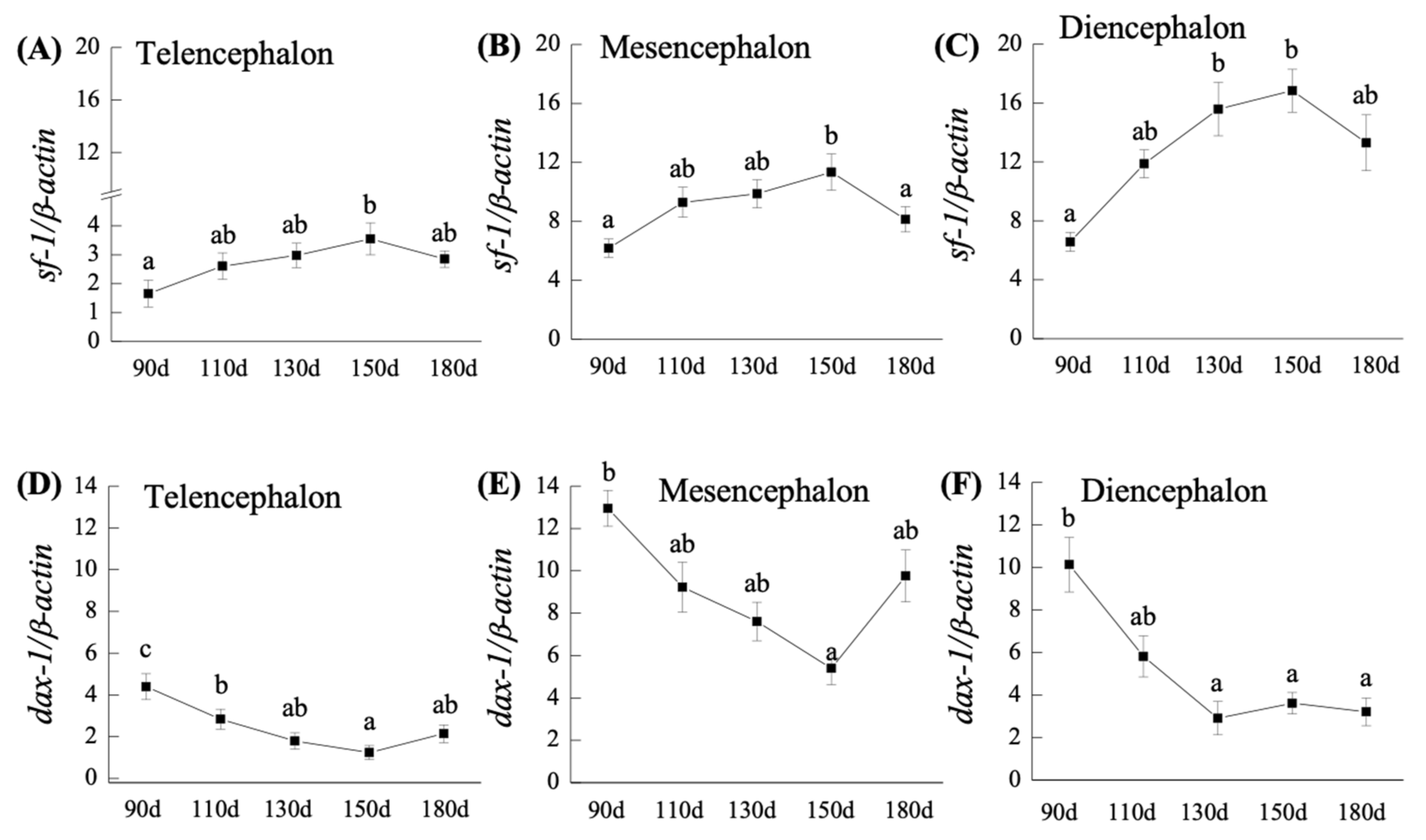
2.2. q-PCR Analysis of sf-1 and dax-1 Transcripts in the Brain of Orange-Spotted Grouper during Developmental Ages

2.2.1. sf-1 mRNA Expression

The neuroanatomy section of the early brain is shown in Figure 1. In the telencephalon, mesencephalon, and diencephalon, sf-1 mRNA levels gradually increased from 110- to 150-dah, although transcript levels were found to be low at 180-dah (Figure 2A–C). In the telencephalon, mesencephalon, and diencephalon, sf-1 mRNA expression was significantly (p < 0.05) higher from 110- to 150-dah compared to 90-dah. Furthermore, when compared to the telencephalon and mesencephalon, the level of mRNA in the diencephalon was higher (Figure 2A–C). The levels of sf-1 mRNA in the diencephalon and mesencephalon at 150-dah were 2.5- and 1.8-fold greater, respectively, than the levels at 90-dah (Figure 2A–C).

2.2.2. dax-1 mRNA Expression

Unlike sf-1 expression, dax-1 mRNA expression was significantly (p < 0.05) higher at 90-dah and subsequently declined from 110- to 150-dah in the telencephalon and mesencephalon, and 110- to 130-dah in the diencephalon (Figure 2D–F). In 90-dah, the amounts of dax-1 transcripts in the telencephalon, mesencephalon, and diencephalon increased significantly (p < 0.05). When compared to the levels in the telencephalon at 90-dah, higher expression of dax-1 in the mesencephalon and diencephalon was 2.2- and 1.6-fold higher, respectively (Figure 2D–F). Furthermore, higher expression of dax-1 mRNA was identified in the mesencephalon at 180-dah, which was 4- and 2.6-fold higher than the levels in the telencephalon and diencephalon at 180-dah, respectively (Figure 2E).

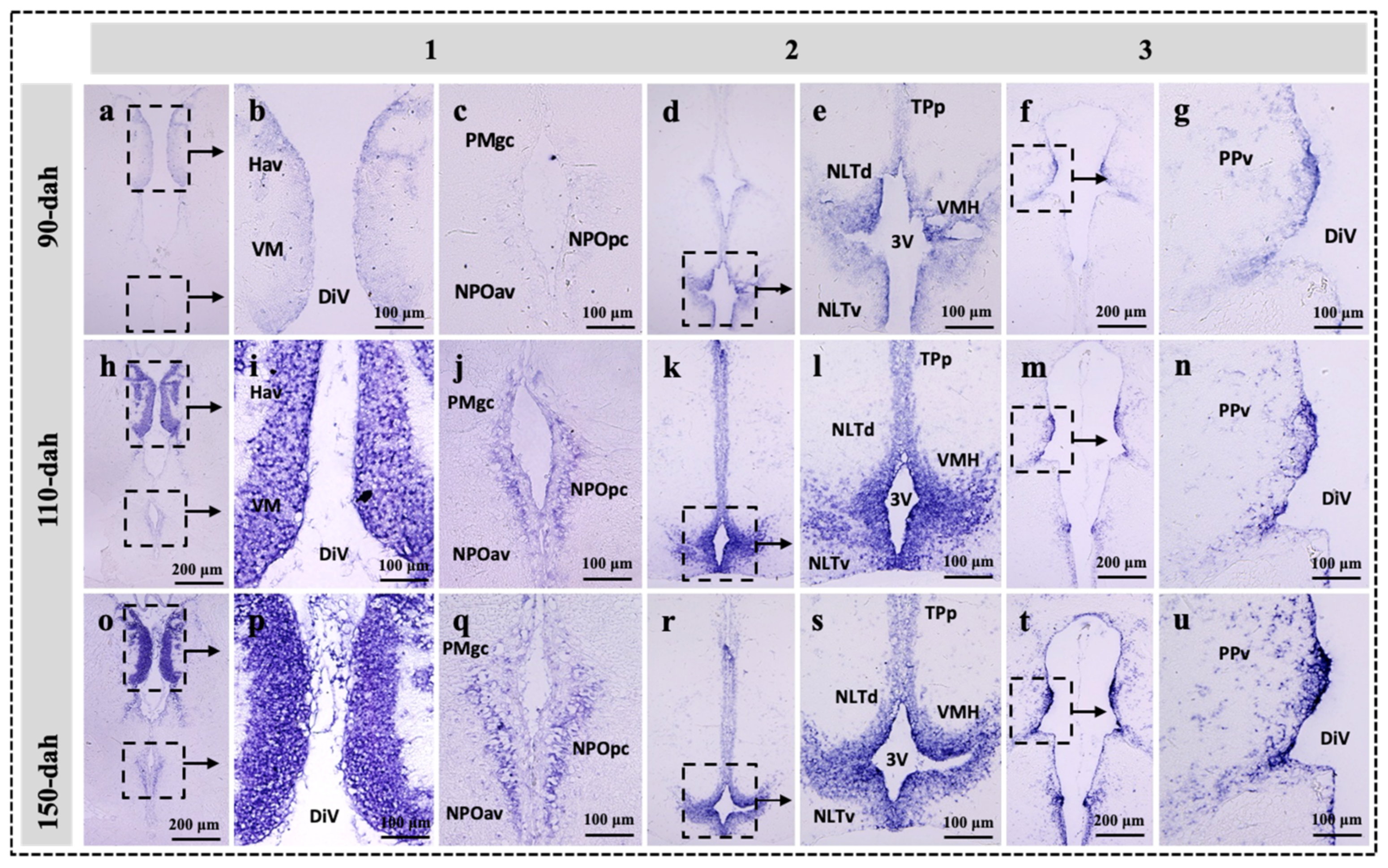
2.3. Localization of sf-1, dax-1 and star Transcripts in the Brain of the Protogynous Orange-Grouper during Different Stages of Developmental

Three hypothalamic brain areas were selected for the in situ hybridization analysis corresponding to the series Sections 1, 2, and 3, the most enriched expression zone for the genes star, sf-1, and dax-1 (Figure 1). The developmental mRNA distribution pattern of star, sf-1, and dax-1 in the hypothalamic areas of the grouper brain was demonstrated at 90-, 110-, and 150-dah (Figure 3, Figure 4 and Figure 5). star, sf-1, and dax-1 transcripts were found in the ventral habenular nucleus, ventromedial thalamic nucleus, diencephalic ventricle, anteroventral part of the parvocellular preoptic nucleus, gigantocellular part of the magnocellular preoptic nucleus, and lateral tuberal nucleus, dorsal and lateral parts of the hypothalamic nucleus (Figure 3a–u and Figure 5a–u). Localization of star (at 110-dah) (Figure 3h–n) and sf-1 (at 110- and 150-dah) (Figure 4h–u) transcriptional signals were primarily found in cells of the ventral zone of the periventricular hypothalamus. However, dax-1 transcripts were higher at 90-dah (Figure 5a–g) compared to 110-dah (Figure 5h–n) and 150-dah (Figure 5o–u) in the ventral zone of the periventricular hypothalamus (Figure 5o–u). Parallel brain sections hybridized with the sense probes revealed no positive mRNA signals (Figure S4).

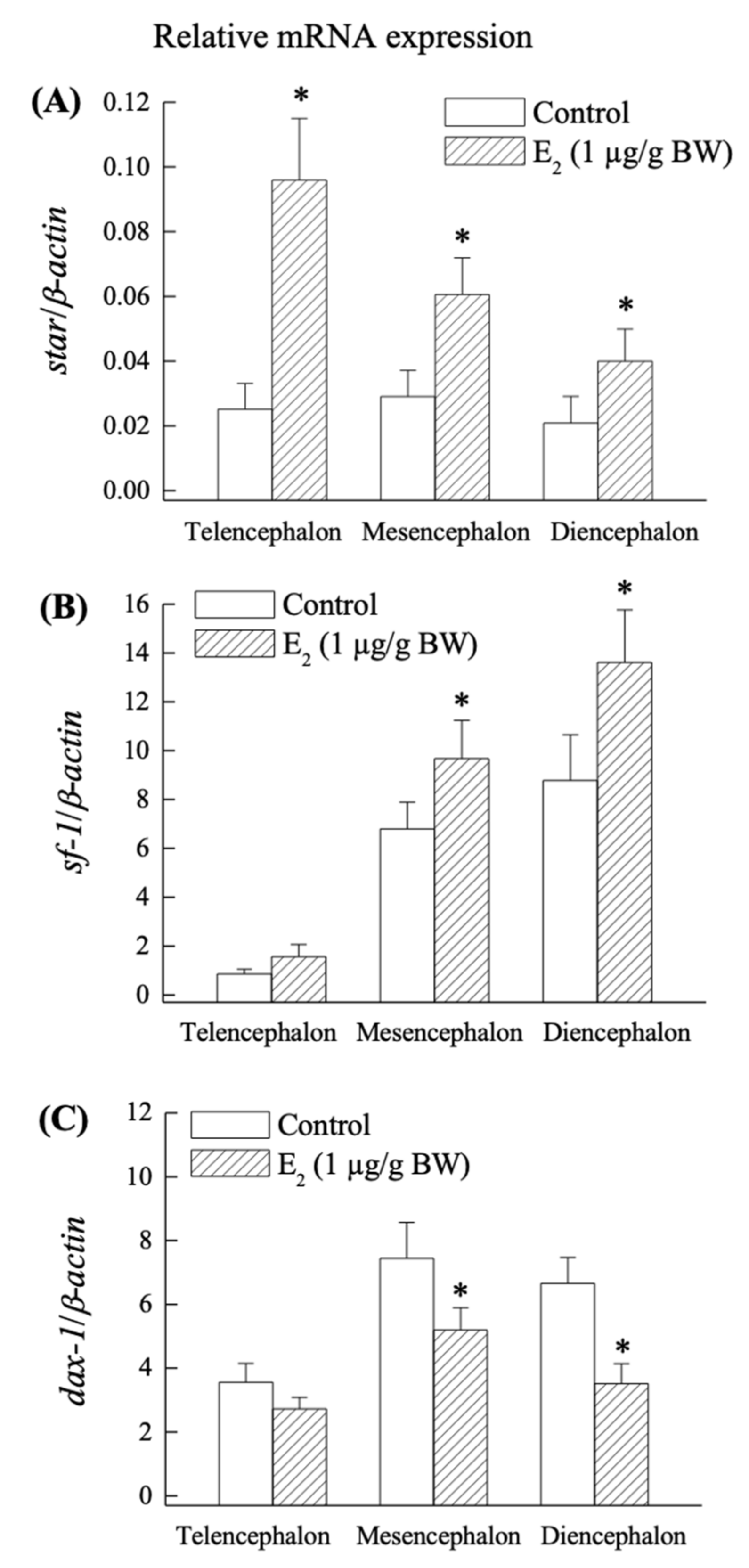
2.4. Effect of E2 on star, sf-1 and dax-1 mRNA Expression in the Grouper Brain In Vivo

Exogenous E2 stimulated star expression in all regions of the brain (Figure 6A), but sf-1 expression was significantly (p < 0.05) higher in the mesencephalon and diencephalon but not in the telencephalon, according to the q-PCR results (Figure 6B). In contrast to the telencephalon, E2 significantly (p < 0.05) reduced dax-1 expression in the mesencephalon and diencephalon (Figure 6C).

3. Discussion

This all-female brain of juvenile orange-spotted grouper (a protogynous species) provides a unique model to study early brain development. We have shown the developmental expression and localization of steroidogenic factors such as sf-1 and dax-1 transcripts in the early female brain of the orange-spotted grouper at different developmental ages in comparison to another key steroidogenic enzyme gene (star). The peak expression of sf-1 and star genes, as well as those cellular levels and effects of exogenous E2 in the early brain, were the most notable findings of this study. In comparison to sf-1 expression, dax-1 expression was lower in the early development of the grouper brain. These intriguing findings suggest that sf-1 may have a regulatory role in steroidogenesis in the early brain of orange-spotted grouper.
sf-1 is an obvious candidate gene to explain the developmental expression pattern in neural estrogen synthesis in the early female brain, as it regulates the transcription of various target genes involved in steroidogenesis. The expression profiles of star, sf-1, and dax-1 are correlated to the time at which most of the other key steroidogenic genes (cyp11a1, hsd3b1, cyp17a1, and cyp19a1b) and estrogen receptors (erα, erβ1, erβ2, and gpr30) exhibited maximal expression in the early female grouper brain [4,6]. In contrast to this scenario, lower expression of dax-1 was found in the early brain during gonadal sex differentiation when sf-1 and other key steroidogenic enzymes (including Cyp19a1b activity) were high, indicating that the grouper brain has a functional peak of neurosteroidogenis that may be regulated by sf-1 and dax-1 (Figure 7). As a result, at 110-dah, decreased expression of dax-1 and increased expression of sf-1, as well as significantly increased expression of other key steroidogenic genes, may suggest that these orphan nuclear receptor genes play an important role in brain steroidogenesis during the early developmental period.
Until now, available studies in the brain have shown that the expression and localization of orphan nuclear receptors in response to the development of the hypothalamus [19,51], rather than the regulation of brain steroid synthesis during early brain development, as compared to the gonad and interrenal organ [45,46,52]. Our in situ hybridization study revealed that sf-1 and star transcripts were highly expressed at 110- to 150-dah and 110-dah, respectively; while dax-1 exhibited higher transcripts at 90-dah and a decreasing trend towards 150-dah in cells of the ventral habenular nucleus, ventromedial thalamic nucleus, and ventromedial hypothalamus. In monosex rainbow trout, Oncorhynchus mykiss, sf-1 mRNA expression was localized in the mediobasal hypothalamus [7]. The expression of SF-1 in the hippocampus of marmosets and rats has been examined using immunohistochemistry [11]. Furthermore, in the marmoset and rat, all StAR-positive cells were also SF-1 positive, demonstrating that the expression of SF-1 and StAR has a functional relationship [11]. SF-1 and DAX-1 immunoreactive cells were discovered in the mouse VMH and pituitary throughout development [23]. Knockout mice deficient in SF-1 have profound defects in the VMH, strongly suggesting the presence of SF-1 target genes at this site [51]. Therefore, our findings revealed that the hypothalamus is a key site for the expression and function of sf-1 and dax-1. As a result, the intensive mRNA localization of star, sf-1, and dax-1 in the hypothalamic areas of the grouper brain agrees with these previous investigations, indicating that the functional linkage of these genes is plausible for steroidogenesis and brain development.
Furthermore, the current study highlights the in vivo effect of exogenous E2 on the regulation of star, sf-1, and dax-1 in the grouper early brain. E2 is the most biologically prominent and active member of the estrogen family of steroids, and it has a wide range of effects on the developing brain [53]. According to a recent study, the peak expression of numerous neurosteroidogenic genes in the brain of black porgy at the time of gonadal differentiation is mediated by both E2-independent and E2-dependent pathways [54]. E2 exposure was found to up-regulate star mRNA in the brain and gonad of the self-fertilizing fish, Kryptolebias marmoratus [55]. On the other hand, the xenoestrogen, diethylstilbestrol exposure decreased in the total amount of sf-1 mRNA in the unborn rat testis but not in fetal ovaries [56]. Holt (1989) [57] proposed the possibility of E2, stimulating the transport of cholesterol to the inner mitochondrial membrane. This is consistent with our findings, which reveal that E2 exposure increases the expression of sf-1 and star mRNA.
Our in vivo results showed that the exogenous E2 up-regulated star (telencephalon, mesencephalon, and diencephalon) and sf-1 (mesencephalon and diencephalon) transcripts compared to their control counterparts. This upregulation could be due to E2 binding with nuclear estrogen receptors (ers) and ers/sf-1 express in the same cells, or an estrogen-responsive element (ERE) located on the promoter region of the star and sf-1 in the brain. These proposals are unclear, and further research is needed to determine whether the star, sf-1, and ers co-express in the same cells. E2 exposure, on the other hand, reduced dax-1 mRNA levels in the mesencephalon and diencephalon. The overexpression of sf-1 mRNA and the downregulation of dax-1 mRNA are strongly related to the results of brain steroidogenesis and E2 exposure, owing to the opposing functions of dax-1 and sf-1 in steroidogenesis. As a result, for E2 upregulation of steroidogenesis in the brain, sf-1 is more significant than dax-1. We previously reported that during early development, E2, aromatase enzyme activity, cyp19a1b, and estrogen receptors (er, er1, and er2) are all at their greatest levels in the brains of orange-spotted grouper and black porgy [2,3,4,5,6]. Thus, our findings imply that during the early developmental period, there is a local production of neurosteroids, particularly neuroestrogens, that is tightly regulated in the preparation for the peak of functional brain growth (neurogenesis).
Therefore, this current and our previous studies [4,5,6] show the temporal expression and localization of sf-1 and dax-1 transcripts in the early brain along with neurosteroidogenic related genes (star, cyp11a1, cyp17a1, and cyp19a1b) in the mono-female sex-differentiated fish (Figure 7). sf-1 transcripts were increased in the early brain as other key steroidogenic genes and E2 [2,3,4,58]; however, dax-1 had an antagonistic expression pattern during these time periods, suggesting that dax-1 may play a negative role in the early brain development of female grouper. Indeed, the star, sf-1, and dax-1 mRNAs were found in abundance in the cells of the ventromedial thalamic nucleus and the mediobasal hypothalamus, and these three genes were mostly found in the same regions, implying that their functions in steroidogenesis during brain development are closely related. Furthermore, exogenous E2 also increased the expression of star, cyp19a1b, and sf-1 in the brain, indicating that these genes are primarily involved in E2 synthesis. This finding led to a better understanding of the transcriptional regulation of sf-1 and dax-1 as molecular players for steroidogenesis during development (Figure 7). Therefore, the balance of sf-1 and dax-1 expression is critical in the regulation of brain development and sex differentiation.

4. Materials & Methods

4.1. Experimental Fish

Orange-spotted female grouper (E. coioides) were used in the present study. The experimental fish were acclimated to the pond condition of the University culture station in a seawater and natural light system (salinity of 33 ppt; temperatures ranged from 20–24 °C). The fish were fed ad libitum with a commercial food (Fwu Sou Feed Co., Taichung, Taiwan). All procedures and investigations were approved by the National Taiwan Ocean University Institutional Animal Care and Use Committee and were performed in accordance with standard guiding principles.
Three batches of orange-spotted groupers were used for the experiment. For the gene expression analysis, fish (n = 8 per age group) were obtained at 90-, 110-, 130-, 150-, and 180-dah. To investigate the localization of genes in hypothalamic brain regions, fish with 90-, 110-, and 150-dah were obtained. To investigate the effects of exogenous E2 on gene expression, 110-dah fish were obtained. The fish were anesthetized with 0.05% ethylene glycol monophenyl ether before being decapitated. The brain was dissected in the same manner as previously described for the orange-spotted grouper and black porgy [4,6,54,59,60]. Brain samples, including telencephalon (including the olfactory bulb, telencephalon, and part of the preoptic area; located between the anterior commissure and the optic chiasm), mesencephalon, and diencephalon (including mesencephalon, thalamus, epithalamus, subthalamus, and hypothalamus), were immediately frozen in liquid nitrogen and stored at −80 °C until RNA isolation. Another batch of whole-brain tissues was fixed in a 4% paraformaldehyde solution for histological/cellular analysis.

4.1.1. Experiment 1: Gene Expression Profiles during Gonadal Sex Differentiation

In order to examine the gene expression profiles in the brain during different developmental ages and gonadal sex differentiation, brain samples (n = 8) were collected from 90-dah (body weight, BW = 11.6 ± 0.5 g, body length, BL = 9.1 ± 0.2 cm), 110-dah (BW = 24.5 ± 0.8 g, BL = 11.7 ± 0.1 cm), 130-dah (BW = 28.1 ± 1.1 g, BL = 12.3 ± 0.2 cm), 150-dah fish (BW = 31.2 ± 1.2 g, BL = 12.9 ± 0.2 cm), and 180-dah (BW = 48.1 ± 2.8 g, BL = 15.3 ± 0.3 cm). The mRNA expression of the genes was measured using q-PCR.

4.1.2. Experiment 2: Localization of sf-1, dax-1 and Star Gene Expression in the Hypothalamus of Orange-Spotted Grouper

We used in situ hybridization in distinct hypothalamic brain regions to determine the anatomical localization of cells that express the star, sf-1, and dax-1 genes (see detailed methods below).

4.1.3. Experiment 3: In Vivo Effects of E2 on the Expression of mRNAs Encoding sf-1, dax-1, and Star in the Brain of Grouper

In order to further investigate the effects of E2 on the mRNAs expression of sf-1, dax-1 and star, 110-dah female grouper for each group (BW = 32.3 ± 1.8 g, BL = 13.5 ± 0.25 cm) were divided into two groups, with n = 8 fish in each of the following experimental groups: control (vehicle alone, coconut oil; Sigma, St. Louis, MO, USA) and E2 treatment (1 µg/g BW; Sigma). On day 1 and 5, the fish received an intramuscular injection (two injections in total). Fish telencephalon, mesencephalon, and diencephalon were collected 24 h (day 6) after the 2nd injection (day 5) and stored at −80 °C until further use. Q-PCR was used to examine changes in the mRNA expression of the corresponding genes in the brains of control and E2-treated groups.

4.2. RNA Extraction, First-Strand cDNA and Molecular Cloning of Genes in the Grouper Brain

Total RNA was extracted from the telencephalon, mesencephalon, and diencephalon (90- to 180-dah) using the TRIzol reagent method (Gibco BRL; Grand Island, NY, USA) according to the manufacturer’s instructions. The quality and concentrations of RNA were assessed by spectrophotometry and checked by running an aliquot (1 μg) on a 1.8% agarose-formaldehyde gel. Single-stranded cDNA was constructed from total RNA using Invitrogen reagents (Invitrogen, Carlsbad, CA, USA), oligo (DT) 12–18 primers and Superscript II reverse transcriptase (Gibco BRL) in a 20 µL reaction volume with an incubation at 42 °C for 60 min, 37 °C for 15 min, and 70 °C for 15 min. The resulting cDNAs were used as a template for subsequent PCR amplification. The PCR reaction was performed in a final volume of 25 μL reaction containing 2.5 μL of 10× reaction buffer (200 mM Tris–HCl, pH 8.4, 500 mM KCl), 1μL of 10 mM dNTP, 1 μL of 2 mM MgCl2, 0.5 μL each of 10 μM forward and reverse primer, respectively (Mission Biotech Co., Ltd., Taipei, Taiwan), 0.2 μL superscript enzyme (Invitrogen, Carlsbad, CA, USA), and 1 μL cDNA. The reaction conditions for degenerate PCR were as follows: 94 °C for 5 min, 35 cycles of 94 °C for 30 s, 50 °C for 30 s, 72 °C for 30 s and 72 °C for 7 min. Each PCR product was electrophoresed on 1.5% agarose gel and the fragment showing the predicted molecular weight was then excised using Gel-MTM Gel Extraction system kit (Viogene, Bio 101, La Jolla, CA, USA). Extracted cDNAs were ligated into the pGEM-T Easy vector (Promega, Madison, WI, USA) and transformed into Escherichia coli competent cells following the manufacturer’s instruction. The Plasmid containing the insert was sequenced and compared with the NCBI database using BLAST. The genes star (GenBank accession no. GU929702), sf-1 (JQ320496), and dax-1 (GU929703) were cloned from the grouper brain, which was deduced from the conserved regions of other teleosts (Figures S1–S3).

4.3. Preparation of Brain Tissue for In Situ Hybridization

For in situ hybridization experiments, 90-, 110-, and 150-old female grouper brains were collected to define star, sf-1, and dax-1 mRNA signals in the brain. For in situ hybridization, we chose three hypothalamic areas with the most enriched expression zones for the genes star, sf-1, and dax-1 (Figure 1). Groupers were anesthetized by immersion in ethylene glycol monophenyl ether (0.05%). Grouper brains were fixed overnight in a 4% paraformaldehyde in PBS (phosphate-buffered saline) at 4 °C. Upon fixation, the skull was removed and the brain was carefully dissected out and stored overnight in fresh fixative at 4 °C. The brain was then rinsed several times in PBS. Later, the fixed brain was dehydrated in a series of alcohols, embedded in paraffin, and then cross-sections (5–6 µm) were collected on APTES-treated slides (3-aminopropyltriethoxysilane, Sigma) diluted in acetone.

4.4. Synthesis of Star, sf-1 and dax-1 RNA Probes

The PCR product of the target gene from the plasmid DNA containing respective inserts of the genes in the pGEM-T Easy vector was generated with 50 U DNA polymerase (New England Biolabs, Ipswich, MA, USA) for DNA amplification with in situ hybridization primers (Table 1). The PCR products were purified using a kit (PCR-Advanced Clean-Up Kit, Viogene). In vitro transcription was carried out using this purified DNA as a template. Digoxigenin-labeled sense and anti-sense riboprobes were synthesized from a fragment of 751 bp, 570 bp, and 443 bp encoding orange-spotted grouper brain star, sf-1, and dax-1, respectively, by using T7 and SP6 polymerase (Promega). The DNA (1µg) templates were incubated for 3 h at 37 °C in the thermocycler PCR machine (Applied Biosystems, Foster City, CA, USA) in a solution containing transcription buffer (5X), 0.1 M dithiothreitol (DTT), DIG-rNTP mix (10X) (Roche, Penzberg, Germany), an RNase inhibitor (40 U/µL) (Promega) and T7 or SP6 RNA polymerase (20 U/µL) and adjusted to 20 µL final volume with sterile DEPC H2O. The extra template was removed by digesting it with 4 µL of DNase I (10 U/µL) at 37 °C for 30 min. After incubation, the probes were precipitated with 100 µL of LiCl (7.5 M) and 900 µL of isopropanal at −20 °C overnight. The pellets were collected by centrifuging the solution at 12,000 rpm at 4 °C for 30 min and the pellets were re-suspended with 2 µL RNase inhibitor and 98 µL sterile DEPC H2O. The probe quality was checked by spectrophotometry at 260 nm.

4.5. In Situ Hybridization

The in situ hybridization analysis was performed as described in our previous study [6,59,60,61,62,63]. The adjacent sections were rehydrated from xylene to a series of alcohol (100%, 90%, 80%, 70%, and 50% EtOH) and washed in PBS several times. Then, they were treated with proteinase K at 10 µg/mL (Roche, Mannheim, Germany) at room temperature for 8 or 10 min and rinsed in PBST (Tween-20 in phosphate-buffered saline) and PBS several times. The brain sections were then prehybridized in the prehybridization buffer for 1 h at 68 °C and hybridized with the sense or anti-sense digoxigenin-labeled RNA probes (0.5 µg/mL for sf-1 and dax-1, and 1 µg/mL for star) in 200 µL hybridization buffer containing 50% formamide, 5X SSC, 500 µg/mL tRNA, 50 µg/mL heparin, and 0.1% Tween-20 (pH 6.0) at 68 °C overnight. After overnight incubation, the sections were allowed to room temperature for 10 min, rinsed with buffer containing 25% formamide, 1X SCC, and 0.1% Tween-20 at 60 °C for 15 min two times, and blocked with 2% blocking reagent (Roche), 2% normal goat serum and PBT at room temperature for 1 h. Alkaline phosphatase-conjugated sheep anti-digoxigenin antibody (Roche) (dilution 1:1500 at 2% blocking reagent) was applied in the section and then incubated overnight at 4 °C. After washing, NTMT (100 mM NaCl, 100 mM Tris-HCl, pH 9.5, 50 mM Mg2Cl, 0.1% Tween-20) was used to ensure the pH is high. Finally, the NBT/BCIP (nitro blue tetrazolium/5-bromo-4-chloro-indolyl-phosphate) staining (Sigma) was used to detect the mRNA signal of grouper steroidogenic genes in the brain.

4.6. Q-PCR Analysis

Transcripts of grouper star [4], sf-1, and dax-1 were quantified by quantitative real-time PCR analysis in the telencephalon, mesencephalon, and diencephalon at different developmental ages (90- to 180-dah). Q-PCR primers were designed for the analysis of star, sf-1, dax-1, and β-actin with the assistance of Primer Express Software (Applied Biosystems) (Table 1). The expression levels of brain β-actin were not significantly different among development ages (Figure S5). β-actin was used as a housekeeping gene in the q-PCR analysis to calibrate the expression levels of brain genes. Gene quantification of the standards, samples, and control was simultaneously conducted in a q-PCR machine (iQTM Multicolor Real-Time PCR Detection System; Bio-Rad Co., Hercules, CA, USA) with iQTM SYBR green (Bio-Rad Co.), which is a dsDNA minor-groove binding dye, forward and reverse primers and water for the reaction mix in a MicroAmp® 96-well reaction plate. The respective standard curve of the log (transcript concentrations) vs. CT (the calculated fractional cycle number at which the PCR-fluorescence product is detectable above a threshold) was −0.995.

4.7. Statistical Analysis

All values (n = 8) are represented as the mean ± SEM (standard error of mean), and the data were analyzed by one-way ANOVA followed by an S–N–K (Student–Newman–Keuls) multiple comparison test to compare the differences (p < 0.05) in the developmental expression of the genes in different parts of the brain at different ages. Student t-test was also conducted to compare the significant differences (p < 0.05) between control and E2-injected fish.

5. Conclusions

In conclusion, the current study found developmental changes in the mRNA expression and localization of orphan nuclear receptors such as sf-1 and dax-1 in relation to neurosteroidogenic enzyme gene in the early brain of the orange-spotted grouper. It has been suggested that sf-1 and dax-1 play an important role with key enzymes in steroidogenesis in the early brain of the orange-spotted grouper. Furthermore, our findings revealed that E2 increased star and sf-1 mRNA expression in the telencephalon and diencephalon, which is critical for early brain development and steroidogenesis (Figure 7). Overall, this research found that the orange-spotted grouper brain has all the necessary mechanisms for neurosteroidogenesis and, as a result, local E2 synthesis, which may regulate early brain development during gonadal sex differentiation.

Supplementary Materials

The following supporting information can be downloaded at: https://0-www-mdpi-com.brum.beds.ac.uk/article/10.3390/ijms23052614/s1.

Author Contributions

Conceived ideas and designed experiments, G.N.; A.A. and C.-F.C.; performed animal experiment, collected samples, cloning, histology, RNA probe, in situ hybridization, and q-PCR data analysis, G.N. and A.A.; provided intellectual input for the paper, G.N., A.A., Y.A.A., R.T.M. and C.-F.C.; wrote manuscript, G.N. and C.-F.C. All authors have read and agreed to the published version of the manuscript.

Funding

This work was supported through the Annual Funding track by the Deanship of Scientific Research, Vice Presidency for Graduate Studies and Scientific Research, King Faisal University, Saudi Arabia [Project No. AN000222].

Institutional Review Board Statement

All procedures and investigations were approved by the College of Life Science of the National Taiwan Ocean University Institutional Animal Care and Use Committee and performed in accordance with standard guiding principles.

Informed Consent Statement

Not applicable.

Data Availability Statement

Data are contained within this article. Raw data are available on request from the corresponding authors.

Conflicts of Interest

The authors declare no conflict of interest.

References

  1. Plassart-Schiess, E.; Baulieu, E.E. Neurosteroids: Recent findings. Brain Res. Brain Res. Rev. 2001, 37, 133–140. [Google Scholar] [CrossRef]
  2. Tomy, S.; Wu, G.C.; Huang, H.R.; Dufour, S.; Chang, C.F. Developmental expression of key steroidogenic enzymes in the brain of protandrous black porgy fish Acanthopagrus Schlegeli. J. Neuroendocrinol. 2007, 19, 643–655. [Google Scholar] [CrossRef] [PubMed]
  3. Tomy, S.; Wu, G.C.; Huang, H.R.; Chang, C.F. Age-dependent differential expression of genes involved in steroid signalling pathway in the brain of protandrous black porgy Acanthopagrus Schlegeli. Dev. Neurobiol. 2009, 69, 299–313. [Google Scholar] [CrossRef] [PubMed]
  4. Nagarajan, G.; Tsai, Y.J.; Chen, C.Y.; Chang, C.F. Developmental expression of genes involved in neural estrogen biosynthesis and signaling in the brain of the orange-spotted grouper Epinephelus coioides during gonadal sex differentiation. J. Steroid Biochem. Mol. Biol. 2011, 127, 155–166. [Google Scholar] [CrossRef] [PubMed]
  5. Nagarajan, G.; Aruna, A.; Chang, C.F. Increase in estrogen signaling in the early brain of orange-spotted grouper Epinephelus coioides: A mini-review. Fish Physiol. Biochem. 2012, 39, 95–101. [Google Scholar] [CrossRef] [PubMed]
  6. Nagarajan, G.; Aruna, A.; Chang, C.F. Neurosteroidogenic enzymes and their regulation in the early brain of the protogynous grouper Epinephelus coioides during gonadal sex differentiation. Gen. Comp. Endocrinol. 2013, 181, 271–287. [Google Scholar] [CrossRef] [PubMed]
  7. Vizziano-Cantonnet, D.; Anglade, I.; Pellegrini, E.; Gueguen, M.M.; Fostier, A.; Guiguen, Y.; Kah, O. Sexual dimorphism in the brain aromatase expression and activity, and in the central expression of other steroidogenic enzymes during the period of sex differentiation in monosex rainbow trout populations. Gen. Comp. Endocrinol. 2011, 170, 346–355. [Google Scholar] [CrossRef]
  8. Diotel, N.; Do Rego, J.L.; Anglade, I.; Vaillant, C.; Pellegrini, E.; Gueguen, M.M.; Mironov, S.; Vaudry, H.; Kah, O. Activity and expression of steroidogenic enzymes in the brain of adult zebrafish. Eur. J. Neurosci. 2011, 34, 45–56. [Google Scholar] [CrossRef]
  9. Stocco, D.M. The role of StAR protein in steroidogenesis: Challenges for the future. J. Endocrinol. 2000, 164, 247–253. [Google Scholar] [CrossRef]
  10. Bauer, M.P.; Bridgham, J.T.; Langenau, D.M.; Johnson, A.L.; Goetz, F.W. Conservation of steroidogenic acute regulatory (StAR) protein structure and expression in vertebrates. Mol. Cell. Endocrinol. 2000, 168, 119–125. [Google Scholar] [CrossRef]
  11. Wehrenberg, U.; Prange-Kiel, J.; Rune, G.M. Steroidogenic factor-1 expression in marmoset and rat hippocampus: Co-localization with StAR and aromatase. J. Neurochem. 2001, 76, 1879–1886. [Google Scholar] [CrossRef] [PubMed]
  12. Kusakabe, M.; Todo, T.; McQuillan, H.J.; Goetz, F.W.; Young, G. Characterization and expression of steroidogenic acute regulatory protein and MLN64 cDNAs in trout. Endocrinology 2002, 143, 2062–2070. [Google Scholar] [CrossRef]
  13. Arukwe, A. Modulation of brain steroidogenesis by affecting transcriptional changes of steroidogenic acute regulatory (StAR) protein and cholesterol side chain cleavage (P450scc) in juvenile Atlantic salmon (Salmo salar) is a novel aspect of nonylphenol toxicity. Environ. Sci. Technol. 2005, 39, 9791–9798. [Google Scholar] [CrossRef] [PubMed]
  14. Nunez, B.S.; Piermarini, P.M.; Evans, A.N.; Applebaum, S.L. Cloning and characterization of cDNAs encoding steroidogenic acute regulatory protein from freshwater stingrays (Potamotrygon spp.). J. Mol. Endocrinol. 2005, 35, 557–569. [Google Scholar] [CrossRef] [Green Version]
  15. Kocerha, J.; Prucha, M.S.; Kroll, K.J.; Steinhilber, D.; Denslow, N. Regulation of steroidogenic acute regulatory protein transcription in largemouth bass by orphan nuclear receptor signaling pathways. Endocrinology 2010, 151, 341–349. [Google Scholar] [CrossRef] [PubMed] [Green Version]
  16. Lavorgna, G.; Ueda, H.; Clos, J.; Wu, C. FTZ-F1, a steroid hormone receptor-like protein implicated in the activation of fushi tarazu. Science 1991, 252, 848–851. [Google Scholar] [CrossRef]
  17. Wong, M.; Ramayya, M.S.; Chrousos, G.P.; Driggers, P.H.; Parker, K.L. Cloning and sequence analysis of the human gene encoding steroidogenic factor 1. J. Mol. Endocrinol. 1996, 11, 117–126. [Google Scholar] [CrossRef]
  18. Ingraham, H.A.; Lala, D.S.; Ikeda, Y.; Luo, X.; Shen, W.H.; Nachtigal, M.W.; Abbud, R.; Nilson, J.H.; Parker, K.L. The nuclear receptor SF-1 acts multiple levels of the reproductive axis. Genes Dev. 1994, 8, 2302–2312. [Google Scholar] [CrossRef] [PubMed] [Green Version]
  19. Parker, K.L.; Rice, D.A.; Lala, D.S.; Ikeda, Y.; Luo, X.; Wong, M.; Bakke, M.; Zhao, L.; Frigeri, C.; Hanley, N.A.; et al. Steroidogenic factor 1: An essential mediator of endocrine development. Recent Prog. Horm. Res. 2002, 57, 19–36. [Google Scholar] [CrossRef] [Green Version]
  20. Smith, C.A.; Andrews, J.E.; Sinclair, A.H. Gonadal sex differentiation in chicken embryos: Expression of estrogen receptor and aromatase genes. J. Steroid Biochem. Mol. Biol. 1997, 60, 295–302. [Google Scholar] [CrossRef]
  21. Western, P.S.; Harry, J.L.; Marshall Graves, J.A.; Sinclair, A.H. Temperature dependent sex determination in the American alligator: Expression of SF1, WT1 and DAX1 during gonadogenesis. Gene 2000, 241, 223–232. [Google Scholar] [CrossRef]
  22. Hanley, N.A.; Rainey, W.E.; Wilson, D.I.; Ball, S.G.; Parker, K.L. Expression profiles of SF-1, DAX1, and CYP17 in the human fetal adrenal gland: Potential interactions in gene regulation. Mol. Endocrinol. 2001, 15, 57–68. [Google Scholar] [CrossRef] [PubMed]
  23. Ikeda, Y.; Takeda, Y.; Shikayama, T.; Mukai, T.; Hisano, S.; Morohashi, K.I. Comparative localization of Dax-1 and Ad4BP/SF-1 during development of the hypothalamic-pituitary-gonadal axis suggests their closely related and distinct functions. Dev. Dyn. 2001, 220, 363–376. [Google Scholar] [CrossRef] [PubMed]
  24. Hatano, O.; Takayama, K.; Imai, T.; Waterman, M.R.; Takakusu, A.; Omura, T.; Morohashi, K. Sex-dependent expression of a transcription factor, Ad4BP, regulating steroidogenic P-450 genes in the gonads during prenatal and postnatal rat development. Development 1994, 120, 2787–2797. [Google Scholar] [CrossRef] [PubMed]
  25. Wu, G.C.; Tomy, S.; Chang, C.F. The expression of nr0b1 and nr5a4 during gonad development and sex change in protandrous black porgy fish, Acanthopagrus Schlegeli. Biol. Reprod. 2008, 78, 200–210. [Google Scholar] [CrossRef]
  26. Yan, T.; Lu, H.; Sun, C.; Peng, Y.; Meng, F.; Gan, R.; Cui, X.; Wu, C.; Zhang, S.; Yang, Y.; et al. Nr5a homologues in the ricefield eel Monopterus albus: Alternative splicing, tissue-specific expression, and differential roles on the activation of cyp19a1a promoter in vitro. Gen. Comp. Endocrinol. 2021, 312, 113871. [Google Scholar] [CrossRef]
  27. Hofsten, A.H.; Modig, C.; Larsson, A.; Karlsson, J.; Olsson, P.E. Determination of the expression pattern of the dual promoter of zebrafish fushi tarazu factor-1a following microinjections into zebrafish one cell stage embryos. Gen. Comp. Endocrinol. 2005, 142, 222–226. [Google Scholar] [CrossRef]
  28. Ikeda, Y.; Shen, W.H.; Ingraham, H.A.; Parker, K.L. Developmental expression of mouse steroidogenic factor 1, an essential regulator of the steroid hydroxylases. Mol. Endocrinol. 1994, 8, 654–662. [Google Scholar]
  29. Sugawara, T.; Holt, J.A.; Kiriakidou, M.; Strauss, J.F., 3rd. Steroidogenic factor 1-dependent promoter activity of the human steroidogenic acute regulatory protein (StAR) gene. Biochemistry 1996, 35, 9052–9059. [Google Scholar] [CrossRef]
  30. Hu, M.C.; Hsu, N.C.; Pai, C.I.; Wang, C.K.; Chung, B. Functions of the upstream and proximal steroidogenic factor 1 (SF-1)-binding sites in the CYP11A1 promoter in basal transcription and hormonal response. Mol. Endocrinol. 2001, 15, 812–818. [Google Scholar] [CrossRef]
  31. Leers-Sucheta, S.; Morohashi, K.; Mason, J.I.; Melner, M.H. Synergistic activation of the human type II 3beta-hydroxysteroid dehydrogenase/Δ5-Δ4 isomerase promoter by the transcription factor steroidogenic factor-1/adrenal 4-binding protein and phorbol ester. J. Biol. Chem. 1997, 272, 7960–7967. [Google Scholar] [CrossRef] [PubMed] [Green Version]
  32. Lynch, J.P.; Lala, D.S.; Peluso, J.J.; Luo, W.; Parker, K.L.; White, B.A. Steroidogenic factor 1, an orphan nuclear receptor, regulates the expression of the rat aromatase gene in gonadal tissues. Mol. Endocrinol. 1993, 7, 776–786. [Google Scholar] [PubMed] [Green Version]
  33. Watanabe, M.; Tanaka, M.; Kobayashi, D.; Yoshiura, Y.; Oba, Y.; Nagahama, Y. Medaka (Oryzias latipes) FTZ-F1 potentially regulates the transcription of P450 aromatase in ovarian follicles: cDNA cloning and functional characterization. Mol. Cell. Endocrinol. 1999, 149, 221–228. [Google Scholar] [CrossRef]
  34. Yoshiura, Y.; Senthilkumaran, B.; Watanabe, M.; Oba, Y.; Kobayashi, T.; Nagahama, Y. Synergistic expression of Ad4BP/SF-1 and cytochrome P-450 aromatase (ovarian type) in the ovary of Nile tilapia, Oreochromis niloticus, during vitellogenesis suggests transcriptional interaction. Biol. Reprod. 2003, 68, 1545–1553. [Google Scholar] [CrossRef]
  35. Sridevi, P.; Chaitanya, R.K.; Dutta-Gupta, A.; Senthilkumaran, B. FTZ-F1 and FOXL2 up-regulate catfish brain aromatase gene transcription by specific binding to the promoter motifs. Biochim. Biophys. Acta 2012, 1819, 57–66. [Google Scholar] [CrossRef]
  36. Nakamoto, M.; Wang, D.S.; Suzuki, A.; Matsuda, M.; Nagahama, Y.; Shibata, N. Dax1 suppresses P450arom expression in medaka ovarian follicles. Mol. Reprod. Dev. 2007, 74, 1239–1246. [Google Scholar] [CrossRef]
  37. Lalli, E.; Bardoni, B.; Zazopoulos, E.; Wurtz, J.M.; Strom, T.M.; Moras, D.; Sas-sone-Corsi, P. A transcriptional silencing domain in DAX-1 whose mutation causes adrenal hypoplasia congenita. Mol. Endocrinol. 1997, 11, 1950–1960. [Google Scholar] [CrossRef]
  38. Zhang, H.; Thomsen, J.S.; Johansson, L.; Gustafsson, J.A.; Treuter, E. DAX-1 functions as an LXXLL-containing corepressor for activated estrogen receptors. J. Biol. Chem. 2000, 275, 39855–39859. [Google Scholar] [CrossRef] [Green Version]
  39. Hanley, N.A.; Stallings, N.; Schimmer, B.P. Steroidogenic factor 1: An essential mediator of endocrine development. Recent Prog. Horm. Res. 2002, 57, 19–36. [Google Scholar]
  40. Holter, E.; Kotaja, N.; Makela, S.; Strauss, L.; Kietz, S.; Janne, O.A.; Gustafsson, J.A.; Palvimo, J.J.; Treuter, E. Inhibitions of androgen receptor (AR) function by the reproductive orphan nuclear receptor DAX-1. Mol. Endocrinol. 2002, 16, 515–528. [Google Scholar] [CrossRef]
  41. Smith, C.A.; Clifford, V.; Western, P.S.; Wilcox, S.A.; BellK, S.; Sinclair, A.H. Cloning and expression of a DAX1 homologue in the chicken embryo. J. Mol. Endocrinol. 2000, 24, 23–32. [Google Scholar] [CrossRef] [PubMed] [Green Version]
  42. Pilon, N.; Behdjani, R.; Daneau, I.; Lussier, J.G.; Silversides, D.W. Porcine steroidogenic factor-1 gene (pSF-1) expression and analysis of embryonic pig gonads during sexual differentiation. Endocrinology 1998, 139, 3803–3812. [Google Scholar] [CrossRef] [PubMed]
  43. Wang, Z.J.; Jeffs, B.; Ito, M.; Achermann, J.C.; Yu, R.N.; Hales, D.B.; Jameson, J.L. Aromatase (Cyp19) expression is up-regulated by targeted disruption of Dax1. Proc. Natl. Acad. Sci. USA 2001, 98, 7988–7993. [Google Scholar] [CrossRef] [PubMed] [Green Version]
  44. Kawabe, K.; Shikayama, T.; Tsuboi, H.; Oka, S.; Oba, K.; Yanase, T.; Nawata, H.; Morohashi, K. Dax-1 as one of the target genes of Ad4BP/SF-1. Mol. Endocrinol. 1999, 13, 1267–1284. [Google Scholar] [CrossRef]
  45. Zhao, Y.; Yang, Z.; Phelan, J.K.; Wheeler, D.A.; Lin, S.; McCabe, E.R.B. Zebrafish dax1 is required for development of the interrenal organ, the adrenal cortex equivalent. Mol. Endocrinol. 2006, 20, 2630–2640. [Google Scholar] [CrossRef] [Green Version]
  46. Chen, S.; Zhang, H.; Wang, F.; Zhang, W.; Peng, G. nr0b1 (DAX1) mutation in zebrafish causes female-to-male sex reversal through abnormal gonadal proliferation and differentiation. Mol. Cell. Endocrinol. 2016, 433, 105–116. [Google Scholar] [CrossRef]
  47. Yajima, Y.; Vestergaard, M.C.; Takagi, M. Damage in brain development by morpholino knockdown of zebrafish dax1. J. Biosci. Bioeng. 2012, 113, 683–688. [Google Scholar] [CrossRef]
  48. Zhang, W.; Li, Y.; Chen, S.; Zhang, C.; Chen, L.; Peng, G. nr0b1 (DAX1) loss of function in zebrafish causes hypothalamic defects via abnormal progenitor proliferation and differentiation. J. Genet. Genom. 2021, 21, S1673–S8527. [Google Scholar] [CrossRef]
  49. Tsai, Y.J.; Lee, M.F.; Chen, C.Y.; Chang, C.F. Development of gonadal tissue and aromatase function in the protogynous orange-spotted grouper, Epinephelus coioides. Zool. Stud. 2011, 50, 693–704. [Google Scholar]
  50. Zhang, W.; Zhang, Y.; Zhang, L.; Zhao, H.; Li, X.; Huang, H.; Lin, H. The mRNA expression of P450 aromatase, gonadotropin beta-subunits and FTZ-F1 in the orange-spotted grouper (Epinephelus Coioides) during 17α-methyltestosterone-induced precocious sex change. Mol. Reprod. Dev. 2007, 74, 665–673. [Google Scholar] [CrossRef]
  51. Parker, K.L.; Schimmer, B.P. Steroidogenic factor 1: A key determinant of endocrine development and function. Endocr. Rev. 1997, 18, 361–377. [Google Scholar] [CrossRef] [PubMed]
  52. Raju, M.; Balasubramanian, S. Expression analysis and localization of wt1, ad4bp/sf-1 and gata4 in the testis of catfish, Clarias batrachus: Impact of wt1-esiRNA silencing. Mol. Cell. Endocrinol. 2016, 431, 164–176. [Google Scholar]
  53. McCarthy, M.M. Estradiol and the developing brain. Physiol. Rev. 2008, 88, 91–124. [Google Scholar] [CrossRef] [PubMed] [Green Version]
  54. Lin, C.J.; Wu, G.C.; Dufour, S.; Chang, C.F. Activation of the brain-pituitary-gonadotropic axis in the black porgy Acanthopagrus schlegelii during gonadal differentiation and testis development and effect of estradiol treatment. Gen. Comp. Endocrinol. 2019, 281, 17–29. [Google Scholar] [CrossRef] [PubMed]
  55. Rhee, J.S.; Kim, B.M.; Lee, C.J.; Yoon, Y.D.; Lee, Y.M.; Lee, J.S. Bisphenol A modulates expression of sex differentiation genes in the self-fertilizing fish, Kryptolebias marmoratus. Aquat. Toxicol. 2011, 104, 218–229. [Google Scholar] [CrossRef]
  56. Majdic, G.; Sharpe, R.M.; Saunders, P.T. Maternal oestrogen/xenoestrogen exposure alters expression of steroidogenic factor-1 (SF-1/Ad4BP) in the fetal rat testis. Mol. Cell. Endocrinol. 1997, 127, 91–98. [Google Scholar] [CrossRef]
  57. Holt, J.A. Regulation of progesterone production in the rabbit corpus luteum. Biol. Reprod. 1989, 40, 201–208. [Google Scholar] [CrossRef] [Green Version]
  58. Lin, C.J.; Fan-Chiang, Y.C.; Dufour, S.; Chang, C.F. Activation of brain steroidogenesis and neurogenesis during the gonadal differentiation in protandrous black porgy, Acanthopagrus schlegelii. Dev. Neurobiol. 2016, 76, 121–136. [Google Scholar] [CrossRef]
  59. Nagarajan, G.; Aruna, A.; Chang, C.F. Neuropeptide Arginine Vasotocin Positively Affects Neurosteroidogenesis in the Early Brain of Grouper, Epinephelus coioides. J. Neuroendocrinol. 2015, 9, 718–736. [Google Scholar] [CrossRef]
  60. Aruna, A.; Lin, C.J.; Nagarajan, G.; Chang, C.F. Neurohypophysial Hormones Associated with Osmotic Challenges in the Brain and Pituitary of the Euryhaline Black Porgy, Acanthopagrus schlegelii. Cells 2021, 10, 3086. [Google Scholar] [CrossRef]
  61. Aruna, A.; Nagarajan, G.; Chang, C.F. Involvement of corticotrophin-releasing hormone and corticosteroid receptors in the brain-pituitary-gill of tilapia during the course of seawater acclimation. J. Neuroendocrinol. 2012, 24, 818–830. [Google Scholar] [CrossRef] [PubMed]
  62. Aruna, A.; Nagarajan, G.; Chang, C.F. The acute salinity changes activate the dual pathways of endocrine responses in the brain and pituitary of tilapia. Gen. Comp. Endocrinol. 2015, 211, 154–164. [Google Scholar] [CrossRef] [PubMed]
  63. Aruna, A.; Wang, T.P.; Cao, J.C.; Lan, D.S.; Nagarajan, G.; Chang, C.F. Differential expression of hypothalamic and gill-crh system with osmotic stress in the euryhaline black porgy, Acanthopagrus schlegelii. Front. Physiol. 2021, 12, 768122. [Google Scholar] [CrossRef] [PubMed]
Figure 1. Neuroanatomy section (A) the whole brain of the orange-spotted grouper showing different areas of the olfactory bulb, telencephalon, diencephalon, mesencephalon, rhombencephalon and (B) showing three chosen areas of the brain for the in situ hybridization correspond to the series Sections 1, 2, and 3 where most enriched expression zone for the genes star, sf-1, and dax-1. Dashed lines showed the dissection position of transverse section. Hematoxylin and eosin were used to stain brain sections. aNH, anterior neurohypophysis; Chor, commissura horizontalis; Cgus, commissure of the secondary gustatory nuclei; CPN, central pretectal nucleus; Ctec, commissural tecti; Cpost, posterior commis-sure; DAO, dorsal accessory optic nucleus; Dc, central zone of dorsal telencephalic area; DiV, diencephalic ventricle; Dm, medial zone of dorsal telencephalic area; Dp, posterior zone of dorsal telencephalic area; FR, fasiculus retroflexus; Hav, ventral habenular nucleus; IMRF, intermediate reticular hypothalamus; IRF, inferior reticular formation; LCa, lobus caudalis cerebella; LFB, lateral forbrain bundle; LX, vagal lobe; MFN, medial funicular nucleus; NDL, diffuse nucleus of the inferior lobe; NLV, nucleus lateralis nucleus; NPGl, lateral preglomerular nucleus; NRL, nucleus of the lateral recess; NRP, nucleus of the posterior recess; NLTd, lateral tuberal nucleus, dorsal part; NLTv, lateral tuberal nucleus, ventral part; PI, pars intermedia; PMgc, gigantocellular part of the magnocellular preoptic nucleus; pNH, posterior neurohypophysis; PPa, parvocellular preoptic nucleus, anterior part; PPd, proximal pars distalis; PPv, periventricular pretectal nucleus, ventral part; PSp, parvocellularsuperficialpretectalnucleus; PTN, posteriortuberalnu-cleus; SO, secondary octaval population; SRF, superior reticular formation; SV, saccus vasculosus; TeV, tectal ventricle; TeO, tectum opticum; TL, torus longitudinalis; TPp, periventricular nucleus of the posterior tuberculum; Val, lateral division of the valvula cerebella; VM, ventromedial thalamic nucleus; VMH, ventromedial hypothalamus.
Ijms 23 02614 g001
Figure 2. Relative transcripts of sf-1 (AC) and dax-1 (DF) in the telencephalon, mesencephalon, and diencephalon from 90- to 180-dah (days after hatching) during gonadal sex differentiation. Gene expression levels were measured by q-PCR and expressed as mean normalized expression (SEM) of n = 8 fish for each group. Different letters denote statistically significant differences (p < 0.05) in gene expression at various developmental ages.
Ijms 23 02614 g002
Figure 3. Developmental localization of star transcripts in the hypothalamic regions of the grouper brain at 90-, 110-, and 150-dah (day after hatching). Bright field visualization of paraffin brain sections showing the detailed distribution of star transcripts in three consecutive area of the hypothalamus at 90- (ag), 110- (hn), and 150-dah (ou) corresponding to the series 1, 2 and 3 on Figure 1. Note that intense star hybridization signals were detected at 110-dah in the ventromeadial thalamic region (h,i), ventromeadial hypothalamic region (k,l), and periventricular pretectal regions (m,n) compared to the same region at 90 (ag) or 150-dah (ou). The abbreviations are referred to Figure 1 legends.
Ijms 23 02614 g003
Figure 4. Developmental localization of sf-1 transcripts in the hypothalamic regions of the grouper brain at 90-, 110-, and 150-dah (day after hatching). Bright field visualization of paraffin brain sections showing the detailed distribution of sf-1 transcripts in three consecutive area of the hypothalamus at 90- (ag), 110- (hn), and 150-dah (ou) corresponding to the series 1, 2 and 3 on Figure 1. Note that intense sf-1 hybridization signals were detected in 110-dah in the ventromeadial thalamic region (h,i), ventromeadial hypothalamic region (k,l), and periventricular pretectal regions (m,n) compared to the same region at 90 (ag) or 150-dah (ou). The abbreviations are referred to Figure 1 legends.
Ijms 23 02614 g004
Figure 5. Developmental localization of dax-1 transcripts in the hypothalamic regions of the grouper brain at 90-, 110-, and 150-dah (day after hatching). Bright field visualization of paraffin brain sections showing the detailed distribution of dax-1 transcripts in three consecutive area of the hypothalamus at 90- (ag), 110- (hn), and 150-dah (ou) corresponding to the series 1, 2 and 3 on Figure 1. Note that intense dax-1 hybridization signals were detected in 90-dah in the ventromeadial thalamic region (ac), ventromeadial hypothalamic region (d,e), and periventricular pretectal regions (f,g) compared to the same region at 150-dah (ou). The abbreviations are referred to Figure 1 legends.
Ijms 23 02614 g005
Figure 6. In vivo effects of exogenous estradiol (E2, 1 µg/g BW, fish n = 8 (110-dah, day after hatching) for each group were given intramuscular injections on day 1 and 5). Brain tissues were collected on day 6 for the analysis of mRNA expression of star (A), sf-1 (B), and dax-1 (C), as measured by q-PCR analysis in the telencephalon, mesencephalon, and diencephalon. The results are expressed as the mean with standard deviation (SEM). The different letters indicate significant differences (p < 0.05) according to a one-way ANOVA followed by a S–N–K (Student–Newman–Keuls) multiple comparison test. An asterisk (*) represents significant (p < 0.05) differences between the control and E2-injected groups.
Ijms 23 02614 g006
Figure 7. Schematic picture highlighting the proposed hypothesis according to the current and previous studies [4,5,6]. (A) The expression profiles of steroidogenic enzyme genes (star, cyp17a1, and cyp19a1b) [4,5,6], aromatase activity [5], sf-1 and dax-1. (B) Brain estradiol (E2) can act locally in the regulation of steroidogenic enzyme genes [6] and sf-1; and collectively E2-sf-1-steroidogenic enzymes system may stimulate steroidogenesis, neurogenic activity, and early brain development in the juvenile orange-spotted grouper. (+) stimulation and (−) inhibition.
Ijms 23 02614 g007
Table 1. Primer sequence used for q-PCR and in situ hybridization analysis for the genes star, sf-1, and dax-1 in the orange-spotted grouper. The nucleotide (nt) length of RNA probes for in situ hybridization: star, 751 nt; sf-1, 570 nt; dax-1, 443 nt.
GeneOrientationNucleotide Sequence Usage Accession Number
starForward5′-TCAGCACAGGGCTTCATCACTAT-3′q-PCR
Reverse5′-TGCAAAAATGCCTGAGCAAAG-3′q-PCR
Sense5′-CGGCATATGAGGAACATGACAG-3′In-situGU929702
Anti Sense5′-CCACCTGCGTCTGAGAGAG-3′In-situ
sf-1Forward5′-TCGGCTTCGTGAAAAACGT-3′q-PCR
Reverse5′-CTCCTGGGCATGGCTCAA-3′q-PCR
Sense5′-GCTGCAAGGGGTTCTTCAAG-3′In-situJQ320496
Anti Sense5′-CGGGGTACTCTGACTTGATG-3′In-situ
dax-1Forward5′-TGAAGAAGTGCTGGAGTGTAGATATCA-3′q-PCR
Reverse5′-CCCTCCACATCTGGGTTGAA-3′q-PCR
Sense5′-CTCGGCGGTGCTGGTGAAGAC-3′In-situGU929703
Anti Sense5′-GCCGGACGTGCTCGTTGAGAG-3′In-situ
β-actinForward5′-AGGGAGAAGATGACCCAGATCA-3′q-PCRAY510710
Reverse5′-GGGACAGCACAGCCTGCAT-3′q-PCR
Publisher’s Note: MDPI stays neutral with regard to jurisdictional claims in published maps and institutional affiliations.

