Review

From Cancer Therapy to Winemaking: The Molecular Structure and Applications of β-Glucans and β-1, 3-Glucanases

by
Catarina Caseiro
1,2,
Joana Nunes Ribeiro Dias
1,2,
Carlos Mendes Godinho de Andrade Fontes
3 and
Pedro Bule
1,2,*
1
CIISA—Centre for Interdisciplinary Research in Animal Health, Faculty of Veterinary Medicine, University of Lisbon, 1300-477 Lisbon, Portugal
2
Associate Laboratory for Animal and Veterinary Sciences (AL4AnimalS), 1300-477 Lisbon, Portugal
3
NZYTech Genes & Enzymes, 1649-038 Lisbon, Portugal
*
Author to whom correspondence should be addressed.
Int. J. Mol. Sci. 2022, 23(6), 3156; https://0-doi-org.brum.beds.ac.uk/10.3390/ijms23063156
Submission received: 28 February 2022 / Revised: 11 March 2022 / Accepted: 11 March 2022 / Published: 15 March 2022
(This article belongs to the Special Issue Biotechnological Application of Carbohydrate Active Enzymes)

Abstract

:
β-glucans are a diverse group of polysaccharides composed of β-1,3 or β-(1,3-1,4) linked glucose monomers. They are mainly synthesized by fungi, plants, seaweed and bacteria, where they carry out structural, protective and energy storage roles. Because of their unique physicochemical properties, they have important applications in several industrial, biomedical and biotechnological processes. β-glucans are also major bioactive molecules with marked immunomodulatory and metabolic properties. As such, they have been the focus of many studies attesting to their ability to, among other roles, fight cancer, reduce the risk of cardiovascular diseases and control diabetes. The physicochemical and functional profiles of β-glucans are deeply influenced by their molecular structure. This structure governs β-glucan interaction with multiple β-glucan binding proteins, triggering myriad biological responses. It is then imperative to understand the structural properties of β-glucans to fully reveal their biological roles and potential applications. The deconstruction of β-glucans is a result of β-glucanase activity. In addition to being invaluable tools for the study of β-glucans, these enzymes have applications in numerous biotechnological and industrial processes, both alone and in conjunction with their natural substrates. Here, we review potential applications for β-glucans and β-glucanases, and explore how their functionalities are dictated by their structure.

1. Introduction

β-glucans are a large class of natural polysaccharides composed of D-glucose monomers linked through β-glycosidic bonds. Although this definition technically includes β-1,4-glucans (cellulose), the term is mostly reserved for β-1,3 and mixed-linkage β-(1,3–1,4) glucans (MLGs). They are widely distributed in nature and can be found in yeast, fungi, bacteria, seaweeds and cereals, where they fulfil storage, structural and protective roles [1,2,3,4]. The molecular structure of β-glucans can vary depending on their source but, at their core, MLGs are unbranched polysaccharides of both β-1,3 and 1,4 linked glucose units, in varying proportions, whereas β-1,3 glucans consist of linear backbones of β-1,3 linked glucose units, often decorated with β-1,6 linked branches. A large amount of attention has been devoted to the molecular and structural features of β-glucans since they determine the physical properties of these polysaccharides, such as water solubility and rheological behavior, in addition to their affinity towards a variety of β-glucan interacting proteins. As such, most functional aspects of β-glucans are a consequence of backbone length (degree of polymerization, DP), the ratio of 1,3:1,4 bonds or 1,3:1,6 bonds (branching degree), interval of branching and sidechain size [5,6].
In recent decades, the popularity of β-glucans as functional, bioactive and nutraceutical ingredients has steadily increased (Figure 1). Because they can interact with specific receptors present in several micro- and macro-organisms, including humans, they are able to trigger various biological responses, which can be harnessed for therapeutic purposes. The therapeutic effects of these polysaccharides can be either immunomodulatory or metabolic [5,7,8,9]. Metabolic effects, usually observed with MLGs, include modulation of the gut microbiome, improved lipidic and glycemic metabolism, and reduced cholesterol levels. Immunomodulatory effects, which are often associated with β-1,3-glucans, include anti-tumor effects, improved wound healing and alleviation of immune-related conditions. β-glucans have also been shown to be important prebiotics, improving gastrointestinal function by regulating intestinal microbiota [10,11,12,13,14,15].
Given that β-glucan receptors are present in most species, their nutraceutical applications are not restricted to humans. For instance, in animal husbandry they can be administered as a feed additive to boost immunity, reducing the need for antibiotics and improving production performances [16,17,18]. Due to their physicochemical properties, β-glucans also have a wide range of potential applications in other industries (Figure 1). The most popular uses are as a thickening and gelification agent in the food industry, and as ingredients in cosmetic and personal care products due to their soothing, moisturizing and anti-irritant properties [19,20].
β-glucan deconstruction typically results from the action of β-1,3-glucanases, including exo-1,3-β-glucosidases (EC 3.2.1.58), endo-β-1,3-glucanases (EC 3.2.1.39) and endo-1,3(4)-β-glucanases (EC 3.2.1.6). Although the first two are mostly β-1,3-glucan specific, having limited action on MLGs, endo-1,3(4)-β-glucosidases are active on both MLGs and β-1,3-glucans. This results from them being able to hydrolyze both β-1,3 and β-1,4 bonds, as long as the glucose residue whose reducing group is involved in the linkage to be hydrolyzed is itself substituted at C-3 [21,22]. They are different from lichenases or MLGases (EC 3.2.1.73), which are endo-(1,3:1,4)-β-glucosidases that can only hydrolyze β-1,4 bonds that are adjacent to a β-1,3 bond, making them MLG specific [23].
β-1,3-glucanases are widely distributed in nature and are mostly expressed by bacteria, fungi, plants and some invertebrates, where they are involved in various physiological processes [24,25]. Those include energy production, cellular remodeling and growth, defense against fungal pathogens, seed germination, digestion and reproduction [26,27,28,29]. The ability of β-glucanases to hydrolyze β-glucans down to small oligosaccharides, or even glucose, has inspired an array of biotechnological applications (Figure 1). Some popular examples for their use are: in the conversion of lignocellulosic biomass into fermentable sugars for the production of bioethanol; as feed additives to reduce the antinutritional effects of cereal β-glucans; as biocontrol agents against pathogenic fungus in various crops; to control invasive Candida albicans infections; as clarification agents in winemaking; to speed up germination during the malting process; and to avoid the accumulation of barley β-glucans in brewing [30,31,32,33,34].
The substrate specificities of β-glucanases, much like with all hydrolases, are intrinsically related to their tridimensional structure, the shape of their substrate-binding cleft and the constellation of substrate interacting residues. These characteristics will not only dictate substrate preferences but also the final products of digestion. Whether hydrolysis by a particular β-glucanase occurs preferentially on longer or shorter, helical or linear, or highly branched or unbranched polysaccharides, ultimately comes down to a structural compatibility between substrate and catalyst [35,36].
The purpose of this review is to provide an outline of current knowledge about β-glucans and β-glucanases, their biochemical properties, and potential biotechnological and biomedical applications. Other β-glucan interacting proteins, such as carbohydrate-binding modules (CBMs) and Dectin-1, are also briefly reviewed, as they are intrinsically related to the biological functions of β-glucans. Special focus is given to the structural features of these biomolecules and how they modulate the β-glucan:protein interaction, dictating the biological role and potential applications of β-glucans and β-1,3-glucanases.

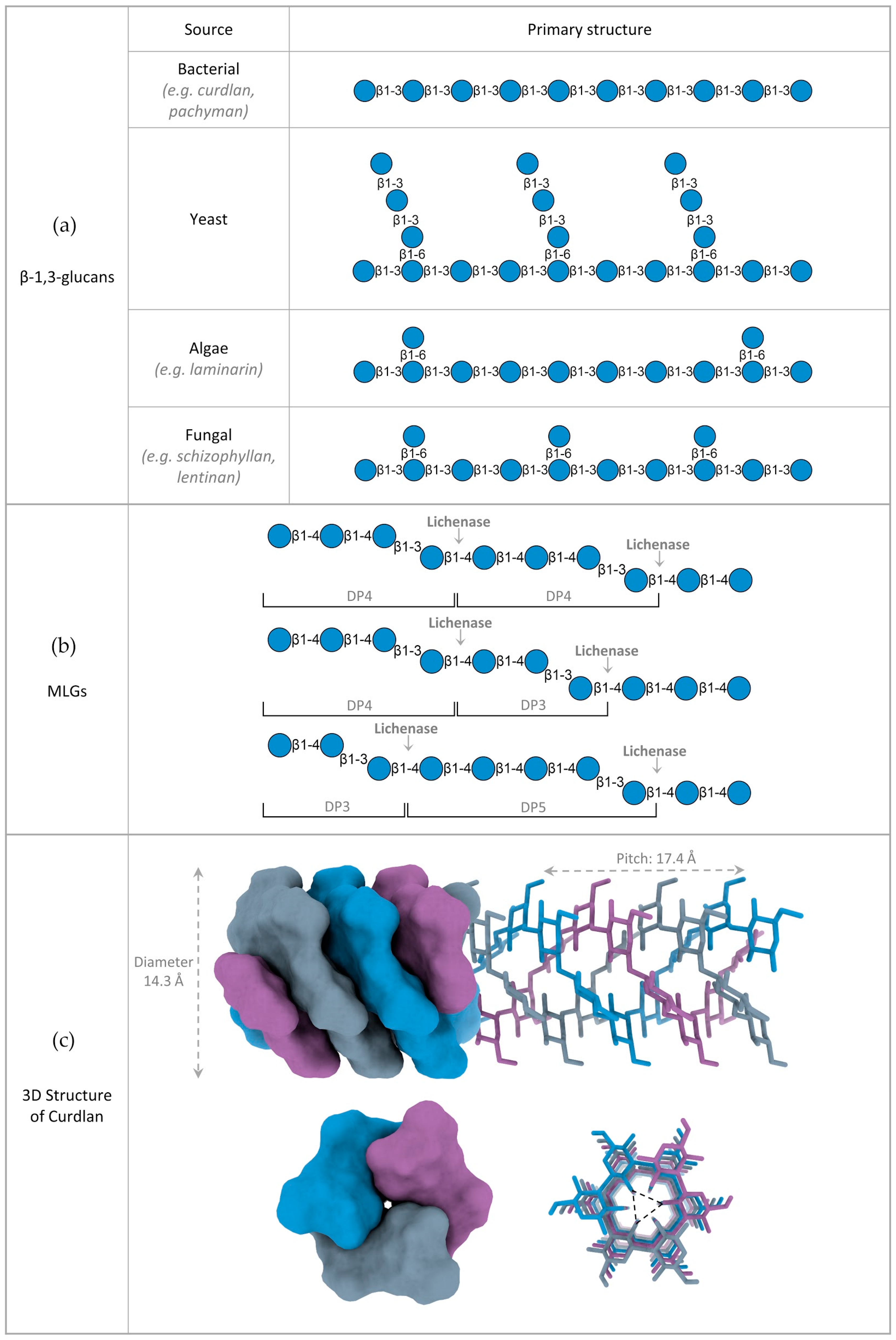
2. Structure of β-Glucans

2.1. Variations in β-Glucan Primary Structure

Depending on the type of linkage present between their glucose monomers, β-glucans are often classified into cereal and non-cereal. Nonetheless, even within each sub-group, the primary structure of β-glucans can vary significantly depending on the source, which translates into distinct physicochemical and functional properties.
Cereal β-glucans is a term often used for mixed-linkage glucans, which are linear and unbranched polymers composed of a mixture of β-1,3 and β-1,4 linked glucose units. The term cereal β-glucan comes from the fact that they were initially thought to be unique to the Poaceae family (grasses). However, evidence has shown that MLGs are also synthesized in other taxa, including plants from the Equisetum genus (horsetail), algae, lichens, fungi and even bacteria [10,37,38,39,40,41]. The ratio between β-1,4 and β-1,3 linkages varies with the species and has a significant impact on MLG’s physicochemical traits. A common tool to evaluate MLG linkage ratio, and its distribution, is the digestion with lichenases (Figure 2b). As mentioned above, lichenases are endo-(1,3:1,4)-β-glucosidases that hydrolyze β-1,4 bonds adjacent to β-1,3 linkages. Depending on the MLG’s composition, lichenase digestion will generate different amounts of end-products with different degrees of polymerization (DP), such as the trisaccharide β-glucosyl-1,4-β-glucosyl-1,3-glucose (G4G3G, DP3) or the tetrasaccharide β-glucosyl-1,4-β-glucosyl-1,4-β-glucosyl-1,3-glucose (G4G4G3G, DP4).
Generally, the most drastic differences occur between different taxonomic groups, likely suggesting different biological roles. For example, enzymatic profiling and linkage analysis of Poaceae MLG reveals a ratio of β-1,4 to β-1,3 linkages ranging from 2.2 to 2.6:1. This translates to a higher proportion of DP3 compared to DP4, with a ratio of 1.5 to 4.5:1, and the occasional occurrence of longer oligosaccharide units [42]. MLGs from horsetails, on the other hand, release mainly DP4 upon lichenase digestion. They are also abundant both on young and older regions of the stem, as opposed to Poaceae MLGs, which are largely absent in mature tissues. This suggests that Equisetum MLGs may have a different function in the cell wall [37,43]. No MLGs have yet been reported in green algae, although structurally similar polysaccharides that include xylose and arabinose have been isolated [44]. On the other hand, red and brown algae, which are more distantly related to plants, both have MLG in their cell walls. This suggests a convergent evolution of the (1,3;1,4)-β-glucan synthase genes. Interestingly, MLG from brown algae is composed exclusively of repeating DP3 units, which creates a highly regular structure with a tendency to self-aggregate [45]. The primary structure of lichen MLGs, often referred to as lichenan, is characterized by a higher DP3:DP4 ratio, although the ratio of β-1,4 and β-1,3 linkages varies significantly between species, ranging from 2.3:1 in Cetraria islandica (Iceland moss), to 0.3:1 in Evernia prunastri (oak moss) [46]. Besides lichens, MLGs with a high percentage of β-1,4 linked glucose have also been reported in non-lichen fungus, such as Aspergillus fumigatus and Neurospora crassa [41,47]. The occurrence of MLG has also been described in both Gram-negative and Gram-positive bacterial species, including Sinorhizobium meliloti and Sarcina ventriculi [38]. Structural profiling revealed that the MLG of S. meliloti consists of repeating units of the disaccharide β-glucosyl-1,3-glucose (DP2) linked by β-1,4 bonds (alternating β-1,4 and β-1,3 linkages), whereas the MLG of S. ventriculi is exclusively composed of repeating DP3 units, similar to that of brown algae [10,38].
The term non-cereal β-glucans is mostly used for β-1,3-glucans, which are polysaccharides with a linear backbone of β-1,3 linked glucoses, often decorated with β-1,6 linked branches. Their overall primary structure can vary significantly, depending on the length of the main chain, the interval of branching and the length of the side chains. These characteristics are mostly dependent on the source of the β-glucans, but other factors such as growth conditions, extraction and purification methods can also have a great impact on β-glucan structure [48,49]. Much like MLGs, the variations in β-1,3-glucan primary structure are more marked between different taxonomic groups. Generally, bacterial β-glucans are unbranched, yeast β-glucans have long β-1,6 linked branches, fungal β-glucans have small β-1,6 linked branches, whereas algal β-glucans are very similar to the fungal ones, but with much smaller DPs (Figure 2a). Curdlan, which was isolated from the bacteria Alcaligenes faecalis, is a linear and essentially unbranched β-1,3-glucan with a DP that can vary between 135 and 455 glucose units [1]. Pachyman, another example of a bacterial β-1,3-glucan, is essentially similar to curdlan, but with a smaller DP (255) and the occasional β-1,6 branch. Both of these are water insoluble. On the other hand, β-glucans extracted from several fungus, such as Lentinus edodes (lentinan), Sclerotium spp. (scleroglucan) and Schizophyllum commune (schizophyllan) also possess a linear β-1,3 backbone, but with β-1,6-linked monoglucose residues approximately every three or five β-1,3-linked glucose residues [50,51,52]. Unlike the bacterial β-glucans, they are mostly water soluble, suggesting that the 1,6-linked monoglucose residues have a role in defining the solubility of β-glucans. The β-glucans present in seaweeds, such as Laminaria digitata, Saccharina longicruris and Durvillaea Antarctica, have a β-1,3 main chain, with a small percentage of β-1,6 glycosidic bonds in the backbone and β-1,6 monoglucose side chain branching [49]. They are smaller than β-glucans from other organisms and do not have a structural function, rather serving as energy storage polysaccharides [49]. Yeast cell wall β-glucans also possess β-1,6 branching, but with a different structure. They have longer branches with additional β-1,3 linked glucoses, sometimes over 50 units long, which can form interchain linkages [49]. As such, the longer the branches, the less water soluble the β-glucans.
Besides branching, the DP of β-1,3-glucans is also a determinant factor in their water solubility, which decreases with size. As such, short branched polysaccharides such as laminarins with a DP of 20–30, are water soluble, whereas unbranched β-1,3-glucans with DPs above 36, such as curdlan, are insoluble due to the cooperative interactions between chains becoming stronger than those between the chains and water molecules [49,53]. Although longer than laminarins, many branched fungal β-1,3-glucans, such as lentinan and schizophyllan, are still water soluble. This solubility depends on the frequency of side branches. However, at high molecular weights (DP > 1000), the branching factor does not seem to compensate for the size factor; hence, large β-glucans from yeast and fungal cell walls, although branched, are water insoluble [54]. In the case of MLGs, water solubility decreases the more β-1,4 linkages are present in the molecule. The most soluble polymers comprise approximately 30% of β-1,3 linkages and 70% of β-1,4 linkages [55].
Ultimately, variations in the chemical structure of the β-glucans will dictate their tertiary structure and consequently affect their biological activities [56].

2.2. 3D Structure of β-Glucans

It has long been established, both by experimental studies and computer-assisted model building, that many long chain β-1,3-glucans form triple-helical structures. Oriented fiber X-ray diffraction [57], solid state 13C-NMR spectroscopy [58], multi-angle laser light scattering [59], fluorescence resonance energy transfer spectroscopy [60] and molecular dynamic simulations [61] suggest a triplex structure composed of parallel strands, stabilized by extensive interstrand and interhelix hydrogen bonds [56]. For example, hydrated curdlan is a triplex of right-handed, six-fold helical chains, meaning that the individual chains of curdlan are constituted by a six-glucose unit per turn. The diameter of the triplex determined from X-ray diffraction experiments was 14.4 Å and the pitch of the helix was estimated to be 17.3 Å (Figure 2c) [57]. Similar structures were also described for the fungal β-glucans schizophyllan and lentinan, albeit with small variations in pitch and diameter [62,63]. In turn, analytical ultracentrifugation and calorimetric analysis showed that short β-1,3-glucans, such as laminarin from Laminaria digitata, whose molecular mass is around 5000 Da, predominantly exist in a single-strand form, with approximately 5% of triple-helical structures [64].
A curdlan triple-helix model, determined by X-ray fiber diffraction, shows interchain hydrogen bonds between the 2-OH groups of each glucose arranged in a triangular pattern. Together with a contribution from hydrophobic forces, these H-bonds are the driving force behind quaternary structure formation (Figure 2c) [49,56].
Optical rotatory dispersion measurements were applied to native and partial degraded curdlan fractions to understand the required chain length for the formation of a triple helix. A DP larger than 200 (molecular mass of 32,000 Da per chain) was found to be necessary to form an ordered structure in dilute alkaline solution (0.1 M). In turn, β-glucans with DPs below 25 are soluble and take a disordered structure in both neutral and alkaline solutions [53].
It has been shown that when β-1,3-glucans are diluted in dimethyl sulfoxide (DMSO) [65], alkaline solutions (pH higher than 12) [63], or submitted to high temperatures (>135 °C) [56,66], the strength of the interchain hydrogen bonds is not sufficient to keep the triplex helix together. As such, under these conditions, β-glucan helixes tend to dissociate into random coils [56].
The capacity of the triple helices to spontaneously renature, when the thermodynamic conditions that favor helix conformation are re-established, has also been examined. For example, alkaline-treated or DMSO-denatured lentinan and schizophyllan adopt a mixture of linear, circular and branched species of triple helix when dialyzed against water [67]. Such conformational variation is not observed in native molecules, which appear as perfect linear triplexes. This may be explained by a simultaneous assembly of the self-stabilizing triple helix structure with biosynthesis, thus suppressing errors in the triplex formation [68]. β-1,3-glucan conformations are closely related with their functional properties and biological activities. Therefore, after the renaturation process, the bioactivities of β-1,3-glucans can be lost or decreased due to structural changes. This means that extraction methods can have a significant impact on β-glucans’ functional profiles [69].

3. β-1,3-Glucanases

β-1,3-glucanases are the main enzymes responsible for β-glucan deconstruction. They are glycoside hydrolases that catalyze the hydrolysis of β-1,3-D-glycosidic bonds and, in some cases, β-1,4-D-glycosidic bonds, in linear or partially branched glucans [70]. They can be divided into endo-glucanases and exo-glucanases, according to the location of the cleaved bonds within the polysaccharide chain. Endo-β-1,3-glucanases cleave β-1-3 glycosidic bonds randomly inside the β-glucan chain, releasing glucan oligosaccharides as main final products, whereas exo-β-1,3-glucanases sequentially release monosaccharides from the non-reducing end of the polysaccharide chain. Exo-β-1,3-glucanases normally reverse the anomeric configuration of the end product during catalysis, whereas endo-β-1,3-glucanase action typically retains the anomeric configuration (via a double-displacement mechanism). As such, some endo-β-1,3-glucanases also have the ability to catalyze the inverse reaction, or transglycosylation reaction, where the β-1,3-oligosaccharides (glycosyl donors) are transferred a glycosyl acceptor, generating new glycosidic bonds [3,71].
β-1,3-glucanases are widely distributed among higher plants, marine invertebrates, fungi, bacteria, archaea and viruses. Mainly due to the several physiological roles played by their natural substrates, the biological functions of β-1,3-glucanases are highly diverse. In bacteria, β-1,3-glucanases are mainly used to degrade polysaccharides into oligo and monosaccharides, which are then used as an energy source [25]. In higher plants, β-glucanases participate in cell differentiation, trafficking of materials through plasmodesmata, in withstanding abiotic stress, in seed development and germination, and are involved in the defense against fungal pathogens by hydrolyzing their cell walls [72]. In fungi, β-1,3-glucanases are engaged in several roles, namely, in the mobilization of β-1,3-glucans when under limited energy conditions, and in yeast cell development and differentiation, and can also act as autolytic enzymes. [70]. In animals, β-1,3-glucanases are found in marine invertebrates, specifically in the Echinodermata, Crustacea and Mollusca phyla. They are mostly present in the digestive tract of marine mollusks and crustaceans, participating in the digestion of algal biomass, and in the eggs of Echinodermata, where they are involved in embryogenesis [25,27]. In viruses, β-glucanases are involved in degrading the host cell wall during viral entry and release [73]. Although mammals do not possess the ability to express β-1,3-glucanases, they still play a determinant role in the regulation of the gastrointestinal microbiome and, consequently, on the maintenance of both gut and general health. In humans, for example, it is widely accepted that the human gut microbiota (HGM) has far-reaching effects on human health and nutrition and is even associated with some diseases, depending on its particular composition. The maintenance of a healthy HGM is dependent on the utilization of otherwise indigestible dietary fiber by individual HGM species, where β-1,3-glucans are included. Bacteroides species, in particular, are predominant autochthonous members of the healthy HGM that can utilize β-glucans as an energy source by expressing several β-1,3-glucanases, allowing them to thrive in a highly competitive ecological niche [21].
β-1,3-glucanases are proteins with a typical molecular mass of around 30 to 50 kDa, but their sizes lay in a wide range of 12 kDa (e.g., Citrus aurantiifolia) up to 230 kDa (e.g., Aspergillus nidulans), with the largest members mostly belonging to eukaryotic species [34,71,74,75,76,77]. Typical optimal pH values for β-1,3-glucanase activity are on the slightly acidic range of 5.0 to 6.5, but can range anywhere between 4.0 (e.g., fungi Lentinula edodes [78]) and 9.0 (e.g., armyworm Spodoptera frugiperda [79]). Fungal β-1,3-glucanases seem to be particularly active on lower pH values (4.0–6.0) [77,78,80]. As with most CAZymes, the optimal temperatures for β-1,3-glucanase activity fall within a wide range, with the lowest temperature values belonging to the β-1,3-glucanases of Aspergillus fumigates (24–40 °C) and the highest to the β-1,3-glucanases of archaea Pyrococcus furiosus (100–105 °C) [74,81]. Overall, the adaptation potential of archaeal enzymes to high temperatures is greater than that of bacteria, which in turn show greater tolerance for hot conditions compared to eukaryotic enzymes. On the other hand, eukaryotic enzymes are the best at adapting to cold conditions [3].

3.1. Classification of β-1,3-Glucanases

β-1,3-glucanases are usually classified either according to their catalytic mechanism or based on primary sequence homology and folding similarities [3]. The Nomenclature Committee of the International Union of Biochemistry and Molecular Biology (IUBMB) and the International Commission on Enzymes or Enzyme Commission (EC) classify enzymes mainly according to the following factors: (i) their catalytic action patterns against specified substrates (endo- or exo-), (ii) the type of linkages they hydrolyze (β-1,3), (iii) the substrate hydrolyzed (β-glucan/β-glucanase) and (iv) the type of reaction catalyzed (hydrolase) [70,82].
According to traditional EC nomenclature, the enzymes which catalyze the hydrolysis of β-1,3-D-glycosidic bonds are subdivided into the following classes: (i) EC 3.2.1.58: exo-β-1,3-glucanases, which cleave D-glucose from the nonreducing end of glucan molecule, (ii) EC 3.2.1.39: endo-β-1,3-glucanases, which require at least two β-1,3 bound glucose residues adjacent to the digested bond and (iii) EC 3.2.1.6: less specific endo-1,3(4)-β-glucanases, which are able to hydrolyze both β-1,3 and β-1,4 bonds, as long as the glucose residue whose reducing group is involved in the linkage to be hydrolyzed is itself substituted at C-3 [3,25].
Many glycoside hydrolases exhibit broad substrate specificity, making it difficult to use a traditional classification. The accumulation of data about the structure of glycoside hydrolases has led to the elaboration of a new system for classification of these enzymes based on the homology of amino acid sequences, their 3D structures and mechanisms of action. The new classification integrated glycoside hydrolases and their homologs into glycoside hydrolase (GH) families (Table 1). At the present time, there are over 170 different families present in the online CAZy database [83]. A glycoside hydrolase is included in a GH family if its primary sequence is homologous to the sequence of at least one already characterized member. Sixty-nine GH families have been grouped into 18 clans due to their common evolutionary origin, 3D structure similarity, conservative arrangement of catalytic residues and similar mechanism of glycosidic bond hydrolysis. The majority of β-1,3-glucanases are distributed between five GH families (5, 16, 17, 55 and 81). Nearly all characterized exo-β-1,3-glucanases (EC 3.2.1.58) are of fungal origin and have been assigned to GH families 5 and 55. In turn, endo-acting laminarinases (EC 3.2.1.39) seem to have a wider structural variety, having members in a wide range of GH families. Nonetheless, all endo-β-1,3-glucanases produced by plants can be assigned to the glycoside hydrolase family 17, whereas most of the bacterial ones, in addition to those from marine mollusks, belong to GH16. Fungal endo-β-1,3-glucanases, on the other hand, are more widely distributed across families but mostly belong to GH81. Classified endo-β-1,3(4)-glucanases (EC 3.2.1.6) are either produced by bacteria or fungi and most have been assigned to GH family 16 (Table 1) [71,76,84,85,86,87].

3.2. Primary Structure of β-1,3-Glucanases

Although some features remain constant, there is a significant degree of diversity between the primary structures of β-1,3-glucanases. As expected, the most conserved parts in β-1,3-glucanase amino acid sequences correspond to the active center and substrate-binding sites. The catalytic center of most endo-β-1,3-glucanases is characterized by the sequence GEIDIXE (with X being a hydrophobic amino acid residue). This sequence contains two important glutamic acid residues that function as the catalytic dyad, with the first assuming the role of catalytic nucleophile and the second that of the proton donor [3,25]. The substrate-binding site is a conserved tryptophan-rich sequence (WPAIWML). These conserved tryptophan residues were shown to promote substrate binding and positioning inside the active center cavity, by means of aromatic stacking interactions with the pyranose rings of glucose residues [71,74].
The importance of other residues for the catalytic activity of endo-β-1,3-glucanases was also shown. Michel et al. (2001) suggest that the conserved aspartic acid of the active site GEIDIXE cooperates with histidine residues in proton trafficking during the deglycosylation step [88]. In endo-β-1,3-glucanases of mollusks, two conserved cysteine residues seem to form disulfide bonds, which are determinant for the thermostability of the molecules [25].

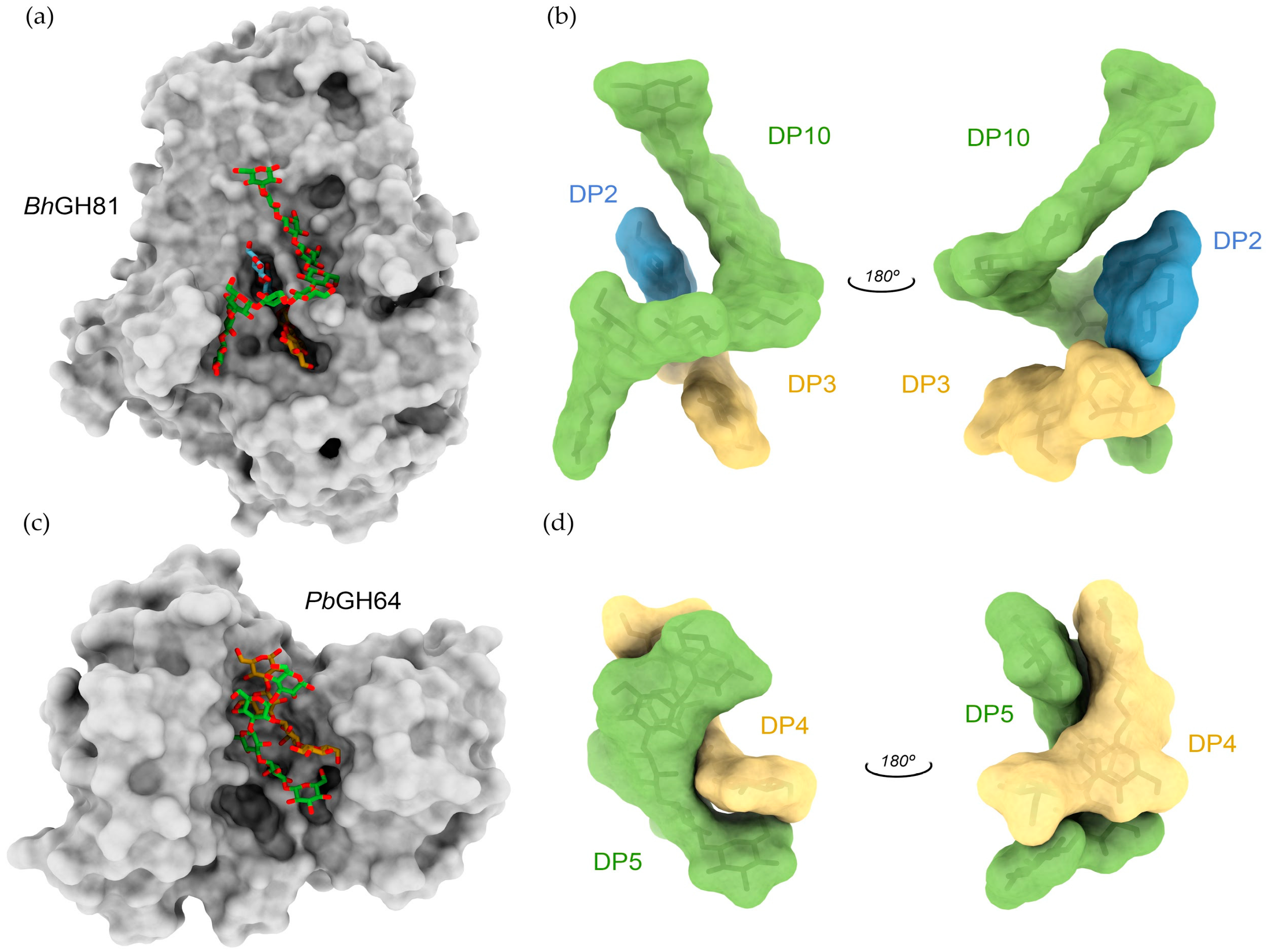
3.3. 3D Structure of β-1,3-Glucanases

Currently, there are 32 experimentally determined 3D structures of β-1,3-glucanases [83]. Although the members of a given GH family can have different primary sequences and substrate specificity, they typically have the same type of fold and the same mechanism of hydrolysis (retaining or inverting). GH16 family members adopt a β-jelly-roll fold, GH5 and 17 families adopt a (β/α)8 (TIM-barrel) fold, GH55s have a β-helix conformation, and GH81s have a (α/α)6 barrel structure [83]. For example, the overall structure of a GH16 endoglucanase from Pyrococcus furiosus (pfLamA) exhibited a classical sandwich-like β-jelly-roll fold formed by two antiparallel sheets facing each other, with seven and eight strands, respectively. This structure is similar to that of BglF, an homologous GH16 endo-β-1,3-glucanase from Nocardiopsis sp. Surprisingly, despite the high degree of homology, BglF shows a preference towards MLG hydrolysis, whereas pfLamA is more active on β-1,3-glucans [89]. The crystal structure of a GH16 family β-1,3-glucanase from Streptomyces sioyaensis showed a sandwich-like β-jelly-roll fold with two disulfide bonds. Cysteine mutants decreased their optimal temperature, which indicates that the disulfide bonds are important to maintain thermostability [90]. In turn, the structures of plant GH17 endo-β-1,3-glucanases reveal a characteristic TIM-barrel fold defined by eight parallel strands in the interior of the protein surrounded by a ring of helices [91]. Despite β-1,3-glucanases of families GH16 and GH17 not having significant sequence similarity and unrelated three-dimensional structures, they show similar substrate specificity and activity, posing as an excellent example of the functional convergent evolution phenomenon [92]. Typically, substrate specificity is dictated by the active center topology, which is defined as a “pocket” or “crater” for exo-type β-1,3-glucanases, and as a “cleft”, “groove” or “tunnel” for endo-type enzymes. In the case of endo-β-1,3-glucanases, the tertiary and quaternary structures of β-1,3-glucans seem to play a particularly important role in substrate recognition. Two great examples of quaternary structure recognition are the GH81 β-1,3-glucanase from Bacillus halodurans (BhGH81) and the GH64 β-1,3-glucanase from Paenibacillus barengoltzii (PbGH64) [35,36]. BhGH81 is an inverting endo-β-1,3-glucanase whose catalytic module adopts an (α/α)6 barrel structure. X-ray data from BhGH81 crystals soaked with laminarin revealed three intertwined β-glucan chains bound to the catalytic site, resembling the triple-helix structure proposed for β-glucans (Figure 3a,b). The authors propose that the ability of BhGH81 to recognize and extensively interact with the large quaternary structure of β-glucans favors a processive mechanism, in which the enzyme is not required to completely release the substrate after bond hydrolysis, but rather slides along the triple-helical structure and continues its catalytic action [36]. Much like BhGH81, PbGH64 is also an inverting endo-β-1,3-glucanase, but with a different tridimensional fold. Nonetheless, the clamp-like binding groove formed between a barrel domain and a mixed α/β domain possesses sufficient area to also be able to bind triple-helical β-1,3-glucan. Co-crystallization of PbGH64 with laminarihexaose revealed two intertwined oligosaccharides in the catalytic groove, which were used to fit the 3D structure of curdlan, confirming PbGH64’s ability to recognize its quaternary structure (Figure 3c,d) [35].

4. Other β-Glucan Binding Proteins

Most biological roles and applications of β-glucans are heavily dependent on their interaction with a wide variety of proteins. These β-glucan binding proteins range from substrate attachment-mediating non-catalytic modules present in β-1,3-glucanases, to cell surface receptors that modulate immune responses towards certain pathogens. They are widely distributed in nature and most seem to share a common feature: ligand configuration is crucial for recognition.
Below we briefly review some of the main β-glucan interacting proteins found in nature, while focusing on their biological role and the mechanisms supporting ligand recognition.

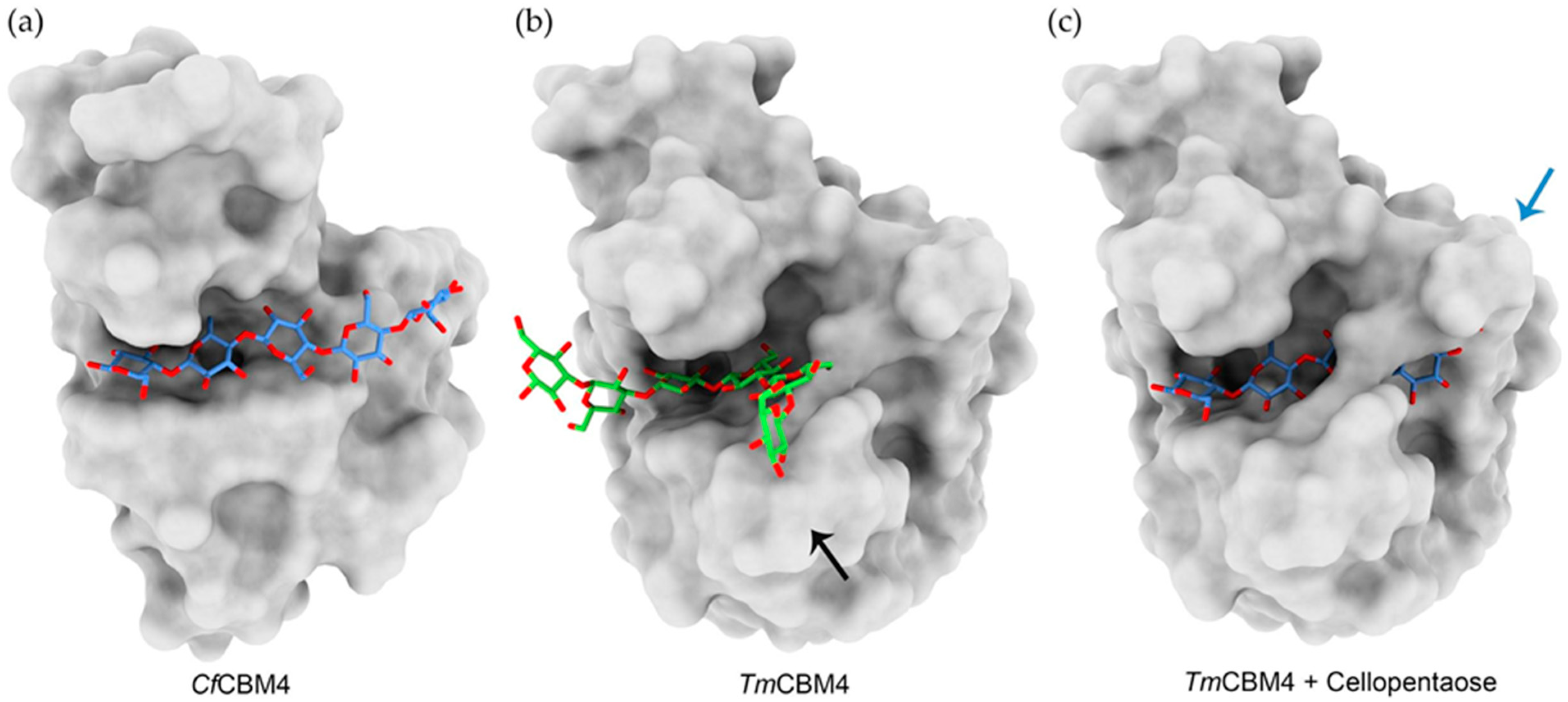
4.1. Carbohydrate Binding Modules
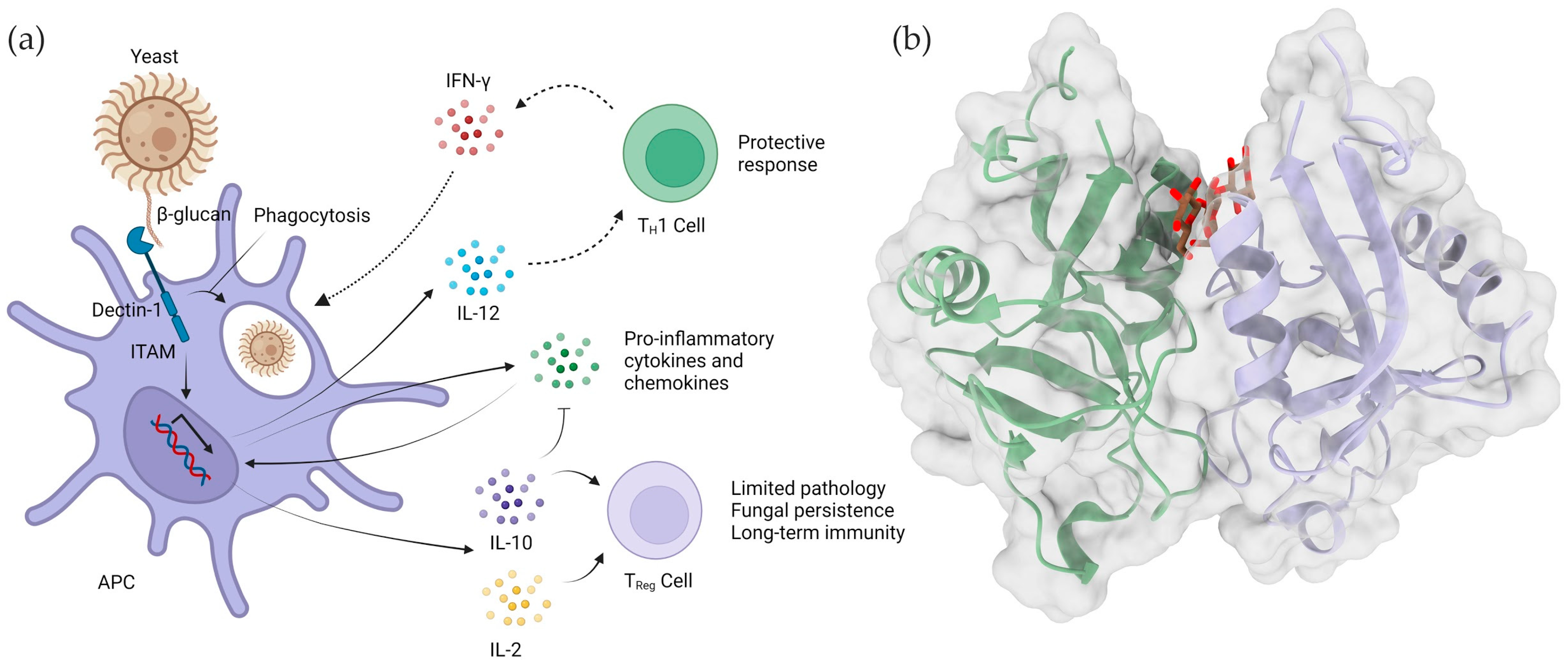
CAZymes are often multi-modular enzymes that possess one or even multiple ancillary modules, in addition to the catalytic one. The most common type of accessory modules are the carbohydrate binding modules (CBMs), which are non-catalytic motifs with the ability to recognize carbohydrates, tethering the enzyme to its cognate substrate [93]. The proximity effect created by the association of an enzyme with a particular substrate through its CBM results in enhanced enzymatic activity of the catalytic module [94]. Like CAZymes, CBMs have also been classified into families according to primary sequence homology. Currently, there are nearly 90 different CBM families, a number that has been steadily growing. Furthermore, CBMs fall into three distinct categories: type A CBMs, which interact with crystalline polysaccharides, mostly cellulose; type B CBMs, which bind to internal regions of single glycan chains; and type C CBMs, which target small saccharides at the ends of complex polysaccharides [95]. Much like CAZymes, the specificities of CBMs cover a variety of different polysaccharide ligands, including β-1,3-glucans and β-1,3-1,4 mixed linked glucans, which are recognized as CBMs from families 4, 6, 13, 32, 39, 43, 53, 54 and 56 [96,97,98,99,100,101]. Although CBMs from families 65, 72, 76, 79, 80, 81 and 85 have also shown affinity towards MLGs, no β-1,3-glucan binding has been reported. The fact that these can also bind to cellulose suggests that they recognize the 1,4 linked portions of the MLGs rather than the 1,3 linked regions [102,103].
The binding specificities of CBM modules are usually related to the reported activity of their parent enzymes, which is why β-1,3-glucanases often possess one or more β-1,3-glucan-binding CBMs. Much like with endo-β-1,3-glucanases, structural analyses of β-1,3-glucan–specific CBMs have shown that one of the main drivers of ligand preference is the recognition of the β-1,3-glucan helical structure. A notable example is the family 4 CBM for Thermotoga maritima (TmCBM4), a member of a CBM family with various ligand specificities. Its tridimensional structure was addressed by molecular replacement using the CBM4 from Cellulomonas fimi (CfCBM4), which implies high homology between both CBMs. In fact, the overall structural folds of both CBMs are very similar and the main ligand binding residues are conserved between the two. However, these CBMs recognize quite different polysaccharides: CfCBM4 is cellulose specific, whereas TmCBM4 only recognizes β-1,3-glucans [96]. The reason for such similar proteins within the same CBM family having distinct specificities comes down to differences in the shape of the polysaccharide binding groove, which is more “U-shaped” in TmCBM4 (Figure 4a,b), perfectly fitting the natural helical conformation of β-1,3-glucans. Moreover, an additional loop prevents any linear polysaccharide, such as cellulose, from fitting into TmCBM4’s active site, further contributing to its β-1,3-glucan specificity (Figure 4c) [96].
More recently, studies on family 56 CBMs found that these modules are capable of recognizing both soluble and insoluble β-1,3-glucans, but do not bind β-1,3-glucooligosaccharides. Analysis of its tridimensional structure allowed the identification of features consistent with binding of the triple-helical quaternary structure of β-1,3-glucopolysaccharides, explaining the lack of affinity for the linear β-1,3-glucooligosaccharides. Furthermore, ITC analysis showed a relatively low number of CBM56 binding sites in laminarin isolated from Laminaria digitata, which is consistent with the fact that only ~5% of laminarin from this source forms triple helices [84].

4.2. Dectin-1

Dectin-1 is a 28 KDa type II transmembrane protein that can bind to β-1,3 glucans, with or without β-1,6 branching. The name Dectin-1 comes from “Dendritic-cell-associated C-type lectin-1”, due to it being initially described as a dendritic cell receptor. However, Dectin-1 was later discovered to be implicated in the elimination of fungal pathogens by macrophages, neutrophils and dendritic cells, and to be expressed on most innate immune system cells [104,105]. Dectin-1 is composed of an extracellular lectin domain capable of β-glucan recognition, a transmembrane region and a cytosolic domain with an immunoreceptor tyrosine-based activation motif (ITAM), which mediates a ligand-induced activation of a signaling response, resulting in the formation of a vast array of immune modulators (Figure 5) [106].
Dectin-1’s extracellular lectin domain is responsible for carbohydrate recognition, binding to β-1,3 glucans in a chain length-dependent fashion, with a seemingly higher affinity for ligands with larger DP [107]. Although the mechanism of the chain length-dependent interaction is unclear, increased β-glucan chain length correlates with increased secondary structure formation. As such, it is likely that the conformation of Dectin-1’s ligand-binding site is capable of more easily accommodating those quaternary helical structures, justifying its preference for longer polysaccharides [108]. In addition, 1,6-branching also seems to affect binding to Dectin-1, with branched polysaccharides having increased affinity in comparison to linear ones [109].
Although Dectin-1’s best-described function is as a fungal and yeast pathogen recognition receptor, some studies suggest that it may have a broader function in pathogen recognition, including a role in directing macrophage response to mycobacterial infections [106]. Furthermore, other functional aspects have also been described for Dectin-1, such as binding to the conserved core domain of annexins in apoptotic cells to induce immune tolerance and the recognition of N-glycans decorating IgG antibodies, a feature whose biological importance is still under investigation [110]. Therefore, Dectin-1 seems to be a uniquely versatile lectin-like molecule with multiple roles in immune modulation [49].
Figure 5. Dectin-1 activation by fungal β-glucans. (a) Dectin-1-mediated anti-fungal immunity via β-glucan binding is a consequence of several cellular responses, including fungal uptake and killing [111,112], and the production of pro-inflammatory cytokines and chemokines that lead to immune cell recruitment and activation [113,114]. Dectin-1-mediated recognition also stimulates the production of interleukin-12 (IL-12), which elicits a protective Thelper 1 (Th1)-cell response with the production of interferon-γ (IFN-γ), thereby activating fungal killing by the phagocytes [113,115]. In dendritic cells, Dectin-1 activation can also induce the production of IL-10 and IL-2 [115], leading to the development of regulatory T cells, which prevent pathological inflammation and promote long-term immunity [116]. IL-10 also inhibits the production of pro-inflammatory cytokines and chemokines (based on a figure by Brown, G. 2006 [117]). Panel (b) shows a cartoon representation of the 3D structure of dimer of two Dectin-1 lectin modules (green and blue), with a trapped β-glucan oligosaccharide between the two units, in brown (PDB code: 2cl8).
Ijms 23 03156 g005

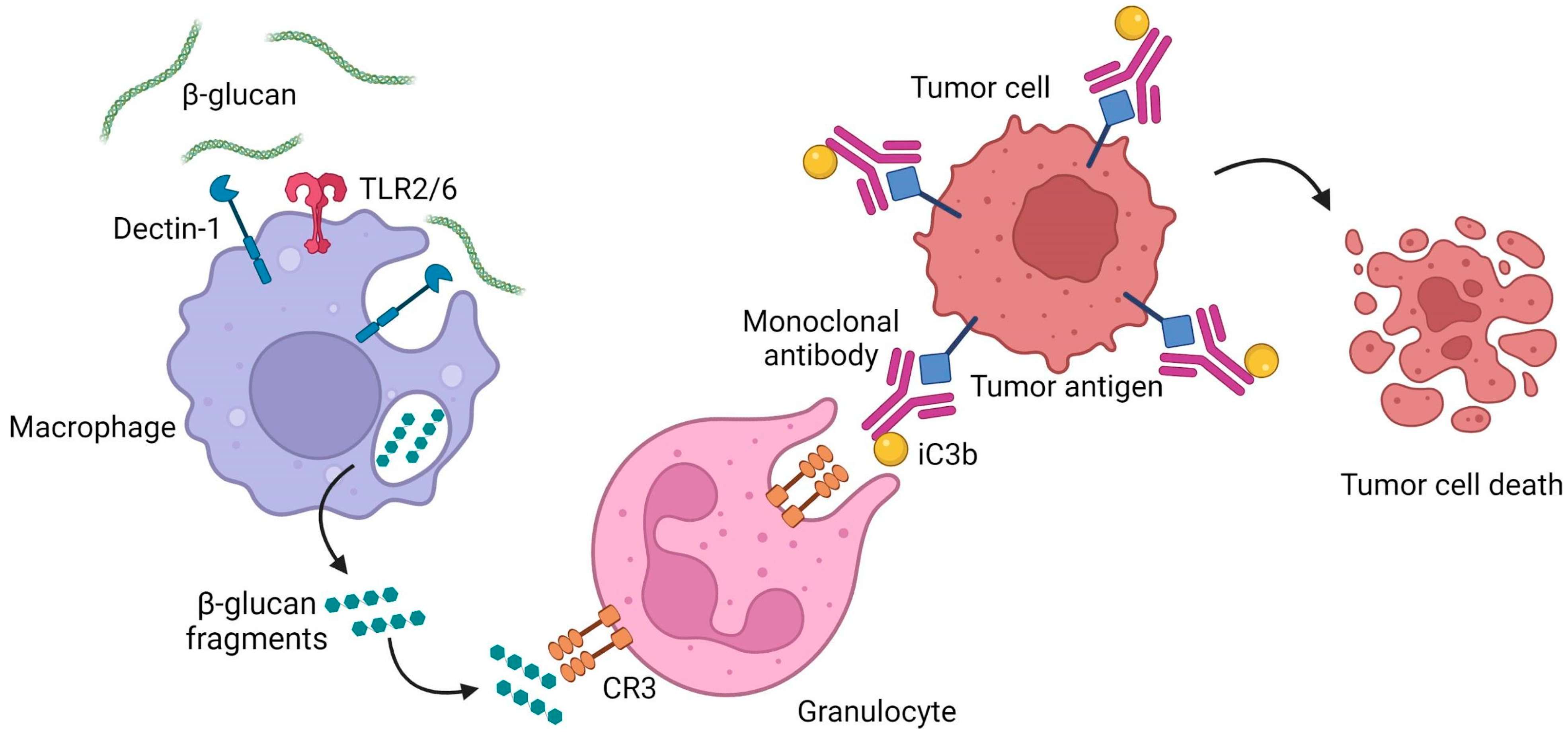
4.3. Complement Receptor 3

Complement receptor 3 (CR3) is a receptor belonging to the β2-integrin family, consisting of a heterodimer between the αM (CD11b) and β2 (CD18) subunits. It is one of the most versatile receptors expressed by NK-cells, neutrophils and lymphocytes, mediating adhesion, chemotaxis and phagocytosis, both in a complement-dependent and -independent manner [118,119].
CR3’s CD11b subunit possesses a lectin-like site, which is capable of recognizing β-glucans. Interestingly, this lectin domain is involved in a double recognition mechanism that triggers immune cells, leading to a cytotoxic response. β-glucans first bind with high affinity to the lectin site and the overlapping I-domain of CD11b, changing CR3’s conformation, priming it for recognition of complement component iC3b [120,121]. Circulating immune cells with primed CR3 will then bind to iC3b-opsonized cells, which can be of any type, including cancer cells tagged with monoclonal antibody (mAb) and coated with iC3b, leading to their elimination through cytotoxicity [122]. Together with Dectin 1’s activation, these are currently the two best described mechanisms of β-glucan-mediated immunomodulation in vertebrates.

4.4. β-glucan Binding Proteins in Invertebrates

Unlike vertebrates, insects and crustaceans lack the ability to develop acquired immunity, relying on their innate immune system for defense against external pathogens. Many of the pathways involved in arthropod immunity are triggered upon the binding of proteins, described as β-(1-3)-glucan recognition proteins (βGRPs), with β-glucans expressed on the surface of pathogens. These βGRPs comprise one of the best characterized families of pattern recognition receptors in invertebrates and are widely distributed among species [123]. The βGRP/Gram-negative binding protein 3 (GNBP3) is one of the best studied examples of βGRPs involved in insect immunity. It uses its β-glucan binding capabilities to detect fungal invasions, triggering a proteolytic cascade that eventually leads to the activation of the Toll pathway and subsequent production of antimicrobial peptides. βGRP/GNBP3 β-glucan binding occurs via its immunoglobulin-like N-terminal domain (N-GNBP3), which is highly conserved among insect species [101,124,125,126]. The molecular mechanism of the interaction is still controversial, as different studies suggest distinct β-glucan binding sites. Although NMR titration experiments and mutational analysis of the silkworm’s (Bombyx mori) GNBP3 suggested that β-glucan binds to the concave surface, its structure in complex with laminarihexaose reveals the ligand bound on the convex surface [101,125]. Nonetheless, even though GNBP3 has a high degree of conservation across species, there is evidence suggesting the existence of two groups with distinct binding specificities. A recent study on βGRP/GNBP3s from four different insect species (Bombyx mori, Plodia interpunctella, Tribolium castaneum and Tenebrio molita), using solid phase ELISA assays, revealed that one group (comprising Bombyx mori and Plodia interpunctella) preferentially binds to triple-helical β-glucans, whereas the other (Tribolium castaneum and Tenebrio molita) is not capable of recognizing the quaternary structures, but is able to bind alkaline-treated β-glucans with a partially open structure [127]. Although these results suggest that ligand conformation is crucial for recognition by βGRP/GNBP3 and that the preferred configuration is different for an individual βGRP/GNBP3, the structural mechanism behind preference remains to be determined.
Another well-known β-glucan binding protein is the Factor G from the horseshoe crab. This hemolymph coagulation factor consists of an heterodimer with an α-subunit (72 kDa) and a β-subunit (37 kDa), which are autocatalytically cleaved into the active form upon binding to β-glucan [128]. The α-subunit is responsible for the recognition of β-glucan, whereas the β-subunit is a serine protease that becomes activated when factor G binds to β-glucan. Two distinct β-glucan recognition sites are present in factor G and, although both can independently bind to ligands with a wide range of DP, proper activation of the clotting cascade requires interaction with long chain β-glucans through both sites [129]. Therefore, this protein functions as a biosensor for the longer (1,3)-β-glucan present on pathogenic fungi, which is why it is used as a diagnostic reagent for the detection of fungal infections in humans [130].
As in the case of most β-glucan interacting proteins described above, the natural triple-helical structure of β-glucan seems to be a key factor for the interaction, with the activation of Factor G increasing over 100-fold after alkaline treatment of β-glucan [131].

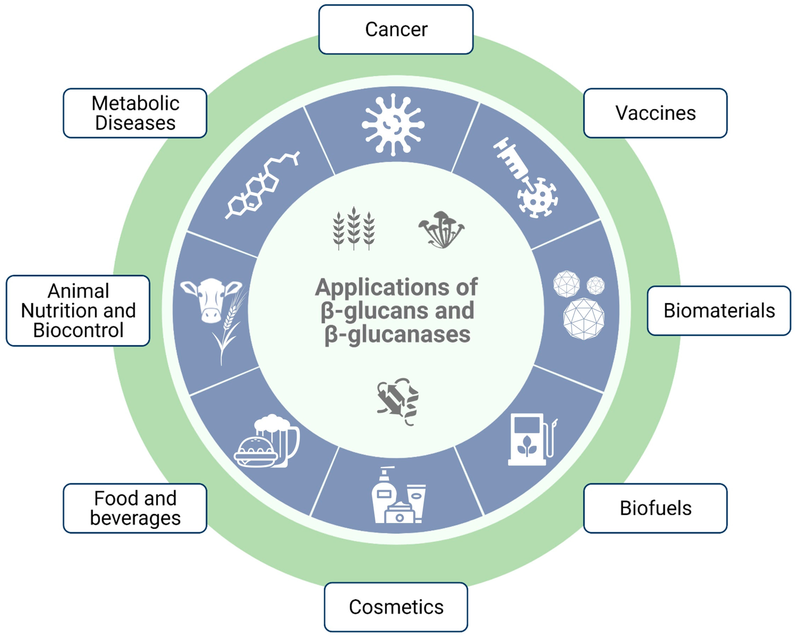
5. Applications of β-Glucans

β-glucans are biologically active biopolymers that can interact with several protein receptors, present both in micro- and macro-organisms, eliciting a wide spectrum of biological responses. These responses often result in important bioactivities including immunomodulating, anticancer, antioxidant, cholesterol-lowering and prebiotic effects [5,7,8,9,132]. Naturally, it is not surprising that numerous studies have tried to harness the potential therapeutic properties of β-glucans, which can be generally divided into immunomodulatory or metabolic properties [10,11,12,13,14,15,133,134].
Due to their physicochemical properties, β-glucans also have a wide range of potential applications in several industries, such as a thickening and gelification agent in the food industry, and as a soothing, moisturizing and skin regenerating agent in the cosmetic and personal care industry [19,20]. They have also inspired several ingenious biotechnological and biomedical solutions, ranging from hydrogel scaffolds for 3D cell cultures to drug delivering nanoparticles [135,136].
Here, we summarize some important biomedical, biotechnological and industrial applications of β-glucans.

5.1. Immunomodulating Activity

The anecdotal recognition of the immune-modulating properties of β-glucans as the main active component of mushrooms has long been well established. However, its effects in promoting immune response and boosting resistance to infections have only been scientifically proven in recent decades [137,138]. The exact mechanisms by which β-glucans activate the immune system are complex, depending on many factors that have not yet been fully revealed [14]. Unfortunately, it does not help that many of the studies fail to mention the exact origin of the β-glucans used, although it seems that most immunomodulatory trials are performed with fungal β-1,3-glucans.
Despite that, β-glucans’ ability to modulate the immune system has been associated with its interaction with several PRRs present on the surface of various immune cells, including monocytes, macrophages, neutrophils and natural killer (NK) cells. Since β-glucans are not produced by mammalian systems, these polysaccharides are recognized by the host’s innate immune response as foreign, acting as biological response modifiers [139]. To date, several different β-glucan PRRs have been described, including complement receptor 3 (CR3), scavenger receptors, lactosylceramide and Dectin-1 [140,141,142,143]. The interaction between β-glucans and PRRs triggers a signal transduction that, depending on the immune cell type, boosts a specific immune response [137].
For instance, research in this field revealed that neutrophil modulation by β-glucans is mainly CR3 dependent, whereas Dectin-1 is the most predominant β-glucan receptor on macrophages [122,144]. By binding to the lectin site of the CR3 on phagocytes and NK cells, β-glucans activate this receptor, promoting cytotoxicity against iC3b-opsonized target cells [145]. In turn, the interaction of Dectin-1 on macrophages with β-glucans triggers a downstream signaling pathway, activating phagocytosis, ROS generation, microbial killing and cytokine production [142,146].
Initially, it was thought that β-glucans’ immunomodulating properties were solely associated with the stimulation of T cell-independent immune responses [147]. However, there is increasing evidence of the influence of β-glucan in T cell differentiation. Because they consist of polysaccharides having both positive and negative charges, β-glucans can activate CD4+ T cells through the MHC-II endocytic pathway [148]. After being processed into smaller molecular weight carbohydrates, they can bind to MHC-II inside the antigen-presenting cells and are subsequently presented to Thelper cells. In addition, β-glucans immune-related responses lead to the polarization of T cells. For example, dendritic cells activated by the bacterial β-glucan curdlan have been shown to promote Th1, Th17, and cytotoxic T lymphocyte priming and differentiation [149,150], and Treg conversion into IL-17 producing T cells [151] via the Dectin-1–dependent pathway. Moreover, the yeast zymosan β-glucan seems to stimulate regulatory antigen-presenting cells, leading to Treg differentiation [152,153].
Notably, recent studies have demonstrated the ability of β-glucan to prime innate immune cells in a robust manner, a concept defined as trained innate immunity, in which a secondary immune stimulus involves a heightened activation of innate immune cells [154,155]. Evidence has demonstrated a stronger innate response from monocytes and macrophages after β-glucan exposure, with improved antimicrobial and inflammatory properties consequent on Dectin-1/toll-like receptor (TLR) activation [156]. However, trained innate immune cells show less specificity and duration of memory, when compared to the classical acquired immunity promoted by antigen-specific responses.
These regulatory effects on immune response are considered one of the most promising biological functions of β-glucans, making these compounds appealing immunomodulating agents for therapeutic interventions.

5.1.1. Anticancer Activity

The first evidence of the antitumor activity of β-glucans was published almost 40 years ago [157]. Since then, several studies have proven the potent anticancer activity of β-glucans against various types of malignancies, including lung, breast and gastrointestinal cancers [158,159,160,161,162,163,164,165,166]. This promising clinical efficacy culminated in the approval of lentinan as a biological response modifier for cancer therapy in Japan, and its clinical use for over 30 years [167].
Lentinan has been considered an adjunctive therapy to radio/chemotherapy for patients diagnosed with solid tumors, improving response rate, one-year survival, performance status, quality of life and radio/chemotherapy toxicity [168]. A clinical trial evaluated the intravenous administration of lentinan extract in combination with two chemotherapeutic compounds (5-FU and Tegafur) in patients with advanced or recurrent stomach, colorectal and breast cancers. Lentinan treatment in combination with chemotherapeutic agents promoted life span prolongation and was associated with the improvement in host immune response [169].
The anticancer effects of β-glucan-based therapies were also clinically associated with immune function regulation. Schizophyllan was used in the treatment of advanced cervical cancer, improving the function of Thelper lymphocytes and enhancing the IL-2/IL-2R system [163]. Schizophyllan combined with chemotherapy enhanced the long-term survival rate of patients with ovarian cancer [170]. Further clinical trials evaluated β-glucans therapeutic efficacy in patients with cancer, and as an adjunctive therapy in patients receiving chemotherapy to limit suppression of hematopoiesis. Data gathered from a clinical trial demonstrated that β-glucans can be safely administered to patients with advanced malignancies receiving chemotherapy and that this adjunctive therapy may ameliorate adverse effects of chemotherapy on blood counts [171]. Furthermore, a significant reduction in chemotherapy-associated side effects such as loss of appetite, alopecia, emotional instability and general weakness was reported in β-glucan supplemented patients, leading to a general improvement in quality of life [172,173,174,175,176].
β-glucans antitumor responses appear to vary depending on their origin, structure and composition [177]. For example, zymosan, a yeast-derived mixture of β-1,3-glucan and protein complexes, elicits immune responses by enhancing the number and function of macrophages while activating the complement system [178]. Lentinan triggers antitumor immune responses by improving the lymphokine-activated killer cell activity and NK cell activity [179]. A yeast-derived particulate β-glucan promotes antitumor immune responses by triggering pro-inflammatory cytokine secretion and stimulating innate immune effector cell activation [180]. Overall, these findings support the notion that β-glucan-based agents mediate antitumor immune responses through different mechanisms.
One of the most interesting β-glucan-mediated antitumor immune responses is the possibility of using β-glucans to trigger complement-dependent antitumor cytotoxicity [177]. Complement is a key component of the innate immunity against β-glucan presenting microorganisms. Because these molecules are not expressed by tumor cells, tumor cells are not capable of triggering CR3-dependent cellular cytotoxicity [181]. As such, by activating the CR3 receptor on innate cells, such as macrophages, dendritic cells, natural killer cells and neutrophils, β-glucans have the ability to prime leukocytes to mediate a specific cytotoxic immune response against previously iC3b-coated tumor cells by circulating anti-tumor antibodies. Therefore, β-glucans may function as a potent adjuvant for cancer mAb therapy to elicit a novel granulocyte and tissue macrophage-mediated tumor-killing mechanism that is not activated by mAb therapy alone [182] (Figure 6).
Several murine syngeneic tumors [182,183] and human carcinoma xenograft models [184,185,186] have attested to the significant therapeutic efficacy of combined β-glucan and anti-tumor mAbs therapy. Moreover, there are multiple clinical trials for β-glucan-based cancer immunotherapies, many of which combine β-glucans with mAbs (Table 2) [187]. A phase I clinical trial enrolling 20 patients with chronic lymphocytic leukemia investigated the combined treatment using mAbs with PGG glucan to study the hypothesis that β-glucans would augment the cytotoxic activity of the innate immune system when administered in association with mAbs. Data showed that treatment was well-tolerated by patients, which justified a further phase II study. Interestingly, while monotherapy with rituximab and alemtuzumab rarely achieved a complete response or a sustained response, its combination with β-glucans resulted in a high complete response rate. Although this study gave hope that the addition of β-glucans may improve the response to mAb therapies, due to the study’s small size, the preliminary data did not allow conclusions to be drawn [188]. A similar clinical trial was conducted based on the same scientific hypothesis using the same PGG glucan in combination with carboplatin/paclitaxel chemotherapy plus an EGFR-targeted antibody (cetuximab) in untreated stage IIIB/IV non-small cell lung cancer patients. Results demonstrated that the addition of β-glucans was well tolerated and significantly improved objective response rate in enrolled patients [189].
This concept has been also investigated using a combination therapy with β-glucans and immune checkpoint inhibitors, and has obtained encouraging results in preclinical studies. The combination treatment with β-glucans and an anti-PD-1 antibody elicited a coordinated immune response, which translated to a promising antitumor response in a syngeneic cancer murine model [190]. The benefit of β-glucans has also been evaluated in association with pembrolizumab in various phase II studies in cancer patients. Recently, a clinical trial studied the effect of combining PGG with pembrolizumab in subjects with chemotherapy-resistant metastatic triple negative breast carcinoma [191]. The combination of Imprime (a soluble yeast derived β-glucan) and this anti-PD1 antibody provided promising response rates and overall survival. Moreover, biopsy analyses consistently showed activation of both myeloid and T cells with extensive infiltration into tumor tissue. By acting as an immune-modulator of the innate and adaptive response, β-glucans have the potential to alter the tumor microenvironment towards an immunostimulatory phenotype, stimulating and improving clinical response to immune checkpoint inhibitors [192].
Another advantage of β-glucans in cancer immunotherapy is their synergistic action with cancer vaccines. By acting as potent adjuvants for tumor vaccines, β-glucans can elicit potent cytotoxic T cell responses along with humoral responses. The combination of a bivalent ganglioside vaccine with β-glucan was investigated in a phase I clinical trial for high-risk pediatric neuroblastoma in second or later remission. Data showed promising serological and minimal residual disease responses and that the vaccine/β-glucan treatment was well tolerated [193].
More recently, the promising immune modulatory effect of β-glucans has encouraged the generation of glucan-based multifunctional nanomedicine systems for targeted delivery of glucan in combination with therapeutic drugs to tumor cells. β-glucans were incorporated as carriers in a novel targeted delivery system loaded with doxorubicin conjugated to trastuzumab antibody for the treatment of HER2+ breast xenotransplant tumors in mice. These β-1,3-glucan-doxorubicin-targeted nanoparticles demonstrated superior tumor inhibition compared to the control groups [194].
Hence, considering its proven safety and low toxicity profile, along with the success of emerging combinatorial approaches for cancer treatment involving these molecules, β-glucans have the potential to revolutionize future strategies in immuno-oncology.
Table 2. Ongoing and completed clinical trials involving β-glucans for cancer treatment, according to the clinicaltrials.gov database (as of 25 February 2022) [195].
MoleculeCancer TypeStatusIdentifier
Yeast derived β-glucan + glutamine + immunoglobulinMetastatic cancersPhase II/IIINCT04710290
Yeast derived Soluble Beta-GlucanAdvanced solid tumorsPhase INCT01910597
Soluble β-1,3-1,6-glucan + standard antibody and chemotherapyBreast cancerPhase I/IINCT00533364
Soluble β-glucan + rituximab + COP/CHOPNon-Hodgkin’s lymphomaPhase INCT00533728
Particulate β-glucanOral squamous cell carcinomaNot ApplicableNCT04387682
β-glucan MM-10-001Locally advanced or metastatic non-small cell lung cancerPhase INCT00857025
β-glucan (Imucell WGP)Non-small cell lung cancerNot ApplicableNCT00682032
β-glucan + pembrolizumabMelanoma stage III/IVNot ApplicableNCT04513028
β-glucan + monoclonal antibody 3F8NeuroblastomaPhase INCT00037011
β -glucan + monoclonal antibody 3F8NeuroblastomaPhase INCT00492167
β-glucan + isotretinoin + sargramostim + monoclonal antibody 3F8NeuroblastomaPhase IINCT00089258
β-glucan + granulocyte-macrophage colony stimulating factor + bivalent vaccine + adjuvant OPT-821NeuroblastomaPhase IINCT04936529
β-glucan + bivalent vaccine + adjuvant OPT-821NeuroblastomaPhase I/IINCT00911560
Imprime PGG β-glucan + rituximab + alemtuzumabChronic lymphocytic leukemiaPhase I/IINCT01269385
Imprime PGG β-glucan + pembrolizumabMalignant Neoplasm of BreastPhase IINCT05159778
Imprime PGG β-glucan + pembrolizumabAdvanced melanoma
Triple-negative breast cancer
Phase IINCT02981303
Imprime PGG C + rituximabRelapsed/refractory indolent B cell non-Hodgkin lymphomasPhase IINCT02086175

5.1.2. Anti-Infective, Anti-Inflammatory and Wound Healing Properties

β-Glucans have also shown interesting biological effects, such as anti-microbial, anti-inflammatory and wound healing properties. Several studies suggested that β-glucan supplementation improved mucosal innate immunity and reduced upper respiratory tract infection incidence and symptom severity in pediatric patients [196,197,198]. β-glucan supplementation may be also associated with defense against infection in adults and the elderly [199,200]. In particular, there is some evidence that adults suffering from allergies benefit from β-glucan supplementation to reduce symptom severity [201]. Very few studies have been performed on autoimmune diseases; however, an extract derived from Agaricus blazei was tested in inflammatory bowel diseases, with modest results on inflammatory cytokines or clinical symptoms [202,203]. Moreover, β-glucans boost wound repair by increasing the infiltration of macrophages, which promote tissue granulation, collagen deposition and skin re-epithelialization [204]. Human clinical studies provided evidence that β-glucan can accelerate healing of chronic wounds. The application of a 3% cream containing curdlan caused a 55% reduction in ulcers after 90 days of treatment [205]. Another clinical study demonstrated that the utilization of soluble yeast β-1,3-glucan allowed 59% of total ulcers to be healed compared to 37% of the control group by week 12 [206].

5.2. Metabolic Activity

Beyond their immunomodulatory effects, β-glucans, particularly MLGs, also present important metabolic and gastro-intestinal effects through the modulation of gut microbiota, alteration of lipid and glucose metabolism, and reduction in cholesterol. These beneficial effects led to a variety of studies regarding β-glucans as potential therapies for metabolic syndrome, obesity and diet regulation, and gastrointestinal conditions, such as irritable bowel, and to lower cardiovascular and diabetes risk [7]. Remarkably, evidence supporting the beneficial role of oat β-glucans led the US Food and Drug Administration (FDA) to approve the use of health claims linking oat products with the reduction in coronary heart disease risk [207].
β-glucans decrease the blood postprandial glycemic and insulin peak by creating a barrier in the small intestine that hinders glucose absorption [192]. Furthermore, there is increasing evidence of β-glucans’ influence on the activation of metabolic pathways through phosphatidylinositol 3-kinase (PI3K)/serine-threonine kinase (Akt), a key signaling pathway of the pathogenesis of diabetes [208]. A systematic review and meta-analysis conducted by Zurbau et al. (2021) confirmed that adding oat β-glucans to carbohydrate-containing meals reduces glycemic and insulinemic responses [209].
In turn, the cholesterol lowering effects of β-glucans are proposed to be mainly mediated by the gel-forming properties of β-glucans, which modulates bile acid and cholesterol metabolism. β-glucans attenuate the intestinal uptake of dietary cholesterol while preventing bile acid reabsorption that subsequently increases the demand for de novo synthesis of bile acids from cholesterol catabolism, which contributes to lowering the LDL fraction of circulating cholesterol [210]. However, more recently, the impact of β-glucans activity on gut microbiota modulation, particularly on those bacterial species that influence bile acid metabolism and production of short chain fatty acids, has been implicated in the regulation of cholesterol homeostasis [211]. Several randomized-controlled trials and subsequent meta-analyses reported a significant correlation between the consumption of oats or oat β-glucans and lower LDL cholesterol levels, while also reporting other enhanced markers of cardiovascular disease risk [212,213,214,215].
In addition, given their non-digestible and non-absorptive characteristics in the human gastrointestinal tract, β-glucans have been suggested to be dietary fibers with potential prebiotic properties [216]. β-glucans are reported to accelerate bowel transit, increase fecal bulk and frequency, and to positively influence gut microbiota regulation, preventing irritable bowel syndrome, diverticular diseases and colon cancer [7]. The growth of normal intestinal microbiota (Lactobacilli and Bifidobacteria species) are supported by β-glucans in vivo and in vitro models [217]. Even though strong evidence of the prebiotic effect of β-glucans in humans is still lacking, β-glucans prebiotic potential was confirmed in a clinical trial conducted by Mitsou et al. (2010). This work reported that daily intake of a barley β-glucan was well tolerated and had a bifidogenic effect, boosting the Bifidobacteria number to a detectable level [218].
Altogether, these results clearly demonstrate the potential that inclusion of β-glucans on diets may have in reducing the prevalence of cardiovascular disease, diabetes and gastrointestinal diseases, and its associated healthcare costs.

5.3. Industrial Applications of β-Glucans

Due to their unique physicochemical properties (solubility, viscosity and gelation), β-glucans have found potential applications in a wide range of industrial sectors, such as the agronomy, cosmetic, pharmaceutical and food industries. In the food industry, β-glucans are very popular thickening agents, but can also be used with many other purposes, such as: additives in the preparation of frozen sweet foods, improving the texture of cakes and the shape retention of ice creams [219,220]; to reduce the leaching out of soluble ingredients and promote softening in pasta noodles preparation [221]; as fat substitutes and gelling agents in the preparation of meat products [222,223]; and as components in the preparation of edible films for food packaging [224]. Another popular application for β-glucans is in the formulation of low-calorie foods [225,226]. For example, a high-fiber and low-calorie substitute for wheat flour was developed using β-glucan-enriched materials from the mushroom Lentinus edodes [227]. The authors suggested that this product can be used to bake cakes containing 1 g of β-glucan per serving with quality attributes akin to those of traditional formulations. Due to the potential metabolic effects of cereal β-glucans, they have also been widely explored as ingredients in the formulation of low-fat and cholesterol-lowering products, including salad dressings [228], milk [229], yogurt [230] and cheese [231]. In addition to the food industry, the cosmetics and personal care industry also make extensive use of β-glucans. Due to their soothing, moisturizing and anti-irritant properties, β-glucans have been a common ingredient in protective creams, ointments and powders, with oat β-glucan being particularly popular. For example, “Avenacare” is a liquid form of active oat β-glucan, naturally extracted from Swedish oats using a chemical-free technology [20]. There is also evidence showing that baker’s yeast β-glucan might promote keratinocyte growth, resulting in increased skin regeneration and protection against damage, and that low doses of laminarin from L. digitata provide antioxidant protection to the skin [232,233].
Due to the immunomodulating aspects of β-glucans, in addition to their ability to improve the gut microbiome and promote gastro-intestinal health, some studies suggest the use of β-glucans as feed ingredients in animal husbandry. There is evidence that they are capable of enhancing the natural immunity of animals, promoting productivity and decreasing the need for antibiotics [234,235,236]. The inclusion of yeast cell β-glucans in poultry diets was shown to increase the intestinal clearance of pathogens by protecting intestinal barriers, stimulating phagocytosis and suppressing pathogen invasion [237,238,239,240]. Supplementation of piglet diets with Sacharomyces cerevisiae β-1,3-glucans increased production performances and resulted in significant stimulation of phagocytosis by both peripheral blood monocytes and neutrophils, and improved responses to endotoxin challenge [241]. In aquaculture, where the vaccination process can be laborious and impractical, β-glucans have also been proposed as potential agents for prophylaxis and disease management [235]. In summary, with current efforts to eliminate the use of antibiotics and growth promoters in the livestock industry, the inclusion of β-glucans in feed may be a viable option, offering both natural growth stimulation and immune protection.

5.4. Biotechnological and Biomedical Applications of β-Glucans

Beyond their use as immunostimulants and metabolic enhancers, β-glucans have also had their properties explored in the development of new biotechnological solutions. Notably, there are a number of recent studies exploring the potential of β-glucans as a novel support for cell culture and as scaffolds to create 3D tissue constructs [135,242,243,244]. Martins et al. (2018) developed photopolymerizable microparticles from methacrylated laminarin, using a microfluidics device, which were incorporated with platelet lysates and further conjugated with an adhesive peptide [242]. The microparticles were seeded with mouse fibroblast L929 cells and the results showed enhanced cell adhesiveness to the laminarin microparticles and an increased proliferation rate after 11 days. Moreover, expanded cells provided the link for microparticle aggregation, resulting in self-assembled robust 3D structures. In another work, a 3D gel composite scaffold was designed using laminarin hydrogel and graphene foam. This composite scaffold exhibited a reinforced toughness and was utilized for 3D culture of human mesenchymal stem cells. The graphene foam supported cell attachment and cell spreading, while the laminarin hydrogel conjugated with an adhesive peptide regulated cell migration and formed an interconnected cellular network in the pores of the graphene foam [135]. In both studies, the authors suggest that their systems have the capacity to produce large tissue-engineered constructs, and thus to be applied in tissue engineering and regenerative medicine. The use of bulk hydrogels in cell culture poses severe limitations, such as oxygen, nutrient and metabolite diffusion. In an attempt to overcome these limitations, self-feeding laminarin-based hydrogels with immobilized β-glucanases were assembled and used to support the growth of 3D tumor and human stem cell cultures, while simultaneously providing a continuous source of glucose for the metabolic activity of cells, through laminarin degradation [245].
A number of recent studies have focused on the development of β-glucan-based drug delivery systems, either as hydrogels or for the encapsulation of therapeutically important biological agents, such as siRNA, peptides and DNA, which can lead to a new approach to immunization and cancer immunotherapy [136,246,247,248]. Notably, in an experimental glioblastoma stem cell therapy study, the utilization of β-1,3-glucan as an outer shell coating for paclitaxel-loaded chitosan nanoparticles was able to enhance chemotherapeutic potency, overcome systemic toxicities and improve drug bioavailability [249]. Another study suggested a multifunctional laminarin-based nanomedicine carrier biomaterial with dual pH/redox sensitivity that has the potential to be applied as a new drug delivery system for cancer therapy. The system achieved great results in delivering a photosensitizer for photodynamic therapy in breast cancer [250]. β-glucans are also a promising alternative to aluminum as vaccine adjuvants, due to their natural ability to promote immunogenicity [251]. Moreover, β-glucan can be prepared in a particulate form, consisting of spherical hollow particles of β-glucan, which can be loaded with different compounds, including peptide antigens, functioning as both as adjuvant and antigen carriers [252,253]. Another interesting study took advantage of the quaternary triple helix structure of β-glucans to develop schizophyllan–antigenic peptide complexes, by replacing one of the strands in the triple helix with a polyadenylic acid chain linked to a peptide. The complex was able to induce a strong antigen-specific cytotoxic T lymphocyte activity, resulting in an increased immunogenicity with a much lower peptide dose. This is due to the targeting effect of schizophyllan towards antigen-presenting cells through the interaction with β-glucan receptors, a strategy that can be used in the development of potent vaccines against infectious diseases and cancer [248]. In summary, there are myriad potential biotechnological and biomedical applications for β-glucans, but much of the research is still in its infancy. Nonetheless, it is evident that there is a growing interest in these subjects, which is reflected in the ever-increasing number of studies.

5.5. Applications of β-1,3-Glucanases

Much like their natural substrates, β-1,3-glucanases have found a number of applications in many industrial processes and serve as inspiration for several biotechnological solutions. They have been extensively studied for the use in the conversion of β-1,3-glucan containing lignocellulosic biomass, namely of algal origin, into fermentable sugars to be used in bioethanol production [80,254]. They offer a more ecological and energy -efficient alternative to classical methods such as chemical extraction at high temperatures, although the costs of producing the enzymes at a large scale are currently prohibitive [255]. Another popular application for β-glucanases is as a feed additive in animal production to improve growth and performance. Barley- and oat-based diets can significantly increase the viscosity of the digesta in monogastric animals, with poultry being particularly affected. This leads to slower gastrointestinal transit and decreased feed intake and nutrient absorption. In order to overcome these antinutritive effects, feed supplementation with β-glucanases has become a common option [31,256,257].
Given that β-1,3-glucans are major components of the fungal cell wall, β-1,3-glucanases possess an important antifungal activity. As such, they have attracted a great deal of attention as potential non-chemical biocontrol agents against fungal pathogens in crops [70,80]. Members of the genus Trichoderma are especially well known for their mycoparasitic activities against various phytopathogenic fungi. By combining β-1,3-glucanases from Trichoderma harzianum with chitinases, it was possible to achieve a potent antifungal activity against the phytopathogenic fungus Botrytis cinera or “grey mold”, which is responsible for serious economic losses to both field- and greenhouse-grown crops [258]. Because β-1,3-glucanases have no cytotoxic effects towards animal cells, their antifungal activity may also pose a potential and safe solution for the treatment of pathogenic fungal infections, particularly those caused by Candida albicans [34].
In brewing, the increased viscosity caused by barley β-glucans can cause a number of problems, notably reduced rates of wort separation and beer filtration, and also the formation of hazes, gels and precipitates. As such, β-1,3-glucanases are extensively used in brewing to avoid the accumulation of barley β-glucans, decreasing wort viscosity, improving filtration and promoting the elimination of unwanted beer turbidity. They have also been shown to be able to speed up germination during the malting process without compromising beer quality and organoleptic features [30,259].
In the wine industry, commercial glucanases are especially used in the stabilization (clarification, filtration) and aging (maturation on lees) steps. During the winemaking process, the presence of lactic acid bacteria (mainly Pediococcus spp.) and the mold Botrytis cinerea can lead to a type of spoilage called ropiness. “Ropy” wines are a result of the production of exopolysaccharide slimes by these contaminating species, which are responsible for hampering clarification, causing filtration problems and leading to an unacceptable mouthfeel [260]. These slimes are mostly composed of high molecular weight β-1,3-glucan, which is why there are numerous commercial enzyme preparations containing β-glucanases, mostly from Trichoderma species, specifically produced to reduce the viscosity of musts and wine caused by these colloidal polysaccharides [261]. Other frequent wine contaminants are wild yeasts, which often lead to the development of off-flavors, decreasing wine quality. Since β-glucans are a major component of the yeast cell wall, β-1,3-glucanase supplementation is often used to control the development of wild species. A β-1,3-glucanase from Delftia tsuruhatensis has demonstrated the potential to both prevent slime production and undesirable yeast growth during vinification, improving the organoleptic properties of wine. This enzyme is particularly interesting because it is highly active under wine-relevant parameters, such as elevated ethanol, sulfite, and phenol concentrations, and at low pH values [262]. Certainly, one of the most interesting features of exogenous β-glucanase supplementation during vinification is its ability to improve the effects of the traditional “bâtonnage” technique, used during the “sur lie” (on lees) aging process. The “bâtonnage” consists in stirring the settled lees back into the wine, which are riddled with dead yeast. This allows the cellular constituents of starter yeasts, mainly mannoproteins, to be mixed with the wine. These mannoproteins, present in the cell wall of the yeasts, are associated with very positive quality and technological wine traits. Using β-glucanase supplementation, it is possible to exert a strong lysogenic action on the yeast, further promoting the release of mannoproteins into the wine (Figure 7). This post-fermentative treatment aims to improve the wine’s organoleptic properties by increasing its body, mouthfeel and creaminess, but it also reduces the risk of microbial spoilage, promotes protein and tartaric salt stabilization, reduces haziness, and improves the wine’s overall aging potential [261,263].
β-1,3-glucanases are also very useful tools in several biotechnological processes, such as the preparation of fungal cell protoplasts for fungal gene function studies [264], in the transformation and extraction of proteins in yeast protein expression systems [259], or even as part of biosensors for the detection of laminarin [265]. In summary, the potential applications of β-1,3-glucanases are many, which is a reflection of their multiple roles in biology, in addition to the importance of their natural substrates.

6. Final Remarks

β-glucans are a diverse class of complex polysaccharides synthesized by multiple organisms across many taxa. There are extensive structural differences between β-glucans, mostly as a result of their distinct sources, but also due to the extraction and purification methods used to obtain them. These differences have profound implications for the physicochemical and functional profiles of β-glucans. There is an abundance of literature attesting to the many applications of β-glucans, with particular emphasis on their therapeutic potential, which varies from antitumor and immunomodulatory effects, to improved glycemic control, cholesterol-lowering and probiotic properties. The multiple registered clinical trials using β-glucans for various pathologies are a clear testament to a longstanding interest in these compounds. Nonetheless, there are still significant barriers to their clinical use. The main issue is that the true extent of the relationship between molecular structure and functional profile remains to be clarified. Despite the many published papers on the biological activities of β-glucans, comparing results is often difficult, as the majority of authors used polysaccharides from different sources, with different sizes and, therefore, different physicochemical properties. It is then imperative to provide further characterization of β-glucans, and to optimize isolation and purification procedures, so that a proper structure–function relationship can be established. Only then will it be possible to identify the most promising compounds for clinical testing. β-glucanases, in addition to having enormous potential for application in many industrial and biotechnological processes, are an invaluable tool to study the structure of β-glucans. By having prior knowledge of a β-glucanase mechanism of action, it is possible to infer about a polysaccharide structure by analyzing the products of its hydrolysis. In turn, a proper understanding of a β-glucanase’s mechanism of action requires the use of a properly characterized substrate. The two are intrinsically connected, and their full biological and biotechnological potential can only be unlocked with systematic, methodical and comparable structure–function studies.

Author Contributions

C.C.—conceptualization, original draft preparation; J.N.R.D.—original draft preparation and visualization; C.M.G.d.A.F.—reviewing and editing; P.B.—conceptualization, original draft preparation, visualization, reviewing and editing. All authors have read and agreed to the published version of the manuscript.

Funding

The authors acknowledge FCT-Fundação para a Ciência e Tecnologia, through the Centro de Investigação Interdisciplinar em Sanidade Animal (CIISA) grant UIDB/00276/2020 and through the Associate Laboratory for Animal and Veterinary Sciences (AL4AnimalS) grant LA/P/0059/2020. They also thank ANI-Agência Nacional de Investigação for grant LISBOA-01-0247-FEDER-047033. C.C. is funded by an individual PhD scholarship from FCT (SFRH/BD/147152/2019).

Institutional Review Board Statement

Not applicable.

Informed Consent Statement

Not applicable.

Data Availability Statement

Not applicable.

Acknowledgments

Figures were created using the Biorender platform, (Available online: www.biorender.com accessed on 25 February 2021). Molecular graphics and analyses performed with UCSF ChimeraX, developed by the Resource for Biocomputing, Visualization, and Informatics at the University of California, San Francisco, with support from National Institutes of Health R01-GM129325 and the Office of Cyber Infrastructure and Computational Biology, National Institute of Allergy and Infectious Diseases [266].

Conflicts of Interest

The authors declare that the research was conducted in the absence of any commercial or financial relationships that could be construed as a potential conflict of interest.

References

  1. Saito, H.; Misaki, A.; Harada, T. A Comparison of the Structure of Curdlan and Pachyman. Agric. Biol. Chem. 1968, 32, 1261–1269. [Google Scholar] [CrossRef]
  2. Du, B.; Meenu, M.; Liu, H.; Xu, B. A Concise Review on the Molecular Structure and Function Relationship of β-Glucan. Int. J. Mol. Sci. 2019, 20, 4032. [Google Scholar] [CrossRef] [PubMed] [Green Version]
  3. Usoltseva, R.V.; Belik, A.A.; Kusaykin, M.I.; Malyarenko, O.S.; Zvyagintseva, T.N.; Ermakova, S.P. Laminarans and 1,3-β-D-Glucanases. Int. J. Biol. Macromol. 2020, 163, 1010–1025. [Google Scholar] [CrossRef] [PubMed]
  4. Murphy, E.J.; Rezoagli, E.; Major, I.; Rowan, N.; Laffey, J.G. β-Glucans. Encyclopedia 2021, 1, 831–847. [Google Scholar] [CrossRef]
  5. Wang, Q.; Sheng, X.; Shi, A.; Hu, H.; Yang, Y.; Liu, L.; Fei, L.; Liu, H. β-Glucans: Relationships between Modification, Conformation and Functional Activities. Molecules 2017, 22, 257. [Google Scholar] [CrossRef] [Green Version]
  6. da Silva Milhorini, S.; Simas, F.F.; Smiderle, F.R.; Inara de Jesus, L.; Rosado, F.R.; Longoria, E.L.; Iacomini, M. β-Glucans from the Giant Mushroom Macrocybe Titans: Chemical Characterization and Rheological Properties. Food Hydrocoll. 2022, 125, 107392. [Google Scholar] [CrossRef]
  7. Murphy, E.J.; Rezoagli, E.; Major, I.; Rowan, N.J.; Laffey, J.G. β-Glucan Metabolic and Immunomodulatory Properties and Potential for Clinical Application. JoF 2020, 6, 356. [Google Scholar] [CrossRef]
  8. Ahnen, R.T.; Jonnalagadda, S.S.; Slavin, J.L. Role of Plant Protein in Nutrition, Wellness, and Health. Nutr. Rev. 2019, 77, 735–747. [Google Scholar] [CrossRef]
  9. Frank, J.; Fukagawa, N.K.; Bilia, A.R.; Johnson, E.J.; Kwon, O.; Prakash, V.; Miyazawa, T.; Clifford, M.N.; Kay, C.D.; Crozier, A.; et al. Terms and Nomenclature Used for Plant-Derived Components in Nutrition and Related Research: Efforts toward Harmonization. Nutr. Rev. 2020, 78, 451–458. [Google Scholar] [CrossRef]
  10. Chang, S.-C.; Saldivar, R.K.; Liang, P.-H.; Hsieh, Y.S.Y. Structures, Biosynthesis, and Physiological Functions of (1,3;1,4)-β-d-Glucans. Cells 2021, 10, 510. [Google Scholar] [CrossRef]
  11. Vetvicka, V.; Vannucci, L.; Sima, P.; Richter, J. Beta Glucan: Supplement or Drug? From Laboratory to Clinical Trials. Molecules 2019, 24, 1251. [Google Scholar] [CrossRef] [PubMed] [Green Version]
  12. Kofuji, K.; Aoki, A.; Tsubaki, K.; Konishi, M.; Isobe, T.; Murata, Y. Antioxidant Activity of β-Glucan. ISRN Pharm. 2012, 2012, 1–5. [Google Scholar] [CrossRef] [PubMed] [Green Version]
  13. Yamada, J.; Hamuro, J.; Hatanaka, H.; Hamabata, K.; Kinoshita, S. Alleviation of Seasonal Allergic Symptoms with Superfine β-1,3-Glucan: A Randomized Study. J. Allergy Clin. Immunol. 2007, 119, 1119–1126. [Google Scholar] [CrossRef] [PubMed]
  14. Han, B.; Baruah, K.; Cox, E.; Vanrompay, D.; Bossier, P. Structure-Functional Activity Relationship of β-Glucans From the Perspective of Immunomodulation: A Mini-Review. Front. Immunol. 2020, 11, 658. [Google Scholar] [CrossRef] [Green Version]
  15. Sivieri, K.; de Oliveira, S.M.; de Souza Marquez, A.; Pérez-Jiménez, J.; Diniz, S.N. Insights on β-Glucan as a Prebiotic Coadjuvant in the Treatment of Diabetes Mellitus: A Review. Food Hydrocoll. Health 2022, 2, 100056. [Google Scholar] [CrossRef]
  16. Vetvicka, V.; Vannucci, L.; Sima, P. The Effects of β-Glucan on Fish Immunity. N. Am. J. Med. Sci. 2013, 5, 580. [Google Scholar] [CrossRef] [Green Version]
  17. Schwartz, B.; Vetvicka, V. Review: β-Glucans as Effective Antibiotic Alternatives in Poultry. Molecules 2021, 26, 3560. [Google Scholar] [CrossRef]
  18. Byrne, K.A.; Loving, C.L.; McGill, J.L. Innate Immunomodulation in Food Animals: Evidence for Trained Immunity? Front. Immunol. 2020, 11, 1099. [Google Scholar] [CrossRef]
  19. Ahmad, A.; Anjum, F.M.; Zahoor, T.; Nawaz, H.; Dilshad, S.M.R. Beta Glucan: A Valuable Functional Ingredient in Foods. Crit. Rev. Food Sci. Nutr. 2012, 52, 201–212. [Google Scholar] [CrossRef]
  20. Du, B.; Bian, Z.; Xu, B. Skin Health Promotion Effects of Natural Beta-Glucan Derived from Cereals and Microorganisms: A Review. Phytother. Res. 2014, 28, 159–166. [Google Scholar] [CrossRef]
  21. Déjean, G.; Tamura, K.; Cabrera, A.; Jain, N.; Pudlo, N.A.; Pereira, G.; Viborg, A.H.; Van Petegem, F.; Martens, E.C.; Brumer, H. Synergy between Cell Surface Glycosidases and Glycan-Binding Proteins Dictates the Utilization of Specific Beta(1,3)-Glucans by Human Gut Bacteroides. mBio 2020, 11, e00095-20. [Google Scholar] [CrossRef] [Green Version]
  22. Labourel, A.; Jam, M.; Legentil, L.; Sylla, B.; Hehemann, J.-H.; Ferrières, V.; Czjzek, M.; Michel, G. Structural and Biochemical Characterization of the Laminarinase Zg LamC GH16 from Zobellia Galactanivorans Suggests Preferred Recognition of Branched Laminarin. Acta Crystallogr. D Biol. Crystallogr. 2015, 71, 173–184. [Google Scholar] [CrossRef]
  23. Terra, W.R.; Ferreira, C. Biochemistry of Digestion. In Comprehensive Molecular Insect Science; Elsevier: Amsterdam, The Netherlands, 2005; pp. 171–224. ISBN 978-0-444-51924-5. [Google Scholar]
  24. Mackay, R.M.; Baird, S.; Dove, M.J.; Erratt, J.A.; Gines, M.; Moranelli, F.; Nasim, A.; Willick, G.E.; Yaguchi, M.; Seligy, V.L. Glucanase Gene Diversity in Prokaryotic and Eukaryotic Organisms. Biosystems 1985, 18, 279–292. [Google Scholar] [CrossRef]
  25. Sova, V.V.; Pesentseva, M.S.; Zakharenko, A.M.; Kovalchuk, S.N.; Zvyagintseva, T.N. Glycosidases of Marine Organisms. Biochemistry 2013, 78, 746–759. [Google Scholar] [CrossRef]
  26. Dong, J.-Z.; Dunstan, D.I. Endochitinase and ?-1,3-Glucanase Genes Are Developmentally Regulated during Somatic Embryogenesis InPicea Glauca. Planta 1997, 201, 189–194. [Google Scholar] [CrossRef] [PubMed]
  27. Sova, V.V.; Shirokova, N.I.; Kusaykin, M.I.; Scobun, A.S.; Elyakova, L.A.; Zvyagintseva, T.N. β-1,3-Glucanase from Unfertilized Eggs of the Sea Urchin Strongylocentrotus Intermedius. Comparison with β-1,3-Glucanases of Marine and Terrestrial Mollusks. Biochemistry 2003, 68, 529–533. [Google Scholar] [CrossRef]
  28. Carvalho, V.S.D.; Gómez-Delgado, L.; Curto, M.Á.; Moreno, M.B.; Pérez, P.; Ribas, J.C.; Cortés, J.C.G. Analysis and Application of a Suite of Recombinant Endo-β(1,3)-d-Glucanases for Studying Fungal Cell Walls. Microb. Cell Fact 2021, 20, 126. [Google Scholar] [CrossRef]
  29. Wu, Q.; Dou, X.; Wang, Q.; Guan, Z.; Cai, Y.; Liao, X. Isolation of β-1,3-Glucanase-Producing Microorganisms from Poria Cocos Cultivation Soil via Molecular Biology. Molecules 2018, 23, 1555. [Google Scholar] [CrossRef] [Green Version]
  30. Brazil, C.; de Oliveira, D.F.; Duarte, R.A.; Galo, J.M.; Lucchetta, L.; da Costa dos Santos, E.; Hashimoto, E.H. β-Glucanase Addition in Brewing Malt Produced by Reduced Time of Germination. Braz. Arch. Biol. Technol. 2019, 62, e19180315. [Google Scholar] [CrossRef]
  31. Karunaratne, N.D.; Classen, H.L.; Ames, N.P.; Bedford, M.R.; Newkirk, R.W. Effects of Diet Hulless Barley and Beta-Glucanase Levels on Ileal Digesta Soluble Beta-Glucan Molecular Weight and Carbohydrate Fermentation in Laying Hens. Poult. Sci. 2022, 101, 101735. [Google Scholar] [CrossRef]
  32. Claus, H.; Mojsov, K. Enzymes for Wine Fermentation: Current and Perspective Applications. Fermentation 2018, 4, 52. [Google Scholar] [CrossRef] [Green Version]
  33. de Marco, J.L.; Felix, C.R. Purification and Characterization of a Beta-Glucanase Produced by Trichoderma Harzianum Showing Biocontrol Potential. Braz. Arch. Biol. Technol. 2007, 50, 21–29. [Google Scholar] [CrossRef]
  34. Bai, L.; Kim, J.; Son, K.-H.; Shin, D.-H.; Ku, B.-H.; Kim, D.Y.; Park, H.-Y. Novel Anti-Fungal d-Laminaripentaose-Releasing Endo-β-1,3-Glucanase with a RICIN-like Domain from Cellulosimicrobium Funkei HY-13. Biomolecules 2021, 11, 1080. [Google Scholar] [CrossRef] [PubMed]
  35. Qin, Z.; Yang, D.; You, X.; Liu, Y.; Hu, S.; Yan, Q.; Yang, S.; Jiang, Z. The Recognition Mechanism of Triple-Helical β-1,3-Glucan by a β-1,3-Glucanase. Chem. Commun. 2017, 53, 9368–9371. [Google Scholar] [CrossRef]
  36. Pluvinage, B.; Fillo, A.; Massel, P.; Boraston, A.B. Structural Analysis of a Family 81 Glycoside Hydrolase Implicates Its Recognition of β-1,3-Glucan Quaternary Structure. Structure 2017, 25, 1348–1359. [Google Scholar] [CrossRef] [Green Version]
  37. Sørensen, I.; Pettolino, F.A.; Wilson, S.M.; Doblin, M.S.; Johansen, B.; Bacic, A.; Willats, W.G.T. Mixed-Linkage (1→3),(1→4)-β-d-Glucan Is Not Unique to the Poales and Is an Abundant Component of Equisetum Arvense Cell Walls. Plant J. 2008, 54, 510–521. [Google Scholar] [CrossRef]
  38. Pérez-Mendoza, D.; Rodríguez-Carvajal, M.Á.; Romero-Jiménez, L.; de Araujo Farias, G.; Lloret, J.; Gallegos, M.T.; Sanjuán, J. Novel Mixed-Linkage β-Glucan Activated by c-Di-GMP in Sinorhizobium meliloti. Proc. Natl. Acad. Sci. USA 2015, 112, E757–E765. [Google Scholar] [CrossRef] [Green Version]
  39. Olafsdottir, E.S.; Ingólfsdottir, K. Polysaccharides from Lichens: Structural Characteristics and Biological Activity. Planta Med. 2001, 67, 199–208. [Google Scholar] [CrossRef] [Green Version]
  40. Lechat, H.; Amat, M.; Mazoyer, J.; Buleon, A.; Lahaye, M. Structure and distribution of glucomannan and sulfated glucan in the cell walls of the red alga Kappaphycus alvarezii (Gigartinales, Rhodophyta). J. Phycol. 2000, 36, 891–902. [Google Scholar] [CrossRef]
  41. Ao, J.; Free, S.J. Genetic and Biochemical Characterization of the GH72 Family of Cell Wall Transglycosylases in Neurospora Crassa. Fungal Genet. Biol. 2017, 101, 46–54. [Google Scholar] [CrossRef] [Green Version]
  42. Burton, R.A.; Fincher, G.B. (1,3;1,4)-β-D-Glucans in Cell Walls of the Poaceae, Lower Plants, and Fungi: A Tale of Two Linkages. Mol. Plant 2009, 2, 873–882. [Google Scholar] [CrossRef] [PubMed] [Green Version]
  43. Fry, S.C.; Nesselrode, B.H.W.A.; Miller, J.G.; Mewburn, B.R. Mixed-linkage (1→3,1→4)-β-d-glucan Is a Major Hemicellulose of Equisetum (Horsetail) Cell Walls. New Phytol. 2008, 179, 104–115. [Google Scholar] [CrossRef] [PubMed]
  44. Eder, M.; Tenhaken, R.; Driouich, A.; Lütz-Meindl, U. Occurrence and characterization of arabinogalactan-like proteins and hemicelluloses in Micrasterias (Streptophyta) 1. J. Phycol. 2008, 44, 1221–1234. [Google Scholar] [CrossRef] [PubMed]
  45. Salmeán, A.A.; Duffieux, D.; Harholt, J.; Qin, F.; Michel, G.; Czjzek, M.; Willats, W.G.T.; Hervé, C. Insoluble (1 → 3), (1 → 4)-β-D-Glucan Is a Component of Cell Walls in Brown Algae (Phaeophyceae) and Is Masked by Alginates in Tissues. Sci. Rep. 2017, 7, 2880. [Google Scholar] [CrossRef]
  46. Honegger, R.; Haisch, A. Immunocytochemical Location of the (1→3) (1→4)-β-Glucan Lichenin in the Lichen-Forming Ascomycete Cetraria islandica (Icelandic Moss) 1. New Phytol. 2001, 150, 739–746. [Google Scholar] [CrossRef]
  47. Fontaine, T.; Simenel, C.; Dubreucq, G.; Adam, O.; Delepierre, M.; Lemoine, J.; Vorgias, C.E.; Diaquin, M.; Latgé, J.-P. Molecular Organization of the Alkali-Insoluble Fraction OfAspergillus Fumigatus Cell Wall. J. Biol. Chem. 2000, 275, 27594–27607. [Google Scholar] [CrossRef] [PubMed] [Green Version]
  48. Henrion, M.; Francey, C.; Lê, K.-A.; Lamothe, L. Cereal B-Glucans: The Impact of Processing and How It Affects Physiological Responses. Nutrients 2019, 11, 1729. [Google Scholar] [CrossRef] [PubMed] [Green Version]
  49. Manabe, N.; Yamaguchi, Y. 3D Structural Insights into β-Glucans and Their Binding Proteins. Int. J. Mol. Sci. 2021, 22, 1578. [Google Scholar] [CrossRef]
  50. Sasaki, T.; Takasuka, N. Further Study of the Structure of Lentinan, an Anti-Tumor Polysaccharide from Lentinus Edodes. Carbohydr. Res. 1976, 47, 99–104. [Google Scholar] [CrossRef]
  51. Nanba, H.; Kubo, K. Antitumor Substance Extracted from Grifola. U.S. Patent 5,854,404, 29 December 1998. [Google Scholar]
  52. Tabata, K.; Ito, W.; Kojima, T.; Kawabata, S.; Misaki, A. Ultrasonic Degradation of Schizophyllan, an Antitumor Polysaccharide Produced by Schizophyllum Commune Fries. Carbohydr. Res. 1981, 89, 121–135. [Google Scholar] [CrossRef]
  53. Ogawa, K.; Tsurugi, J.; Watanabe, T. The Dependence of the Conformation of a (1→3)-β-D-Glucan on Chain-Length in Alkaline Solution. Carbohydr. Res. 1973, 29, 397–403. [Google Scholar] [CrossRef]
  54. Legentil, L.; Paris, F.; Ballet, C.; Trouvelot, S.; Daire, X.; Vetvicka, V.; Ferrières, V. Molecular Interactions of β-(1→3)-Glucans with Their Receptors. Molecules 2015, 20, 9745–9766. [Google Scholar] [CrossRef] [PubMed]
  55. Gajdošová, A.; Petruláková, Z.; Havrlentová, M.; Červená, V.; Hozová, B.; Šturdík, E.; Kogan, G. The Content of Water-Soluble and Water-Insoluble β-d-Glucans in Selected Oats and Barley Varieties. Carbohydr. Polym. 2007, 70, 46–52. [Google Scholar] [CrossRef]
  56. Sletmoen, M.; Stokke, B.T. Higher Order Structure of (1,3)-β-D-Glucans and Its Influence on Their Biological Activities and Complexation Abilities. Biopolymers 2008, 89, 310–321. [Google Scholar] [CrossRef] [PubMed]
  57. Chuah, C.T.; Sarko, A.; Deslandes, Y.; Marchessault, R.H. Packing Analysis of Carbohydrates and Polysaccharides. Part 14. Triple-Helical Crystalline Structure of Curdlan and Paramylon Hydrates. Macromolecules 1983, 16, 1375–1382. [Google Scholar] [CrossRef]
  58. Yoshioka, Y.; Uehara, N.; Saito, H. Conformation-Dependent Change in Antitumor Activity of Linear and Branched (1.RAR.3)-.BETA.-D-Glucans on the Basis of Conformational Elucidation by Carbon-13 Nuclear Magnetic Resonance Spectroscopy. Chem. Pharm. Bull. 1992, 40, 1221–1226. [Google Scholar] [CrossRef] [Green Version]
  59. Kulicke, W.-M.; Lettau, A.I.; Thielking, H. Correlation between Immunological Activity, Molar Mass, and Molecular Structure of Different (1→3)-β-d-Glucans. Carbohydr. Res. 1997, 297, 135–143. [Google Scholar] [CrossRef]
  60. Young, S.-H.; Dong, W.-J.; Jacobs, R.R. Observation of a Partially Opened Triple-Helix Conformation in 1→3-β-Glucan by Fluorescence Resonance Energy Transfer Spectroscopy. J. Biol. Chem. 2000, 275, 11874–11879. [Google Scholar] [CrossRef] [Green Version]
  61. Okobira, T.; Miyoshi, K.; Uezu, K.; Sakurai, K.; Shinkai, S. Molecular Dynamics Studies of Side Chain Effect on the β-1,3- d -Glucan Triple Helix in Aqueous Solution. Biomacromolecules 2008, 9, 783–788. [Google Scholar] [CrossRef]
  62. Kashiwagi, Y.; Norisuye, T.; Fujita, H. Triple Helix of Schizophyllum Commune Polysaccharide in Dilute Solution. 4. Light Scattering and Viscosity in Dilute Aqueous Sodium Hydroxide. Macromolecules 1981, 14, 1220–1225. [Google Scholar] [CrossRef]
  63. Bluhm, T.L.; Sarko, A. The Triple Helical Structure of Lentinan, a Linear β-(1 → 3)-D-Glucan. Can. J. Chem. 1977, 55, 293–299. [Google Scholar] [CrossRef]
  64. Oda, M.; Tanabe, Y.; Noda, M.; Inaba, S.; Krayukhina, E.; Fukada, H.; Uchiyama, S. Structural and Binding Properties of Laminarin Revealed by Analytical Ultracentrifugation and Calorimetric Analyses. Carbohydr. Res. 2016, 431, 33–38. [Google Scholar] [CrossRef] [PubMed]
  65. Norisuye, T.; Yanaki, T.; Fujita, H. Triple Helix of Aschizophyllum Commune Polysaccharide in Aqueous Solution. J. Polym. Sci. Polym. Phys. Ed. 1980, 18, 547–558. [Google Scholar] [CrossRef]
  66. Yanaki, T.; Tabata, K.; Kojima, T. Melting Behaviour of a Triple Helical Polysaccharide Schizophyllan in Aqueous Solution. Carbohydr. Polym. 1985, 5, 275–283. [Google Scholar] [CrossRef]
  67. Zhang, X.; Zhang, L.; Xu, X. Morphologies and Conformation Transition Oflentinan in Aqueous NaOH Solution. Biopolymers 2004, 75, 187–195. [Google Scholar] [CrossRef]
  68. Bluhm, T.L.; Deslandes, Y.; Marchessault, R.H.; Pérez, S.; Rinaudo, M. Solid-State and Solution Conformation of Scleroglucan. Carbohydr. Res. 1982, 100, 117–130. [Google Scholar] [CrossRef]
  69. Guo, X.; Kang, J.; Xu, Z.; Guo, Q.; Zhang, L.; Ning, H.; Cui, S.W. Triple-Helix Polysaccharides: Formation Mechanisms and Analytical Methods. Carbohydr. Polym. 2021, 262, 117962. [Google Scholar] [CrossRef]
  70. Martin, K.; McDougall, B.M.; McIlroy, S.; Jayus; Chen, J.; Seviour, R.J. Biochemistry and Molecular Biology of Exocellular Fungal β-(1,3)- and β-(1,6)-Glucanases. FEMS Microbiol. Rev. 2007, 31, 168–192. [Google Scholar] [CrossRef]
  71. Li, Z.; Liu, W.; Lyu, Q. Biochemical Characterization of a Novel Endo-1,3-β-Glucanase from the Scallop Chlamys Farreri. Mar. Drugs 2020, 18, 466. [Google Scholar] [CrossRef]
  72. Balasubramanian, V.; Vashisht, D.; Cletus, J.; Sakthivel, N. Plant β-1,3-Glucanases: Their Biological Functions and Transgenic Expression against Phytopathogenic Fungi. Biotechnol. Lett. 2012, 34, 1983–1990. [Google Scholar] [CrossRef]
  73. Sun, L.; Gurnon, J.R.; Adams, B.J.; Graves, M.V.; Van Etten, J.L. Characterization of a β-1,3-Glucanase Encoded by Chlorella Virus PBCV-1. Virology 2000, 276, 27–36. [Google Scholar] [CrossRef] [PubMed] [Green Version]
  74. Gueguen, Y.; Voorhorst, W.G.B.; van der Oost, J.; de Vos, W.M. Molecular and Biochemical Characterization of an Endo-β-1,3-Glucanase of the Hyperthermophilic Archaeon Pyrococcus furiosus. J. Biol. Chem. 1997, 272, 31258–31264. [Google Scholar] [CrossRef] [PubMed] [Green Version]
  75. Hien, N.H.; Fleet, G.H. Separation and Characterization of Six (1 Leads to 3)-Beta-Glucanases from Saccharomyces Cerevisiae. J. Bacteriol. 1983, 156, 1204–1213. [Google Scholar] [CrossRef] [Green Version]
  76. Rotchanapreeda, T.; Kumsang, Y.; Sae-Chew, P.; Rujirawat, T.; Lohnoo, T.; Yingyong, W.; Payattikul, P.; Reamtong, O.; Krajaejun, T. Expression, Purification, and Characterization of the Recombinant Exo-1,3-β-Glucanase (Exo1) of the Pathogenic Oomycete Pythium insidiosum. Heliyon 2020, 6, e04237. [Google Scholar] [CrossRef] [PubMed]
  77. Rodríguez-Mendoza, J.; Santiago-Hernández, A.; Alvarez-Zúñiga, M.T.; Gutiérrez-Antón, M.; Aguilar-Osorio, G.; Hidalgo-Lara, M.E. Purification and Biochemical Characterization of a Novel Thermophilic Exo-β-1,3-Glucanase from the Thermophile Biomass-Degrading Fungus Thielavia Terrestris Co3Bag1. Electron. J. Biotechnol. 2019, 41, 60–71. [Google Scholar] [CrossRef]
  78. Sakamoto, Y.; Nakade, K.; Konno, N. Endo-β-1,3-Glucanase GLU1, from the Fruiting Body of Lentinula Edodes, Belongs to a New Glycoside Hydrolase Family. Appl. Environ. Microbiol. 2011, 77, 8350–8354. [Google Scholar] [CrossRef] [Green Version]
  79. Bragatto, I.; Genta, F.A.; Ribeiro, A.F.; Terra, W.R.; Ferreira, C. Characterization of a β-1,3-Glucanase Active in the Alkaline Midgut of Spodoptera Frugiperda Larvae and Its Relation to β-Glucan-Binding Proteins. Insect Biochem. Mol. Biol. 2010, 40, 861–872. [Google Scholar] [CrossRef]
  80. El-Shora, H.M.; El-Sharkawy, R.M.; Khateb, A.M.; Darwish, D.B. Production and Immobilization of β-Glucanase from Aspergillus Niger with Its Applications in Bioethanol Production and Biocontrol of Phytopathogenic Fungi. Sci. Rep. 2021, 11, 21000. [Google Scholar] [CrossRef]
  81. Hartl, L.; Gastebois, A.; Aimanianda, V.; Latgé, J.-P. Characterization of the GPI-Anchored Endo β-1,3-Glucanase Eng2 of Aspergillus Fumigatus. Fungal Genet. Biol. 2011, 48, 185–191. [Google Scholar] [CrossRef] [Green Version]
  82. Co, R.; Hug, L.A. A Need for Improved Cellulase Identification from Metagenomic Sequence Data. Appl. Environ. Microbiol. 2020, 87, e01928-20. [Google Scholar] [CrossRef]
  83. Lombard, V.; Golaconda Ramulu, H.; Drula, E.; Coutinho, P.M.; Henrissat, B. The Carbohydrate-Active Enzymes Database (CAZy) in 2013. Nucleic Acids Res. 2014, 42, D490–D495. [Google Scholar] [CrossRef] [PubMed] [Green Version]
  84. Hettle, A.; Fillo, A.; Abe, K.; Massel, P.; Pluvinage, B.; Langelaan, D.N.; Smith, S.P.; Boraston, A.B. Properties of a Family 56 Carbohydrate-Binding Module and Its Role in the Recognition and Hydrolysis of β-1,3-Glucan. J. Biol. Chem. 2017, 292, 16955–16968. [Google Scholar] [CrossRef] [PubMed] [Green Version]
  85. Kalyani, D.C.; Reichenbach, T.; Aspeborg, H.; Divne, C. A Homodimeric Bacterial Exo-β-1,3-Glucanase Derived from Moose Rumen Microbiome Shows a Structural Framework Similar to Yeast Exo-β-1,3-Glucanases. Enzym. Microb. Technol. 2021, 143, 109723. [Google Scholar] [CrossRef] [PubMed]
  86. Takashima, T.; Taku, T.; Yamanaka, T.; Fukamizo, T.; Numata, T.; Ohnuma, T. Crystal Structure and Biochemical Characterization of CJP38, a β-1,3-Glucanase and Allergen of Cryptomeria Japonica Pollen. Mol. Immunol. 2019, 116, 199–207. [Google Scholar] [CrossRef] [PubMed]
  87. Millet, N.; Moya-Nilges, M.; Sachse, M.; Krijnse Locker, J.; Latgé, J.; Mouyna, I. Aspergillus Fumigatus Exoβ(1-3)Glucanases Family GH55 Are Essential for Conidial Cell Wall Morphogenesis. Cell. Microbiol. 2019, 21, e13102. [Google Scholar] [CrossRef] [PubMed]
  88. Michel, G.; Chantalat, L.; Duee, E.; Barbeyron, T.; Henrissat, B.; Kloareg, B.; Dideberg, O. The κ-Carrageenase of P. Carrageenovora Features a Tunnel-Shaped Active Site. Structure 2001, 9, 513–525. [Google Scholar] [CrossRef] [Green Version]
  89. Ilari, A.; Fiorillo, A.; Angelaccio, S.; Florio, R.; Chiaraluce, R.; van der Oost, J.; Consalvi, V. Crystal Structure of a Family 16 Endoglucanase from the Hyperthermophile Pyrococcus Furiosus- Structural Basis of Substrate Recognition: Substrate Recognition in Family 16 Endoglucanase. FEBS J. 2009, 276, 1048–1058. [Google Scholar] [CrossRef]
  90. Feng, J.; Xu, S.; Feng, R.; Kovalevsky, A.; Zhang, X.; Liu, D.; Wan, Q. Identification and Structural Analysis of a Thermophilic β-1,3-Glucanase from Compost. Bioresour. Bioprocess. 2021, 8, 102. [Google Scholar] [CrossRef]
  91. Wojtkowiak, A.; Witek, K.; Hennig, J.; Jaskolski, M. Two High-Resolution Structures of Potato Endo-1,3-β-Glucanase Reveal Subdomain Flexibility with Implications for Substrate Binding. Acta Crystallogr. D Biol. Crystallogr. 2012, 68, 713–723. [Google Scholar] [CrossRef]
  92. Fibriansah, G.; Masuda, S.; Koizumi, N.; Nakamura, S.; Kumasaka, T. The 1.3 Å Crystal Structure of a Novel Endo-β-1,3-Glucanase of Glycoside Hydrolase Family 16 from Alkaliphilic Nocardiopsis Sp. Strain F96. Proteins 2007, 69, 683–690. [Google Scholar] [CrossRef]
  93. Gilbert, H.J.; Knox, J.P.; Boraston, A.B. Advances in Understanding the Molecular Basis of Plant Cell Wall Polysaccharide Recognition by Carbohydrate-Binding Modules. Curr. Opin. Struct. Biol. 2013, 23, 669–677. [Google Scholar] [CrossRef] [PubMed]
  94. Herve, C.; Rogowski, A.; Blake, A.W.; Marcus, S.E.; Gilbert, H.J.; Knox, J.P. Carbohydrate-Binding Modules Promote the Enzymatic Deconstruction of Intact Plant Cell Walls by Targeting and Proximity Effects. Proc. Natl. Acad. Sci. USA 2010, 107, 15293–15298. [Google Scholar] [CrossRef] [PubMed] [Green Version]
  95. Fontes, C.M.G.A.; Gilbert, H.J. Cellulosomes: Highly Efficient Nanomachines Designed to Deconstruct Plant Cell Wall Complex Carbohydrates. Annu. Rev. Biochem. 2010, 79, 655–681. [Google Scholar] [CrossRef] [PubMed]
  96. Boraston, A.B.; Nurizzo, D.; Notenboom, V.; Ducros, V.; Rose, D.R.; Kilburn, D.G.; Davies, G.J. Differential Oligosaccharide Recognition by Evolutionarily-Related β-1,4 and β-1,3 Glucan-Binding Modules. J. Mol. Biol. 2002, 319, 1143–1156. [Google Scholar] [CrossRef]
  97. Van Bueren, A.L.; Morland, C.; Gilbert, H.J.; Boraston, A.B. Family 6 Carbohydrate Binding Modules Recognize the Non-Reducing End of β-1,3-Linked Glucans by Presenting a Unique Ligand Binding Surface. J. Biol. Chem. 2005, 280, 530–537. [Google Scholar] [CrossRef] [Green Version]
  98. Tamashiro, T.; Tanabe, Y.; Ikura, T.; Ito, N.; Oda, M. Critical Roles of Asp270 and Trp273 in the α-Repeat of the Carbohydrate-Binding Module of Endo-1,3-β-Glucanase for Laminarin-Binding Avidity. Glycoconj. J. 2012, 29, 77–85. [Google Scholar] [CrossRef]
  99. Martín-Cuadrado, A.B.; del Dedo, J.E.; de Medina-Redondo, M.; Fontaine, T.; del Rey, F.; Latgé, J.P.; de Aldana, C.R.V. The Schizosaccharomyces pombe Endo-1,3-β-Glucanase Eng1 Contains a Novel Carbohydrate Binding Module Required for Septum Localization. Mol. Microbiol. 2008, 69, 188–200. [Google Scholar] [CrossRef]
  100. Dvortsov, I.A.; Lunina, N.A.; Chekanovskaya, L.A.; Schwarz, W.H.; Zverlov, V.V.; Velikodvorskaya, G.A. Carbohydrate-Binding Properties of a Separately Folding Protein Module from β-1,3-Glucanase Lic16A of Clostridium Thermocellum. Microbiology 2009, 155, 2442–2449. [Google Scholar] [CrossRef] [Green Version]
  101. Kanagawa, M.; Satoh, T.; Ikeda, A.; Adachi, Y.; Ohno, N.; Yamaguchi, Y. Structural Insights into Recognition of Triple-Helical β-Glucans by an Insect Fungal Receptor. J. Biol. Chem. 2011, 286, 29158–29165. [Google Scholar] [CrossRef] [Green Version]
  102. Duan, C.-J.; Feng, Y.-L.; Cao, Q.-L.; Huang, M.-Y.; Feng, J.-X. Identification of a Novel Family of Carbohydrate-Binding Modules with Broad Ligand Specificity. Sci. Rep. 2016, 6, 19392. [Google Scholar] [CrossRef] [Green Version]
  103. Venditto, I.; Luis, A.S.; Rydahl, M.; Schückel, J.; Fernandes, V.O.; Vidal-Melgosa, S.; Bule, P.; Goyal, A.; Pires, V.M.R.; Dourado, C.G.; et al. Complexity of the Ruminococcus Flavefaciens Cellulosome Reflects an Expansion in Glycan Recognition. Proc. Natl. Acad. Sci. USA 2016, 113, 7136–7141. [Google Scholar] [CrossRef] [PubMed] [Green Version]
  104. Taylor, P.R.; Brown, G.D.; Reid, D.M.; Willment, J.A.; Martinez-Pomares, L.; Gordon, S.; Wong, S.Y.C. The β-Glucan Receptor, Dectin-1, Is Predominantly Expressed on the Surface of Cells of the Monocyte/Macrophage and Neutrophil Lineages. J. Immunol. 2002, 169, 3876–3882. [Google Scholar] [CrossRef] [PubMed] [Green Version]
  105. Ariizumi, K.; Shen, G.-L.; Shikano, S.; Xu, S.; Ritter, R.; Kumamoto, T.; Edelbaum, D.; Morita, A.; Bergstresser, P.R.; Takashima, A. Identification of a Novel, Dendritic Cell-Associated Molecule, Dectin-1, by Subtractive CDNA Cloning. J. Biol. Chem. 2000, 275, 20157–20167. [Google Scholar] [CrossRef] [PubMed] [Green Version]
  106. Schorey, J.; Lawrence, C. The Pattern Recognition Receptor Dectin-1: From Fungi to Mycobacteria. CDT 2008, 9, 123–129. [Google Scholar] [CrossRef] [PubMed]
  107. Palma, A.S.; Feizi, T.; Zhang, Y.; Stoll, M.S.; Lawson, A.M.; Díaz-Rodríguez, E.; Campanero-Rhodes, M.A.; Costa, J.; Gordon, S.; Brown, G.D.; et al. Ligands for the β-Glucan Receptor, Dectin-1, Assigned Using “Designer” Microarrays of Oligosaccharide Probes (Neoglycolipids) Generated from Glucan Polysaccharides. J. Biol. Chem. 2006, 281, 5771–5779. [Google Scholar] [CrossRef] [PubMed] [Green Version]
  108. Hanashima, S.; Ikeda, A.; Tanaka, H.; Adachi, Y.; Ohno, N.; Takahashi, T.; Yamaguchi, Y. NMR Study of Short β(1-3)-Glucans Provides Insights into the Structure and Interaction with Dectin-1. Glycoconj. J. 2014, 31, 199–207. [Google Scholar] [CrossRef] [PubMed]
  109. Brown, J.; O’Callaghan, C.A.; Marshall, A.S.J.; Gilbert, R.J.C.; Siebold, C.; Gordon, S.; Brown, G.D.; Jones, E.Y. Structure of the Fungal β-Glucan-Binding Immune Receptor Dectin-1: Implications for Function. Protein Sci. 2007, 16, 1042–1052. [Google Scholar] [CrossRef]
  110. Manabe, Y.; Marchetti, R.; Takakura, Y.; Nagasaki, M.; Nihei, W.; Takebe, T.; Tanaka, K.; Kabayama, K.; Chiodo, F.; Hanashima, S.; et al. The Core Fucose on an IgG Antibody Is an Endogenous Ligand of Dectin-1. Angew. Chem. Int. Ed. 2019, 58, 18697–18702. [Google Scholar] [CrossRef]
  111. Steele, C.; Marrero, L.; Swain, S.; Harmsen, A.G.; Zheng, M.; Brown, G.D.; Gordon, S.; Shellito, J.E.; Kolls, J.K. Alveolar Macrophage–Mediated Killing of Pneumocystis Carinii f. Sp. Muris Involves Molecular Recognition by the Dectin-1 β-Glucan Receptor. J. Exp. Med. 2003, 198, 1677–1688. [Google Scholar] [CrossRef] [Green Version]
  112. Gantner, B.N.; Simmons, R.M.; Underhill, D.M. Dectin-1 Mediates Macrophage Recognition of Candida Albicans Yeast but Not Filaments. EMBO J. 2005, 24, 1277–1286. [Google Scholar] [CrossRef] [Green Version]
  113. Gantner, B.N.; Simmons, R.M.; Canavera, S.J.; Akira, S.; Underhill, D.M. Collaborative Induction of Inflammatory Responses by Dectin-1 and Toll-like Receptor 2. J. Exp. Med. 2003, 197, 1107–1117. [Google Scholar] [CrossRef] [PubMed] [Green Version]
  114. Brown, G.D.; Herre, J.; Williams, D.L.; Willment, J.A.; Marshall, A.S.J.; Gordon, S. Dectin-1 Mediates the Biological Effects of β-Glucans. J. Exp. Med. 2003, 197, 1119–1124. [Google Scholar] [CrossRef] [PubMed] [Green Version]
  115. Rogers, N.C.; Slack, E.C.; Edwards, A.D.; Nolte, M.A.; Schulz, O.; Schweighoffer, E.; Williams, D.L.; Gordon, S.; Tybulewicz, V.L.; Brown, G.D.; et al. Syk-Dependent Cytokine Induction by Dectin-1 Reveals a Novel Pattern Recognition Pathway for C Type Lectins. Immunity 2005, 22, 507–517. [Google Scholar] [CrossRef] [PubMed] [Green Version]
  116. Romani, L. Immunity to Fungal Infections. Nat. Rev. Immunol. 2004, 4, 11–24. [Google Scholar] [CrossRef]
  117. Brown, G.D. Dectin-1: A Signalling Non-TLR Pattern-Recognition Receptor. Nat. Rev. Immunol. 2006, 6, 33–43. [Google Scholar] [CrossRef]
  118. Ross, G.D.; Větvička, V. CR3 (CD11b, CD18): A Phagocyte and NK Cell Membrane Receptor with Multiple Ligand Specificities and Functions. Clin. Exp. Immunol. 2008, 92, 181–184. [Google Scholar] [CrossRef]
  119. Le Cabec, V.; Cols, C.; Maridonneau-Parini, I. Nonopsonic Phagocytosis of Zymosan and Mycobacterium kansasii by CR3 (CD11b/CD18) Involves Distinct Molecular Determinants and Is or Is Not Coupled with NADPH Oxidase Activation. Infect. Immun. 2000, 68, 4736–4745. [Google Scholar] [CrossRef] [Green Version]
  120. Xia, Y.; Borland, G.; Huang, J.; Mizukami, I.F.; Petty, H.R.; Todd, R.F.; Ross, G.D. Function of the Lectin Domain of Mac-1/Complement Receptor Type 3 (CD11b/CD18) in Regulating Neutrophil Adhesion. J. Immunol. 2002, 169, 6417–6426. [Google Scholar] [CrossRef] [Green Version]
  121. Tsikitis, V.L.; Morin, N.A.; Harrington, E.O.; Albina, J.E.; Reichner, J.S. The Lectin-Like Domain of Complement Receptor 3 Protects Endothelial Barrier Function from Activated Neutrophils. J. Immunol. 2004, 173, 1284–1291. [Google Scholar] [CrossRef]
  122. van Bruggen, R.; Drewniak, A.; Jansen, M.; van Houdt, M.; Roos, D.; Chapel, H.; Verhoeven, A.J.; Kuijpers, T.W. Complement Receptor 3, Not Dectin-1, Is the Major Receptor on Human Neutrophils for β-Glucan-Bearing Particles. Mol. Immunol. 2009, 47, 575–581. [Google Scholar] [CrossRef]
  123. Brown, G.D.; Gordon, S. Immune Recognition of Fungal β-Glucans: Immune Recognition of Fungal β-Glucans. Cell. Microbiol. 2005, 7, 471–479. [Google Scholar] [CrossRef] [PubMed]
  124. Dai, H.; Hiromasa, Y.; Takahashi, D.; VanderVelde, D.; Fabrick, J.A.; Kanost, M.R.; Krishnamoorthi, R. An Initial Event in the Insect Innate Immune Response: Structural and Biological Studies of Interactions between β-1,3-Glucan and the N-Terminal Domain of β-1,3-Glucan Recognition Protein. Biochemistry 2013, 52, 161–170. [Google Scholar] [CrossRef] [PubMed] [Green Version]
  125. Takahasi, K.; Ochiai, M.; Horiuchi, M.; Kumeta, H.; Ogura, K.; Ashida, M.; Inagaki, F. Solution Structure of the Silkworm ΒGRP/GNBP3 N-Terminal Domain Reveals the Mechanism for β-1,3-Glucan-Specific Recognition. Proc. Natl. Acad. Sci. USA 2009, 106, 11679–11684. [Google Scholar] [CrossRef] [PubMed] [Green Version]
  126. Mishima, Y.; Quintin, J.; Aimanianda, V.; Kellenberger, C.; Coste, F.; Clavaud, C.; Hetru, C.; Hoffmann, J.A.; Latgé, J.-P.; Ferrandon, D.; et al. The N-Terminal Domain of Drosophila Gram-Negative Binding Protein 3 (GNBP3) Defines a Novel Family of Fungal Pattern Recognition Receptors. J. Biol. Chem. 2009, 284, 28687–28697. [Google Scholar] [CrossRef] [Green Version]
  127. Adachi, Y.; Ishii, M.; Kanno, T.; Tetsui, J.; Ishibashi, K.I.; Yamanaka, D.; Miura, N.; Ohno, N. N-Terminal (1→3)-β-d-Glucan Recognition Proteins from Insects Recognize the Difference in Ultra-Structures of (1→3)-β-d-Glucan. Int. J. Mol. Sci. 2019, 20, 3498. [Google Scholar] [CrossRef] [Green Version]
  128. Seki, N.; Muta, T.; Oda, T.; Iwaki, D.; Kuma, K.; Miyata, T.; Iwanaga, S. Horseshoe Crab (1,3)-Beta-D-Glucan-Sensitive Coagulation Factor, G. A Serine Protease Zymogen Heterodimer with Similarities to Beta-Glucan-Binding Proteins. J. Biol. Chem. 1994, 269, 1370–1374. [Google Scholar] [CrossRef]
  129. Takaki, Y.; Seki, N.; Kawabata, S.; Iwanaga, S.; Muta, T. Duplicated Binding Sites for (1→3)-β-d-Glucan in the Horseshoe Crab Coagulation Factor G. J. Biol. Chem. 2002, 277, 14281–14287. [Google Scholar] [CrossRef] [Green Version]
  130. Theel, E.S.; Doern, C.D. Point-Counterpoint: β-d-Glucan Testing Is Important for Diagnosis of Invasive Fungal Infections. J. Clin. Microbiol. 2013, 51, 3478–3483. [Google Scholar] [CrossRef] [Green Version]
  131. Saitô, H.; Yoshioka, Y.; Uehara, N.; Aketagawa, J.; Tanaka, S.; Shibata, Y. Relationship between Conformation and Biological Response for (1→3)-β-d-Glucans in the Activation of Coagulation Factor G from Limulus Amebocyte Lysate and Host-Mediated Antitumor Activity. Demonstration of Single-Helix Conformation as a Stimulant. Carbohydr. Res. 1991, 217, 181–190. [Google Scholar] [CrossRef]
  132. Duysburgh, C.; Van den Abbeele, P.; Kamil, A.; Fleige, L.; De Chavez, P.J.; Chu, Y.; Barton, W.; O’Sullivan, O.; Cotter, P.D.; Quilter, K.; et al. In Vitro–in Vivo Validation of Stimulatory Effect of Oat Ingredients on Lactobacilli. Pathogens 2021, 10, 235. [Google Scholar] [CrossRef]
  133. Fuller, R.; Butt, H.; Noakes, P.S.; Kenyon, J.; Yam, T.S.; Calder, P.C. Influence of Yeast-Derived 1,3/1,6 Glucopolysaccharide on Circulating Cytokines and Chemokines with Respect to Upper Respiratory Tract Infections. Nutrition 2012, 28, 665–669. [Google Scholar] [CrossRef]
  134. Fang, J.; Wang, Y.; Lv, X.; Shen, X.; Ni, X.; Ding, K. Structure of a β-Glucan from Grifola Frondosa and Its Antitumor Effect by Activating Dectin-1/Syk/NF-ΚB Signaling. Glycoconj. J. 2012, 29, 365–377. [Google Scholar] [CrossRef] [PubMed]
  135. Feng, L.; Hao, Y.; Zhu, M.; Zhai, Y.; Yang, L.; Liu, Y.; Cheng, G. Incorporation of Laminarin-Based Hydrogel with Graphene Foam To Enhance the Toughness of Scaffold and Regulate the Stem Cell Behavior. ACS Biomater. Sci. Eng. 2019, 5, 5295–5304. [Google Scholar] [CrossRef] [PubMed]
  136. De Smet, R.; Demoor, T.; Verschuere, S.; Dullaers, M.; Ostroff, G.R.; Leclercq, G.; Allais, L.; Pilette, C.; Dierendonck, M.; De Geest, B.G.; et al. β-Glucan Microparticles Are Good Candidates for Mucosal Antigen Delivery in Oral Vaccination. J. Control. Release 2013, 172, 671–678. [Google Scholar] [CrossRef] [PubMed]
  137. Goodridge, H.S.; Wolf, A.J.; Underhill, D.M. β-Glucan Recognition by the Innate Immune System. Immunol. Rev. 2009, 230, 38–50. [Google Scholar] [CrossRef] [PubMed]
  138. Riggi, S.J.; Di Luzio, N.R. Identification of a Reticuloendothelial Stimulating Agent in Zymosan. Am. J. Physiol. Leg. Content 1961, 200, 297–300. [Google Scholar] [CrossRef] [PubMed]
  139. Soltanian, S.; Stuyven, E.; Cox, E.; Sorgeloos, P.; Bossier, P. Beta-Glucans as Immunostimulant in Vertebrates and Invertebrates. Crit. Rev. Microbiol. 2009, 35, 109–138. [Google Scholar] [CrossRef] [PubMed]
  140. Brown, G.D.; Gordon, S. A New Receptor for β-Glucans. Nature 2001, 413, 36–37. [Google Scholar] [CrossRef]
  141. Taylor, P.R.; Tsoni, S.V.; Willment, J.A.; Dennehy, K.M.; Rosas, M.; Findon, H.; Haynes, K.; Steele, C.; Botto, M.; Gordon, S.; et al. Dectin-1 Is Required for β-Glucan Recognition and Control of Fungal Infection. Nat. Immunol. 2007, 8, 31–38. [Google Scholar] [CrossRef]
  142. Rice, P.J.; Kelley, J.L.; Kogan, G.; Ensley, H.E.; Kalbfleisch, J.H.; Browder, I.W.; Williams, D.L. Human Monocyte Scavenger Receptors Are Pattern Recognition Receptors for (1-->3)-Beta-D-Glucans. J. Leukoc. Biol. 2002, 72, 140–146. [Google Scholar]
  143. Zimmerman, J.W.; Lindermuth, J.; Fish, P.A.; Palace, G.P.; Stevenson, T.T.; DeMong, D.E. A Novel Carbohydrate-Glycosphingolipid Interaction between a β-(1–3)-Glucan Immunomodulator, PGG-Glucan, and Lactosylceramide of Human Leukocytes. J. Biol. Chem. 1998, 273, 22014–22020. [Google Scholar] [CrossRef] [PubMed] [Green Version]
  144. Brown, G.D.; Taylor, P.R.; Reid, D.M.; Willment, J.A.; Williams, D.L.; Martinez-Pomares, L.; Wong, S.Y.C.; Gordon, S. Dectin-1 Is A Major β-Glucan Receptor On Macrophages. J. Exp. Med. 2002, 196, 407–412. [Google Scholar] [CrossRef] [PubMed] [Green Version]
  145. Vetvicka, V.; Thornton, B.P.; Ross, G.D. Soluble Beta-Glucan Polysaccharide Binding to the Lectin Site of Neutrophil or Natural Killer Cell Complement Receptor Type 3 (CD11b/CD18) Generates a Primed State of the Receptor Capable of Mediating Cytotoxicity of IC3b-Opsonized Target Cells. J. Clin. Investig. 1996, 98, 50–61. [Google Scholar] [CrossRef] [PubMed] [Green Version]
  146. Sato, T.; Iwabuchi, K.; Nagaoka, I.; Adachi, Y.; Ohno, N.; Tamura, H.; Seyama, K.; Fukuchi, Y.; Nakayama, H.; Yoshizaki, F.; et al. Induction of Human Neutrophil Chemotaxis by Candida Albicans-Derived -1,6-Long Glycoside Side-Chain-Branched -Glucan. J. Leukoc. Biol. 2006, 80, 204–211. [Google Scholar] [CrossRef] [PubMed]
  147. Bohn, J.A.; BeMiller, J.N. (1→3)-β-d-Glucans as Biological Response Modifiers: A Review of Structure-Functional Activity Relationships. Carbohydr. Polym. 1995, 28, 3–14. [Google Scholar] [CrossRef]
  148. Cobb, B.A.; Wang, Q.; Tzianabos, A.O.; Kasper, D.L. Polysaccharide Processing and Presentation by the MHCII Pathway. Cell 2004, 117, 677–687. [Google Scholar] [CrossRef]
  149. LeibundGut-Landmann, S.; Osorio, F.; Brown, G.D.; Reis e Sousa, C. Stimulation of Dendritic Cells via the Dectin-1/Syk Pathway Allows Priming of Cytotoxic T-Cell Responses. Blood 2008, 112, 4971–4980. [Google Scholar] [CrossRef] [Green Version]
  150. LeibundGut-Landmann, S.; Groß, O.; Robinson, M.J.; Osorio, F.; Slack, E.C.; Tsoni, S.V.; Schweighoffer, E.; Tybulewicz, V.; Brown, G.D.; Ruland, J.; et al. Syk- and CARD9-Dependent Coupling of Innate Immunity to the Induction of T Helper Cells That Produce Interleukin 17. Nat. Immunol. 2007, 8, 630–638. [Google Scholar] [CrossRef]
  151. Osorio, F.; LeibundGut-Landmann, S.; Lochner, M.; Lahl, K.; Sparwasser, T.; Eberl, G.; Reis e Sousa, C. DC Activated via Dectin-1 Convert Treg into IL-17 Producers. Eur. J. Immunol. 2008, 38, 3274–3281. [Google Scholar] [CrossRef]
  152. Dillon, S. Yeast Zymosan, a Stimulus for TLR2 and Dectin-1, Induces Regulatory Antigen-Presenting Cells and Immunological Tolerance. J. Clin. Investig. 2006, 116, 916–928. [Google Scholar] [CrossRef]
  153. Karumuthil-Melethil, S.; Perez, N.; Li, R.; Vasu, C. Induction of Innate Immune Response through TLR2 and Dectin 1 Prevents Type 1 Diabetes. J. Immunol. 2008, 181, 8323–8334. [Google Scholar] [CrossRef] [PubMed] [Green Version]
  154. Netea, M.G.; Quintin, J.; van der Meer, J.W.M. Trained Immunity: A Memory for Innate Host Defense. Cell Host Microbe 2011, 9, 355–361. [Google Scholar] [CrossRef] [PubMed] [Green Version]
  155. Quintin, J. Fungal Mediated Innate Immune Memory, What Have We Learned? Semin. Cell Dev. Biol. 2019, 89, 71–77. [Google Scholar] [CrossRef] [PubMed]
  156. Quintin, J.; Saeed, S.; Martens, J.H.A.; Giamarellos-Bourboulis, E.J.; Ifrim, D.C.; Logie, C.; Jacobs, L.; Jansen, T.; Kullberg, B.-J.; Wijmenga, C.; et al. Candida Albicans Infection Affords Protection against Reinfection via Functional Reprogramming of Monocytes. Cell Host Microbe 2012, 12, 223–232. [Google Scholar] [CrossRef] [Green Version]
  157. Nakao, I.; Uchino, H.; Orita, K.; Kaido, I.; Kimura, T.; Goto, Y.; Kondo, T.; Takino, T.; Taguchi, T.; Nakajima, T.; et al. Clinical evaluation of schizophyllan (SPG) in advanced gastric cancer-a randomized comparative study by an envelope method. Gan Kagaku Ryoho 1983, 10, 1146–1159. [Google Scholar]
  158. Kodama, N.; Komuta, K.; Nanba, H. Effect of Maitake (Grifola frondosa) D-Fraction on the Activation of NK Cells in Cancer Patients. J. Med. Food 2003, 6, 371–377. [Google Scholar] [CrossRef]
  159. Standish, L.J.; Wenner, C.A.; Sweet, E.S.; Bridge, C.; Nelson, A.; Martzen, M.; Novack, J.; Torkelson, C. Trametes Versicolor Mushroom Immune Therapy in Breast Cancer. J. Soc. Integr. Oncol. 2008, 6, 122–128. [Google Scholar]
  160. Kashimoto, N.; Ishii, S.; Myojin, Y.; Ushijima, M.; Hayama, M.; Watanabe, H. A Water-Soluble Extract from Cultured Medium of Ganoderma Lucidum (Reishi) Mycelia Attenuates the Small Intestinal Injury Induced by Anti-Cancer Drugs. Oncol. Lett. 2010, 1, 63–68. [Google Scholar] [CrossRef] [Green Version]
  161. Kobayashi, H.; Yoshida, R.; Kanada, Y.; Fukuda, Y.; Yagyu, T.; Inagaki, K.; Kondo, T.; Kurita, N.; Suzuki, M.; Kanayama, N.; et al. Suppressing Effects of Daily Oral Supplementation of Beta-Glucan Extracted from Agaricus Blazei Murill on Spontaneous and Peritoneal Disseminated Metastasis in Mouse Model. J. Cancer Res. Clin. Oncol. 2005, 131, 527–538. [Google Scholar] [CrossRef]
  162. Mao, G.-H.; Zhang, Z.-H.; Fei, F.; Ding, Y.-Y.; Zhang, W.-J.; Chen, H.; Ali, S.S.; Zhao, T.; Feng, W.-W.; Wu, X.-Y.; et al. Effect of Grifola Frondosa Polysaccharide on Anti-Tumor Activity in Combination with 5-Fu in Heps-Bearing Mice. Int. J. Biol. Macromol. 2019, 121, 930–935. [Google Scholar] [CrossRef]
  163. Shimizu, Y.; Hasumi, K.; Masubuchi, K. Augmenting Effect of Sizofiran on the Immunofunction of Regional Lymph Nodes in Cervical Cancer. Cancer 1992, 69, 1184–1194. [Google Scholar] [PubMed]
  164. Meng, X.; Liang, H.; Luo, L. Antitumor Polysaccharides from Mushrooms: A Review on the Structural Characteristics, Antitumor Mechanisms and Immunomodulating Activities. Carbohydr. Res. 2016, 424, 30–41. [Google Scholar] [CrossRef] [PubMed]
  165. Vetvicka, V.; Teplyakova, T.V.; Shintyapina, A.B.; Korolenko, T.A. Effects of Medicinal Fungi-Derived β-Glucan on Tumor Progression. JoF 2021, 7, 250. [Google Scholar] [CrossRef] [PubMed]
  166. Hetland, G.; Tangen, J.-M.; Mahmood, F.; Mirlashari, M.R.; Nissen-Meyer, L.S.H.; Nentwich, I.; Therkelsen, S.P.; Tjønnfjord, G.E.; Johnson, E. Antitumor, Anti-Inflammatory and Antiallergic Effects of Agaricus Blazei Mushroom Extract and the Related Medicinal Basidiomycetes Mushrooms, Hericium Erinaceus and Grifola Frondosa: A Review of Preclinical and Clinical Studies. Nutrients 2020, 12, 1339. [Google Scholar] [CrossRef] [PubMed]
  167. Ina, K.; Kataoka, T.; Ando, T. The Use of Lentinan for Treating Gastric Cancer. ACAMC 2013, 13, 681–688. [Google Scholar] [CrossRef] [PubMed] [Green Version]
  168. Antonelli, M.; Donelli, D.; Firenzuoli, F. Lentinan for Integrative Cancer Treatment: An Umbrella Review. Proceedings 2020, 83, 8733. [Google Scholar] [CrossRef]
  169. Taguchi, T. Effects of lentinan in advanced or recurrent cases of gastric, colorectal, and breast cancer. Gan Kagaku Ryoho 1983, 10, 387–393. [Google Scholar]
  170. Inoue, M.; Tanaka, Y.; Sugita, N.; Yamasaki, M.; Yamanaka, T.; Minagawa, J.; Nakamuro, K.; Tani, T.; Okudaira, Y.; Karita, T.; et al. Improvement of Long-Term Prognosis in Patients with Ovarian Cancers by Adjuvant Sizofiran Immunotherapy: A Prospective Randomized Controlled Study. Biotherapy 1993, 6, 13–18. [Google Scholar] [CrossRef]
  171. Weitberg, A.B. A Phase I/II Trial of Beta-(1,3)/(1,6) D-Glucan in the Treatment of Patients with Advanced Malignancies Receiving Chemotherapy. J. Exp. Clin. Cancer Res. 2008, 27, 40. [Google Scholar] [CrossRef] [Green Version]
  172. Kataoka, H.; Shimura, T.; Mizoshita, T.; Kubota, E.; Mori, Y.; Mizushima, T.; Wada, T.; Ogasawara, N.; Tanida, S.; Sasaki, M.; et al. Lentinan with S-1 and Paclitaxel for Gastric Cancer Chemotherapy Improve Patient Quality of Life. Hepatogastroenterology 2009, 56, 547–550. [Google Scholar]
  173. Gu, X.-M. Combination Therapy with Lentinan Improves Outcomes in Patients with Esophageal Carcinoma. Mol. Med. Rep. 2011, 5, 745–748. [Google Scholar] [CrossRef] [PubMed] [Green Version]
  174. Yamaguchi, Y.; Miyahara, E.; Hihara, J. Efficacy and Safety of Orally Administered Lentinula Edodes Mycelia Extract for Patients Undergoing Cancer Chemotherapy: A Pilot Study. Am. J. Chin. Med. 2011, 39, 451–459. [Google Scholar] [CrossRef] [PubMed]
  175. Nagashima, Y.; Maeda, N.; Yamamoto, S.; Yoshino, S.; Oka, M. Evaluation of Host Quality of Life and Immune Function in Breast Cancer Patients Treated with Combination of Adjuvant Chemotherapy and Oral Administration of Lentinula Edodes Mycelia Extract. OTT 2013, 6, 853. [Google Scholar] [CrossRef] [PubMed] [Green Version]
  176. Ahn, W.-S.; Kim, D.-J.; Chae, G.-T.; Lee, J.-M.; Bae, S.-M.; Sin, J.-I.; Kim, Y.-W.; Namkoong, S.-E.; Lee, I.P. Natural Killer Cell Activity and Quality of Life Were Improved by Consumption of a Mushroom Extract, Agaricus Blazei Murill Kyowa, in Gynecological Cancer Patients Undergoing Chemotherapy. Int. J. Gynecol. Cancer 2004, 14, 589–594. [Google Scholar] [CrossRef] [PubMed]
  177. Vannucci, L.; Krizan, J.; Sima, P.; Stakheev, D.; Caja, F.; Rajsiglova, L.; Horak, V.; Saieh, M. Immunostimulatory Properties and Antitumor Activities of Glucans. Int. J. Oncol. 2013, 43, 357–364. [Google Scholar] [CrossRef] [Green Version]
  178. Ezekowitz, R.A.; Sim, R.B.; Hill, M.; Gordon, S. Local Opsonization by Secreted Macrophage Complement Components. Role of Receptors for Complement in Uptake of Zymosan. J. Exp. Med. 1984, 159, 244–260. [Google Scholar] [CrossRef]
  179. Tani, M.; Tanimura, H.; Yamaue, H.; Tsunoda, T.; Iwahashi, M.; Noguchi, K.; Tamai, M.; Hotta, T.; Mizobata, S. Augmentation of Lymphokine-Activated Killer Cell Activity by Lentinan. Anticancer Res. 1993, 13, 1773–1776. [Google Scholar]
  180. Qi, C.; Cai, Y.; Gunn, L.; Ding, C.; Li, B.; Kloecker, G.; Qian, K.; Vasilakos, J.; Saijo, S.; Iwakura, Y.; et al. Differential Pathways Regulating Innate and Adaptive Antitumor Immune Responses by Particulate and Soluble Yeast-Derived β-Glucans. Blood 2011, 117, 6825–6836. [Google Scholar] [CrossRef]
  181. Gelderman, K.A.; Tomlinson, S.; Ross, G.D.; Gorter, A. Complement Function in MAb-Mediated Cancer Immunotherapy. Trends Immunol. 2004, 25, 158–164. [Google Scholar] [CrossRef]
  182. Hong, F.; Yan, J.; Baran, J.T.; Allendorf, D.J.; Hansen, R.D.; Ostroff, G.R.; Xing, P.X.; Cheung, N.-K.V.; Ross, G.D. Mechanism by Which Orally Administered β-1,3-Glucans Enhance the Tumoricidal Activity of Antitumor Monoclonal Antibodies in Murine Tumor Models. J. Immunol. 2004, 173, 797–806. [Google Scholar] [CrossRef] [Green Version]
  183. Chihara, G.; Maeda, Y.; Hamuro, J.; Sasaki, T.; Fukuoka, F. Inhibition of Mouse Sarcoma 180 by Polysaccharides from Lentinus Edodes (Berk.) Sing. Nature 1969, 222, 687–688. [Google Scholar] [CrossRef] [PubMed]
  184. Li, B.; Allendorf, D.J.; Hansen, R.; Marroquin, J.; Cramer, D.E.; Harris, C.L.; Yan, J. Combined Yeast β-Glucan and Antitumor Monoclonal Antibody Therapy Requires C5a-Mediated Neutrophil Chemotaxis via Regulation of Decay-Accelerating Factor CD55. Cancer Res. 2007, 67, 7421–7430. [Google Scholar] [CrossRef] [PubMed] [Green Version]
  185. Modak, S.; Koehne, G.; Vickers, A.; O’Reilly, R.J.; Cheung, N.-K.V. Rituximab Therapy of Lymphoma Is Enhanced by Orally Administered (1→3),(1→4)-d-β-Glucan. Leuk. Res. 2005, 29, 679–683. [Google Scholar] [CrossRef] [PubMed]
  186. Salvador, C.; Li, B.; Hansen, R.; Cramer, D.E.; Kong, M.; Yan, J. Yeast-Derived β-Glucan Augments the Therapeutic Efficacy Mediated by Anti–Vascular Endothelial Growth Factor Monoclonal Antibody in Human Carcinoma Xenograft Models. Clin. Cancer Res. 2008, 14, 1239–1247. [Google Scholar] [CrossRef] [Green Version]
  187. Zhang, M.; Kim, J.A.; Huang, A.Y.-C. Optimizing Tumor Microenvironment for Cancer Immunotherapy: β-Glucan-Based Nanoparticles. Front. Immunol. 2018, 9, 341. [Google Scholar] [CrossRef] [Green Version]
  188. Zent, C.S.; Call, T.G.; Bowen, D.A.; Conte, M.J.; LaPlant, B.R.; Witzig, T.E.; Ansell, S.M.; Weiner, G.J. Early Treatment of High Risk Chronic Lymphocytic Leukemia with Alemtuzumab, Rituximab and Poly-(1-6)-Beta-Glucotriosyl-(1-3)- Beta-Glucopyranose Beta-Glucan Is Well Tolerated and Achieves High Complete Remission Rates. Leuk. Lymphoma 2015, 56, 2373–2378. [Google Scholar] [CrossRef] [Green Version]
  189. Thomas, M.; Sadjadian, P.; Kollmeier, J.; Lowe, J.; Mattson, P.; Trout, J.R.; Gargano, M.; Patchen, M.L.; Walsh, R.; Beliveau, M.; et al. A Randomized, Open-Label, Multicenter, Phase II Study Evaluating the Efficacy and Safety of BTH1677 (1,3–1,6 Beta Glucan; Imprime PGG) in Combination with Cetuximab and Chemotherapy in Patients with Advanced Non-Small Cell Lung Cancer. Investig. New Drugs 2017, 35, 345–358. [Google Scholar] [CrossRef] [Green Version]
  190. Qiu, X.; Chan, A.S.H.; Jonas, A.B.; Kangas, T.; Ottoson, N.R.; Graff, J.R.; Bose, N. Imprime PGG, a Yeast β-Glucan PAMP Elicits a Coordinated Immune Response in Combination with Anti-PD1 Antibody. J. Immunol. 2016, 196, 214–216. [Google Scholar]
  191. Uhlik, M.T.; Bose, N.; Cox, J.; Mattson, P.; Gargano, M.; O’Day, S.; Borges, V.; Chmielowski, B.; Rao, R.; Abu-Khalaf, M.; et al. Abstract PD1-02: Response and Clinical Benefit Assessment of the Combination of the Dectin-1 Agonist Imprime PGG and Anti-PD-1 Pembrolizumab in Chemotherapy-Resistant Metastatic Triple Negative Breast Cancer (TNBC). Cancer Res. 2020, 80, PD1-02. [Google Scholar] [CrossRef]
  192. Cognigni, V.; Ranallo, N.; Tronconi, F.; Morgese, F.; Berardi, R. Potential Benefit of β-Glucans as Adjuvant Therapy in Immuno-Oncology: A Review. Explor. Target. Anti Tumor Ther. 2021, 2, 122–138. [Google Scholar] [CrossRef]
  193. Kushner, B.H.; Cheung, I.Y.; Modak, S.; Kramer, K.; Ragupathi, G.; Cheung, N.-K.V. Phase I Trial of a Bivalent Gangliosides Vaccine in Combination with β-Glucan for High-Risk Neuroblastoma in Second or Later Remission. Clin. Cancer Res. 2014, 20, 1375–1382. [Google Scholar] [CrossRef] [PubMed] [Green Version]
  194. Nasrollahi, Z.; Mohammadi, S.R.; Mollarazi, E.; Yadegari, M.H.; Hassan, Z.M.; Talaei, F.; Dinarvand, R.; Akbari, H.; Atyabi, F. Functionalized Nanoscale β-1,3-Glucan to Improve Her2+ Breast Cancer Therapy: In Vitro and in Vivo Study. J. Control. Release 2015, 202, 49–56. [Google Scholar] [CrossRef] [PubMed]
  195. Home—ClinicalTrials.Gov. Available online: https://clinicaltrials.gov/ct2/home (accessed on 5 August 2021).
  196. Richter, J.; Svozil, V.; Král, V.; Rajnohová Dobiášová, L.; Vetvicka, V. β-Glucan Affects Mucosal Immunity in Children with Chronic Respiratory Problems under Physical Stress: Clinical Trials. Ann. Transl. Med. 2015, 3, 52. [Google Scholar] [CrossRef] [PubMed]
  197. Richter, J.; Svozil, V.; Král, V.; Rajnohová Dobiášová, L.; Stiborová, I.; Vetvicka, V. Clinical Trials of Yeast-Derived β-(1,3) Glucan in Children: Effects on Innate Immunity. Ann. Transl. Med. 2014, 2, 15. [Google Scholar] [CrossRef] [PubMed]
  198. De Marco Castro, E.; Calder, P.C.; Roche, H.M. Β-1,3/1,6-Glucans and Immunity: State of the Art and Future Directions. Mol. Nutr. Food Res. 2021, 65, 1901071. [Google Scholar] [CrossRef] [PubMed]
  199. Graubaum, H.-J.; Busch, R.; Stier, H.; Gruenwald, J. A Double-Blind, Randomized, Placebo-Controlled Nutritional Study Using an Insoluble Yeast Beta-Glucan to Improve the Immune Defense System. FNS 2012, 03, 738–746. [Google Scholar] [CrossRef] [Green Version]
  200. Fuller, R.; Moore, M.V.; Lewith, G.; Stuart, B.L.; Ormiston, R.V.; Fisk, H.L.; Noakes, P.S.; Calder, P.C. Yeast-Derived β-1,3/1,6 Glucan, Upper Respiratory Tract Infection and Innate Immunity in Older Adults. Nutrition 2017, 39–40, 30–35. [Google Scholar] [CrossRef]
  201. Talbott, S.M.; Talbott, J.A.; Talbott, T.L.; Dingler, E. β-Glucan Supplementation, Allergy Symptoms, and Quality of Life in Self-described Ragweed Allergy Sufferers. Food Sci. Nutr. 2013, 1, 90–101. [Google Scholar] [CrossRef] [Green Version]
  202. Therkelsen, S.P.; Hetland, G.; Lyberg, T.; Lygren, I.; Johnson, E. Cytokine Levels After Consumption of a Medicinal Agaricus Blazei Murill-Based Mushroom Extract, AndoSanTM, in Patients with Crohn’s Disease and Ulcerative Colitis in a Randomized Single-Blinded Placebo-Controlled Study. Scand. J. Immunol. 2016, 84, 323–331. [Google Scholar] [CrossRef] [Green Version]
  203. Therkelsen, S.P.; Hetland, G.; Lyberg, T.; Lygren, I.; Johnson, E. Effect of a Medicinal Agaricus Blazei Murill-Based Mushroom Extract, AndoSanTM, on Symptoms, Fatigue and Quality of Life in Patients with Ulcerative Colitis in a Randomized Single-Blinded Placebo Controlled Study. PLoS ONE 2016, 11, e0150191. [Google Scholar] [CrossRef]
  204. Chaudhari, V.; Buttar, H.S.; Bagwe-Parab, S.; Tuli, H.S.; Vora, A.; Kaur, G. Therapeutic and Industrial Applications of Curdlan With Overview on Its Recent Patents. Front. Nutr. 2021, 8, 646988. [Google Scholar] [CrossRef] [PubMed]
  205. Medeiros, S.D.V.; Cordeiro, S.L.; Cavalcanti, J.E.C.; Melchuna, K.M.; da Silva Lima, A.M.; Filho, I.A.; Medeiros, A.C.; Rocha, K.B.F.; Oliveira, E.M.; Faria, E.D.B.; et al. Effects of Purified Saccharomyces Cerevisiae (1→3)-β-Glucan on Venous Ulcer Healing. Int. J. Mol. Sci. 2012, 13, 8142–8158. [Google Scholar] [CrossRef] [PubMed]
  206. Zykova, S.N.; Balandina, K.A.; Vorokhobina, N.V.; Kuznetsova, A.V.; Engstad, R.; Zykova, T.A. Macrophage Stimulating Agent Soluble Yeast Β-1,3/1,6-glucan as a Topical Treatment of Diabetic Foot and Leg Ulcers: A Randomized, Double Blind, Placebo-controlled Phase II Study. J. Diabetes Investig. 2014, 5, 392–399. [Google Scholar] [CrossRef] [PubMed] [Green Version]
  207. Food and Drug Administration (FDA). Health Claims: Soluble Fibre from Certain Foods and Coronary Heart Disease (CHD); FDA: Silver Spring, MA, USA, 2005; pp. 17162–76150.
  208. Hsu, M.-J.; Lee, S.-S.; Lin, W.-W. Polysaccharide Purified from Ganoderma Lucidum Inhibits Spontaneous and Fas-Mediated Apoptosis in Human Neutrophils through Activation of the Phosphatidylinositol 3 Kinase/Akt Signaling Pathway. J. Leukoc. Biol. 2002, 72, 207–216. [Google Scholar]
  209. Zurbau, A.; Noronha, J.C.; Khan, T.A.; Sievenpiper, J.L.; Wolever, T.M.S. The Effect of Oat β-Glucan on Postprandial Blood Glucose and Insulin Responses: A Systematic Review and Meta-Analysis. Eur. J. Clin. Nutr. 2021, 75, 1540–1554. [Google Scholar] [CrossRef]
  210. McRorie, J.W.; McKeown, N.M. Understanding the Physics of Functional Fibers in the Gastrointestinal Tract: An Evidence-Based Approach to Resolving Enduring Misconceptions about Insoluble and Soluble Fiber. J. Acad. Nutr. Diet. 2017, 117, 251–264. [Google Scholar] [CrossRef] [Green Version]
  211. Joyce, S.A.; Kamil, A.; Fleige, L.; Gahan, C.G.M. The Cholesterol-Lowering Effect of Oats and Oat Beta Glucan: Modes of Action and Potential Role of Bile Acids and the Microbiome. Front. Nutr. 2019, 6, 171. [Google Scholar] [CrossRef]
  212. Tiwari, U.; Cummins, E. Meta-Analysis of the Effect of β-Glucan Intake on Blood Cholesterol and Glucose Levels. Nutrition 2011, 27, 1008–1016. [Google Scholar] [CrossRef]
  213. Whitehead, A.; Beck, E.J.; Tosh, S.; Wolever, T.M. Cholesterol-Lowering Effects of Oat β-Glucan: A Meta-Analysis of Randomized Controlled Trials. Am. J. Clin. Nutr. 2014, 100, 1413–1421. [Google Scholar] [CrossRef] [Green Version]
  214. Ho, H.V.T.; Sievenpiper, J.L.; Zurbau, A.; Blanco Mejia, S.; Jovanovski, E.; Au-Yeung, F.; Jenkins, A.L.; Vuksan, V. The Effect of Oat β -Glucan on LDL-Cholesterol, Non-HDL-Cholesterol and ApoB for CVD Risk Reduction: A Systematic Review and Meta-Analysis of Randomised-Controlled Trials. Br. J. Nutr. 2016, 116, 1369–1382. [Google Scholar] [CrossRef] [Green Version]
  215. Ripsin, C.M.; Keenan, J.M.; Jacobs, D.R.; Elmer, P.J.; Welch, R.R.; Van Horn, L.; Liu, K.; Turnbull, W.H.; Thye, F.W.; Kestin, M. Oat Products and Lipid Lowering. A Meta-Analysis. JAMA 1992, 267, 3317–3325. [Google Scholar] [CrossRef] [PubMed]
  216. Li, X.; Cheung, P.C.K. Application of Natural β -Glucans as Biocompatible Functional Nanomaterials. Food Sci. Hum. Wellness 2019, 8, 315–319. [Google Scholar] [CrossRef]
  217. Zhou, A.L.; Hergert, N.; Rompato, G.; Lefevre, M. Whole Grain Oats Improve Insulin Sensitivity and Plasma Cholesterol Profile and Modify Gut Microbiota Composition in C57BL/6J Mice. J. Nutr. 2015, 145, 222–230. [Google Scholar] [CrossRef] [PubMed] [Green Version]
  218. Mitsou, E.K.; Panopoulou, N.; Turunen, K.; Spiliotis, V.; Kyriacou, A. Prebiotic Potential of Barley Derived β-Glucan at Low Intake Levels: A Randomised, Double-Blinded, Placebo-Controlled Clinical Study. Food Res. Int. 2010, 43, 1086–1092. [Google Scholar] [CrossRef]
  219. Zhang, H.; Zhang, F.; Yuan, R. Applications of Natural Polymer-Based Hydrogels in the Food Industry. In Hydrogels Based on Natural Polymers; Elsevier: Amsterdam, The Netherlands, 2020; pp. 357–410. ISBN 978-0-12-816421-1. [Google Scholar]
  220. Aljewicz, M.; Florczuk, A.; Dąbrowska, A. Influence of β-Glucan Structures and Contents on the Functional Properties of Low-Fat Ice Cream During Storage. Pol. J. Food Nutr. Sci. 2020, 70, 233–240. [Google Scholar] [CrossRef]
  221. Xin, Y.; Lee, J.Y.; Kim, J.; Kim, Y. Effect of Curdlan on Textural and Cooking Qualities of Noodles Made with Tofu. J. Food Process. Preserv. 2018, 42, e13661. [Google Scholar] [CrossRef]
  222. Jiang, S.; Cao, C.; Xia, X.; Liu, Q.; Kong, B. Enhancement of the Textural and Gel Properties of Frankfurters by Adding Thermo-reversible or Thermo-irreversible Curdlan Gels. J. Food Sci. 2019, 84, 1068–1077. [Google Scholar] [CrossRef]
  223. Szpicer, A.; Onopiuk, A.; Półtorak, A.; Wierzbicka, A. The Influence of Oat β-Glucan Content on the Physicochemical and Sensory Properties of Low-Fat Beef Burgers. CyTA J. Food 2020, 18, 315–327. [Google Scholar] [CrossRef]
  224. Qian, Y.; Bian, L.; Wang, K.; Chia, W.Y.; Khoo, K.S.; Zhang, C.; Chew, K.W. Preparation and Characterization of Curdlan/Nanocellulose Blended Film and Its Application to Chilled Meat Preservation. Chemosphere 2021, 266, 128948. [Google Scholar] [CrossRef]
  225. Laroche, C.; Michaud, P. New Developments and Prospective Applications for β (1,3) Glucans. BIOT 2007, 1, 59–73. [Google Scholar] [CrossRef]
  226. Lee, C.H.; Chin, K.B. Changes in Physicochemical Properties of Pork Myofibrillar Protein Combined with Corn Starch and Application to Low-fat Pork Patties. Int. J. Food Sci. Technol. 2020, 55, 157–164. [Google Scholar] [CrossRef]
  227. Kim, J.; Lee, S.M.; Bae, I.Y.; Park, H.-G.; Gyu Lee, H.; Lee, S. (1-3)(1-6)-β-Glucan-Enriched Materials from Lentinus Edodes Mushroom as a High-Fibre and Low-Calorie Flour Substitute for Baked Foods: β-Glucan-Enriched Materials from Lentinus Edodes Mushroom. J. Sci. Food Agric. 2011, 91, 1915–1919. [Google Scholar] [CrossRef] [PubMed]
  228. Kontogiorgos, V.; Biliaderis, C.; Kiosseoglou, V.; Doxastakis, G. Stability and Rheology of Egg-Yolk-Stabilized Concentrated Emulsions Containing Cereal β-Glucans of Varying Molecular Size. Food Hydrocoll. 2004, 18, 987–998. [Google Scholar] [CrossRef]
  229. Sharafbafi, N.; Tosh, S.M.; Alexander, M.; Corredig, M. Phase Behaviour, Rheological Properties, and Microstructure of Oat β-Glucan-Milk Mixtures. Food Hydrocoll. 2014, 41, 274–280. [Google Scholar] [CrossRef]
  230. Rinaldi, L.; Rioux, L.-E.; Britten, M.; Turgeon, S.L. In Vitro Bioaccessibility of Peptides and Amino Acids from Yogurt Made with Starch, Pectin, or β-Glucan. Int. Dairy J. 2015, 46, 39–45. [Google Scholar] [CrossRef]
  231. Tudorica, C.M.; Jones, T.E.R.; Kuri, V.; Brennan, C.S. The Effects of Refined Barleyβ-Glucan on the Physico-Structural Properties of Low-Fat Dairy Products: Curd Yield, Microstructure, Texture and Rheology. J. Sci. Food Agric. 2004, 84, 1159–1169. [Google Scholar] [CrossRef]
  232. Ozanne, H.; Toumi, H.; Roubinet, B.; Landemarre, L.; Lespessailles, E.; Daniellou, R.; Cesaro, A. Laminarin Effects, a β-(1,3)-Glucan, on Skin Cell Inflammation and Oxidation. Cosmetics 2020, 7, 66. [Google Scholar] [CrossRef]
  233. Zulli, F.; Suter, F.; Biltz, H.; Nissen, H.P. Improving Skin Function with CM-Glucan, a Biological Response Modifier from Yeast. Int. J. Cosmet. Sci. 1998, 20, 79–86. [Google Scholar] [CrossRef]
  234. Anwar, M.I.; Muhammad, F.; Awais, M.M.; Akhtar, M. A Review of β-Glucans as a Growth Promoter and Antibiotic Alternative against Enteric Pathogens in Poultry. Worlds Poult. Sci. J. 2017, 73, 651–661. [Google Scholar] [CrossRef]
  235. Meena, D.K.; Das, P.; Kumar, S.; Mandal, S.C.; Prusty, A.K.; Singh, S.K.; Akhtar, M.S.; Behera, B.K.; Kumar, K.; Pal, A.K.; et al. Beta-Glucan: An Ideal Immunostimulant in Aquaculture (a Review). Fish Physiol. Biochem. 2013, 39, 431–457. [Google Scholar] [CrossRef]
  236. Vetvicka, V.; Vannucci, L.; Sima, P. The Effects of β-Glucan on Pig Growth and Immunity. TOBIOCJ 2014, 1, 89–93. [Google Scholar] [CrossRef] [PubMed] [Green Version]
  237. Lowry, V.K.; Farnell, M.B.; Ferro, P.J.; Swaggerty, C.L.; Bahl, A.; Kogut, M.H. Purified β-Glucan as an Abiotic Feed Additive up-Regulates the Innate Immune Response in Immature Chickens against Salmonella Enterica Serovar Enteritidis. Int. J. Food Microbiol. 2005, 98, 309–318. [Google Scholar] [CrossRef]
  238. Huff, G.R.; Huff, W.E.; Farnell, M.B.; Rath, N.C.; Solis de los Santos, F.; Donoghue, A.M. Bacterial Clearance, Heterophil Function, and Hematological Parameters of Transport-Stressed Turkey Poults Supplemented with Dietary Yeast Extract. Poult. Sci. 2010, 89, 447–456. [Google Scholar] [CrossRef] [PubMed]
  239. Shao, Y.; Guo, Y.; Wang, Z. β-1,3/1,6-Glucan Alleviated Intestinal Mucosal Barrier Impairment of Broiler Chickens Challenged with Salmonella Enterica Serovar Typhimurium. Poult. Sci. 2013, 92, 1764–1773. [Google Scholar] [CrossRef] [PubMed]
  240. Cox, C.M.; Sumners, L.H.; Kim, S.; McElroy, A.P.; Bedford, M.R.; Dalloul, R.A. Immune Responses to Dietary β-Glucan in Broiler Chicks during an Eimeria Challenge. Poult. Sci. 2010, 89, 2597–2607. [Google Scholar] [CrossRef] [PubMed]
  241. Eicher, S.D.; McKee, C.A.; Carroll, J.A.; Pajor, E.A. Supplemental Vitamin C and Yeast Cell Wall β-Glucan as Growth Enhancers in Newborn Pigs and as Immunomodulators after an Endotoxin Challenge after Weaning1. J. Anim. Sci. 2006, 84, 2352–2360. [Google Scholar] [CrossRef]
  242. Martins, C.R.; Custódio, C.A.; Mano, J.F. Multifunctional Laminarin Microparticles for Cell Adhesion and Expansion. Carbohydr. Polym. 2018, 202, 91–98. [Google Scholar] [CrossRef]
  243. Salgado, M.; Rodríguez-Rojo, S.; Reis, R.L.; Cocero, M.J.; Duarte, A.R.C. Preparation of Barley and Yeast β-Glucan Scaffolds by Hydrogel Foaming: Evaluation of Dexamethasone Release. J. Supercrit. Fluids 2017, 127, 158–165. [Google Scholar] [CrossRef] [Green Version]
  244. Wang, H.; Xu, Z.; Wu, Y.; Li, H.; Liu, W. A High Strength Semi-Degradable Polysaccharide-Based Hybrid Hydrogel for Promoting Cell Adhesion and Proliferation. J. Mater. Sci. 2018, 53, 6302–6312. [Google Scholar] [CrossRef]
  245. Zargarzadeh, M.; Silva, A.S.; Nunes, C.; Coimbra, M.A.; Custódio, C.A.; Mano, J.F. Self-Glucose Feeding Hydrogels by Enzyme Empowered Degradation for 3D Cell Culture. Mater. Horiz. 2022, 9, 694–707. [Google Scholar] [CrossRef]
  246. Choi, Y.; Jang, J.; Koo, H.-J.; Tanaka, M.; Lee, K.-H.; Choi, J. Alginate-Chitosan Hydrogel Patch with Beta-Glucan Nanoemulsion for Antibacterial Applications. Biotechnol. Bioproc. E 2021, 26, 71–77. [Google Scholar] [CrossRef]
  247. Fujiwara, N.; Izumi, H.; Morimoto, Y.; Sakurai, K.; Mochizuki, S. Complex Consisting of Antisense DNA and β-Glucan Promotes Internalization into Cell through Dectin-1 and Hybridizes with Target MRNA in Cytosol. Cancer Gene Ther. 2019, 26, 32–40. [Google Scholar] [CrossRef] [PubMed]
  248. Mochizuki, S.; Morishita, H.; Sakurai, K. Complex Consisting of β-Glucan and Antigenic Peptides with Cleavage Site for Glutathione and Aminopeptidases Induces Potent Cytotoxic T Lymphocytes. Bioconjugate Chem. 2017, 28, 2246–2253. [Google Scholar] [CrossRef] [PubMed]
  249. Singh, P.K.; Srivastava, A.K.; Dev, A.; Kaundal, B.; Choudhury, S.R.; Karmakar, S. 1, 3β-Glucan Anchored, Paclitaxel Loaded Chitosan Nanocarrier Endows Enhanced Hemocompatibility with Efficient Anti-Glioblastoma Stem Cells Therapy. Carbohydr. Polym. 2018, 180, 365–375. [Google Scholar] [CrossRef] [PubMed]
  250. Yu, Y.; Zou, S.; Wang, K.; Liang, R.; Fan, X.; Wang, B.; Liu, M.; Fang, L.; Liu, W.; Wu, Z.; et al. Synthesis, Characterization and In Vitro Evaluation of Dual PH/Redox Sensitive Marine Laminarin-Based Nanomedicine Carrier Biomaterial for Cancer Therapy. J. Biomed. Nanotechnol. 2018, 14, 1568–1577. [Google Scholar] [CrossRef]
  251. Vetvicka, V.; Vannucci, L.; Sima, P. Β-glucan as a New Tool in Vaccine Development. Scand. J. Immunol. 2020, 91, e12833. [Google Scholar] [CrossRef]
  252. Soares, E.; Cordeiro, R.; Faneca, H.; Borges, O. Polymeric Nanoengineered HBsAg DNA Vaccine Designed in Combination with Β-glucan. Int. J. Biol. Macromol. 2019, 122, 930–939. [Google Scholar] [CrossRef]
  253. Mirza, Z.; Soto, E.R.; Dikengil, F.; Levitz, S.M.; Ostroff, G.R. Beta-Glucan Particles as Vaccine Adjuvant Carriers. In Vaccines for Invasive Fungal Infections; Kalkum, M., Semis, M., Eds.; Springer: New York, NY, USA, 2017; Volume 1625, pp. 143–157. ISBN 978-1-4939-7103-9. [Google Scholar]
  254. Labourel, A.; Jam, M.; Jeudy, A.; Hehemann, J.-H.; Czjzek, M.; Michel, G. The β-Glucanase ZgLamA from Zobellia Galactanivorans Evolved a Bent Active Site Adapted for Efficient Degradation of Algal Laminarin. J. Biol. Chem. 2014, 289, 2027–2042. [Google Scholar] [CrossRef] [Green Version]
  255. Ebhodaghe, S.O.; Imanah, O.E.; Ndibe, H. Biofuels from Microalgae Biomass: A Review of Conversion Processes and Procedures. Arab. J. Chem. 2022, 15, 103591. [Google Scholar] [CrossRef]
  256. Gilani, S.; Gracia, M.I.; Barnard, L.; Dersjant-Li, Y.; Millán, C.; Gibbs, K. Effects of a Xylanase and Beta-Glucanase Enzyme Combination on Growth Performance of Broilers Fed Maize-Soybean Meal-Based Diets. J. Appl. Anim. Nutr. 2021, 9, 77–83. [Google Scholar] [CrossRef]
  257. Józefiak, D.; Rutkowski, A.; Jensen, B.B.; Engberg, R.M. The Effect of β-Glucanase Supplementation of Barley- and Oat-Based Diets on Growth Performance and Fermentation in Broiler Chicken Gastrointestinal Tract. Br. Poult. Sci. 2006, 47, 57–64. [Google Scholar] [CrossRef] [PubMed]
  258. Fuglsang, C.C.; Johansen, C.; Christgau, S.; Adler-Nissen, J. Antimicrobial Enzymes: Applications and Future Potential in the Food Industry. Trends Food Sci. Technol. 1995, 6, 390–396. [Google Scholar] [CrossRef]
  259. Ramos, O.S.; Malcata, F.X. Food-Grade Enzymes. In Comprehensive Biotechnology; Elsevier: Amsterdam, The Netherlands, 2011; pp. 555–569. ISBN 978-0-08-088504-9. [Google Scholar]
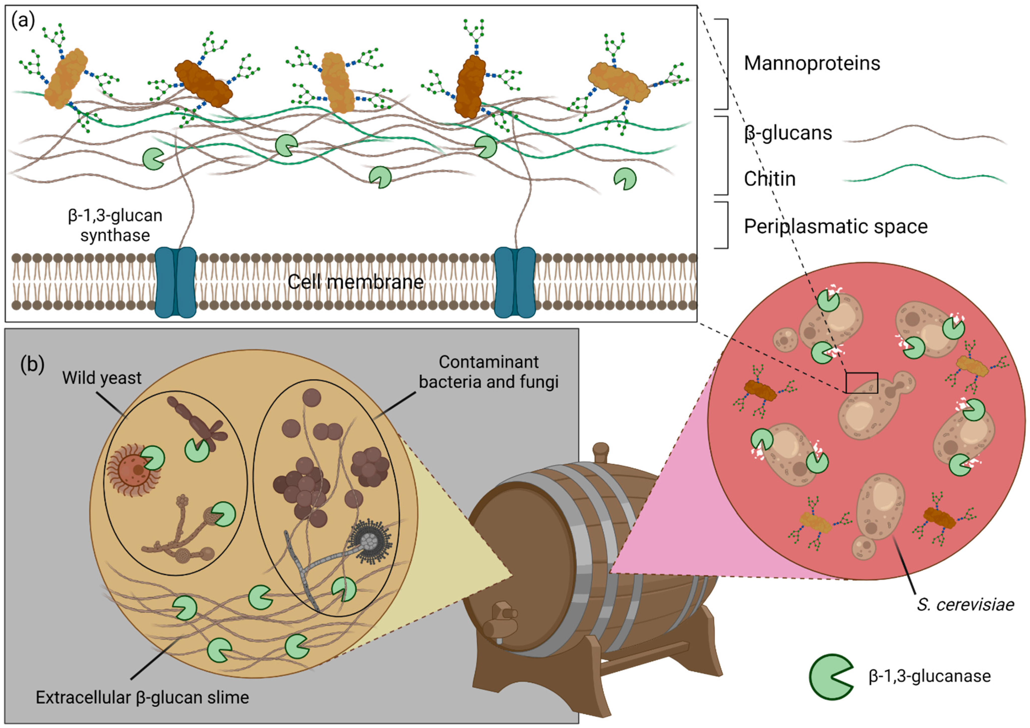
  260. Dimopoulou, M.; Lonvaud-Funel, A.; Dols-Lafargue, M. Polysaccharide Production by Grapes Must and Wine Microorganisms. In Biology of Microorganisms on Grapes, in Must and in Wine; König, H., Unden, G., Fröhlich, J., Eds.; Springer International Publishing: Cham, Switzerland, 2017; pp. 293–314. ISBN 978-3-319-60020-8. [Google Scholar]
  261. Hüfner, E.; Haßelbeck, G. Application of Microbial Enzymes During Winemaking. In Biology of Microorganisms on Grapes, in Must and in Wine; König, H., Unden, G., Fröhlich, J., Eds.; Springer International Publishing: Cham, Switzerland, 2017; pp. 635–658. ISBN 978-3-319-60020-8. [Google Scholar]
  262. Blättel, V.; Larisika, M.; Pfeiffer, P.; Nowak, C.; Eich, A.; Eckelt, J.; König, H. β-1,3-Glucanase from Delftia Tsuruhatensis Strain MV01 and Its Potential Application in Vinification. Appl. Environ. Microbiol. 2011, 77, 983–990. [Google Scholar] [CrossRef] [PubMed] [Green Version]
  263. Martínez, J.M.; Cebrián, G.; Álvarez, I.; Raso, J. Release of Mannoproteins during Saccharomyces Cerevisiae Autolysis Induced by Pulsed Electric Field. Front. Microbiol. 2016, 7, 1435. [Google Scholar] [CrossRef] [PubMed]
  264. Gacto, M.; Vicente-Soler, J.; Cansado, J.; Villa, T.G. Characterization of an Extracellular Enzyme System Produced by Micromonospora Chalcea with Lytic Activity on Yeast Cells. J. Appl. Microbiol. 2000, 88, 961–967. [Google Scholar] [CrossRef] [Green Version]
  265. Miyanishi, N.; Inaba, Y.; Okuma, H.; Imada, C.; Watanabe, E. Amperometric Determination of Laminarin Using Immobilized β-1,3-Glucanase. Biosens. Bioelectron. 2004, 19, 557–562. [Google Scholar] [CrossRef]
  266. Pettersen, E.F.; Goddard, T.D.; Huang, C.C.; Meng, E.C.; Couch, G.S.; Croll, T.I.; Morris, J.H.; Ferrin, T.E. UCSF ChimeraX: Structure Visualization for Researchers, Educators, and Developers. Protein Sci. 2021, 30, 70–82. [Google Scholar] [CrossRef]
Figure 1. The applications of β-glucans and β-1,3-glucanases. Due to their unique physicochemical and bioactive properties, β-glucans and their processing enzymes are attractive molecules with multiple applications, ranging from cancer treatment to brewing.
Ijms 23 03156 g001
Figure 2. Structural features of β-glucans. (a) Primary structure of β-1,3-glucans from different sources. (b) Primary structure of mixed-linkage glucans. The arrows point towards lichenase cleavage sites. Lichenase is an endo-hydrolase that hydrolyses the MLGs into smaller oligomers. Oligosaccharide profiles in DPs vary depending on the taxonomic origin of MLGs. (c) Triple helical quaternary structure of curdlan. Each individual chain is represented in a different color. The pitch and diameter of the helix are represented, as well as the triangular arrangement of interstrand H-bonds.
Ijms 23 03156 g002
Figure 3. Tridimensional structure of BhGH81 and PbGH64 in complex with laminarin oligosaccharides. Gray-colored Van der Waals surface of (a) BhGH81 and (c) PbGH64, with the laminarin oligosaccharides in stick representation bound to the active site, highlighting the β-glucan quaternary structure recognition feature of both enzymes. Panels (c,d) show a detailed view of the pseudo-helical structures formed by the bound oligosaccharides. Each oligosaccharide strand is represented in a different color and identified with their degree of polymerization. In (b), DP2 and DP3 are likely to be the product of hydrolysis of BhGH81 on a DP5 laminarin chain, as this is the structure of an active wild-type enzyme. PDB codes: 5t4g (BhGH81), 5h9y (PbGH64).
Ijms 23 03156 g003
Figure 4. Substrate specificities of different CBM4 are dictated by active site conformation. Gray-colored Van der Waals surfaces of (a) CfCBM4 bound to cellopentaose (blue) and of (b) TmCBM4 bound to laminarihexaose (green). Both ligands are shown in stick representation. TmCBM4 shows a more “U-shaped” binding site as a result of two loop extensions (black and blue arrows). An extra loop (black arrow) on TmCBM4 creates an extra binding surface that interacts with the non-reducing end of the laminarin oligosaccharide. Panel (c) was obtained by superposing the structures of CfCBM4 and TmCBM4, and shows how the second extra loop on TmCBM4 (blue arrow) does not allow the more linear cellulose oligosaccharide to fit into the binding site. PDB codes: 1gu3 (CfCBM4), 1gui (TmCBM4).
Ijms 23 03156 g004
Figure 6. Complement-dependent antitumor cytotoxicity mediated by β-glucans. Due to the lack of β-glucans, tumor cells are not able to induce dual binding to leukocytes, promoting complement-dependent cytotoxicity. However, the introduction of exogenous β-glucans can generate dual-binding of leukocytes to iC3b-positive tumor cells. β-glucans are captured and internalized by the macrophages via the Dectin-1 receptor with or without TLR-2/6. The large β-glucan molecules are fragmented into smaller sized β-glucan fragments within the macrophages and are carried to the marrow and endothelial reticular system and subsequently released. These small β-glucan fragments are eventually recognized by the circulating granulocytes, monocytes or macrophages via the complement receptor (CR)-3, priming leukocytes against tumor cells opsonized with iC3b by monoclonal antibodies.
Ijms 23 03156 g006
Figure 7. Applications of β-1,3-glucanases in the winemaking process. (a) The starter yeasts, usually S. cerevisiae, possess several components in their cell wall, most importantly mannoproteins that, when released into the wine, improve several organoleptic and technological properties of the final product. By hydrolyzing yeast cell wall β-glucans, β-1,3-glucanases promote the release of those mannoproteins. (b) Exogenous β-1,3-glucanases also contribute to improved wine quality and longevity by promoting the lysis of contaminant wild yeast, which are responsible for several off-flavors, and by hydrolyzing the extracellular β-glucan-rich slime produced by contaminant fungi and bacteria, which are the cause of the undesirable wine “ropiness”.
Ijms 23 03156 g007
Table 1. Distribution of characterized β-1,3-glucanases across glycoside hydrolase (GH) families and taxa, according to the CAZy.org database (as of February 2022).
β-1,3-glucanase ClassDomainGH Families
3589161751556481128152157158
EC 3.2.1.58Bacteria 2 2
Fungi 15 13
Plants 2
EC 3.2.1.39Archaea 1
Bacteria 1 273 1533 13
Fungi 21 2 1011
Plants 28 1
Animals 14
EC 3.2.1.6Bacteria3 2211 1
Fungi 6
Unclassified 3
Publisher’s Note: MDPI stays neutral with regard to jurisdictional claims in published maps and institutional affiliations.

