Article

Melatonin Positively Regulates Both Dark- and Age-Induced Leaf Senescence by Reducing ROS Accumulation and Modulating Abscisic Acid and Auxin Biosynthesis in Cucumber Plants

State Key Laboratory of Crop Biology, Key Laboratory of Crop Biology and Genetic Improvement of Horticultural Crops in Huanghuai Region, College of Horticulture Science and Engineering, Shandong Agricultural University, Tai’an 271018, China
*
Author to whom correspondence should be addressed.
These authors contributed equally to this work.
Int. J. Mol. Sci. 2022, 23(7), 3576; https://0-doi-org.brum.beds.ac.uk/10.3390/ijms23073576
Submission received: 10 February 2022 / Revised: 15 March 2022 / Accepted: 16 March 2022 / Published: 25 March 2022
(This article belongs to the Special Issue Response to Environmental Stress in Plants)

Abstract

:
Melatonin (MT), as a signaling molecule, plays a vital role in regulating leaf senescence in plants. This study aimed to verify the antioxidant roles of MT in delaying dark- or age-induced leaf senescence of cucumber plants. The results showed that endogenous MT responds to darkness and overexpression of CsASMT, the key gene of MT synthesis, and delays leaf senescence stimulated by darkness, as manifested by significantly lower malonaldehyde (MDA) and reactive oxygen species (ROS) contents as well as higher activities and gene expression of antioxidant enzymes compared to the control. Moreover, MT suppressed both age- or dark-induced leaf senescence of cucumber, as evidenced by a decrease in senescence-related gene SAG20 and cell-death-related gene PDCD expression and ROS content and an increase in antioxidant capacity and chlorophyll biosynthesis compared with the H2O-treated seedlings. Meanwhile, the suppression of age-induced leaf senescence by melatonin was also reflected by the reduction in abscisic acid (ABA) biosynthesis and signaling pathways as well as the promotion of auxin (IAA) biosynthesis and signaling pathways in cucumber plants in the solar greenhouse. Combining the results of the two separate experiments, we demonstrated that MT acts as a powerful antioxidant to alleviate leaf senescence by activating the antioxidant system and IAA synthesis and signaling while inhibiting ABA synthesis and signaling in cucumber plants.

1. Introduction

Cucumber (Cucumis sativus L.) is an important vegetable crop that is cultivated worldwide. However, leaf senescence induced by age or abiotic stress leads to an obvious decrease in photosynthetic efficiency, which further results in a decline in production because of the shortened harvest time [1,2,3], particularly during the cultivation pattern of one crop per year in a solar greenhouse. Thus, seeking an appropriate method to delay leaf senescence and further unraveling the mechanisms regulating leaf senescence of cucumber plants will help extend their life and improve the harvest yield during cucumber production.
Leaf senescence is generally the first stage of plant senescence, during which a number of degenerative events occur, such as a decrease in metabolic activities, cell death, protein degradation, and decomposition of organoids [4,5,6]. Leaf senescence, characterized by yellowing leaves, results in a decline in photosynthetic efficiency and limits the growth and yield of plants, and it is often accompanied by age or accelerated by abiotic stress, such as chilling, high temperature, drought, and darkness [6,7,8]. Leaf senescence has been reported to be related to the accumulation of reactive oxygen species (ROS), which further results in aggravated oxidative damage to cellular macromolecules [9,10]. For instance, the overaccumulation of superoxide anion (O2) and hydrogen peroxide (H2O2), caused by the upregulation of the expression of respiratory burst oxidase homolog (RBOH) genes, was observed in senescent leaves of Chinese flowering cabbage during storage after harvest [11]. In addition, phytohormone signals are also involved in the regulation of leaf senescence, such as abscisic acid (ABA), jasmonic acid (JA), salicylic acid (SA), auxin (IAA), and cytokinin (CTK), and among these phytohormones, the application of exogenous ABA, JA, and SA enhances leaf senescence, and higher endogenous contents of ABA, JA, and SA are observed in senescent leaves, while IAA and CTK are reported to positively delay leaf senescence [12,13,14,15,16].
Melatonin (MT) is a small indole substance that is ubiquitous in animals, prokaryotes, and plants. MT, as a new type of hormone-like substance, was first discovered in the bovine pineal gland in 1958 [17]. Numerous publications have reported that MT not only regulates root development and promotes the growth of plants [18] but is also a powerful antioxidant that can delay leaf senescence induced by age or abiotic stresses [19,20,21,22], and the main mechanism of melatonin is directly scavenging ROS or activating antioxidant enzymes by upregulating mRNA abundance and promoting the ascorbate–glutathione (AsA–GSH) cycle to eliminate excess ROS [23]. For instance, Shi reported that MT delayed dark-induced leaf senescence by improving antioxidant and AsA–GSH cycle systems to decrease the accumulation of ROS and inhibit the expression of SAGs [24]. Meanwhile, the downregulation of RBOH (RbohB, RbohC, RbohD, RbohD2, and RbohE) was observed in melatonin-treated Chinese flowering cabbage to inhibit RBOH-mediated ROS production [11]. In addition, regarding the specific mechanism of action, the function of MT in the regulation of leaf senescence is related to other phytohormones [25]. For instance, exogenous application of melatonin repressed the expression of ABA biosynthetic genes and ABA signal-related genes ABF1, ABF4, and ABI5 to decrease ABA content and delayed cabbage senescence [26], and CTK was reported to be activated by melatonin to alleviate leaf senescence [7].
Thus, the application of MT may be an effective approach to delay leaf senescence and finally increase the yield in greenhouse vegetable production; however, the role of MT in regulating senescence is not as clear as that of other phytohormones. In our previous study, we found that MT significantly improved the chilling tolerance of cucumber by enhancing photosynthetic carbon assimilation [27]. In this study, we constructed an overexpression vector of the key MT biosynthetic gene ASMT and found that the overexpression of ASMT obviously upregulated the mRNA abundance of antioxidant enzyme genes and promoted the accumulation of AsA and GSH, which further decreased the excess ROS content in dark-induced senescent cucumber leaves. More importantly, we also found that the application of melatonin could also improve the chlorophyll content and downregulate SAG20 and the cell death of leaves at 28, 35, and 42 days of leaf age, which ultimately delayed the leaf senescence of cucumber plants in the solar greenhouse. These results will be valuable for increasing yields in cucumber plants and will provide a theoretical basis for the high-efficiency application of MT during vegetable cultivation.

2. Results

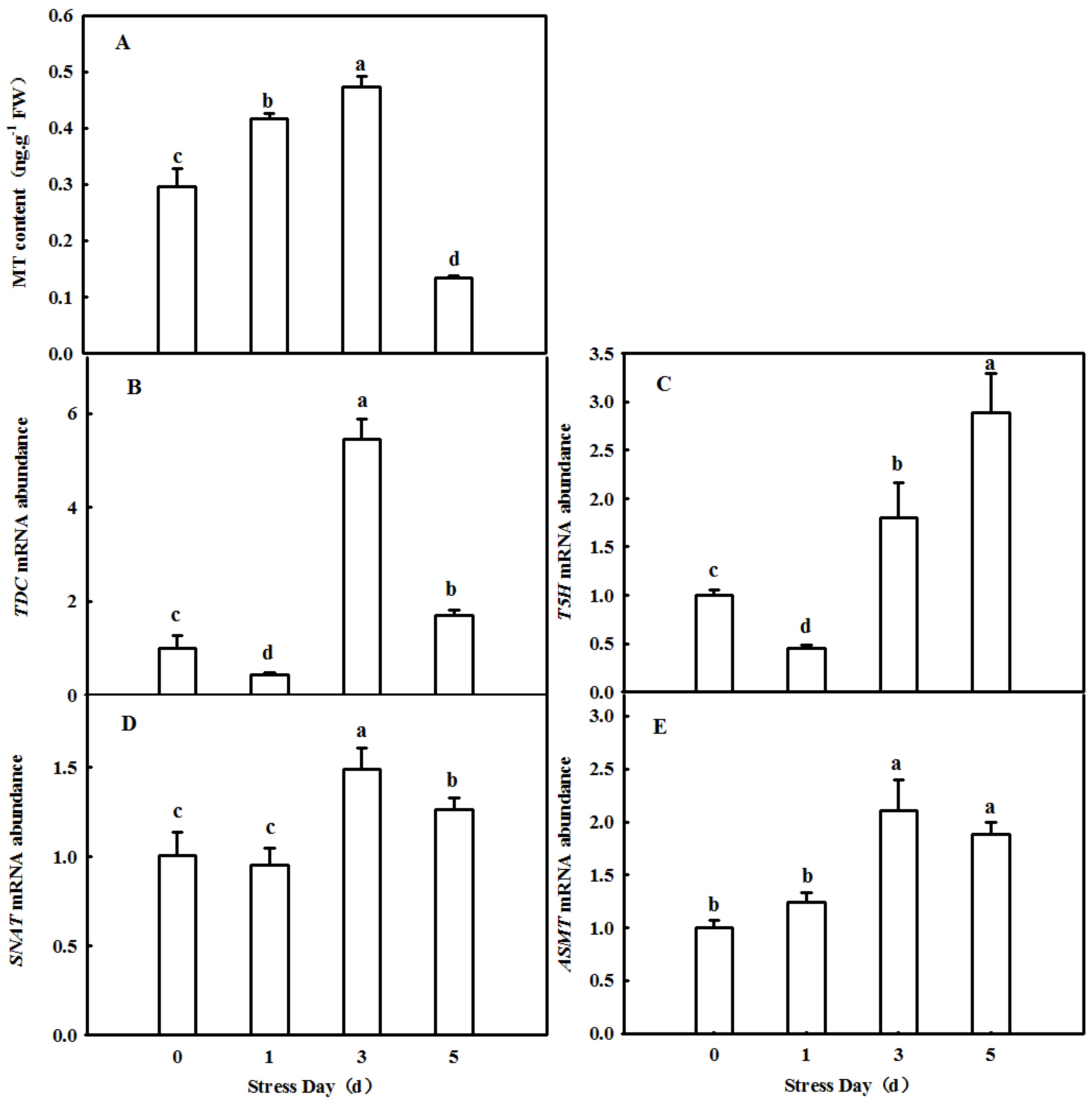
2.1. Dynamic Changes in Endogenous MT in Cucumber Leaves in Which Senescence Was Induced by Darkness

To investigate the effect of senescence factors on the content of melatonin in cucumber leaves, a dark environment was used to induce rapid senescence. In cucumber leaves, the content of melatonin first increased and then decreased with the extension of treatment time. After 3 days of treatment, the content of melatonin increased to the highest point, 0.4723 ng·g−1, and then decreased to the lowest point, 0.1333 ng·g−1 (Figure 1A). In addition, we also found a change in the trend of mRNA abundance of TDC, T5H, SNAT, and ASMT, which are the key melatonin synthesis genes, and Figure 1B–E shows that these changes were consistent with the melatonin content. This suggests that darkness induces melatonin synthesis and indirectly affects melatonin content.

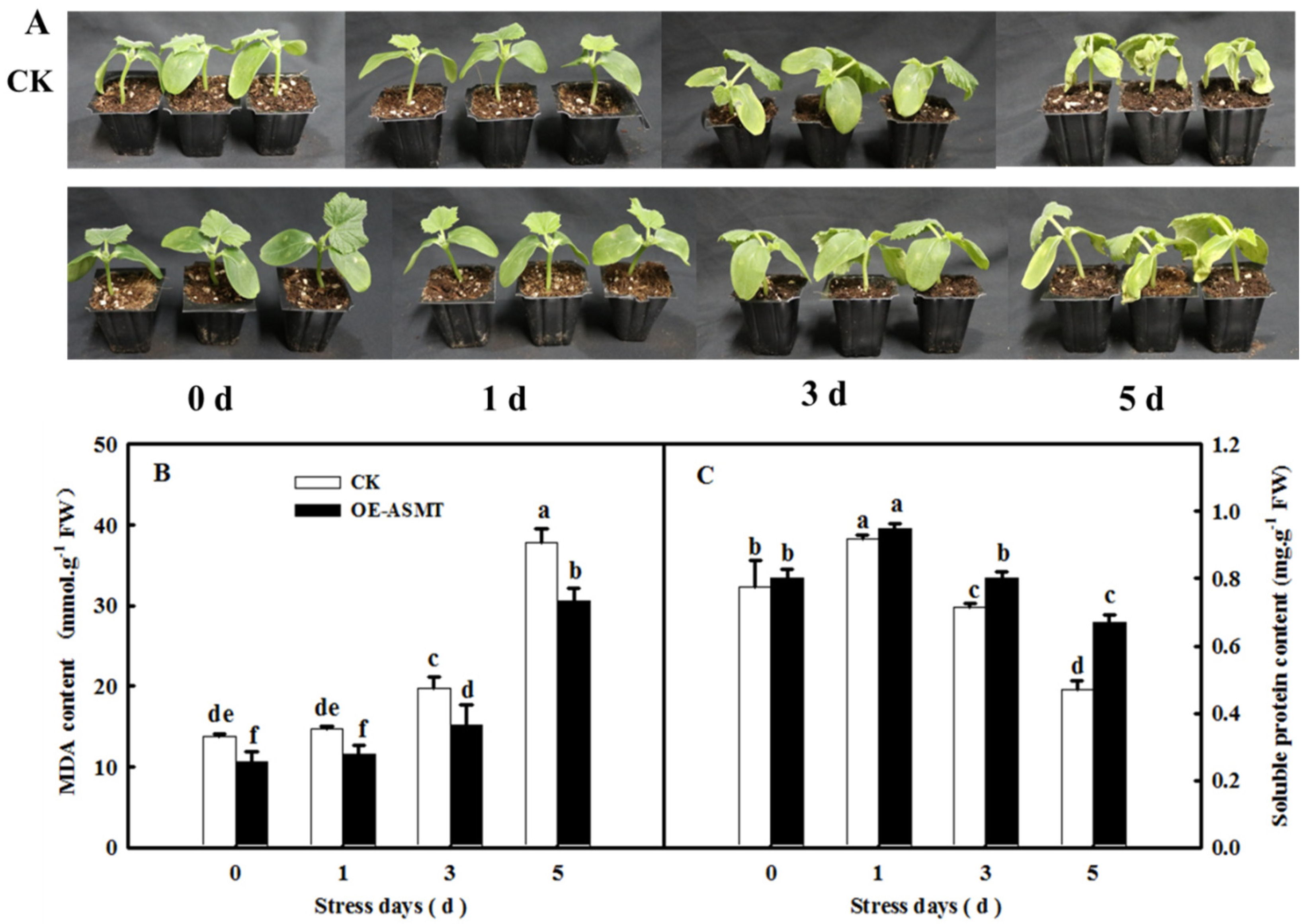
2.2. Effects of ASMT Overexpression on Dark-Induced Cucumber Leaf Senescence

To further understand the important role of MT in leaf senescence, we constructed a transient overexpression vector of CsASMT, a key gene for MT synthesis, and the endogenous MT content was indeed increased (data not shown). As shown in Figure 2A, after 5 days of dark stress, the cotyledons of cucumber seedlings in the control group showed yellow coloring and serious wilting. Moreover, the soluble protein content of cucumber seedlings in the control was significantly lower than that in the OE-ASMT seedlings, and there was a significant difference after treatment for 3 days. After treatment for 5 days, the soluble protein content of the control seedlings decreased by 180% compared with that in the OE-ASMT seedlings (Figure 2C). Overexpression of CsASMT also had a more obvious effect on alleviating MDA accumulation, the difference was obvious at the early stage of dark stress, and the difference increased with the extension of treatment time (Figure 2B). These results further demonstrated the positive regulation of MT on cucumber leaf senescence.

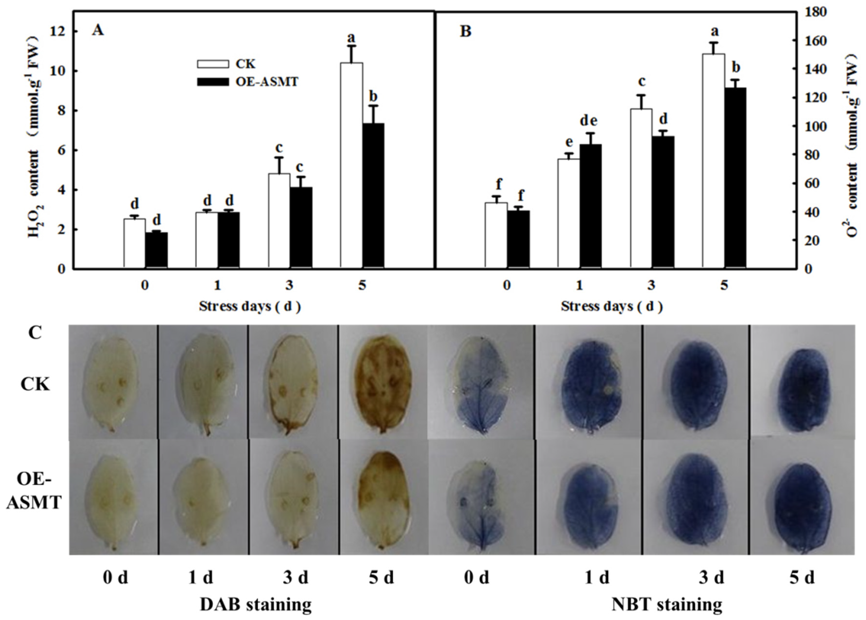
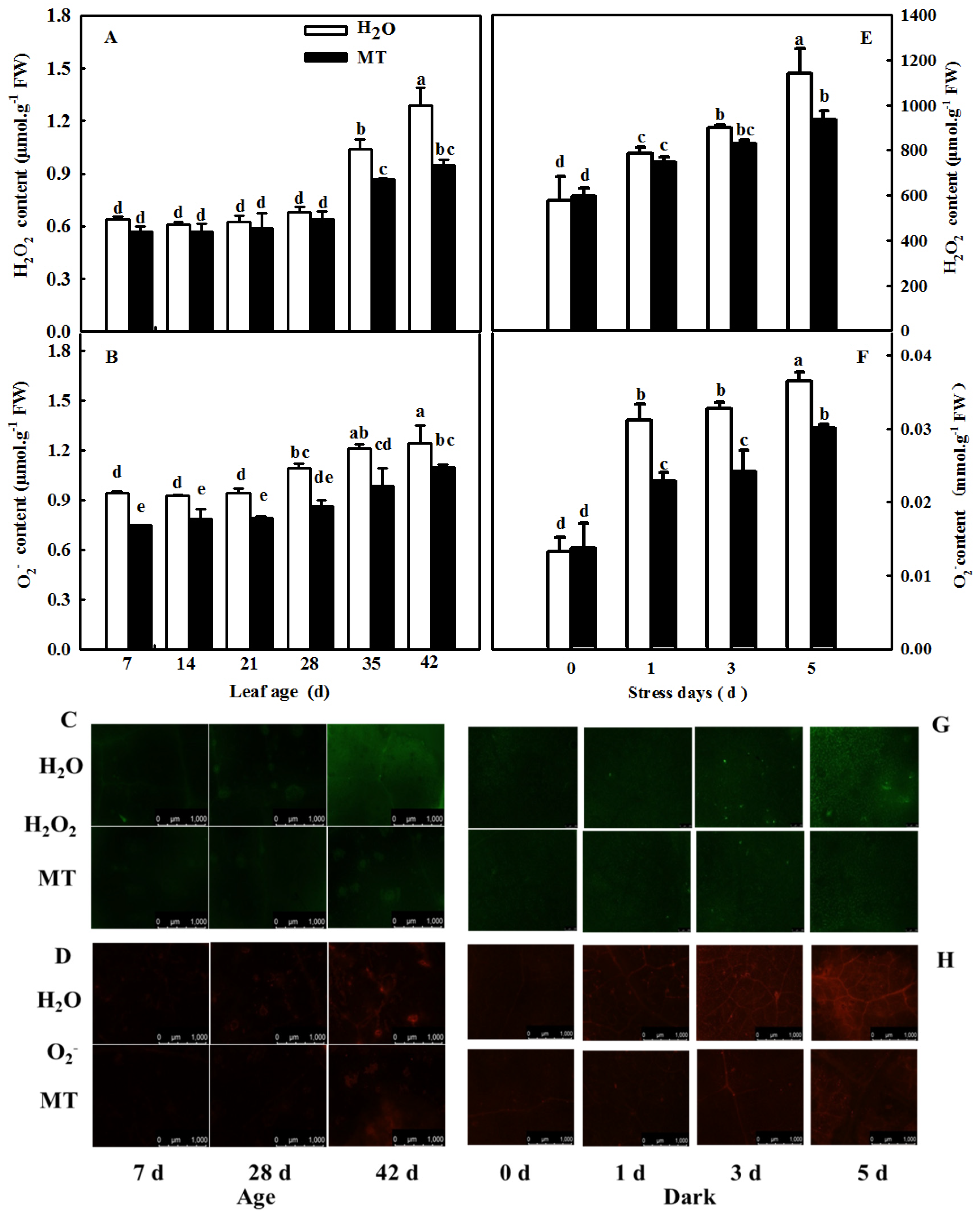
2.3. Effects of ASMT Overexpression on ROS Content in Cucumber Leaves in Which Senescence Was Induced by Darkness

As shown in Figure 3A, the H2O2 and O2 contents of cucumber cotyledons in control and CsASMT overexpression plants increased gradually (Figure 3A,B), and after 5 days of darkness, the contents of H2O2 and O2 in leaves of overexpressed CsASMT plants were significantly lower than those of the control, decreasing by 29.5% and 15.7%, respectively. In accordance with the change in ROS content, the diaminobenzidine (DAB) and nitroblue tetrazolium (NBT) staining (Figure 3C) results confirmed that ROS increased significantly in both treatments and that ROS content was higher than that in CsASMT-overexpressing plants under darkness.

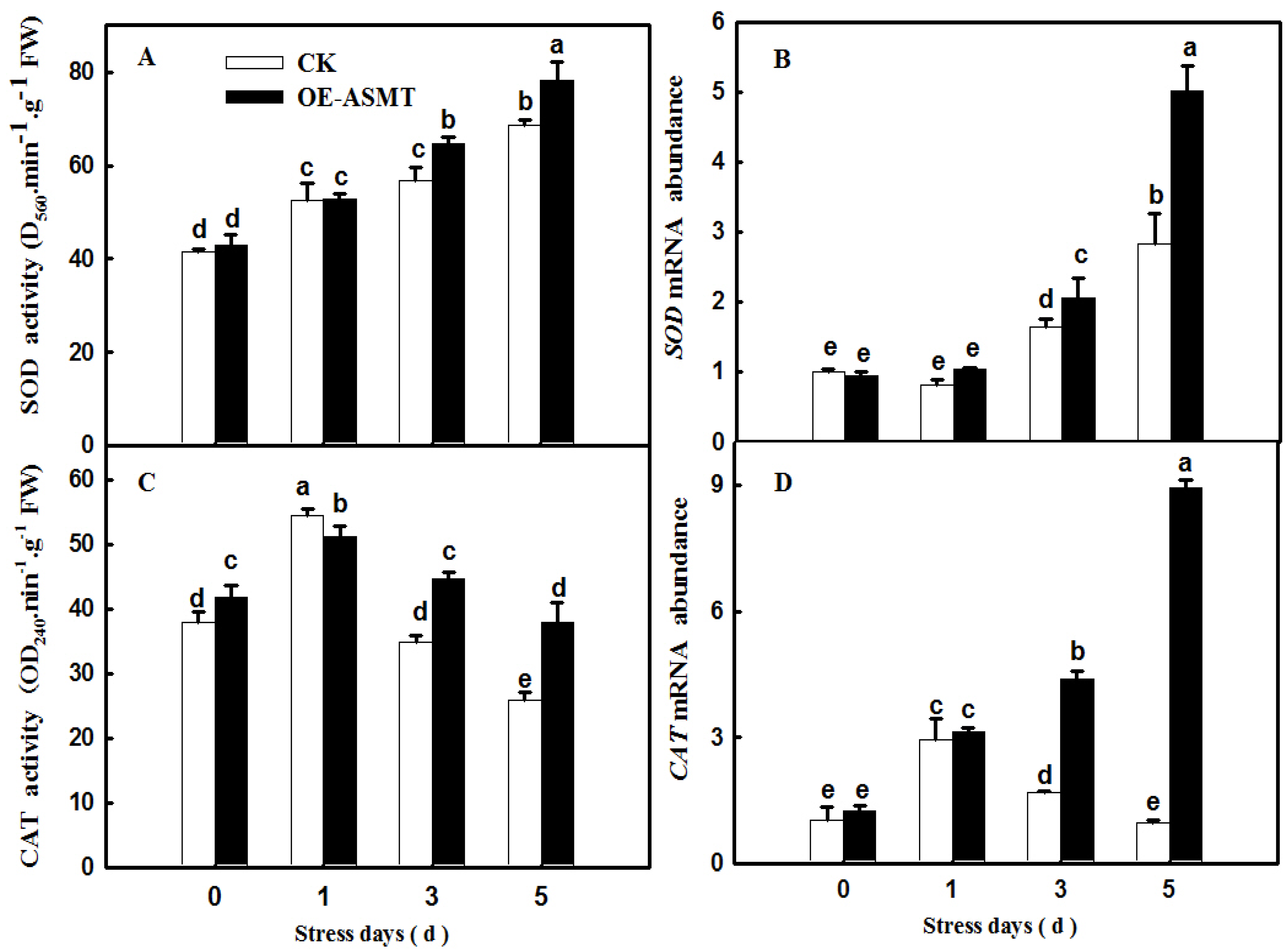
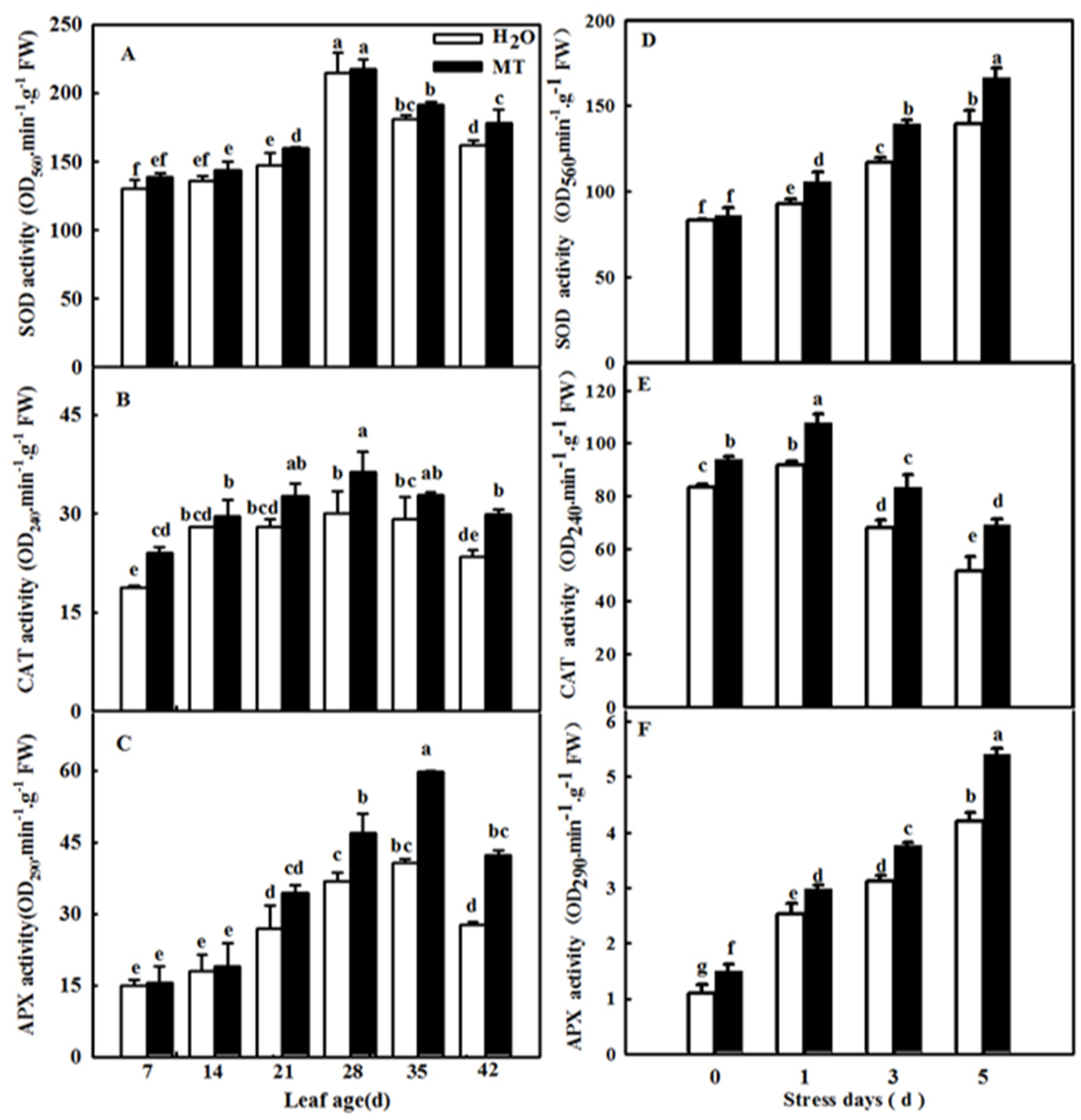
2.4. Effects of ASMT Overexpression on Antioxidant Enzyme Activity in Cucumber Leaves in Which Senescence Was Induced by Darkness

To further explore the effect of CsASMT overexpression on the antioxidant defense system of cucumber under darkness, we further measured the activities and relative mRNA expression levels of superoxide dismutase (SOD) and catalase (CAT). The data showed that darkness induced the activities of SOD and CAT, and their activities in CsASMT overexpression seedlings were obviously higher than those in the control. Meanwhile, the overexpression of ASMT also increased the mRNA abundance of SOD and CAT compared with the control seedlings under dark conditions (Figure 4). The results implied that MT could effectively improve the activity and expression of antioxidant enzymes and further alleviate the accumulation of ROS.

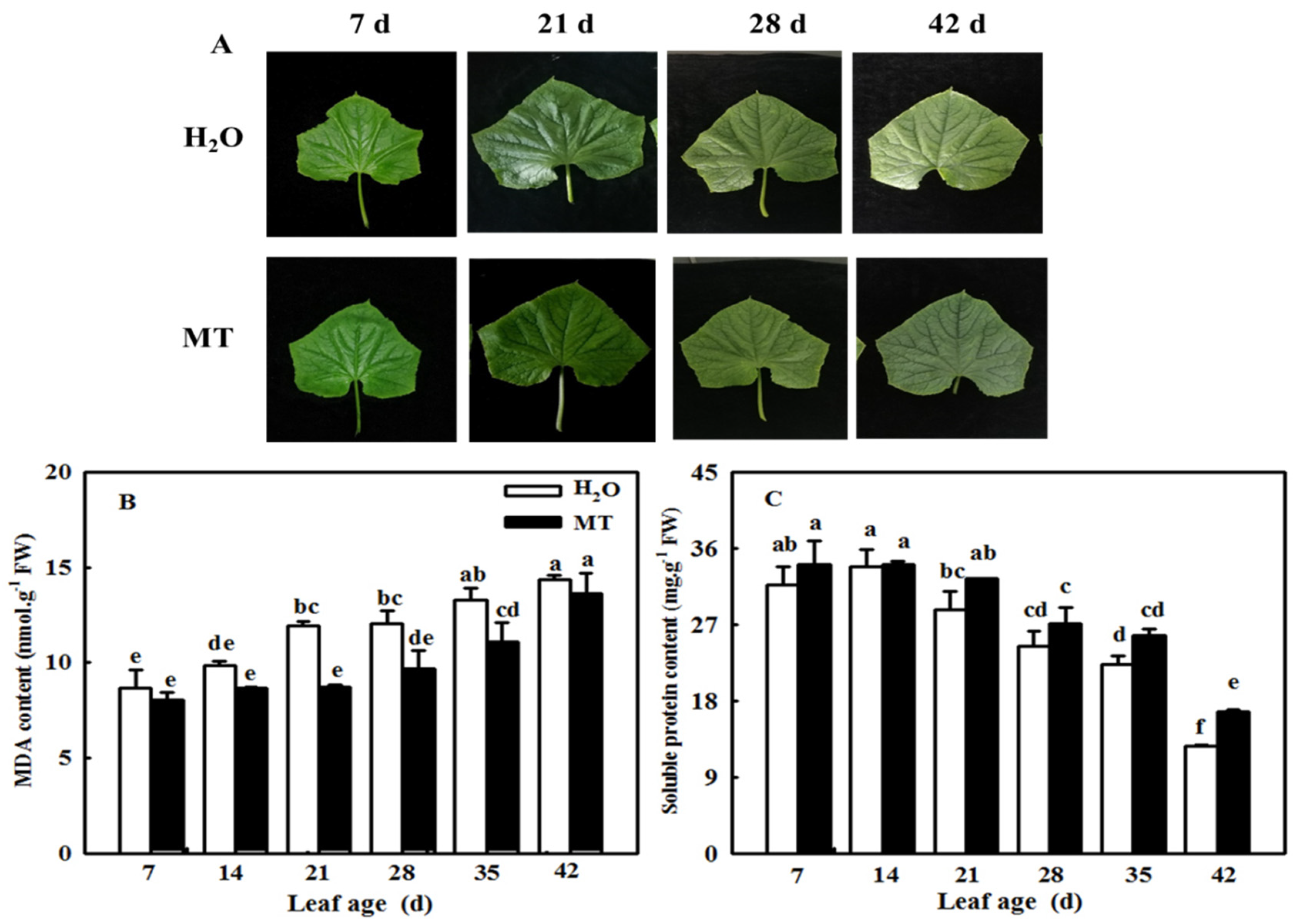
2.5. MT Delays the Cucumber Leaf Senescence Induced by Age

To further verify whether the application of MT could be used during cucumber production, we observed the leaf phenotype and measured the change in MDA and soluble protein contents in leaves of different ages. As shown in Figure 5, the leaves of cucumber were obviously yellow-green, showed obvious signs of senescence at 42 days of age, and showed a significant increase in MDA content and a decrease in soluble protein content. However, the application of MT notably showed more green leaves, lower MDA content, and higher protein contents compared with the H2O treatment. For instance, the soluble protein content was 31.4% higher than that of the control at 42 days of leaf age.

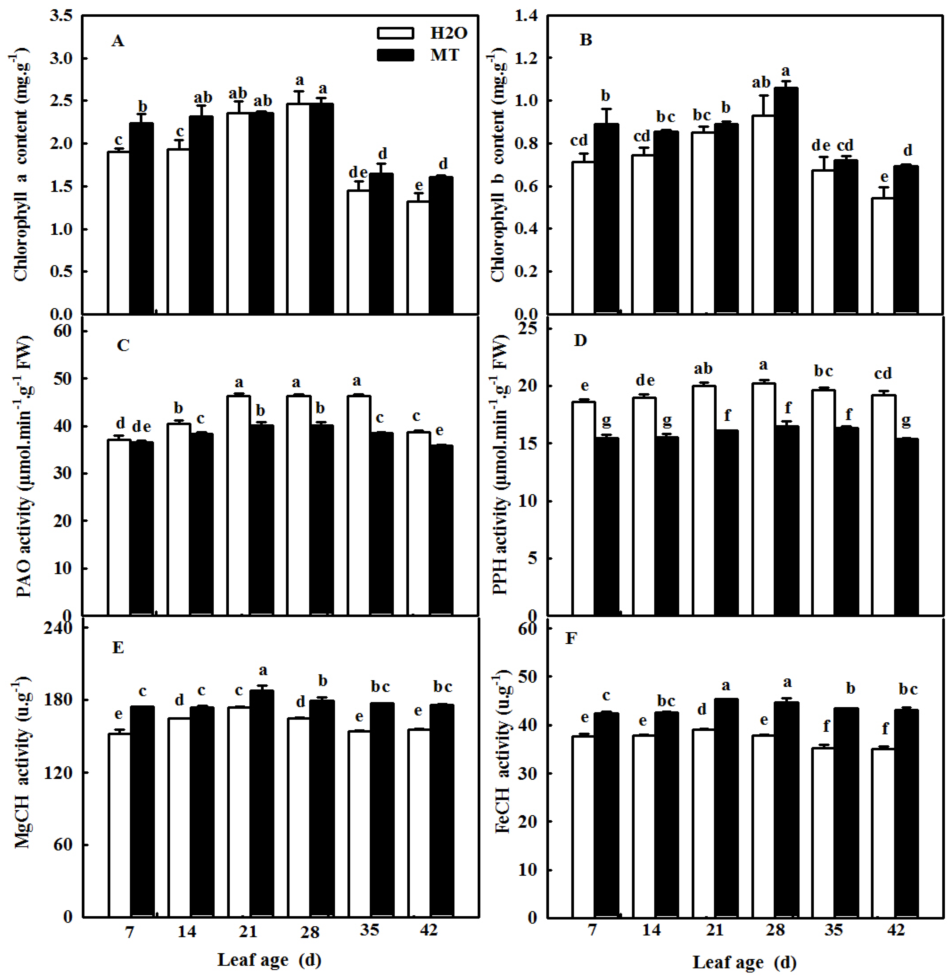
2.6. MT Regulates Chlorophyll Metabolism Enzyme Activity to Maintain Chlorophyll Content during Cucumber Leaf Senescence

Yellowing is a notable phenomenon during leaf senescence. To prove whether MT could relieve the decrease in chlorophyll during leaf senescence induced by age, we observed changes in not only the activity of chlorophyll synthetase but also chlorophyll degradation enzymes. Figure 6A shows that the chlorophyll contents of cucumber leaves first increased and then decreased at the leaf age of 35 days, which implied that the leaves began to be senescent. Meanwhile, we found that the activities of PAO, PPH, MgCH, and FeCH showed a similar trend in the chlorophyll contents with increasing leaf age; however, the application of MT maintained higher MgCH and FeCH activities, which were related to chlorophyll synthesis, and suppressed PAO and PPH activities, which were related to chlorophyll degradation (Figure 6C–F). These results signify the importance of MT in regulating the balance of chlorophyll synthesis and degradation to delay leaf senescence in cucumber.

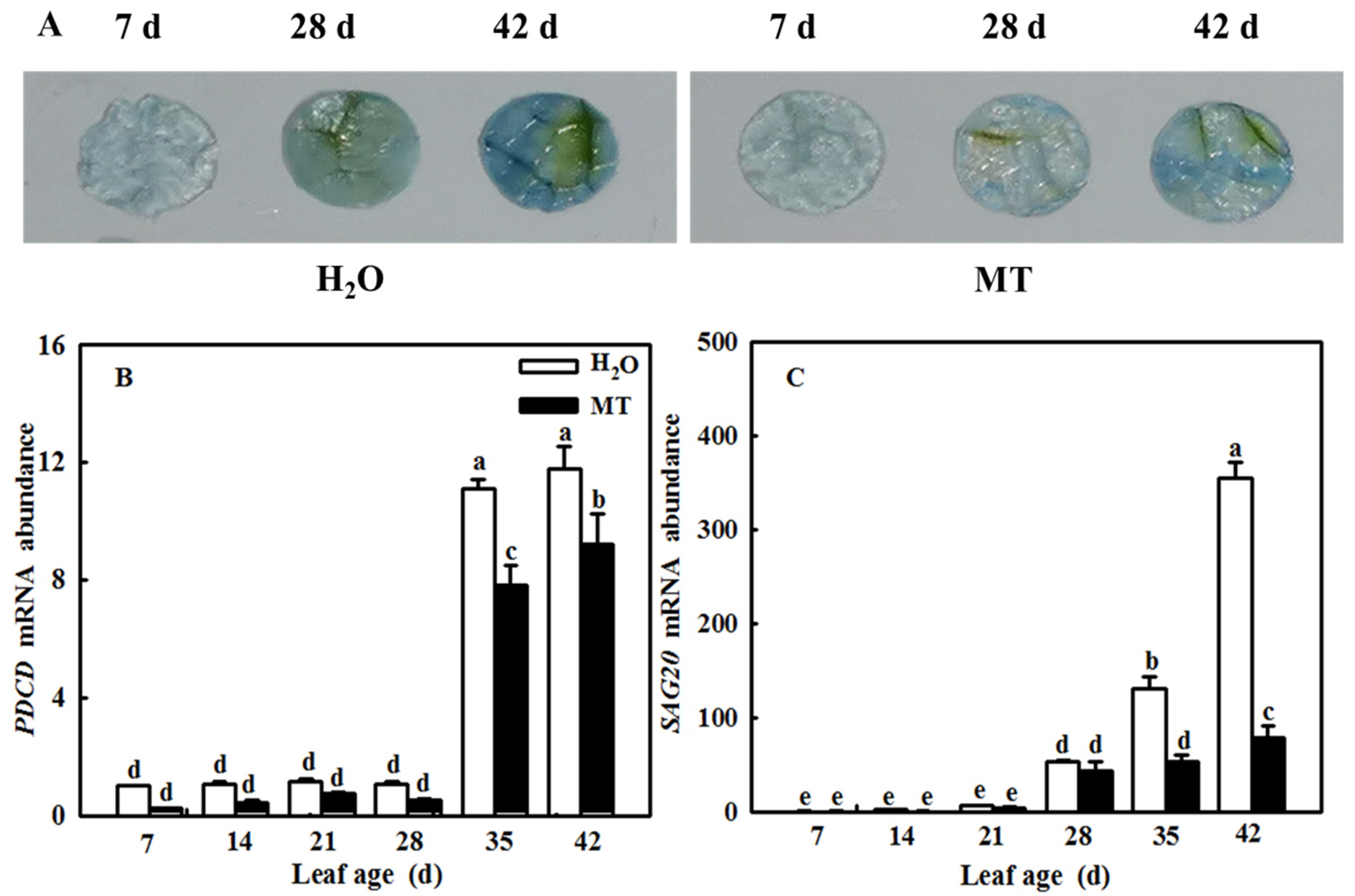
2.7. MT Downregulates Cell-Death- and Senescence-Related Gene mRNA Abundance to Delay Leaf Senescence Induced by Age

During the leaf senescence process, SAG mRNA levels were significantly upregulated, and plants started cell death. Here, we found that leaf age induced the relative mRNA expression of SAG20 and PDCD (Figure 7A,B), which gradually increased with increasing leaf age. Importantly, MT apparently alleviated cell death and downregulated the SAG20 mRNA level compared with the H2O-treated plants. The staining of programmed cell death was in agreement with the change in PDCD mRNA levels (Figure 7C).

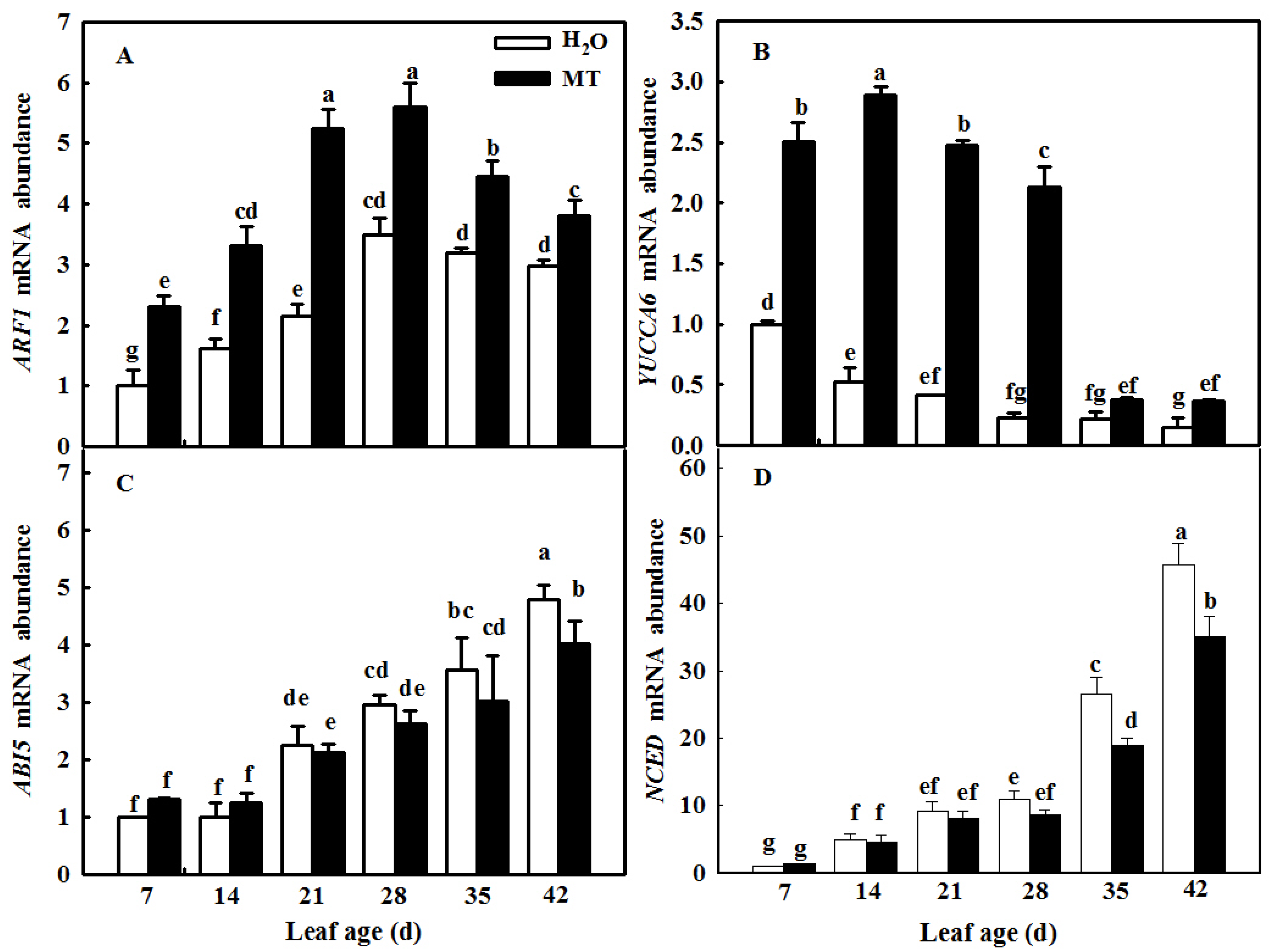
2.8. MT Modulates IAA and ABA Signals and Synthesis to Delay Leaf Senescence Induced by Age

Considering the importance of phytohormones for leaf senescence, we measured the mRNA abundance of IAA- and ABA-related genes in both H2O- and MT-treated leaves at different leaf ages. With the increase in age, ARF1 and YUCCA6 mRNA abundance levels first increased and then decreased at the leaf age of 35 days, when leaves began to be senescent; however, ARF1 and YUCCA6 mRNA abundance levels in MT were notably higher than those of H2O treatment (Figure 8A,B), implying that MT could significantly alleviate the decrease in IAA in age-induced senescent leaves. In contrast to the change in IAA, the mRNA abundance of the ABA-related genes ABI5 and NCED increased during the leaf development process (Figure 8C,D), but there was no significant difference between the H2O and MT treatments before the leaf age of 28 days, and ABI5 and NCED mRNA abundance levels in the MT treatment were obviously lower than those in the H2O treatment after the leaf age of 35 days, implying that MT inhibited ABA synthesis and signal transduction in senescent cucumber leaves.

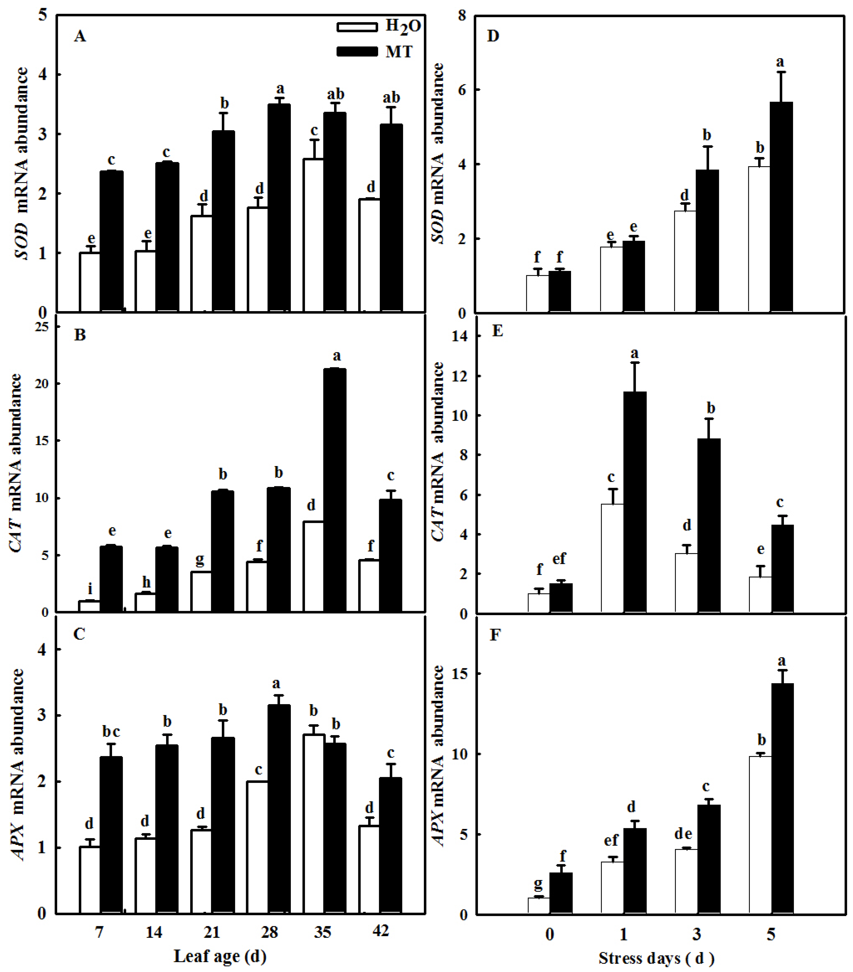
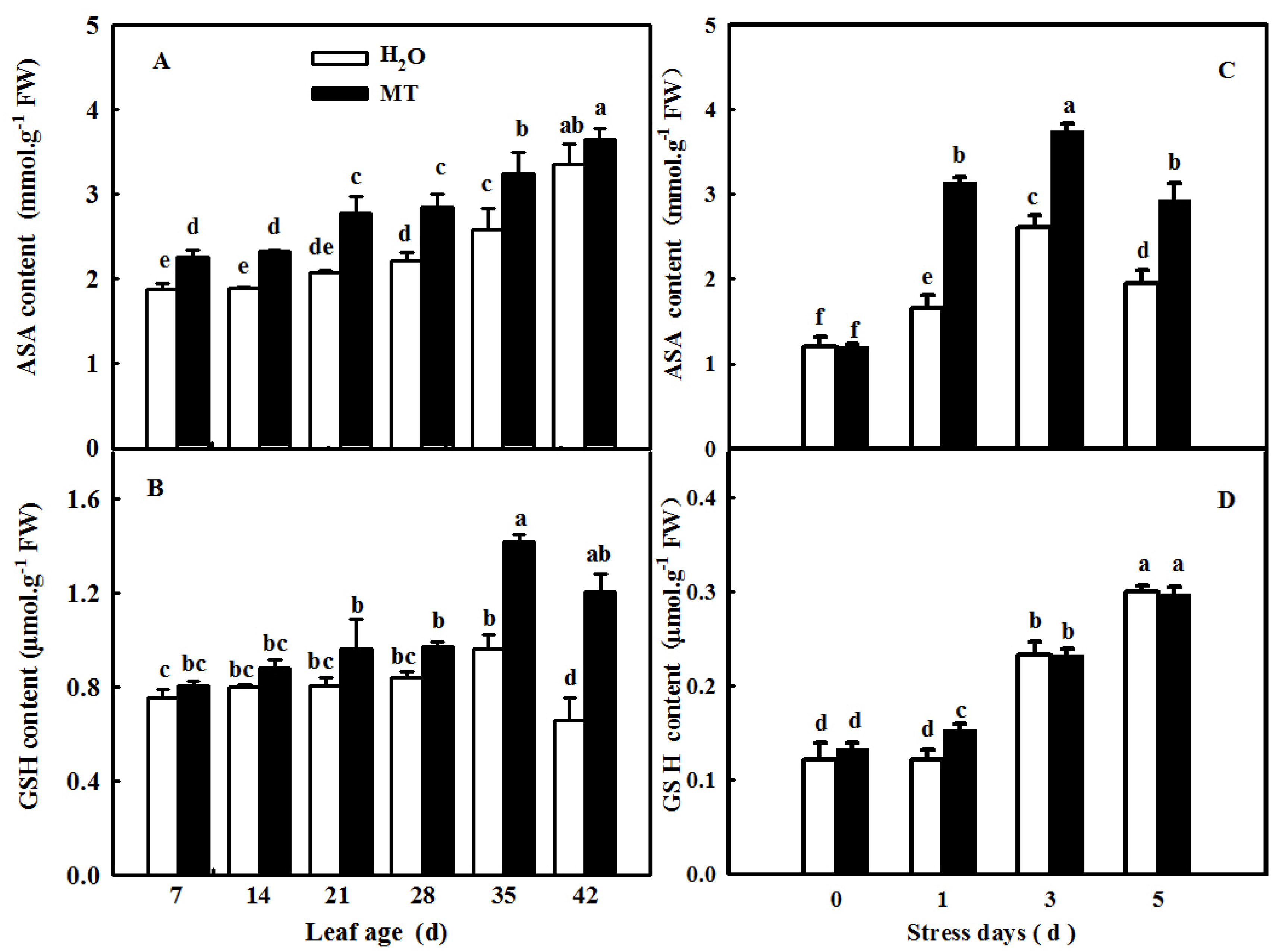
2.9. MT-Delayed Leaf Senescence Is Associated with Increased ROS-Scavenging Activity

Through the measurement of the H2O2 content and O2 production rate in leaves during natural senescence within 42 days or in leaves in which senescence was induced by darkness, it can be seen that the contents of H2O2 and the O2 production rate in leaves gradually increased during senescence induced by age or darkness, but the application of exogenous MT significantly inhibited the accumulation of ROS in both experiments compared with the H2O treatment (Figure 9).
Plants activate the antioxidant system when excess ROS occur during senescence or under abiotic stresses. Here, we observed the effect of exogenous MT on the activities and the relative expression of antioxidant enzymes during the cucumber leaf senescence process. As shown in Figure 10, the application of MT significantly promoted the activities of SOD, CAT, and ascorbate peroxidase (APX) in leaves in which senescence was induced by age or darkness. Moreover, real-time PCR data showed that senescence induced the mRNA expression of SOD, CAT, and APX, and MT-treated seedlings displayed obviously higher relative mRNA expression of SOD, CAT, and APX than H2O-treated seedlings (Figure 11), indicating that MT could upregulate the mRNA abundance of genes to further activate antioxidant enzyme activity. Considering that redox species are involved in ROS scavenging, we also examined the change in ASA and GSH contents of leaves at different ages or treated with darkness sprayed with or without MT. Our results showed that leaf senescence notably increased the contents of ASA and GSH, and this effect of MT on redox species contents was largely higher than that of H2O treatment in leaves in which senescence was induced by age or darkness (Figure 12), which implied that MT could promote the antioxidant capacity to scavenge ROS accumulation caused by senescence induced by age or darkness.

3. Discussion

MT serves as an important signaling molecule in the regulation of leaf senescence induced by abiotic stresses, darkness, and age [19,23,28]. With increasing age, the endogenous MT content of Arabidopsis thaliana increased, and the application of exogenous MT also increased the concentration of MT to further delay leaf senescence [19], implying that the endogenous MT level played a vital role in the regulation of MT on the resistance of plants. TDC, T5H, SNAT, and ASMT or HIOMT were the key genes for MT synthesis, which was edited as a genetic approach to change the MT level [29]. For instance, the overexpression of ASMT significantly alleviated photoinhibition and induced the expression of HSPs to promote the thermotolerance of tomato [30], while the silencing of TDC aggravated the accumulation of cadmium in tomato [31]. Meanwhile, COMT1 overexpression promoted melatonin biosynthesis and contributed to the alleviation of carbendazim phytotoxicity and residues in tomato plants [32]. Usually, leaf senescence induced by darkness is used to simulate natural senescence because leaves with dark-induced senescence show changes in chlorophyll content and a yellow leaf phenotype similar to those during natural senescence [33,34]. Thus, we first studied the effect of ASMT overexpression on leaf senescence induced by darkness, and the data showed that darkness induced the upregulation of CsT5H, CsASMT, and CsTDC mRNA abundance, which further led to an increase in endogenous MT content, implying that MT acts as a regulator during leaf senescence (Figure 1). Overexpression of ASMT dramatically delayed dark-induced leaf senescence compared with the control, as evidenced by the green phenotype and lower contents of MDA and soluble protein (Figure 2). Consistent with this result, according to the data from the field experiment, the application of exogenous MT also notably decreased the MDA and soluble protein contents in senescing cucumber leaves at 28, 35, and 42 days of leaf age in the solar greenhouse (Figure 5).
Previous studies have demonstrated that ROS are involved in the regulation of plant senescence [35,36,37], and excess ROS content leads to oxidative damage to lipids, proteins, and DNA during leaf senescence [38,39]. In this paper, the quantification and histochemical analysis of H2O2 and O2 showed that obvious ROS build-up was observed in senescing cucumber leaves induced by darkness or age, which was lower in ASMT-overexpressing and melatonin-treated cucumber leaves (Figure 3 and Figure 9), and thus, the lower lipid peroxidation product MDA content was also measured in ASMT-overexpressing or MT-treated cucumber leaves, which was in accordance with the results of Tan [11] in Chinese cabbage, and melatonin was thought to be an antioxidant [39,40]. Usually, leaf senescence is considered to be a form of programmed cell death (PCD), and higher transcript levels of senescence-associated genes (SAGs) are induced during this process [4,5]. Programmed cell death is associated with abiotic-triggered oxidative stress caused by excessive production of ROS, such as H2O2 and O2, in cells [41,42]. Our study showed that a corresponding increase in cell death in both trypan blue staining and PDCD mRNA abundance and an upregulation of SAG12 mRNA abundance were observed in senescing cucumber leaves. However, aging cucumber leaves pretreated with melatonin effectively inhibited cell death, which was related to the lower ROS content observed in senescing cucumber leaves induced by darkness or age (Figure 3, Figure 7 and Figure 9). The yellowing of green leaves due to the degradation of chlorophyll (Chl) and Chl–protein complexes is the first observed typical phenomenon during leaf senescence [43], and chlorophyllase1 (CLH1), CLH2, pheophytin pheophorbide hydrolase (PPH), pheophorbide A oxygenase (PAO), and NYC1-like (NOL) play a vital role in chlorophyll degradation, and these are obviously upregulated by senescence-related phytohormones as well as abiotic stresses to accelerate leaf senescence [44,45,46,47,48]. Here, we found a green phenotype and higher chlorophyll content by not only downregulating the activities of PAO and PPH but also activating the activities of MgCH and FeCH, which are the key enzymes contributing to the synthesis of chlorophyll in 28-, 35-, and 42-day-old cucumber leaves in a solar greenhouse (Figure 6). In addition, previous studies have revealed that the activation of the antioxidant defense system is the main mechanism by which MT regulates abiotic stress and leaf senescence [49,50,51]. Indeed, higher mRNA abundance and activities of SOD and CAT were measured in ASMT-overexpressing plants than in control plants after darkness (Figure 4). Similarly, the application of MT also induced the upregulation of SOD, CAT, and APX transcripts and activities as well as the AsA–GSH cycle (Figure 10, Figure 11 and Figure 12) in cucumber leaves compared with H2O-treated cucumber leaves during senescence induced by age or darkness, which was in accordance with the report of Tan [11].
IAA was reported to delay leaf senescence [15], and the differentially expressed genes from transcriptomic data of leaf senescence induced by darkness also proved evidence that IAA was involved in the regulation of senescence [52]. Elevated auxin levels caused by overexpression of key auxin synthesis genes, such as YUCC6, or application of exogenous IAA obviously delayed leaf senescence [53,54]. Furthermore, the change in the transcriptional level of IAA signaling transcription factors, such as AUXIN RESPONSE FACTOR 2 (ARF2) and AUXIN RESISTANT 3 (AXR3)/INDOLE-3-ACETIC ACID INDUCIBLE 17 (IAA17), also affected leaf senescence [19,55]. Contrary to the role of IAA, a high concentration of ABA accelerates leaf senescence [56]. Different lines of evidence highlight that ABA content or signaling is promoted or activated by leaf senescence induced by age or abiotic stress [7,57]. Consistent with the results of previous studies, our data also showed that the application of MT significantly activated the biosynthesis and signaling pathways of IAA while inhibiting the biosynthesis and signaling genes involved in ABA (Figure 8) in cucumber leaves in which senescence was induced by age, which further decreased the generation of ROS [39,58].
In summary, MT induced chilling tolerance in cucumber seedlings, as shown by the decrease in stress-induced electrolyte leakage, the decreased contents of H2O2 and MDA, and the production rate of O2, which occurred partially due to the induction of antioxidant metabolism. On the other hand, MT treatment maintained a high photosynthetic carbon assimilation capacity and increased the activity of the PSII reaction center and electron transfer efficiency, thus alleviating the damage to the photosynthetic apparatus under chilling stress and increasing the chilling tolerance of cucumber seedlings.
In summary, the endogenous MT response to leaf senescence is induced by darkness. The overexpression of ASMT or the application of MT significantly increased antioxidant capacity to decrease the contents of MDA and H2O2 and the production rate of O2, adjusted the balance of chlorophyll synthesis and degradation to increase the chlorophyll content, alleviated the decrease in IAA signaling and synthesis, decreased ABA signaling and synthesis, and finally delayed the leaf senescence induced by darkness or aging, as evidenced by lower cell death and SAG20 mRNA abundance.

4. Materials and Methods

4.1. Cucumber Transient Transformation

The overexpression vector transformation and cucumber transient transformation were performed according to the method of Meng [59]. Briefly, the CDS region of CsASMT was subcloned into the pBI121 vector, and then the recombinant plasmid was transformed into A. tumefaciens strain EHA105. Moreover, EHA105 cells containing the pBI121-CsASMT plasmid were cultured to OD600 = 0.8, which was diluted to OD600 = 0.4 when the bacterial solution was injected into 7-day-old cucumber cotyledons using a syringe without a needle. After 24 h of darkness, the cucumber seedlings were transferred to normal conditions (day/night 25 °C/18 °C, 600 μmol m−2·s−1 PFD) for 1 day. Then, the seedlings were treated with darkness and sampled at 0, 1, 3, and 5 days.
For the field experiment, cucumber plants (Cucumis sativus L. ‘Jinyou 35’) were planted in a solar greenhouse at the two-leaf stage and were routinely managed. The leaves were labeled at 0, 7, 14, 21, 28, 35, and 42 days of leaf age, and the day on which the four leaves emerged from the growing tip was recorded, and 100 μmol·L−1 MT was sprayed every 7 days at the four-leaf stage. The plants treated with H2O were used as the control. Then, we sampled 0-, 7-, 14-, 21-, 28-, 35-, and 42-day-old leaves for the following analysis. To avoid leaf senescence caused by mutual shade between leaves, cucumber plants were grown close to the ground without hanging.

4.2. Measurement of MDA, Soluble Protein, and Chlorophyll Contents

MDA content was estimated with the thiobarbituric acid (TBA) colorimetric method as described by [60]. The soluble protein and chlorophyll contents were measured according to the method reported by Zhao [61].

4.3. Key Chlorophyll Degradation Enzyme Activities

The activities of PPH and PAO enzymes were determined according to the instructions of the kits (H-60393, H-60401) manufactured by Suzhou Keming Biotechnology Co., Ltd., Suzhou, China; MgCH and FeCH enzyme activities were determined according to the instructions of the kit produced by Suzhou Keming Biotechnology Co., Ltd., Suzhou, China.

4.4. Measurement of ROS Content

H2O2 and superoxide anion (O2) contents were measured at 0, 1, 3, and 5 days after darkness and at 7, 14, 21, 28, 35, and 42 days of leaf age in cucumber leaves in a solar greenhouse. The quantitation of H2O2 content was determined by the plant H2O2 Assay Kit (A064-1, Nanjing Jiancheng Bioengineering Institute of China, Nanjing, China). The O2 production rate was determined using the method described by Wang [62].
Cellular H2O2 was detected with an inverted fluorescence microscope using the H2O2 fluorescent probe 2′,7′-dichlorodihydrofluorescein diacetate (H2DCFDA) (MCE, Cat. No. HY-D0940, Shanghai, China), and O2 was visualized with dihydroethidium (DHE) (Fluorescence Biotechnology Co. Ltd., Cat. No. 15200, Beijing, China) according to our previous paper [63,64].

4.5. NBT and DAB Staining

NBT staining of O2 was performed according to the method of Jabs et al. (1996) [65] with minor modifications. The fresh leaves were washed with distilled water, immersed in 0.5 mM NBT in a vacuum, and stained at 28 °C for 1 h. Then, the leaves were boiled in a mixed ethanol:lactic acid:glycerol (3:1:1) solution to remove pigments, and O2 was visualized by blue-purple coloration. DAB staining of H2O2 was carried out as described by Thordal-Christensen et al. (1997) [66]. The cleaned fresh leaves were soaked in 1 mM DAB staining solution (pH 3.8) in a vacuum and stained at 28 °C for 8 h. Then, the leaves were boiled in a mixed ethanol:lactic acid:glycerol (3:1:1) solution to remove pigments, and H2O2 was visualized by reddish-brown coloration.

4.6. Antioxidant Enzyme Activity Assay

Superoxide dismutase (SOD) activity was measured according to Beyer and Fridovich’s method [67]. The CAT activity was measured according to the method described by Chance [68]. The ascorbate peroxidase (APX) activity was measured according to the method described by Nakano and Asada [69], and the catalase (CAT) activity was assayed in accordance with the method of Li [70].

4.7. Determination of the Redox Substance Contents

The GSH content was measured according to the instructions of the GSH-2-W kit (Suzhou KeMing Bioengineering Institute, Suzhou, China). The AsA content was tested according to the method described by Li et al. [71].

4.8. Trypan Blue Staining for Cell Death

Different leaf-age cucumber leaves were collected for the observation of cell death and made into discs 1.5 cm in diameter. Then, the discs were transferred to trypan blue staining solution (2.5 mg/mL trypan blue, 25% lactic acid, 23% water phenol, 25% glycerinum) according to the method of Yin et al. [72].

4.9. Measurements of MT Content

Cucumber leaves treated with H2O and aged 7, 14, 21, 28, 35, and 42 days were taken and immediately frozen in liquid nitrogen. The frozen leaves were dried in a vacuum freeze dryer and ground into frozen powder under liquid nitrogen freezing, and 0.1 g of powder was weighed for MT extraction [27]. A 10 μL extract was then tested on an HPLC–MS system (Thermo Fisher Scientific, TSQ Quantum Access, Waltham, MA, USA) with the test conditions set according to Bian [73].

4.10. RNA Extraction and Gene Expression Analysis

An RNA TRIzol kit was used for total RNA extraction from sample cucumber leaves (Tiangen, Beijing, China), and cDNA was obtained according to the instructions of the reverse transcription system. For real-time quantitative PCR (RT-qPCR), the reaction was performed with a TransStart TipTop Green qPCR Super Mix (Cwbio, Beijing, China) using a LightCycler 480 II system (Roche, Penzberg, Germany), and the cucumber ß-actin gene (XM_011659465) was used as an internal reference gene. The RT-qPCR primers are shown in Table 1.

4.11. Statistical Analysis

The experiments were designed with at least three replicates, which contained 5–10 plants. The data are presented as the mean ± the standard deviation (SD). All data were analyzed statistically using DPS software. Statistical analysis of the values was performed by Duncan’s multiple range test (DMRT), and comparisons with p < 0.05 were considered significantly different.

Author Contributions

Conceptualization, T.J. and H.B.; methodology, T.J., K.L. and Y.W.; software, T.J. and K.L.; validation, K.L. and Y.W.; formal analysis, T.J. and H.B.; investigation, T.J. and K.L.; data curation, T.J.; writing—original draft preparation, T.J.; writing—review and editing, H.B.; visualization, X.A.; project administration, H.B. and X.A. All authors have read and agreed to the published version of the manuscript.

Funding

This research was funded by the National Key Research and Development Program of China (2019YFD1000300), the Major Science and Technology Innovation of Shandong Province in China (2019JZZY010715), and The Special Fund of Vegetable Industrial Technology System of Shandong Province (SDAIT-05). And the APC was funded by the National Key Research and Development Program of China (2019YFD1000300).

Institutional Review Board Statement

Not applicable.

Informed Consent Statement

Not applicable.

Data Availability Statement

The original contributions presented in the study are included in the article, further inquiries can be directed to the corresponding authors.

Conflicts of Interest

The authors declare no conflict of interest.

References

  1. Sun, J.L.; Sui, X.L.; Huang, H.Y.; Wang, S.H.; Wei, Y.X.; Zhang, Z.X. Low Light Stress Down-Regulated Rubisco Gene Expression and Photosynthetic Capacity During Cucumber (Cucumis sativus L.) Leaf Development. J. Integr. Agric. 2014, 13, 997–1007. [Google Scholar] [CrossRef] [Green Version]
  2. Liu, H.; Gao, Y.; Gao, C.; Liu, S.; Zhang, J.; Chen, G.; Zhang, S.; Wu, F. Study of the physiological mechanism of delaying cucumber senescence by wheat intercropping pattern. J. Plant Physiol. 2019, 234, 154–166. [Google Scholar] [CrossRef] [PubMed]
  3. Wu, H.Y.; Dong, F.Q.; Liu, L.A.; Shi, L.; Zhang, W.F.; Jiang, C.D. Dorsoventral variation in photosynthesis during leaf senescence probed by chlorophyll a fluorescence induction kinetics in cucumber and maize plants. Photosynthetica 2020, 58, 479–487. [Google Scholar] [CrossRef] [Green Version]
  4. Betania, F.Q.; Yoo-Sun, N.; Edward, H.; Richard, M.A. Molecular aspects of leaf senescence. Trends Plant Sci. 2000, 5, 278–282. [Google Scholar]
  5. Nooden, L.D.; Penny, J.P. Correlative controls of senescence and plant death in Arabidopsis thaliana Brassicaceae. J. Exp. Bot. 2001, 52, 2151–2159. [Google Scholar] [CrossRef]
  6. Thakur, N.; Sharma, V.; Kishore, K. Leaf senescence, an overview. Indian J. Plant Physiol. 2019, 21, 225–238. [Google Scholar]
  7. Zhang, J.; Shi, Y.; Zhang, X.; Du, H.; Xu, B.; Huang, B. Melatonin suppression of heat-induced leaf senescence involves changes in abscisic acid and cytokinin biosynthesis and signaling pathways in perennial ryegrass (Lolium perenne L.). Environ. Exp. Bot. 2017, 138, 36–45. [Google Scholar]
  8. Ma, X.; Zhang, J.; Burgess, P.; Rossi, S.; Huang, B. Interactive effects of melatonin and cytokinin on alleviating drought induced leaf senescence in creeping bentgrass (Agrostis stolonifera). Environ. Exp. Bot. 2018, 145, 1–11. [Google Scholar] [CrossRef]
  9. Petrov, V.; Hille, J.; Mueller-Roeber, B.; Gechev, T.S. ROS-mediated abiotic stress-induced programmed cell death in plants. Front. Plant Sci. 2015, 6, 69–78. [Google Scholar] [CrossRef] [Green Version]
  10. Wang, F.; Liu, J.; Zhou, L.; Pan, G.; Li, Z.; Zaidi, S.; Cheng, F. Senescence-specific change in ROS scavenging enzyme activities and regulation of various SOD isozymes to ROS levels in psf mutant rice leaves. Plant Physiol. Biochem. 2016, 109, 248–261. [Google Scholar] [CrossRef]
  11. Tan, X.L.; Zhao, Y.T.; Shan, W.; Kuang, J.F.; Chen, J.Y. Melatonin delays leaf senescence of postharvest Chinese flowering cabbage through ROS homeostasis. Food Res. Int. 2020, 138, 109790. [Google Scholar]
  12. Zhang, K.; Xia, X.; Zhang, Y.; Gan, S.S. An ABA-regulated and golgi-localized protein phosphatase controls water loss during leaf senescence in Arabidopsis. Plant J. 2012, 69, 667–678. [Google Scholar] [PubMed]
  13. Zhang, Y.; Ji, T.; Li, T.; Tian, Y.; Wang, L.; Liu, W. Jasmonic acid promotes leaf senescence through MYC2-mediated repression of CATALASE2 expression in Arabidopsis. Plant Sci. 2020, 299, 110–604. [Google Scholar]
  14. Yu, X.; Xu, Y.; Yan, S. Salicylic acid and ethylene coordinately promote leaf Senescence. J. Integr. Plant Biol. 2021, 63, 823–827. [Google Scholar] [CrossRef] [PubMed]
  15. Kim, J.I.; Murphy, A.S.; Baek, D.; Lee, S.W.; Narasimhan, M.L. YUCCA6 over-expression demonstrates auxin function in delaying leaf senescence in Arabidopsis thaliana. J. Exp. Bot. 2011, 62, 3981–3992. [Google Scholar] [CrossRef] [PubMed]
  16. Oshry, M.; Evyatar, S.; Štěpán, K.; Petr, T.; Asaph, A.; Rivka, E. Silicon promotes cytokinin biosynthesis and delayssenescence in Arabidopsis and Sorghum. Plant Cell Environ. 2017, 40, 1189–1196. [Google Scholar]
  17. Lerner, A.B.; Case, J.D.; Takahashi, Y.; Lee, T.H.; Mori, W. Isolation of melatonin, the pineal gland factor that lightens melanocytes. J. Am. Chem. Soc. 1958, 80, 2587. [Google Scholar] [CrossRef]
  18. Serkan, E. Melatonin promotes plant growth by maintaining integration and coordination between carbon and nitrogen metabolisms. Plant Cell Rep. 2019, 38, 1001–1012. [Google Scholar]
  19. Shi, H.; Reiter, R.J.; Tan, D.X.; Chan, Z. INDOLE-3-ACETIC ACID INDUCIBLE 17 positively modulates natural leaf senescence through melatonin-mediated pathway in Arabidopsis. J. Pineal Res. 2015, 58, 26–33. [Google Scholar]
  20. Arnao, M.B.; Hernández-Ruiz, J. Protective effect of melatonin against chlorophyll degradation during the senescence of barley leaves. J. Pineal Res. 2009, 46, 58–63. [Google Scholar]
  21. Arnao, M.B.; Hernández-Ruiz, J. Melatonin, plant growth regulator and/or biostimulator during stress? Trend Plant Sci. 2014, 19, 789–797. [Google Scholar] [CrossRef] [PubMed]
  22. Arnao, M.B.; Hernández-Ruiz, J. Functions of melatonin in plants, a review. J. Pineal Res. 2015, 59, 133–150. [Google Scholar] [CrossRef] [Green Version]
  23. Zhang, N.; Sun, Q.; Li, H.; Li, X.; Cao, Y.; Zhang, H.; Guo, Y.D. Melatonin improved anthocyanin accumulation by regulating gene expressions and resulted in high reactive oxygen species scavenging capacity in cabbage. Front. Plant Sci. 2016, 7, 197. [Google Scholar] [CrossRef] [PubMed]
  24. Shi, X.Y.; Xu, S.S.; Mu, D.; Sadeghnezhad, E.; Li, Q.; Ma, Z.H.; Zhao, L.X.; Zhang, Q.D.; Wang, L.X. Exogenous Melatonin Delays Dark-Induced Grape Leaf Senescence by Regulation of Antioxidant System and Senescence Associated Genes (SAGs). Plants 2019, 8, 366. [Google Scholar] [CrossRef] [PubMed] [Green Version]
  25. Gong, B.; Shi, Q. Review of Melatonin in Horticultural Crops. Sci. Agric. Sin. 2017, 50, 2326–2337. [Google Scholar]
  26. Tan, X.L.; Fan, Z.Q.; Kuang, J.F.; Lu, W.J.; Reiter, R.J.; Lakshmanan, P.; Su, X.G.; Zhou, J.; Chen, J.Y.; Shan, W. Melatonin delays leaf senescence of Chinese flowering cabbage by suppressing ABFs-mediated abscisic acid biosynthesis and chlorophyll degradation. J. Pineal Res. 2019, 67, e12570. [Google Scholar] [CrossRef] [PubMed]
  27. Feng, Y.Q.; Fu, X.; Han, L.J.; Xu, C.X.; Liu, C.Y.; Bi, H.G.; Ai, X.Z. Nitric Oxide Functions as a Downstream Signal for Melatonin-Induced Cold Tolerance in Cucumber Seedlings. Front. Plant Sci. 2021, 12, 686545. [Google Scholar] [CrossRef]
  28. Liang, C.Z.; Zheng, G.Y.; Li, W.Z.; Wang, Y.; Hu, B.; Wang, H.; Wu, H.; Qian, Y.; Zhu, X.-G.; Tan, D.-X.; et al. Melatonin delays leaf senescence and enhances salt stress tolerance in rice. J. Pineal Res. 2015, 59, 91–101. [Google Scholar] [CrossRef]
  29. Park, W.J. Melatonin as an endogenous plant regulatory signal: Debates and perspectives. J. Pineal Res. 2011, 54, 143–149. [Google Scholar] [CrossRef]
  30. Xu, W.; Cai, S.Y.; Zhang, Y.; Wang, Y.; Ahammed, G.J.; Xia, X.J.; Shi, K.; Zhou, Y.H.; Yu, J.Q.; Reiter, R.J.; et al. Melatonin enhances thermotolerance by promoting cellular protein protection in tomato plants. J. Pineal Res. 2016, 61, 457–469. [Google Scholar] [CrossRef]
  31. Li, M.Q.; Hasan, M.K.; Li, C.X.; Ahammed, G.J.; Xia, X.J.; Shi, K.; Zhou, Y.H.; Reiter, R.J.; Yu, J.Q.; Xu, M.X.; et al. Melatonin mediates selenium-induced tolerance to cadmium stress in tomato plants. J. Pineal Res. 2016, 61, 291–302. [Google Scholar] [CrossRef] [PubMed]
  32. Yan, Y.; Sun, S.; Zhao, N.; Yang, W.; Shi, Q.; Gong, B. COMT1 overexpression resulting in increased melatonin biosynthesis contributes to the alleviation of carbendazim phytotoxicity and residues in tomato plants. Environ. Pollut. 2019, 252, 51–61. [Google Scholar] [CrossRef] [PubMed]
  33. Weaver, L.M.; Gan, S.; Quirino, B.; Amasino, R.M. A comparison of the expression patterns of several senescence-associated genes in response to stress and hormone treatment. Plant Mol. Biol. 1998, 37, 455–469. [Google Scholar] [CrossRef]
  34. Yu, J.; Zhang, Y.; Di, C.; Zhang, Q.; Zhang, K.; Wang, C.; You, Q.; Yan, H.; Dai, S.Y.; Yuan, J.S.; et al. JAZ7 negatively regulates dark-induced leaf senescence in Arabidopsis. J. Exp. Bot. 2016, 67, 751–762. [Google Scholar] [CrossRef] [PubMed] [Green Version]
  35. Zhang, Z.; Liu, C.; Guo, Y. Wheat Transcription Factor TaSNAC11-4B Positively Regulates Leaf Senescence through Promoting ROS Production in Transgenic Arabidopsis. Int. J. Mol. Sci. 2020, 21, 7672. [Google Scholar] [CrossRef]
  36. Niu, F.; Cui, X.; Zhao, P.; Sun, M.; Yang, B.; Deyholos, M.K.; Li, Y.; Zhao, X.; Jiang, Y. WRKY42 transcription factor positively regulates leaf senescence through modulating SA and ROS synthesis in Arabidopsis thaliana. Plant J. 2020, 104, 171–184. [Google Scholar] [CrossRef]
  37. Asad, M.A.U.; Wang, F.B.; Ye, Y.; Guan, X.Y.; Zhou, L.J.; Han, Z.Y.; Pan, G.; Cheng, F.M. Contribution of ABA metabolism and ROS generation to sugar starvation-induced senescence of rice leaves. Plant Growth Regul. 2021, 95, 241–257. [Google Scholar] [CrossRef]
  38. Ribeiro, C.W.; Korbes, A.P.; Garighan, J.A.; Jardim-Messeder, D.; Carvalho, F.E.; Sousa, R.H.; Caverzan, A.; Teixeira, F.K.; Silveira, J.A.G.; Margis-Pinheiro, M. Rice peroxisomal ascorbate peroxidase knockdown affects ros signaling and triggers early leaf senescence. Plant Sci. 2017, 263, 55–65. [Google Scholar] [CrossRef]
  39. Choudhary, A.; Kumar, A.; Kaur, N. ROS and oxidative burst: Roots in plant development. Plant Divers. 2020, 42, 33–43. [Google Scholar] [CrossRef]
  40. Zhao, Y.-Q.; Zhang, Z.-W.; Chen, Y.-E.; Ding, C.-B.; Yuan, S.; Reiter, R.J.; Yuan, M. Melatonin: A potential agent in delaying leaf senescence. Crit. Rev. Plant Sci. 2021, 40, 1–22. [Google Scholar] [CrossRef]
  41. Krasensky, J.; Jonak, C. Drought, salt and temperature stress-induced metabolic rearrangements and regulatory networks. J. Exp. Bot. 2012, 63, 1593–1608. [Google Scholar] [CrossRef] [PubMed] [Green Version]
  42. Maricruz, R.; Donald, J.H.; Eduardo, V.C. Abiotic stress triggers ROS-mediated programmed cell death in banana (Musa sp., AAA group, Cavendish sub-group) fruit. Sci. Hortic. 2022, 293, 110748. [Google Scholar]
  43. Chen, Y.; Yamori, W.; Tanaka, A.; Tanaka, R.; Ito, H. Degradation of the photosystem II core complex is independent of chlorophyll degradation mediated by Stay-Green Mg2+ dechelatase in Arabidopsis. Plant Sci. 2021, 307, 110–902. [Google Scholar] [CrossRef] [PubMed]
  44. Takamiya, K.I.; Tsuchiya, T.; Ohta, H. Degradation pathway(s) of chlorophyll, what has gene cloning revealed? J. Trends Plant Sci. 2000, 5, 426–431. [Google Scholar] [CrossRef]
  45. Zhang, W.; Liu, T.Q.; Ren, G.D.; Hörtensteiner, S.; Zhou, Y.M.; Cahoon, E.B.; Zhang, C.Y. Chlorophyll degradation, the tocopherol biosynthesis-related phytol hydrolase in Arabidopsis seeds is still missing. Plant Physiol. 2014, 166, 70–79. [Google Scholar] [CrossRef] [Green Version]
  46. Liu, L.; Li, H.X.; Zeng, H.L.; Cai, Q.S.; Zhou, X.; Yin, C.X. Exogenous jasmonic acid and cytokinin antagonistically regulate rice flag leaf senescence by mediating chlorophyll degradation, membrane deterioration, and senescence-associated genes expression. J. Plant Growth Regul. 2016, 35, 366–376. [Google Scholar] [CrossRef]
  47. Yu, G.H.; Xie, Z.N.; Zhang, J.; Lei, S.S.; Lin, W.J.; Xu, B.; Huang, B.R. NOL-mediated functional stay-green traits in perennial ryegrass (Lolium perenne L.) involving multifaceted molecular factors and metabolic pathways regulating leaf senescence. Plant J. 2021, 106, 1219–1232. [Google Scholar] [CrossRef]
  48. Rossi, S.; Chapman, C.; Huang, B. Suppression of heat-induced leaf senescence by γ-aminobutyric acid, proline, and ammonium nitrate through regulation of chlorophyll degradation in creeping bentgrass. Environ. Exp. Bot. 2020, 177, 104–116. [Google Scholar] [CrossRef]
  49. Zhang, J.; Li, H.; Xu, B.; Li, J.; Huang, B. Exogenous Melatonin Suppresses Dark-Induced Leaf Senescence by Activating the Superoxide Dismutase-Catalase Antioxidant Pathway and Down-Regulating Chlorophyll Degradation in Excised Leaves of Perennial Ryegrass (Lolium perenne L.). Front. Plant Sci. 2016, 7, 1500. [Google Scholar] [CrossRef] [Green Version]
  50. Wang, M.; Zhang, T.; Ding, F. Exogenous Melatonin Delays Methyl Jasmonate-Triggered Senescence in Tomato Leaves. Agronomy 2019, 9, 795. [Google Scholar] [CrossRef] [Green Version]
  51. Chen, Y.; Li, R.; Ge, J.; Liu, J.; Wang, W.; Xu, M.; Zhang, R.; Hussain, S.; Wei, H.; Dai, Q. Exogenous melatonin confers enhanced salinity tolerance in rice by blocking the ROS burst and improving Na+/K+ homeostasis. Environ. Exp. Bot. 2021, 189, 104–530. [Google Scholar] [CrossRef]
  52. Fan, J.; Lou, Y.; Shi, H.; Chen, L.; Cao, L. Transcriptomic analysis of dark-induced senescence in bermudagrass (Cynodon dactylon). Plants 2019, 8, 614. [Google Scholar] [CrossRef] [PubMed] [Green Version]
  53. Bernd, M.-R.; Salma, B. Auxin and Its Role in Plant Senescence. J. Plant Growth Regul. 2014, 33, 21–33. [Google Scholar]
  54. Goren-Saglam, N.; Harrison, E.; Breeze, E.; Oz, G.; Buchanan-Wollaston, V. Analysis of the impact of indole-3-acetic acid (IAA) on gene expression during leaf senescence in Arabidopsis thaliana. Physiol. Mol. Biol. Plants 2020, 26, 733–745. [Google Scholar] [CrossRef]
  55. Feng, G.; Xu, Q.; Wang, Z.; Zhuoma, Q. AINTEGUMENTA negatively regulates age-dependent leaf senescence downstream of AUXIN RESPONSE FACTOR 2 in Arabidopsis thaliana. Plant Biotechnol. 2016, 33, 71–76. [Google Scholar] [CrossRef] [Green Version]
  56. Lee, I.C.; Hong, S.W.; Whang, S.S.; Lim, P.O.; Nam, H.G.; Koo, J.C. Age-Dependent Action of an ABA-Inducible Receptor Kinase, RPK1, as a Positive Regulator of Senescence in Arabidopsis Leaves. Plant Cell Physiol. 2011, 52, 651–662. [Google Scholar] [CrossRef] [Green Version]
  57. Zhao, Y.; Chan, Z.; Gao, J.; Xing, L.; Cao, M.; Yu, C.; Hu, Y.; You, J.; Shi, H.; Zhu, Y.; et al. ABA receptor PYL9 promotes drought resistance and leaf senescence. Proc. Natl. Acad. Sci. USA 2016, 113, 1949–1954. [Google Scholar] [CrossRef] [Green Version]
  58. Cha, J.-Y.; Kim, M.R.; Jung, I.J.; Kang, S.B.; Park, H.J.; Kim, M.G.; Dae-Jin, Y.; Kim, W.-Y. The thiol reductase activity of YUCCA6 mediates delayed leaf senescence by regulating genes involved in auxin redistribution. Front. Plant Sci. 2016, 7, 626. [Google Scholar] [CrossRef] [Green Version]
  59. Meng, X.N.; Yu, Y.; Zhao, J.Y.; Cui, N.; Song, T.F.; Yang, Y.; Fan, H.Y. The two translationally controlled tumor protein genes, CsTCTP1 and CsTCTP2, are negative modulators in the Cucumis sativus defense response to Sphaerotheca fuliginea. Front. Plant Sci. 2018, 9, 544. [Google Scholar] [CrossRef] [Green Version]
  60. Heath, R.L.; Packer, L. Photoperoxidation in isolated chloroplasts: I. Kinetics and stoichiometry of fatty acid peroxidation. Arch. Biochem. Biophys. 1968, 125, 189–198. [Google Scholar] [CrossRef]
  61. Zhao, S.J.; Shi, G.A.; Dong, X.C. Experimental Technique of Plant Physiology; Agricultural Science and Technology Press of China: Beijing, China, 2002; pp. 55–57. [Google Scholar]
  62. Wang, A.G. Quantitative relation between the reaction of hydroxylamine and superoxide anion radicals in plants. Plant Physiol. Commun. 1990, 26, 55–57. [Google Scholar]
  63. Liu, F.; Zhang, X.; Cai, B.; Pan, D.; Fu, X.; Bi, H.; Ai, X. Physiological response and transcription profiling analysis reveal the role of glutathione in H2S-induced chilling stress tolerance of cucumber seedlings. Plant Sci. 2020, 291, 110363. [Google Scholar] [CrossRef] [PubMed]
  64. Zhang, X.; Feng, Y.; Jing, T.; Liu, X.; Ai, X.; Bi, H. Melatonin Promotes the Chilling Tolerance of Cucumber Seedlings by Regulating Antioxidant System and Relieving Photoinhibition. Front. Plant Sci. 2021, 12, 789617. [Google Scholar] [CrossRef]
  65. Jabs, T.; Dietrich, R.A.; Dangl, J.L. Initiation of runaway cell death in an Arabidopsis mutant by extracellular superoxide. Science 1996, 27, 1853–1856. [Google Scholar] [CrossRef]
  66. Thordal-Christensen, H.; Zhang, Z.; Wei, Y.; Collinge, D.B. Subcellular localization of H2O2 in plants: H2O2 accumulation in papillae and hypersensitive response during the barley-powdery mildew interaction. Plant J. 1997, 11, 1187–1194. [Google Scholar] [CrossRef]
  67. Beyer, W.F.; Fridovich, I. Assaying for superoxide dismutase activity: Some large consequences of minor changes in conditions. Anal. Biochem. 1987, 161, 559–566. [Google Scholar] [CrossRef]
  68. Chance, B.; Maehly, A.C. Assay of catalase and peroxidases. Methods Enzym. 1995, 2, 764–775. [Google Scholar]
  69. Nakano, Y.; Asada, K. Purification of ascorbate peroxidase in spinach chloroplasts; its inactivation in ascorbate-depleted medium and reactivation by monodehydroascorbate radical. Plant Cell Physiol. 1987, 28, 131–140. [Google Scholar]
  70. Li, H.S.; Sun, Q.; Zhao, S.J.; Zhang, W.H. Experimental Principles and Techniques of Plant Physiology and Biochemistry; Higher Education Press: Beijing, China, 2000. [Google Scholar]
  71. Li, Y.-M.; He, X.-R.; Li, Q.-M.; Liu, B.-B.; Li, S.-H.; Ai, X.-Z.; Wei, M.; Zhang, D.-L. Effect of CO2 enrichment on antioxidant system in cucumber seedling root system under drought stress. Plant Physiol. J. 2019, 55, 1011–1019. [Google Scholar]
  72. Yin, Z.C.; Chen, J.; Zeng, L.R.; Goh, M.; Leung, H.; Khush, G.S.; Wang, G.L. Characterizing rice lesion mimic mutants and identifying a mutant with broad-spectrum resistance to rice blast and bacterial blight. Mol. Plant-Microbe Interact. 2000, 13, 869–876. [Google Scholar] [CrossRef] [Green Version]
  73. Bian, F.; Xiao, Q.; Hao, G.; Sun, Y.; Lu, W.; Zhai, H. Effect of root-applied melatonin on endogenous melatonin and chlorophyll fluorescence characteristics in grapevine under NaCl stress. Sci. Agric. Sin. 2018, 51, 952–963. [Google Scholar]
Figure 1. The variation of endogenous MT (A) and mRNA expression of TDC (B), T5H (C), SNAT (D), and ASMT (E) in leaves of cucumber plants under darkness. The two-leaf stage cucumber seedlings were treated with dark condition (day/night temperature: 25 °C/18 °C). The 2nd leaf samples were taken at 0, 1, 3, and 5 days. All values shown are mean ± SD (n = 3). a–d indicate that mean values are significantly different among samples (p < 0.05).
Ijms 23 03576 g001
Figure 2. The effect of CsASMT transient overexpression on senescence of leaves under dark condition. (A) The phenotype of cotyledons; (B) MDA content; (C) soluble protein content. The CsASMT transient overexpression plants with two cotyledons were displaced into growth chambers and treated with dark condition (day/night temperature: 25 °C/18 °C). The cotyledons were collected for the determination of MDA and soluble protein contents at 0, 1, 3, and 5 days. All values shown are mean ± SD (n = 3). a–f indicate that mean values are significantly different among samples (p < 0.05).
Ijms 23 03576 g002
Figure 3. The effect of CsASMT transient overexpression on ROS content of leaves under dark condition. (A) H2O2 content; (B) O2 content; (C) DAB and NBT staining. The CsASMT transient overexpression plants with two cotyledons were displaced into growth chambers and treated with dark condition (day/night temperature: 25 °C/18 °C). The cotyledons were collected for ROS content determination at 0, 1, 3, and 5 days. All values shown are mean ± SD (n = 3). a–f indicate that mean values are significantly different among samples (p < 0.05).
Ijms 23 03576 g003
Figure 4. The effect of CsASMT transient overexpression on the activities and relative mRNA expressions of antioxidant enzyme in cucumber leaves under dark condition. (A) SOD activity; (B) the relative expression of SOD; (C) CAT activity; (D) the relative expression of CAT. The CsASMT transient overexpression plants with two cotyledons were displaced into growth chambers and treated with dark condition (day/night temperature: 25 °C/18 °C). The cotyledons were collected for the determination of antioxidant enzyme activity and related mRNA at 0, 1, 3, and 5 days. All values shown are mean ± SD (n = 3). a–e indicate that mean values are significantly different among samples (p < 0.05).
Ijms 23 03576 g004
Figure 5. The effect of exogenous MT on MDA and protein contents in different age leaves of cucumber plants in a solar greenhouse. (A) Phenotype of leaves at different ages; (B) MDA content; (C) soluble protein content. At the three-leaf stage, the new leaf at the top was labeled 0 days and then labeled at 7, 14, 21, 28, 35, and 42 days. Meanwhile, cucumber plants were sprayed with 100 μmol·L−1 MT every 7 days, and plants treated with H2O were the control. The different leaf-age leaves were sampled for MDA and protein content determination. All values shown are mean ± SD (n = 3). a–f indicate that mean values are significantly different among samples (p < 0.05).
Ijms 23 03576 g005
Figure 6. The effect of exogenous MT on chlorophyll metabolism in different age leaves of cucumber plants in a solar greenhouse. (A) Chlorophyll a content; (B) chlorophyll b content; (C) PAO activity; (D) PPH activity; (E) MgCH activity; (F) FeCH activity. At the three-leaf stage, the new leaf at the top was labeled 0 days and then labeled at 7, 14, 21, 28, 35, and 42 days. Meanwhile, cucumber plants were sprayed with 100 μmol·L−1 MT every 7 days, and plants treated with H2O were the control. The different leaf-age leaves were sampled for the determination of chlorophyll contents and relative enzyme activities. All values shown are mean ± SD (n = 3). a–g indicate that mean values are significantly different among samples (p < 0.05).
Ijms 23 03576 g006
Figure 7. The effect of exogenous MT on cell-death- and senescence-related gene mRNA abundance in different age leaves of cucumber plants in a solar greenhouse. (A) The stain of programmed cell death; (B) PDCD mRNA abundance; (C) SAG20 mRNA abundance. At the three-leaf stage, the new leaf at the top was labeled 0 days and then labeled at 7, 14, 21, 28, 35, and 42 days. Meanwhile, cucumber plants were sprayed with 100 μmol·L−1 MT every 7 days, and plants treated with H2O were the control. The different leaf-age leaves were sampled for trypan blue staining and gene mRNA abundance determination. All values shown are mean ± SD (n = 3). a–e indicate that mean values are significantly different among samples (p < 0.05).
Ijms 23 03576 g007
Figure 8. The effect of exogenous MT on IAA-/ABA-related gene mRNA abundance in different age leaves of cucumber plants in a solar greenhouse. (A) ARF1 mRNA abundance; (B) YUCCA6 mRNA abundance; (C) ABI5 mRNA abundance; (D) NCED mRNA abundance. At the three-leaf stage, the new leaf at the top was labeled 0 days and then labeled at 7, 14, 21, 28, 35, and 42 days. Meanwhile, cucumber plants were sprayed with 100 μmol L−1 MT every 7 days, and plants treated with H2O were the control. The different leaf-age leaves were sampled for gene mRNA abundance determination. All values shown are mean ± SD (n = 3). a–g indicate that mean values are significantly different among samples (p < 0.05).
Ijms 23 03576 g008
Figure 9. The effect of exogenous MT on ROS content in senescent leaves of cucumber plants induced by age or darkness. (A) H2O2 content in different age leaves; (B), O2 content in different age leaves; (C) inverted microscope imaging of H2O2 in different age leaves; (D) inverted microscope imaging of O2 in different age leaves (E) H2O2 content in leaves under darkness; (F) O2 content in leaves under darkness; (G) inverted microscope imaging of H2O2 in leaves under darkness; (H) inverted microscope imaging of O2 in leaves under darkness. At the two-leaf stage, the cucumber seedlings were planted in a solar greenhouse or started to be treated with MT. At the three-leaf stage, the new leaf at the top of cucumber plants was labeled 0 days and then labeled at 7, 14, 21, 28, 35, and 42 days in a solar greenhouse; plants were sprayed with 100 μmol·L−1 MT every 7 days, and plants treated with H2O were the control. Meanwhile, the seedlings were treated with H2O and 100 μmol·L−1 MT, respectively, 2 times, and then seedlings were displaced into growth chambers and treated with dark condition (day/night temperature: 25 °C/18 °C). The 2nd leaf samples were taken at 0, 1, 3, and 5 days. All values shown are mean ± SD (n = 3). a–e indicate that mean values are significantly different among samples (p < 0.05).
Ijms 23 03576 g009
Figure 10. The effect of exogenous MT on antioxidant enzyme activity in senescent leaves of cucumber plants induced by age or darkness. (AC) SOD, CAT, and APX activities in different age leaves; (DF) SOD, CAT, and APX activities in leaves under darkness. At the two-leaf stage, the cucumber seedlings were planted in a solar greenhouse or started to be treated with MT. At the three-leaf stage, the new leaf at the top of cucumber plants was labeled 0 days and then labeled at 7, 14, 21, 28, 35, and 42 days in a solar greenhouse; plants were sprayed with 100 μmol·L−1 MT every 7 days, and plants treated with H2O were the control. Meanwhile, the seedlings were treated with H2O and 100 μmol·L−1 MT, respectively, 2 times, and then seedlings were displaced into growth chambers and treated with dark condition (day/night temperature: 25 °C/18 °C). The 2nd leaf samples were taken at 0, 1, 3, and 5 days. All values shown are mean ± SD (n = 3). a–g indicate that mean values are significantly different among samples (p < 0.05).
Ijms 23 03576 g010
Figure 11. The effect of exogenous MT on the mRNA abundance of antioxidant enzyme genes in senescent leaves of cucumber plants induced by age or darkness. (AC) SOD, CAT, and APX mRNA abundance in different age leaves; (DF) SOD, CAT, and APX mRNA abundance in leaves under darkness. At the two-leaf stage, the cucumber seedlings were planted in a solar greenhouse or started to be treated with MT. At the three-leaf stage, the new leaf at the top of cucumber plants was labeled 0 days and then labeled at 7, 14, 21, 28, 35, and 42 days in a solar greenhouse; plants were sprayed with 100 μmol·L−1 MT every 7 days, and plants treated with H2O were the control. Meanwhile, the seedlings were treated with H2O and 100 μmol·L−1 MT, respectively, 2 times, and then seedlings were displaced into growth chambers and treated with dark condition (day/night temperature: 25 °C/18 °C). The 2nd leaf samples were taken at 0, 1, 3, and 5 days. All values shown are mean ± SD (n = 3). a–i indicate that mean values are significantly different among samples (p < 0.05).
Ijms 23 03576 g011
Figure 12. The effect of exogenous MT on the ASA and GSH contents in senescent leaves of cucumber plants induced by age or darkness. (A,B) ASA and GSH contents in different age leaves; (C,D) ASA and GSH contents in leaves under darkness. At the two-leaf stage, the cucumber seedlings were planted in a solar greenhouse or started to be treated with MT. At the three-leaf stage, the new leaf at the top of cucumber plants was labeled 0 days and then labeled at 7, 14, 21, 28, 35, and 42 days in a solar greenhouse; plants were sprayed with 100 μmol·L−1 MT every 7 days, and plants treated with H2O were the control. Meanwhile, the seedlings were treated with H2O and 100 μmol·L−1 MT, respectively, 2 times, and then seedlings were displaced into growth chambers and treated with dark condition (day/night temperature: 25 °C/18 °C). The 2nd leaf samples were taken at 0, 1, 3, and 5 days. All values shown are mean ± SD (n = 3). a–f indicate that mean values are significantly different among samples (p < 0.05).
Ijms 23 03576 g012
Table 1. The primers for RT-PCR.
GenesAccession NumbersPrimer Pairs (5′–3′)
ASMTXM_004144879ATTGGAAGTTTAGTTGATGTGGGA
AGCATCAGCCTTGGGAATGGAAT
TDCXM_004135488ATAAATGGTTCTTCTCGGCGCCAG
GTTAATCATATTCGACTTCTGGT
T5HXM_004140201AGCTTGTGCAGGCTACCAACT
GAACGTTGGAACAAACTTGTG
SNATXM_011655429AGTCCCCTGTTTCAGAGGAGAAT
AGATTCCGATAAAACTCTACCAC
SAG20XM_004149882CAGACCTGGAGTGGTGGTTC
GCCGGAGATCTGTCACAACA
PDCDXM_011661791AGATGATGATGACGACGATG-
CAGCCTTGCTTGGAAATAG
PAOXM_031884976GGGCATTGAAAACTGGAAGA
TTACTTGGCGATCAAAAATGG
PPHXM_011661125GCAATGTGACGCCCTTAACT
CATCGAACAGGTCATTGGTG
SODXM_011660217GGAAAGATGTGAAGGCTGTGG
GCACCATGTTGTTTTCCAGCAG
CATXM_001308916AATGGCCGGAGGATGTGA
CCAACGACATAGAGAAAGCCAAG
APXXM_001280706GTGCTACCCTGTTGTGAGTG
AACAGCGATGTCAAGGCCAT
ARF1XM_011656213CCAGATCCTCCCCTTCCTGA
GTCATCCGCATGCCTCCTAA
YUCCA6XM_004150231GGGACACTGCAAGATTCGGA
GCTTGACGTTTCAGCCGTTT
NCEDXM_004147720TGGTGAACCGAAATCTACTTG
CGAAGGCTAAGATGTGGC
ABI5XM_011651278GGAATTGCTTTTCAGCGGCA
ACTCCATTGGCATTCAGCGA
Publisher’s Note: MDPI stays neutral with regard to jurisdictional claims in published maps and institutional affiliations.

