
The Predictive Role of Plasma Biomarkers in the Evolution of Aortopathies Associated with Congenital Heart Malformations


Abstract
:1. Introduction
- Syndromic—associated with genetic syndromes (Marfan, Loeys-Dietz, Ehler-Danlos type IV and Turner) and presenting an increased risk of dissection;
- Primary—associated with CHD, including aortic coarctation, conotruncal abnormalities (persistent truncus arterious, great vessels transposition, double outlet right ventricle, tetralogy of Fallot, double outlet right ventricle), left heart hypoplastic syndrome or bicuspid aortic valve (BAV);
- Secondary—a consequence of surgical intervention (arterial switch, ROSS or Fontan procedure).
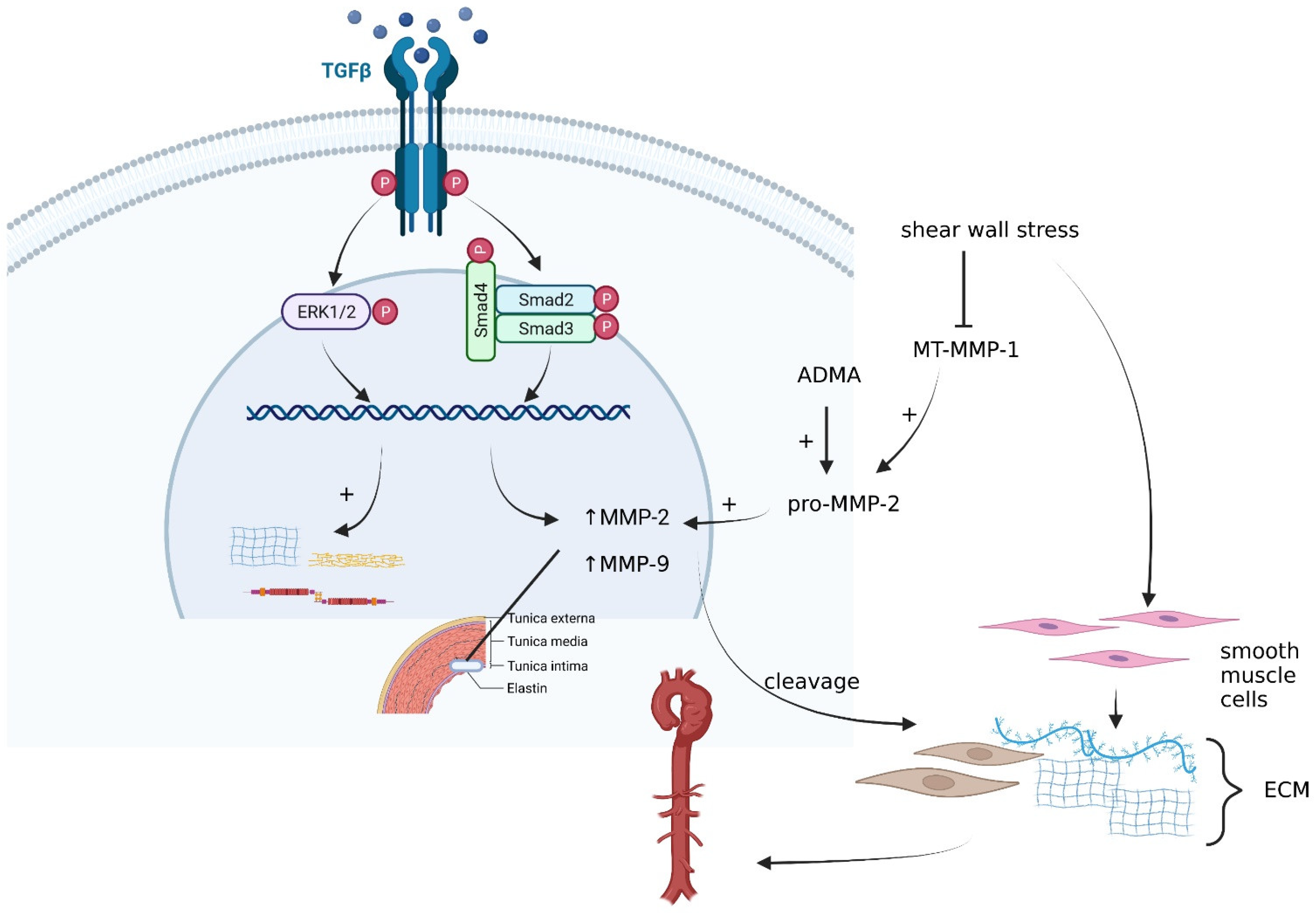
1.1. Matrix Metalloproteinases (MMPs)
1.2. Asymmetric Dimethylarginine (ADMA)
1.3. Soluble Receptor for Advanced Glycation End-Products (sRAGE)
1.4. Transforming Growth Factor Beta (TGF-β)
2. Future Perspectives
3. Conclusions
Author Contributions
Funding
Institutional Review Board Statement
Informed Consent Statement
Data Availability Statement
Conflicts of Interest
Abbreviations
| ADMA | Asymmetric dimethylarginine |
| AGE | Advanced glycation end-product |
| BAV | Bicuspid aortic valve |
| CHD | Congenital heart disease |
| CT | Computed tomography |
| ECM | Extracellular matrix |
| esRAGE | Endogenous secretory RAGE |
| MMP | Matrix metalloproteinase |
| MRI | Magnetic resonance imaging |
| MT-MMP | Membrane-type matrix metalloproteinase |
| NF-κB | Nuclear-factor kappa-B |
| NO | Mitric oxide |
| Nos3 | Nitric oxide synthase-3 enzyme |
| SMC | Smooth muscle cell |
| SNP | Single nucleotide polymorphisms |
| sRAGE | Soluble receptor for advanced glycation end-products |
| TAV | Tricuspid aortic valve |
| TAWSS | Time average wall shear stress |
| TGF-β | Transforming growth factor beta |
| TIMP | Tissue inhibitor of metalloproteinase |
| TLR | Toll-like receptor |
| TOE | Transoesophageal echocardiography |
| TOF | Tetralogy of Fallot |
| TTE | Transthoracic echocardiography |
| VSD | Ventricular septal defect |
| WSS | Wall shear stress |
References
- Habchi, K.M.; Ashikhmina, E.; Vieira, V.M.; Shahram, J.T.; Isselbacher, E.M.; Sundt, T.M.; Shekar, P.; Muehlschlegel, J.D.; Bicuspid Aortic Valve Consortium; Body, S.C. Association between bicuspid aortic valve morphotype and regional dilatation of the aortic root and trunk. Int. J. Cardiovasc. Imaging 2017, 33, 341–349. [Google Scholar] [CrossRef] [PubMed]
- Nussbaumer, C.; Bouchardy, J.; Blanche, C.; Piccini, D.; Pavon, A.-G.; Monney, P.; Stuber, M.; Schwitter, J.; Rutz, T. 2D cine vs. 3D self-navigated free-breathing high-resolution whole heart cardiovascular magnetic resonance for aortic root measurements in congenital heart disease. J. Cardiovasc. Magn. Reson. 2021, 23, 65. [Google Scholar] [CrossRef] [PubMed]
- Niaz, T.; Poterucha, J.T.; Johnson, J.N.; Craviari, C.; Nienaber, T.; Palfreeman, J.; Cetta, F.; Hagler, D.J. Incidence, morphology, and progression of bicuspid aortic valve in pediatric and young adult subjects with coexisting congenital heart defects. Congenit. Hearth Dis. 2017, 12, 261–269. [Google Scholar] [CrossRef] [PubMed]
- Silberbach, M.; Roos-Hesselink, J.W.; Andersen, N.H.; Braverman, A.C.; Brown, N.; Collins, R.T.; De Backer, J.; Eagle, K.A.; Hiratzka, L.F.; Johnson, W.H.; et al. Cardiovascular Health in Turner Syndrome: A Scientific Statement from the American Heart Association. Circ. Genom. Precis. Med. 2018, 11, e000048. [Google Scholar] [CrossRef] [PubMed] [Green Version]
- Zarate, Y.A.; Sellars, E.; LePard, T.; Carlo, W.F.; Tang, X.; Collins, R.T. Aortic dilation in pediatric patients. Eur. J. Pediatr. 2015, 174, 1585–1592. [Google Scholar] [CrossRef]
- Tan, J.L.; Gatzoulis, M.A.; Ho, S.Y. Aortic root disease in tetralogy of Fallot. Curr. Opin. Cardiol. 2006, 21, 569–572. [Google Scholar] [CrossRef] [PubMed]
- Carlo, W.F.; McKenzie, E.D.; Slesnick, T.C. Root Dilation in Patients with Truncus Arteriosus. Congenit. Hearth Dis. 2011, 6, 228–233. [Google Scholar] [CrossRef] [PubMed]
- Goda, M.; Gewillig, M.; Eyskens, B.; Heying, R.; Cools, B.; Rega, F.; Meyns, B. Mechanism of autograft insufficiency after the Ross operation in children. Cardiol. Young 2013, 23, 523–529. [Google Scholar] [CrossRef] [PubMed] [Green Version]
- Cotts, T.B.; Salciccioli, K.B.; Swanson, S.K.; Yetman, A.T. Aortopathy in Congenital Heart Disease. Cardiol. Clin. 2020, 38, 325–336. [Google Scholar] [CrossRef]
- Simpson, J.M.; Pushparajah, K. Dilatation of the Aorta in Bicuspid Aortic Valve Disease. Circ. Cardiovasc. Imaging 2020, 13, e010448. [Google Scholar] [CrossRef]
- Erbel, R.; Aboyans, V.; Boileaul, C.; Bossone, E.; Bartolomeo, R.D.; Eggebrecht, H.; Evangelista, A.; Falk, V.; Frank, H.; Gaemperli, O.; et al. ESC Guidelines on the diagnosis and treatment of aortic diseases: Document Covering Acute and Chronic Aortic Diseases of the Thoracic and Abdominal Aorta of the Adult. The Task Force for the Diagnosis and Treatment of Aortic Diseases of the European Society of Cardiology (ESC). Eur. Heart J. 2014, 35, 2873–2926. [Google Scholar] [PubMed] [Green Version]
- Flachskampf, F.A.; Badano, L.; Daniel, W.G.; Feneck, R.O.; Fox, K.F.; Fraser, A.G.; Pasquet, A.; Pepi, M.; De Isla, L.P.; Zamorano, J.L.; et al. Recommendations for transoesophageal echocardiography: Update 2010. Eur. J. Echocardiogr. 2010, 11, 557–576. [Google Scholar] [CrossRef] [PubMed] [Green Version]
- Evangelista, A.; Aguilar, R.; Cuellar, H.; Thomas, M.; Laynez, A.; Rodríguez-Palomares, J.; Mahía, P.; Gonzàlez-Alujas, T.; García-Dorado, D. Usefulness of real-time three-dimensional transoesophageal echocardiography in the assessment of chronic aortic dissection. Eur. J. Echocardiogr. 2011, 12, 272–277. [Google Scholar] [CrossRef] [PubMed] [Green Version]
- Einstein, A.J.; Weiner, S.D.; Bernheim, A.; Kulon, M.; Bokhari, S.; Johnson, L.L.; Moses, J.W.; Balter, S. Multiple Testing, Cumulative Radiation Dose, and Clinical Indications in Patients Undergoing Myocardial Perfusion Imaging. JAMA 2010, 304, 2137–2144. [Google Scholar] [CrossRef] [PubMed] [Green Version]
- Kramer, C.M.; Barkhausen, J.; Bucciarelli-Ducci, C.; Flamm, S.D.; Kim, R.J.; Nagel, E. Standardized cardiovascular magnetic resonance imaging (CMR) protocols: 2020 update. J. Cardiovasc. Magn. Reson. 2020, 22, 17. [Google Scholar] [CrossRef] [PubMed]
- Hagendorff, A.; Stoebe, S.; Tayal, B. A systematic approach to 3D echocardiographic assessment of the aortic root. Glob. Cardiol. Sci. Pract. 2018, 2018, 12. [Google Scholar] [CrossRef]
- Bendeck, M.P.; Keeley, F.W.; Langille, B.L. Perinatal accumulation of arterial wall constituents: Relation to hemodynamic changes at birth. Am. J. Physiol. Circ. Physiol. 1994, 267, H2268–H2279. [Google Scholar] [CrossRef]
- Kelleher, C.M.; McLean, S.E.; Mecham, R.P. Vascular Extracellular Matrix and Aortic Development. Curr. Top. Dev. Biol. 2004, 62, 153–188. [Google Scholar] [CrossRef]
- Parks, W.C.; Roby, J.D.; Wu, L.C.; Gross, L.E. Cellular Expression of Tropoelastin mRNA Splice Variants. Matrix 1992, 12, 156–162. [Google Scholar] [CrossRef]
- Ikonomidis, J.S.; Ivey, C.R.; Wheeler, J.B.; Akerman, A.W.; Rice, A.; Patel, R.K.; Stroud, R.E.; Shah, A.A.; Hughes, C.G.; Ferrari, G.; et al. Plasma biomarkers for distinguishing etiologic subtypes of thoracic aortic aneurysm disease. J. Thorac. Cardiovasc. Surg. 2013, 145, 1326–1333. [Google Scholar] [CrossRef] [Green Version]
- Harrison, O.J.; Cagampang, F.; Ohri, S.K.; Torrens, C.; Salhiyyah, K.; Modi, A.; Moorjani, N.; Whetton, A.D.; Townsend, P.A. Candidate plasma biomarkers for predicting ascending aortic aneurysm in bicuspid aortic valve disease. J. Cardiothorac. Surg. 2018, 13, 76. [Google Scholar] [CrossRef] [PubMed]
- Tzemos, N.; Lyseggen, E.; Silversides, C.; Jamorski, M.; Tong, J.H.; Harvey, P.; Floras, J.; Siu, S. Endothelial Function, Carotid–Femoral Stiffness, and Plasma Matrix Metalloproteinase-2 in Men with Bicuspid Aortic Valve and Dilated Aorta. J. Am. Coll. Cardiol. 2010, 55, 660–668. [Google Scholar] [CrossRef] [PubMed]
- Gavriliuk, N.D.; Druzhkova, T.A.; Irtyuga, O.B.; Zhloba, A.A.; Subbotina, T.F.; Uspenskiy, V.; Alexeyeva, N.P.; Moiseeva, O.M. Asymmetric Dimethylarginine in Patients with Ascending Aortic Aneurysms. AORTA 2016, 4, 219–225. [Google Scholar] [CrossRef]
- Branchetti, E.; Bavaria, J.E.; Grau, J.B.; Shaw, R.E.; Poggio, P.; Lai, E.K.; Desai, N.D.; Gorman, J.H.; Gorman, R.C.; Ferrari, G. Circulating Soluble Receptor for Advanced Glycation End Product Identifies Patients with Bicuspid Aortic Valve and Associated Aortopathies. Arter. Thromb. Vasc. Biol. 2014, 34, 2349–2357. [Google Scholar] [CrossRef] [PubMed] [Green Version]
- Hillebrand, M.; Millot, N.; Sheikhzadeh, S.; Rybczynski, M.; Gerth, S.; Kölbel, T.; Keyser, B.; Kutsche, K.; Robinson, P.N.; Berger, J.; et al. Total Serum Transforming Growth Factor-β1 Is Elevated in the Entire Spectrum of Genetic Aortic Syndromes. Clin. Cardiol. 2014, 37, 672–679. [Google Scholar] [CrossRef] [PubMed] [Green Version]
- Borger, M.; Fedak, P.W.; Stephens, E.H.; Gleason, T.G.; Girdauskas, E.; Ikonomidis, J.S.; Khoynezhad, A.; Siu, S.; Verma, S.; Hope, M.D.; et al. The American Association for Thoracic Surgery consensus guidelines on bicuspid aortic valve-related aortopathy: Full online-only version. J. Thorac. Cardiovasc. Surg. 2018, 156, e41–e74. [Google Scholar] [CrossRef] [PubMed]
- Wang, Y.; Wu, B.; Dong, L.; Wang, C.; Wang, X.; Shu, X. Circulating matrix metalloproteinase patterns in association with aortic dilatation in bicuspid aortic valve patients with isolated severe aortic stenosis. Hearth Vessel. 2016, 31, 189–197. [Google Scholar] [CrossRef]
- Drapisz, S.; Góralczyk, T.; Jamka-Miszalski, T.; Olszowska, M.; Undas, A. Nonstenotic bicuspid aortic valve is associated with elevated plasma asymmetric dimethylarginine. J. Cardiovasc. Med. 2013, 14, 446–452. [Google Scholar] [CrossRef]
- Merkx, R.; Duijnhouwer, A.L.; Vink, E.; Roos-Hesselink, J.W.; Schokking, M. Aortic Diameter Growth in Children with a Bicuspid Aortic Valve. Am. J. Cardiol. 2017, 120, 131–136. [Google Scholar] [CrossRef] [Green Version]
- Martínez-Micaelo, N.; Ligero, C.; Antequera-González, B.; Junza, A.; Yanes, O.; Alegret, J.M. Plasma Metabolomic Profiling Associates Bicuspid Aortic Valve Disease and Ascending Aortic Dilation with a Decrease in Antioxidant Capacity. J. Clin. Med. 2020, 9, 2215. [Google Scholar] [CrossRef]
- Agewall, S. Matrix metalloproteinases and cardiovascular disease. Eur. Hearth J. 2006, 27, 121–122. [Google Scholar] [CrossRef] [PubMed] [Green Version]
- Dollery, C.M.; McEwan, J.R.; Henney, A.M. Matrix Metalloproteinases and Cardiovascular Disease. Circ. Res. 1995, 77, 863–868. [Google Scholar] [CrossRef] [PubMed]
- Birkedal-Hansen, H. Proteolytic remodeling of extracellular matrix. Curr. Opin. Cell Biol. 1995, 7, 728–735. [Google Scholar] [CrossRef]
- Itoh, Y. Membrane-type matrix metalloproteinases: Their functions and regulations. Matrix Biol. 2015, 44–46, 207–223. [Google Scholar] [CrossRef]
- Ikonomidis, J.S.; Jones, J.A.; Barbour, J.R.; Stroud, R.E.; Clark, L.L.; Kaplan, B.S.; Zeeshan, A.; Bavaria, J.E.; Gorman, J.H., 3rd; Spinale, F.G.; et al. Expression of matrix metalloproteinases and endogenous inhibitors within ascending aortic aneurysms of patients with bicuspid or tricuspid aortic valves. J. Thorac. Cardiovasc. Surg. 2007, 133, 1028–1036. [Google Scholar] [CrossRef] [Green Version]
- Ravn, H.B.; Falk, E. Histopathology of plaque rupture. Cardiol. Clin. 1999, 17, 263–270. [Google Scholar] [CrossRef]
- Tamarina, N.A.; McMillan, W.D.; Shively, V.P.; Pearce, W.H. Expression of matrix metalloproteinases and their inhibitors in aneurysms and normal aorta. Surgery 1997, 122, 264–272. [Google Scholar] [CrossRef]
- Thompson, R.W.; Holmes, D.R.; Mertens, R.A.; Liao, S.; Botney, M.D.; Mecham, R.P.; Welgus, H.G.; Parks, W.C. Production and localization of 92-kilodalton gelatinase in abdominal aortic aneurysms. An elastolytic metalloproteinase expressed by aneurysm-infiltrating macrophages. J. Clin. Investig. 1995, 96, 318–326. [Google Scholar] [CrossRef] [Green Version]
- Wang, H.; Keiser, J.A. Expression of membrane-type matrix metalloproteinase in rabbit neointimal tissue and its correlation with matrix-metalloproteinase-2 activation. J. Vasc. Res. 1998, 35, 45–54. [Google Scholar] [CrossRef]
- Pasta, S.; Agnese, V.; Gallo, A.; Cosentino, F.; Di Giuseppe, M.; Gentile, G.; Raffa, G.M.; Maalouf, J.F.; Michelena, H.I.; Bellavia, D.; et al. Shear Stress and Aortic Strain Associations with Biomarkers of Ascending Thoracic Aortic Aneurysm. Ann. Thorac. Surg. 2020, 110, 1595–1604. [Google Scholar] [CrossRef]
- Li, T.; Li, X.; Liu, X.; Yang, J.; Ma, C. The elevated expression of TLR4 and MMP9 in human abdominal aortic aneurysm tissues and its implication. BMC Cardiovasc. Disord. 2021, 21, 378. [Google Scholar] [CrossRef] [PubMed]
- Koullias, G.J.; Korkolis, D.P.; Ravichandran, P.; Psyrri, A.; Hatzaras, I.; Elefteriades, J.A. Tissue microarray detection of matrix metalloproteinases, in diseased tricuspid and bicuspid aortic valves with or without pathology of the ascending aorta. Eur. J. Cardio-Thoracic Surg. 2004, 26, 1098–1103. [Google Scholar] [CrossRef] [PubMed] [Green Version]
- Fondard, O.; Detaint, D.; Iung, B.; Choqueux, C.; Adle-Biassette, H.; Jarraya, M.; Hvass, U.; Couetil, J.-P.; Henin, D.; Michel, J.-B.; et al. Extracellular matrix remodelling in human aortic valve disease: The role of matrix metalloproteinases and their tissue inhibitors. Eur. Hearth J. 2005, 26, 1333–1341. [Google Scholar] [CrossRef] [PubMed]
- Longo, G.M.; Xiong, W.; Greiner, T.C.; Zhao, Y.; Fiotti, N.; Baxter, B.T. Matrix metalloproteinases 2 and 9 work in concert to produce aortic aneurysms. J. Clin. Investig. 2002, 110, 625–632. [Google Scholar] [CrossRef]
- Jacob, M.P. Extracellular matrix remodeling and matrix metalloproteinases in the vascular wall during aging and in pathological conditions. Biomed. Pharmacother. 2003, 57, 195–202. [Google Scholar] [CrossRef]
- Ishii, T.; Asuwa, N. Collagen and elastin degradation by matrix metalloproteinases and tissue inhibitors of matrix metalloproteinase in aortic dissection. Hum. Pathol. 2000, 31, 640–646. [Google Scholar] [CrossRef]
- Sinha, I.; Bethi, S.; Cronin, P.; Williams, D.M.; Roelofs, K.; Ailawadi, G.; Henke, P.K.; Eagleton, M.J.; Deeb, G.M.; Patel, H.J.; et al. A biologic basis for asymmetric growth in descending thoracic aortic aneurysms: A role for matrix metalloproteinase 9 and 2. J. Vasc. Surg. 2006, 43, 342–348. [Google Scholar] [CrossRef] [Green Version]
- McMillan, W.D.; Tamarina, N.A.; Cipollone, M.; Johnson, D.A.; Parker, M.A.; Pearce, W.H. Size Matters: The Relationship between MMP-9 Expression and Aortic Diameter. Circulation 1997, 96, 2228–2232. [Google Scholar] [CrossRef]
- Liao, M.; Zou, S.; Bao, Y.; Jin, J.; Yang, J.; Liu, Y.; Green, M.; Yang, F.; Qu, L. Matrix metalloproteinases are regulated by MicroRNA 320 in macrophages and are associated with aortic dissection. Exp. Cell Res. 2018, 370, 98–102. [Google Scholar] [CrossRef]
- Zhang, X.; Wu, D.; Choi, J.C.; Minard, C.G.; Hou, X.; Coselli, J.S.; Shen, Y.H.; LeMaire, S.A. Matrix metalloproteinase levels in chronic thoracic aortic dissection. J. Surg. Res. 2014, 189, 348–358. [Google Scholar] [CrossRef] [Green Version]
- van der Bom, T.; Bouma, B.; Meijboom, F.J.; Zwinderman, A.H.; Mulder, B.J. The prevalence of adult congenital heart disease, results from a systematic review and evidence based calculation. Am. Hearth J. 2012, 164, 568–575. [Google Scholar] [CrossRef] [PubMed]
- Jackson, V.; Olsson, T.; Kurtovic, S.; Folkersen, L.; Paloschi, V.; Wågsäter, D.; Franco-Cereceda, A.; Eriksson, P. Matrix metalloproteinase 14 and 19 expression is associated with thoracic aortic aneurysms. J. Thorac. Cardiovasc. Surg. 2012, 144, 459–466. [Google Scholar] [CrossRef] [PubMed] [Green Version]
- Spinale, F.G.; Sapp, A.A. Cardiovascular Risk and Matrix Metalloproteinase Polymorphisms. Circ. Cardiovasc. Genet. 2017, 10, e001958. [Google Scholar] [CrossRef] [PubMed] [Green Version]
- Gorący, I.; Grudniewicz, S.; Safranow, K.; Ciechanowicz, A.; Jakubiszyn, P.; Gorący, A.; Brykczyński, M. Genetic Polymorphisms of MMP1, MMP9, COL1A1, and COL1A2 in Polish Patients with Thoracic Aortopathy. Dis. Markers 2020, 2020, 9567239. [Google Scholar] [CrossRef] [PubMed]
- D’Oria, M.; Di Girolamo, F.G.; Calvagna, C.; Gorgatti, F.; Altamura, N.; Lepidi, S.; Biolo, G.; Fiotti, N. Remodeling of abdominal aortic aneurysm sac following endovascular aortic repair: Association with clinical, surgical, and genetic factors. Cardiovasc. Pathol. 2022, 58, 107405. [Google Scholar] [CrossRef]
- Duellman, T.; Warren, C.L.; Matsumura, J.; Yang, J. Analysis of multiple genetic polymorphisms in aggressive-growing and slow-growing abdominal aortic aneurysms. J. Vasc. Surg. 2014, 60, 613–621. [Google Scholar] [CrossRef] [Green Version]
- Liu, O.; Xie, W.; Qin, Y.; Jia, L.; Zhang, J.; Xin, Y.; Guan, X.; Li, H.; Gong, M.; Liu, Y.; et al. MMP-2 gene polymorphisms are associated with type A aortic dissection and aortic diameters in patients. Medicine 2016, 95, e5175. [Google Scholar] [CrossRef]
- Corbitt, H.; Gutierrez, J.; Silberbach, M.; Maslen, C.L. The genetic basis of Turner syndrome aortopathy. Am. J. Med Genet. Part C Semin. Med Genet. 2019, 181, 117–125. [Google Scholar] [CrossRef]
- Stabouli, S.; Kotsis, V.; Maliachova, O.; Printza, N.; Chainoglou, A.; Christoforidis, A.; Taparkou, A.; Dotis, J.; Farmaki, E.; Zafeiriou, D. Matrix metalloproteinase −2, −9 and arterial stiffness in children and adolescents: The role of chronic kidney disease, diabetes, and hypertension. Int. J. Cardiol. Hypertens. 2020, 4, 100025. [Google Scholar] [CrossRef]
- Cui, J.Z.; Harris, K.C.; Raedschelders, K.; Hollander, Z.; Potts, J.E.; De Souza, A.; Kiess, M.; McManus, B.M.; Bernatchez, P.; Raffin, L.A.; et al. Aortic Dimensions, Biophysical Properties, and Plasma Biomarkers in Children and Adults with Marfan or Loeys-Dietz Syndrome. CJC Open 2020, 3, 585–594. [Google Scholar] [CrossRef]
- Cheng, K.-S.; Liao, Y.-C.; Chen, M.-Y.; Kuan, T.-C.; Hong, Y.-H.; Ko, L.; Hsieh, W.-Y.; Wu, C.-L.; Chen, M.-R.; Lin, C.-S. Circulating Matrix Metalloproteinase-2 and -9 Enzyme Activities in the Children with Ventricular Septal Defect. Int. J. Biol. Sci. 2013, 9, 557–563. [Google Scholar] [CrossRef] [PubMed] [Green Version]
- Fedak, P.; de Sa, M.P.; Verma, S.; Nili, N.; Kazemian, P.; Butany, J.; Strauss, B.H.; Weisel, R.D.; David, T.E. Vascular matrix remodeling in patients with bicuspid aortic valve malformations: Implications for aortic dilatation. J. Thorac. Cardiovasc. Surg. 2003, 126, 797–805. [Google Scholar] [CrossRef] [Green Version]
- Aicher, D.; Urbich, C.; Zeiher, A.; Dimmeler, S.; Schäfers, H.-J. Endothelial Nitric Oxide Synthase in Bicuspid Aortic Valve Disease. Ann. Thorac. Surg. 2007, 83, 1290–1294. [Google Scholar] [CrossRef] [PubMed]
- Chen, H.-H.; Wang, D.L. Nitric Oxide Inhibits Matrix Metalloproteinase-2 Expression via the Induction of Activating Transcription Factor 3 in Endothelial Cells. Mol. Pharmacol. 2004, 65, 1130–1140. [Google Scholar] [CrossRef] [Green Version]
- Ali, O.A.; Chapman, M.; Nguyen, T.H.; Chirkov, Y.Y.; Heresztyn, T.; Mundisugih, J.; Horowitz, J.D. Interactions between inflammatory activation and endothelial dysfunction selectively modulate valve disease progression in patients with bicuspid aortic valve. Heart 2014, 100, 800–805. [Google Scholar] [CrossRef] [PubMed]
- Satılmışoğlu, M.H.; Diker, V.; Taşbulak, Ö.; Diker, M.; Birand, A.; Kaya, M.; Iyigün, T.; Eksik, A. Increased plasma asymmetric dimethylarginine level is associated with ascending aorta dilatation: A case-control study. Kardiol. Pol. 2017, 75, 1020–1026. [Google Scholar] [CrossRef] [Green Version]
- Böger, R.H. Asymmetric Dimethylarginine, an Endogenous Inhibitor of Nitric Oxide Synthase, Explains the “L-Arginine Paradox” and Acts as a Novel Cardiovascular Risk Factor. J. Nutr. 2004, 134, 2842S–2847S. [Google Scholar] [CrossRef] [PubMed]
- Park, K.-H.; Park, W.J. Endothelial Dysfunction: Clinical Implications in Cardiovascular Disease and Therapeutic Approaches. J. Korean Med Sci. 2015, 30, 1213–1225. [Google Scholar] [CrossRef] [Green Version]
- Valkonen, V.-P.; Päivä, H.; Salonen, J.T.; Lakka, T.; Lehtimäki, T.; Laakso, J.; Laaksonen, R. Risk of acute coronary events and serum concentration of asymmetrical dimethylarginine. Lancet 2001, 358, 2127–2128. [Google Scholar] [CrossRef]
- Nemeth, Z.; Cziráki, A.; Szabados, S.; Biri, B.; Kéki, S.; Koller, A. Elevated Levels of Asymmetric Dimethylarginine (ADMA) in the Pericardial Fluid of Cardiac Patients Correlate with Cardiac Hypertrophy. PLoS ONE 2015, 10, e0135498. [Google Scholar] [CrossRef] [Green Version]
- Huemer, M.; Simma, B.; Mayr, D.; Mühl, A.; Rami, B.; Schober, E.; Ulmer, H.; Zanier, U.; Bodamer, O.A. Low Levels of Asymmetric Dimethylarginine in Children with Diabetes Mellitus Type I Compared with Healthy Children. J. Pediatr. 2011, 158, 602–606.e1. [Google Scholar] [CrossRef] [PubMed]
- Hsu, C.-N.; Tain, Y.-L. Asymmetric Dimethylarginine (ADMA) in Pediatric Renal Diseases: From Pathophysiological Phenomenon to Clinical Biomarker and Beyond. Children 2021, 8, 837. [Google Scholar] [CrossRef] [PubMed]
- Sarkar, A.; Prasad, K.; Ziganshin, B.A.; Elefteriades, J.A. Reasons to Investigate the Soluble Receptor for Advanced Glycation End-Product (sRAGE) Pathway in Aortic Disease. AORTA 2013, 1, 210–217. [Google Scholar] [CrossRef] [PubMed] [Green Version]
- Barlovic, M.C.T.A.K.J.-D.D.P.; Thomas, M.; Jandeleit-Dahm, K. Cardiovascular Disease: What’s All the AGE/RAGE About? Cardiovasc. Hematol. Disord. Targets 2010, 10, 7–15. [Google Scholar] [CrossRef]
- Ramasamy, R.; Yan, S.F.; Schmidt, A.M. The RAGE Axis and Endothelial Dysfunction: Maladaptive Roles in the Diabetic Vasculature and Beyond. Trends Cardiovasc. Med. 2005, 15, 237–243. [Google Scholar] [CrossRef] [PubMed]
- Lindsey, J.B.; Cipollone, F.; Abdullah, S.M.; Mcguire, D.K. Receptor for advanced glycation end-products (RAGE) and soluble RAGE (sRAGE): Cardiovascular implications. Diabetes Vasc. Dis. Res. 2009, 6, 7–14. [Google Scholar] [CrossRef]
- Wendt, T.; Harja, E.; Bucciarelli, L.; Qu, W.; Lu, Y.; Rong, L.L.; Jenkins, D.G.; Stein, G.; Schmidt, A.M.; Yan, S.F. RAGE modulates vascular inflammation and atherosclerosis in a murine model of type 2 diabetes. Atherosclerosis 2006, 185, 70–77. [Google Scholar] [CrossRef]
- Galatioto, J.; Lai, E.; Bintanel-Morcillo, M.; Wang, T.; Ferrari, G.; Pyeritz, R.E.; Bavaria, J.E.; Branchetti, E. Abstract 307: Circulating sRAGE is Associated with Aortic Dysfunction in Mice Models of Thoracic Aortic Aneurysm. Arter. Thromb. Vasc. Biol. 2017, 37, A307. [Google Scholar] [CrossRef]
- Prasad, K.; Sarkar, A.; Zafar, M.A.; Shoker, A.; Moselhi, H.E.; Tranquilli, M.; Ziganshin, B.A.; Elefteriades, J.A. Advanced Glycation End Products and its Soluble Receptors in the Pathogenesis of Thoracic Aortic Aneurysm. AORTA 2016, 4, 1–10. [Google Scholar] [CrossRef] [Green Version]
- Jia, H.; Kang, L.; Lu, S.; Chen, Z.; Shen, J.; Huang, B.; Zou, Y.; Sun, Y. Circulating soluble receptor of advanced glycation end product is associated with bicuspid aortic aneurysm progression via NF-κB pathway. Interact. Cardiovasc. Thorac. Surg. 2021, 34, 274–282. [Google Scholar] [CrossRef]
- Diekmann, F.; Chouvarine, P.; Sallmon, H.; Meyer-Kobbe, L.; Kieslich, M.; Plouffe, B.; Murthy, S.; Lichtinghagen, R.; Legchenko, E.; Hansmann, G. Soluble Receptor for Advanced Glycation End Products (sRAGE) Is a Sensitive Biomarker in Human Pulmonary Arterial Hypertension. Int. J. Mol. Sci. 2021, 22, 8591. [Google Scholar] [CrossRef] [PubMed]
- Cheung, Y.-F.; Chow, P.-C.; So, E.K.-F.; Chan, K.-W. Circulating Transforming Growth Factor-β and Aortic Dilation in Patients with Repaired Congenital Heart Disease. Sci. Rep. 2019, 9, 162. [Google Scholar] [CrossRef] [PubMed] [Green Version]
- Tingting, T.; Wenjing, F.; Qian, Z.; Hengquan, W.; Simin, Z.; Zhisheng, J.; Shunlin, Q. The TGF-β pathway plays a key role in aortic aneurysms. Clin. Chim. Acta 2020, 501, 222–228. [Google Scholar] [CrossRef] [PubMed]
- Wang, Y.; Ait-Oufella, H.; Herbin, O.; Bonnin, P.; Ramkhelawon, B.; Taleb, S.; Huang, J.; Offenstadt, G.; Combadiere, C.; Rénia, L.; et al. TGF-β activity protects against inflammatory aortic aneurysm progression and complications in angiotensin II–infused mice. J. Clin. Investig. 2010, 120, 422–432. [Google Scholar] [CrossRef] [PubMed] [Green Version]
- Takeda, N.; Hara, H.; Fujiwara, T.; Kanaya, T.; Maemura, S.; Komuro, I. TGF-β Signaling-Related Genes and Thoracic Aortic Aneurysms and Dissections. Int. J. Mol. Sci. 2018, 19, 2125. [Google Scholar] [CrossRef] [PubMed] [Green Version]
- Bertolino, P.; Deckers, M.; Lebrin, F.; Dijke, P.T. Transforming Growth Factor-β Signal Transduction in Angiogenesis and Vascular Disorders. Chest 2005, 128, 585S–590S. [Google Scholar] [CrossRef]
- Verrecchia, F.; Mauviel, A. TGF-β and TNF-α: Antagonistic cytokines controlling type I collagen gene expression. Cell. Signal. 2004, 16, 873–880. [Google Scholar] [CrossRef]
- Jones, J.A.; Spinale, F.G.; Ikonomidis, J.S. Transforming Growth Factor-β Signaling in Thoracic Aortic Aneurysm Development: A Paradox in Pathogenesis. J. Vasc. Res. 2009, 46, 119–137. [Google Scholar] [CrossRef] [Green Version]
- Dai, J.; Losy, F.; Guinault, A.-M.; Pages, C.; Anegon, I.; Desgranges, P.; Becquemin, J.-P.; Allaire, E. Overexpression of Transforming Growth Factor-β1 Stabilizes Already-Formed Aortic Aneurysms: A First Approach to Induction of Functional Healing by Endovascular Gene Therapy. Circulation 2005, 112, 1008–1015. [Google Scholar] [CrossRef]
- El-Hamamsy, I.; Yacoub, M.H. Cellular and molecular mechanisms of thoracic aortic aneurysms. Nat. Rev. Cardiol. 2009, 6, 771–786. [Google Scholar] [CrossRef]
- Romaniello, F.; Mazzaglia, D.; Pellegrino, A.; Grego, S.; Fiorito, R.; Ferlosio, A.; Chiariello, L.; Orlandi, A. Aortopathy in Marfan syndrome: An update. Cardiovasc. Pathol. 2014, 23, 261–266. [Google Scholar] [CrossRef] [PubMed]
- Angelov, S.N.; Hu, J.H.; Wei, H.; Airhart, N.; Shi, M.; Dichek, D.A. TGF-β (Transforming Growth Factor-β) Signaling Protects the Thoracic and Abdominal Aorta from Angiotensin II-Induced Pathology by Distinct Mechanisms. Arter. Thromb. Vasc. Biol. 2017, 37, 2102–2113. [Google Scholar] [CrossRef] [PubMed] [Green Version]
- Dietz, H.C. TGF-β in the pathogenesis and prevention of disease: A matter of aneurysmic proportions. J. Clin. Investig. 2010, 120, 403–406. [Google Scholar] [CrossRef] [PubMed]
- Hara, H.; Maemura, S.; Fujiwara, T.; Takeda, N.; Ishii, S.; Yagi, H.; Suzuki, T.; Harada, M.; Toko, H.; Kanaya, T.; et al. Inhibition of transforming growth factor-β signaling in myeloid cells ameliorates aortic aneurysmal formation in Marfan syndrome. PLoS ONE 2020, 15, e0239908. [Google Scholar] [CrossRef]
- Rueda-Martínez, C.; Lamas, O.; Carrasco-Chinchilla, F.; Robledo-Carmona, J.; Porras, C.; Sánchez-Espín, G.; Navarro, M.J.; Fernández, B. Increased blood levels of transforming growth factor ? in patients with aortic dilatation. Interact. Cardiovasc. Thorac. Surg. 2017, 25, 571–574. [Google Scholar] [CrossRef] [Green Version]
- Li, W.; Li, Q.; Jiao, Y.; Qin, L.; Ali, R.; Zhou, J.; Ferruzzi, J.; Kim, R.W.; Geirsson, A.; Dietz, H.C.; et al. Tgfbr2 disruption in postnatal smooth muscle impairs aortic wall homeostasis. J. Clin. Investig. 2014, 124, 755–767. [Google Scholar] [CrossRef] [Green Version]
- Paloschi, V.; Gådin, J.R.; Khan, S.; Björck, H.M.; Du, L.; Maleki, S.; Roy, J.; Lindeman, J.H.; Mohamed, S.A.; Tsuda, T.; et al. Aneurysm Development in Patients with a Bicuspid Aortic Valve is not Associated with Transforming Growth Factor-β Activation. Arter. Thromb. Vasc. Biol. 2015, 35, 973–980. [Google Scholar] [CrossRef] [Green Version]
- Forte, A.; Bancone, C.; Cobellis, G.; Buonocore, M.; Santarpino, G.; Fischlein, T.J.; Cipollaro, M.; De Feo, M.; Della Corte, A. A Possible Early Biomarker for Bicuspid Aortopathy: Circulating Transforming Growth Factor β-1 to Soluble Endoglin Ratio. Circ. Res. 2017, 120, 1800–1811. [Google Scholar] [CrossRef]
- Sun, F.; Hayama, E.; Nakanishi, T. Abstract 2606: Expression of Transforming Growth Factor-Beta in the Aorta of Congenital Heart Disease. Circulation 2008, 118, S_752. [Google Scholar] [CrossRef]
- Zanjani, K.S.; Niwa, K. Aortic dilatation and aortopathy in congenital heart diseases. J. Cardiol. 2013, 61, 16–21. [Google Scholar] [CrossRef] [Green Version]
- Brooke, B.S.; Habashi, J.P.; Judge, D.; Patel, N.; Loeys, B.; Dietz, H.C. Angiotensin II Blockade and Aortic-Root Dilation in Marfan’s Syndrome. N. Engl. J. Med. 2008, 358, 2787–2795. [Google Scholar] [CrossRef] [PubMed] [Green Version]
- Shiina, Y.; Niwa, K. Cardio-Ankle Vascular Index (CAVI) and Plasma Transforming Growth Factor-β1 (TGF-β1) Level Correlate with Aortopathy in Adults with Repaired Tetralogy of Fallot. Pediatr. Cardiol. 2016, 38, 338–343. [Google Scholar] [CrossRef] [PubMed]
- Hill, J.C.; Billaud, M.; Richards, T.D.; Kotlarczyk, M.P.; Shiva, S.; Phillippi, J.A.; Gleason, T.G. Layer-specific Nos3 expression and genotypic distribution in bicuspid aortic valve aortopathy. Eur. J. Cardio-Thoracic Surg. 2022, ezac237. [Google Scholar] [CrossRef] [PubMed]
- Scola, L.; Giarratana, R.; Marinello, V.; Cancila, V.; Pisano, C.; Ruvolo, G.; Frati, G.; Lio, D.; Balistreri, C. Polymorphisms of Pro-Inflammatory IL-6 and IL-1β Cytokines in Ascending Aortic Aneurysms as Genetic Modifiers and Predictive and Prognostic Biomarkers. Biomolecules 2021, 11, 943. [Google Scholar] [CrossRef] [PubMed]
- Hovsepian, D.M.; Ziporin, S.J.; Sakurai, M.K.; Lee, J.K.; Curci, J.A.; Thompson, R.W. Elevated Plasma Levels of Matrix Metalloproteinase-9 in Patients with Abdominal Aortic Aneurysms: A Circulating Marker of Degenerative Aneurysm Disease. J. Vasc. Interv. Radiol. 2000, 11, 1345–1352. [Google Scholar] [CrossRef]


| Type of Metalloproteinase | Expression Variation | Type of Study/Population Involved | Correlation with Conditions Studied |
|---|---|---|---|
| MMP-1 | Upregulation | Case-control study, 13 patients:
| Abdominal aortic aneurysm—Tamarina et al. [37] |
| Upregulation—significantly among the intima | Study group—21 patients with aortic dissection; in 19 cases, expression in remote sites was assessed. Controls—10 autopsies. | Aortic dissection—Ishii et al. [46] | |
| MMP-2 | Upregulation | Experimental—organ donor tissue. | Athero-occlusive disease, and abdominal aortic aneurysm—Thompson et al. [38] |
Case-control study, 26 patients with aortic stenosis/insufficiency:
| BAV—Koullias et al. [42] | ||
Case-control study:
| BAV—Ikonomidis et al. [35] | ||
Study population:
| BAV and vascular fibrilin-1 deficiency—Fedak et al. [62] | ||
| MMP-2 deficiency | Experimental—mice. | Lack of abdominal aortic aneurysm production—Longo et al. [44] | |
| Upregulation—significantly among the intima | Study group—21 patients with aortic dissection; in 19 cases, expression in remote sites was assessed. Controls—10 autopsies. | Aortic dissection—Ishii et al. [46] | |
| MMP-9 | Upregulation | Case-control study, 13 patients:
| Abdominal aortic aneurysm—Tamarina et al. [37] |
Case-control study, 48 patients:
| Abdominal aortic aneurysm—Li et al. [41] | ||
Case-control study, 26 patients with aortic stenosis/insufficiency:
| BAV, abdominal aortic aneurysm/dissection—Koullias et al. [42] | ||
Case—control study:
| Aortic abdominal aneurysm, particular significant association with moderate size aneurysm—McMillan et al. [48] | ||
Study population:
| BAV and vascular fibrilin-1 deficiency—Fedak et al. [62] | ||
| MMP-9 deficiency | Experimental—mice. | Lack of abdominal aortic aneurysm production—Longo et al. [44] | |
| Upregulation—significantly among the intima | Study group—21 patients with aortic dissection; in 19 cases, expression in remote sites was assessed. Controls—10 autopsies. | Aortic dissection—Ishii et al. [46] | |
| TIMP-1 | Upregulation | Experimental—organ donor tissue. | Abdominal aortic aneurysm—Thompson et al. [38] |
Case—control study, 13 patients:
| Abdominal aortic aneurysm—Tamarina et al. [37] | ||
Case-control study, 26 patients with aortic stenosis/insufficiency:
| BAV in aortic stenosis subjects—Koullias et al. [42] | ||
| Upregulation—significantly among the intima | Study group—21 patients with aortic dissection; in 19 cases, expression in remote sites was assessed. Controls—10 autopsies. | Aortic dissection—Ishii et al. [46] | |
| TIMP-2 | Upregulation | Case—control study, 13 patients:
| Abdominal aortic aneurysm—Tamarina et al. [37] |
| Upregulation—significantly among the intima | Study group—21 patients with aortic dissection’ in 19 cases, expression in remote sites was assessed. Controls—10 autopsies. | Aortic dissection—Ishii et al. [46] |
| Type of Metalloproteinase | Expression Variation | Type of Study/ Population Involved | Correlation with Conditions Studied |
|---|---|---|---|
| MMP-1 | Upregulation | 125 patients with ascending aortic aneurysms | Ascending aortic aneurysm surgery prediction; correlation with WSS and TAWSS—Pasta et al. [40] |
Case-control study:
| Acute aortic dissection—Liao et al. [49] | ||
| MMP-2 | Upregulation | 125 patients with ascending aortic aneurysms | Ascending aortic aneurysm surgery prediction; correlation with WSS and TAWSS—Pasta et al. [40] |
Case-control study:
| Acute aortic dissection—Liao et al. [49] | ||
| MMP-9 | Upregulation | Case-control study:
| Final stages of chronic thoracic aortic aneurysm—Zhang et al. [50] |
Study population (93 subjects):
| Severe, isolated aortic stenosis in BAV patients—Wang Y [27] | ||
Pediatric case-control study (110 patients):
| VSD; spontaneous closure of VSD—Cheng et al. [61] | ||
Case-control study:
| Final stages of chronic thoracic aortic aneurysm—Zhang et al. [50] | ||
| TIMP-1 | Upregulation | 125 patients with ascending aortic aneurysms | Ascending aortic aneurysm surgery prediction; correlation with WSS and TAWSS—Pasta et al. [40] |
Publisher’s Note: MDPI stays neutral with regard to jurisdictional claims in published maps and institutional affiliations. |
© 2022 by the authors. Licensee MDPI, Basel, Switzerland. This article is an open access article distributed under the terms and conditions of the Creative Commons Attribution (CC BY) license (https://creativecommons.org/licenses/by/4.0/).
Share and Cite
Făgărășan, A.; Săsăran, M.O. The Predictive Role of Plasma Biomarkers in the Evolution of Aortopathies Associated with Congenital Heart Malformations. Int. J. Mol. Sci. 2022, 23, 4993. https://0-doi-org.brum.beds.ac.uk/10.3390/ijms23094993
Făgărășan A, Săsăran MO. The Predictive Role of Plasma Biomarkers in the Evolution of Aortopathies Associated with Congenital Heart Malformations. International Journal of Molecular Sciences. 2022; 23(9):4993. https://0-doi-org.brum.beds.ac.uk/10.3390/ijms23094993
Chicago/Turabian StyleFăgărășan, Amalia, and Maria Oana Săsăran. 2022. "The Predictive Role of Plasma Biomarkers in the Evolution of Aortopathies Associated with Congenital Heart Malformations" International Journal of Molecular Sciences 23, no. 9: 4993. https://0-doi-org.brum.beds.ac.uk/10.3390/ijms23094993