
Fluorescent Magnetic Nanoparticles for Bioimaging through Biomimetic Surface Modification
 , , , and
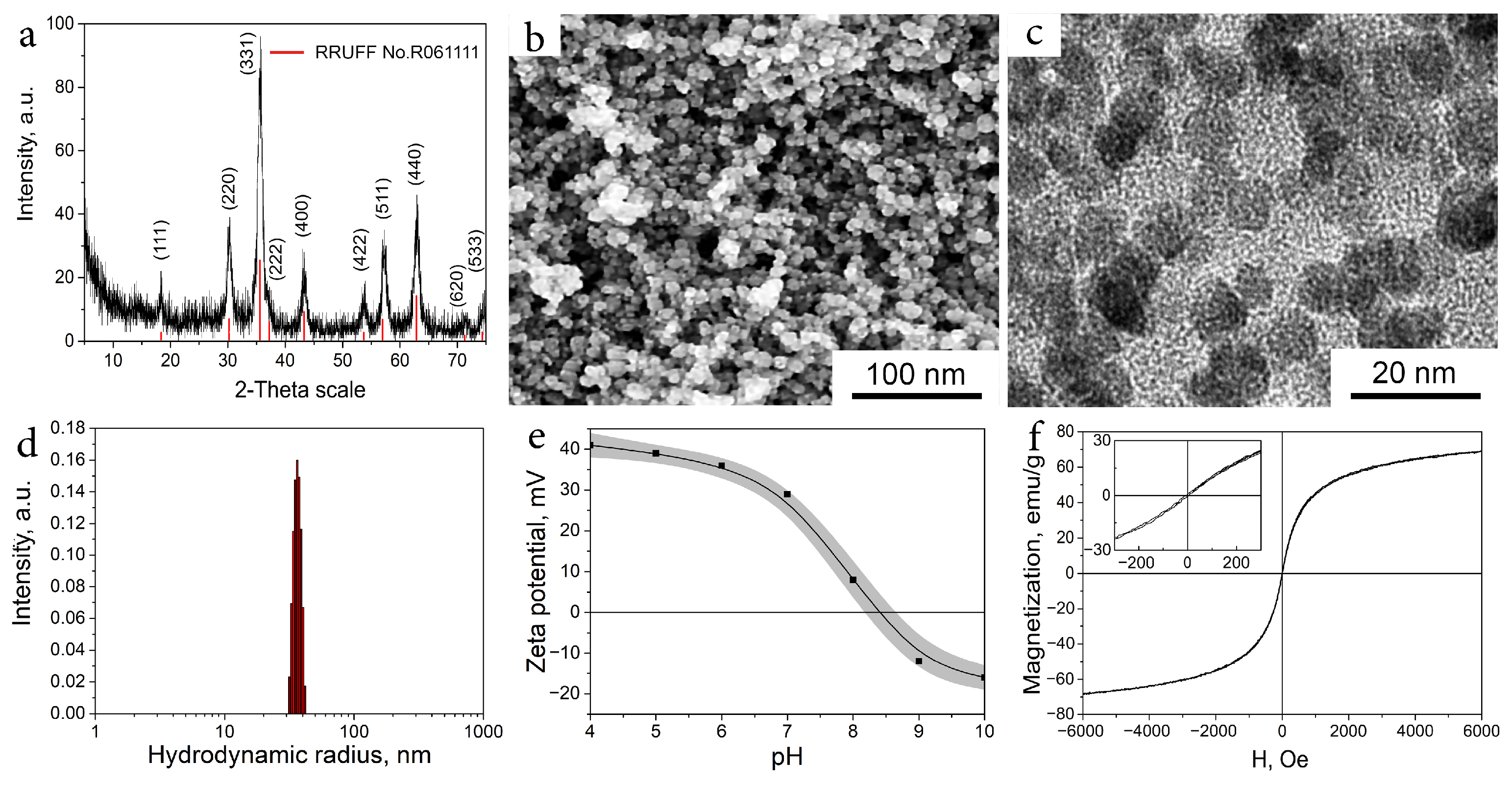
Abstract
:1. Introduction
2. Results
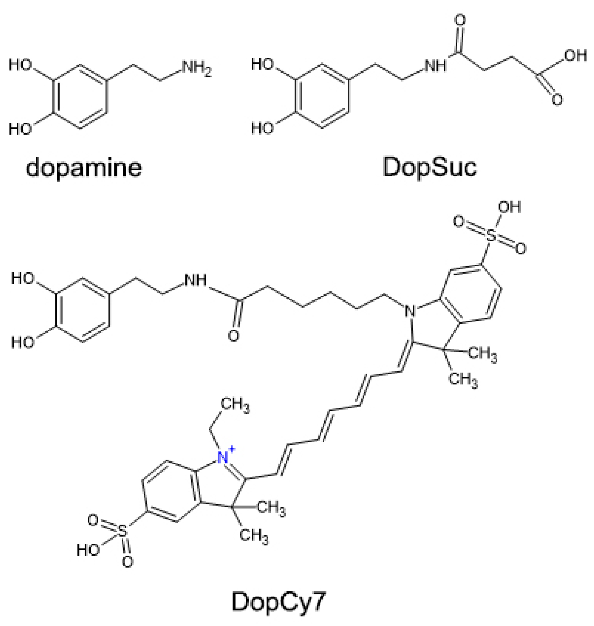
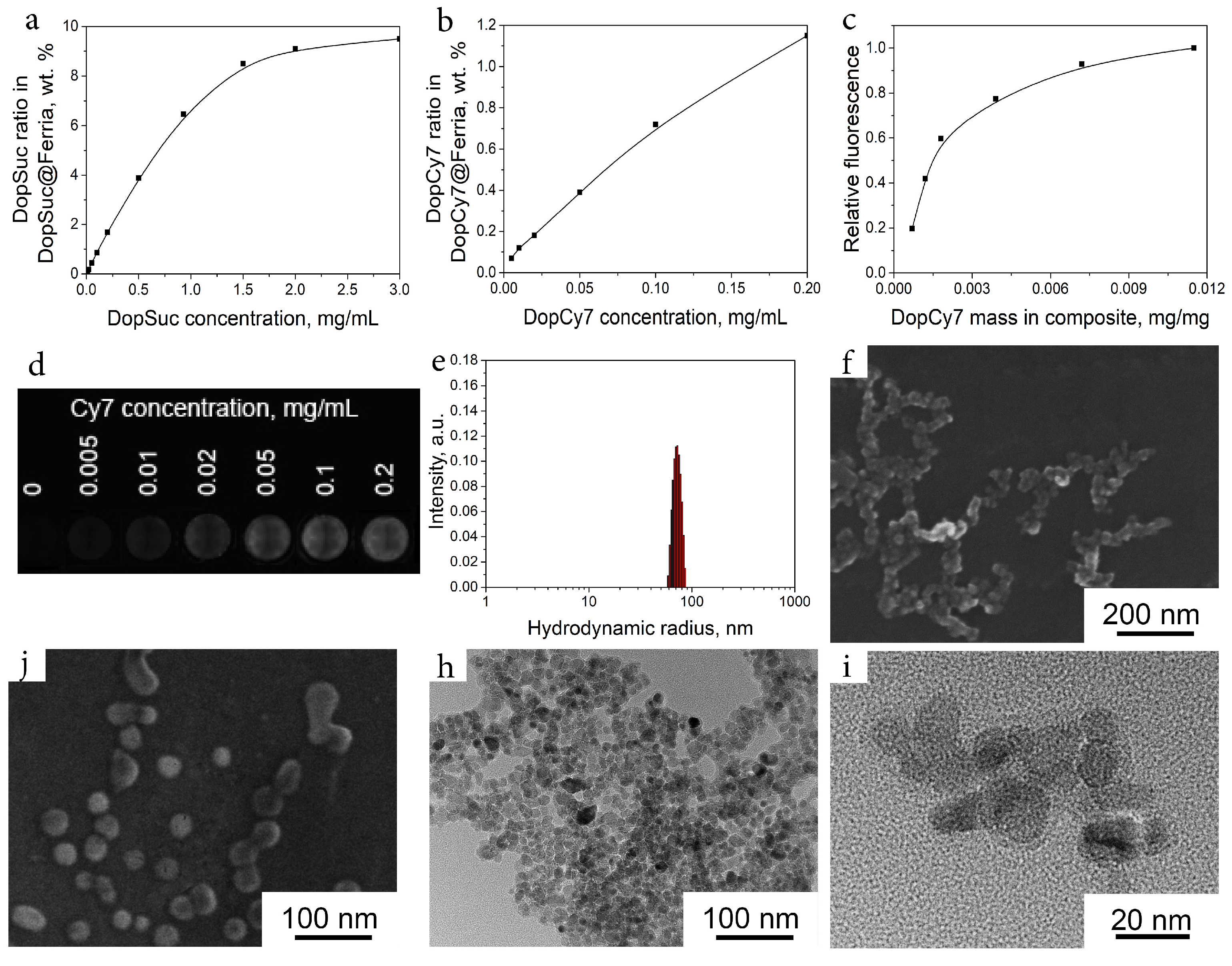
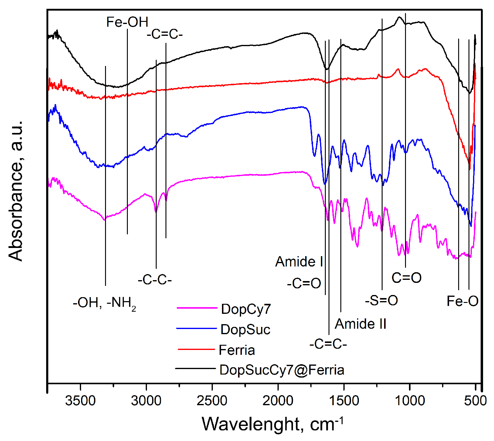
2.1. Synthesis and Modification of Composites
2.2. Biomimetic Modification of MNPs
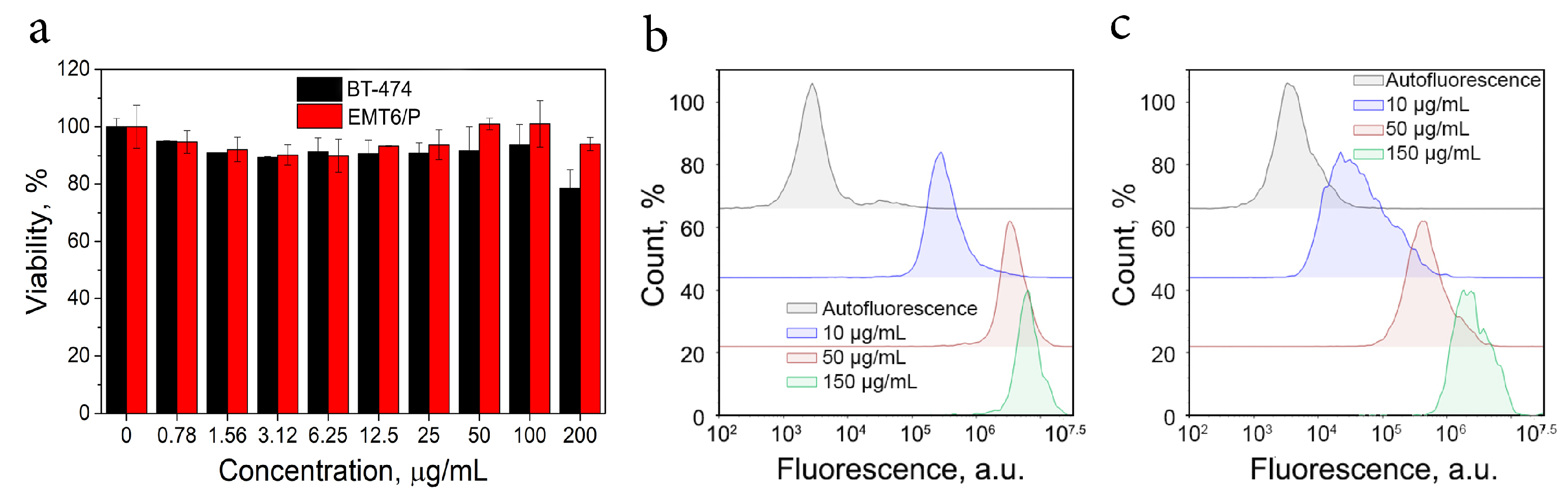
2.3. Bioactivity of Nanocomposite
3. Discussion
4. Materials and Methods
5. Conclusions
Author Contributions
Funding
Institutional Review Board Statement
Informed Consent Statement
Conflicts of Interest
References
- Ventola, C.L. Progress in nanomedicine: Approved and investigational nanodrugs. Pharm. Ther. 2017, 42, 742. [Google Scholar]
- Choi, Y.H.; Han, H.K. Nanomedicines: CURRENT status and future perspectives in aspect of drug delivery and pharmacokinetics. J. Pharm. Investig. 2018, 48, 43–60. [Google Scholar] [CrossRef] [PubMed] [Green Version]
- Huang, L.; Zhao, S.; Fang, F.; Xu, T.; Lan, M.; Zhang, J. Advances and perspectives in carrier-free nanodrugs for cancer chemo-monotherapy and combination therapy. Biomaterials 2021, 268, 120557. [Google Scholar] [CrossRef] [PubMed]
- Drozdov, A.S.; Nikitin, P.I.; Rozenberg, J.M. Systematic review of cancer targeting by nanoparticles revealed a global association between accumulation in tumors and spleen. Int. J. Mol. Sci. 2021, 22, 13011. [Google Scholar] [CrossRef] [PubMed]
- Singh, D.; Dilnawaz, F.; Sahoo, S.K. Challenges of moving theranostic nanomedicine into the clinic. Nanomedicine 2020, 15, 2. [Google Scholar] [CrossRef] [Green Version]
- Martins, J.P.; Das Neves, J.; de la Fuente, M.; Celia, C.; Florindo, H.; Günday-Türeli, N.; Popat, A.; Santos, J.L.; Sousa, F.; Schmid, R.; et al. The solid progress of nanomedicine. Drug Deliv. Transl. Res. 2020, 10, 726–729. [Google Scholar] [CrossRef] [Green Version]
- Gadekar, V.; Borade, Y.; Kannaujia, S.; Rajpoot, K.; Anup, N.; Tambe, V.; Kalia, K.; Tekade, R.K. Nanomedicines accessible in the market for clinical interventions. J. Control. Release 2021, 330, 372–397. [Google Scholar] [CrossRef]
- Mu, Q.; Yu, J.; McConnachie, L.A.; Kraft, J.C.; Gao, Y.; Gulati, G.K.; Ho, R.J. Translation of combination nanodrugs into nanomedicines: Lessons learned and future outlook. J. Drug Target. 2018, 26, 435–447. [Google Scholar] [CrossRef]
- Wang, X.; Hou, Y.; Yao, L.; Gao, M.; Ge, M. Generation, characterization, and application of hierarchically structured self-assembly induced by the combined effect of self-emulsification and phase separation. J. Am. Chem. Soc. 2016, 138, 2090–2093. [Google Scholar] [CrossRef]
- Vinogradov, V.V.; Drozdov, A.S.; Mingabudinova, L.R.; Shabanova, E.M.; Kolchina, N.O.; Anastasova, E.I.; Markova, A.A.; Shtil, A.A.; Milichko, V.A.; Starova, G.L.; et al. Composites based on heparin and MIL-101 (Fe): The drug releasing depot for anticoagulant therapy and advanced medical nanofabrication. J. Mater. Chem. B 2018, 6, 2450–2459. [Google Scholar] [CrossRef]
- Zhong, X.; Yang, K.; Dong, Z.; Yi, X.; Wang, Y.; Ge, C.; Zhao, Y.; Liu, Z. Polydopamine as a biocompatible multifunctional nanocarrier for combined radioisotope therapy and chemotherapy of cancer. Adv. Funct. Mater. 2015, 25, 7327–7336. [Google Scholar] [CrossRef]
- Chen, Z.; Wang, Z.; Gu, Z. Bioinspired and biomimetic nanomedicines. Acc. Chem. Res. 2019, 52, 1255–1264. [Google Scholar] [CrossRef] [PubMed]
- De Oliveira, H.; Thevenot, J.; Lecommandoux, S. Smart polymersomes for therapy and diagnosis: Fast progress toward multifunctional biomimetic nanomedicines. Wiley Interdiscip. Rev. Nanomed. Nanobiotech. 2012, 4, 525–546. [Google Scholar] [CrossRef] [PubMed]
- Drozdov, A.; Prilepskii, A.; Koltsova, E.; Anastasova, E.; Vinogradov, V. Magnetic polyelectrolyte-based composites with dual anticoagulant and thrombolytic properties: Towards optimal composition. J. Sol-Gel Sci. Technol. 2020, 95, 771–782. [Google Scholar] [CrossRef]
- Lee, H.; Dellatore, S.M.; Miller, W.M.; Messersmith, P.B. Mussel-inspired surface chemistry for multifunctional coatings. Science 2007, 318, 426–430. [Google Scholar] [CrossRef] [Green Version]
- Qiu, W.Z.; Yang, H.C.; Xu, Z.K. Dopamine-assisted co-deposition: An emerging and promising strategy for surface modification. Adv. Colloid Interface Sci. 2018, 256, 111–125. [Google Scholar] [CrossRef]
- Ryu, J.H.; Messersmith, P.B.; Lee, H. Polydopamine surface chemistry: A decade of discovery. ACS Appl. Mater. Interfaces 2018, 10, 7523–7540. [Google Scholar] [CrossRef]
- Klosterman, L.; Riley, J.K.; Bettinger, C.J. Control of heterogeneous nucleation and growth kinetics of dopamine-melanin by altering substrate chemistry. Langmuir 2015, 31, 3451–3458. [Google Scholar] [CrossRef]
- Lee, H.A.; Park, E.; Lee, H. Polydopamine and its derivative surface chemistry in material science: A focused review for studies at KAIST. Adv. Mater. 2020, 32, 1907505. [Google Scholar] [CrossRef]
- Liebscher, J.; Mroówczynński, R.; Scheidt, H.A.; Filip, C.; Haădade, N.D.; Turcu, R.; Bende, A.; Beck, S. Structure of polydopamine: A never-ending story? Langmuir 2013, 29, 10539–10548. [Google Scholar] [CrossRef]
- Ball, V. Polydopamine nanomaterials: Recent advances in synthesis methods and applications. Front. Bioeng. Biotechnol. 2018, 6, 109. [Google Scholar] [CrossRef]
- Palladino, P.; Bettazzi, F.; Scarano, S. Polydopamine: Surface coating, molecular imprinting, and electrochemistry—Successful Applications and future perspectives in (bio) analysis. Anal. Bioanal. Chem. 2019, 411, 4327–4338. [Google Scholar] [CrossRef] [PubMed]
- Mazur, M.; Barras, A.; Kuncser, V.; Galatanu, A.; Zaitzev, V.; Turcheniuk, K.V.; Woisel, P.; Lyskawa, J.; Laure, W.; Siriwardena, A.; et al. Iron oxide magnetic nanoparticles with versatile surface functions based on dopamine anchors. Nanoscale 2013, 5, 2692–2702. [Google Scholar] [CrossRef] [PubMed]
- Barclay, T.G.; Hegab, H.M.; Clarke, S.R.; Ginic-Markovic, M. Versatile surface modification using polydopamine and related polycatecholamines: Chemistry, structure, and applications. Adv. Mater. Interfaces 2017, 4, 1601192. [Google Scholar] [CrossRef]
- Lee, H.A.; Ma, Y.; Zhou, F.; Hong, S.; Lee, H. Material-independent surface chemistry beyond polydopamine coating. Accounts Chem. Res. 2019, 52, 704–713. [Google Scholar] [CrossRef]
- Drozdov, A.S.; Ivanovski, V.; Avnir, D.; Vinogradov, V.V. A universal magnetic ferrofluid: Nanomagnetite stable hydrosol with no added dispersants and at neutral pH. J. Colloid Interface Sci. 2016, 468, 307–312. [Google Scholar] [CrossRef] [PubMed]
- Anastasova, E.I.; Puzyrev, D.; Ivanovski, V.; Drozdov, A.S. Magnetically assisted synthesis of porous sol-gel magnetite matrices with structural anisotropy. J. Magn. Magn. Mater. 2020, 503, 166619. [Google Scholar] [CrossRef]
- Zakharzhevskii, M.; Drozdov, A.S.; Kolchanov, D.S.; Shkodenko, L.; Vinogradov, V.V. Test-system for bacteria sensing based on peroxidase-like activity of inkjet-printed magnetite nanoparticles. Nanomaterials 2020, 10, 313. [Google Scholar] [CrossRef] [Green Version]
- Anastasova, E.I.; Ivanovski, V.; Fakhardo, A.F.; Lepeshkin, A.I.; Omar, S.; Drozdov, A.S.; Vinogradov, V.V. A pure magnetite hydrogel: Synthesis, properties and possible applications. Soft Matter 2017, 13, 8651–8660. [Google Scholar] [CrossRef] [Green Version]
- Ivanovski, V.; Shapovalova, O.E.; Drozdov, A.S. Structural Rearrangements of Carbonic Anhydrase Entrapped in Sol-Gel Magnetite Determined by ATR–FTIR Spectroscopy. Int. J. Mol. Sci. 2022, 23, 5975. [Google Scholar] [CrossRef]
- Kuznowicz, M.; Jędrzak, A.; Rębiś, T.; Jesionowski, T. Biomimetic magnetite/polydopamine/β-cyclodextrins nanocomposite for long-term glucose measurements. Biochem. Eng. J. 2021, 174, 108127. [Google Scholar] [CrossRef]
- Yoshino, F.; Amano, T.; Zou, Y.; Xu, J.; Kimura, F.; Furusho, Y.; Chano, T.; Murakami, T.; Zhao, L.; Komatsu, N. Preferential tumor accumulation of polyglycerol functionalized nanodiamond conjugated with cyanine dye leading to near-infrared fluorescence in vivo tumor imaging. Small 2019, 15, 1901930. [Google Scholar] [CrossRef] [PubMed]
- Lunin, A.V.; Korenkov, E.S.; Mochalova, E.N.; Nikitin, M.P. Green Synthesis of Size-Controlled in Vivo Biocompatible Immunoglobulin-Based Nanoparticles by a Swift Thermal Formation. ACS Sustain. Chem. Eng. 2021, 9, 13128–13134. [Google Scholar] [CrossRef]
- Zelepukin, I.V.; Yaremenko, A.V.; Ivanov, I.N.; Yuryev, M.V.; Cherkasov, V.R.; Deyev, S.M.; Nikitin, P.I.; Nikitin, M.P. Long-term fate of magnetic particles in mice: A comprehensive study. ACS Nano 2021, 15, 11341–11357. [Google Scholar] [CrossRef]
- Nikitin, M.P.; Orlov, A.; Sokolov, I.; Minakov, A.; Nikitin, P.; Ding, J.; Bader, S.; Rozhkova, E.; Novosad, V. Ultrasensitive detection enabled by nonlinear magnetization of nanomagnetic labels. Nanoscale 2018, 10, 11642–11650. [Google Scholar] [CrossRef] [Green Version]
- Cheng, W.; Zeng, X.; Chen, H.; Li, Z.; Zeng, W.; Mei, L.; Zhao, Y. Versatile polydopamine platforms: Synthesis and promising applications for surface modification and advanced nanomedicine. ACS Nano 2019, 13, 8537–8565. [Google Scholar] [CrossRef]
- Volov, A.; Shkodenko, L.; Koshel, E.; Drozdov, A.S. Bio-Inspired Surface Modification of Magnetite Nanoparticles with Dopamine Conjugates. Nanomaterials 2022, 12, 2230. [Google Scholar] [CrossRef]
- Stueber, D.D.; Villanova, J.; Aponte, I.; Xiao, Z.; Colvin, V.L. Magnetic nanoparticles in biology and medicine: Past, present, and future trends. Pharmaceutics 2021, 13, 943. [Google Scholar] [CrossRef]
- Andreeva, Y.; Drozdov, A.; Solovyeva, A.; Fakhardo, A.; Vinogradov, V. Polyelectrolyte-based magnetic photonic crystals with anticoagulant activity. Mater. Today Chem. 2020, 17, 100292. [Google Scholar] [CrossRef]
- Montenegro, J.M.; Grazu, V.; Sukhanova, A.; Agarwal, S.; Jesus, M.; Nabiev, I.; Greiner, A.; Parak, W.J. Controlled antibody/(bio-) conjugation of inorganic nanoparticles for targeted delivery. Adv. Drug Deliv. Rev. 2013, 65, 677–688. [Google Scholar] [CrossRef]
- Marques, A.; Costa, P.; Velho, S.; Amaral, M. Functionalizing nanoparticles with cancer-targeting antibodies: A comparison of strategies. J. Control. Release 2020, 320, 180–200. [Google Scholar] [CrossRef] [PubMed]
- Shipunova, V.; Nikitin, M.; Nikitin, P.; Deyev, S. MPQ-cytometry: A magnetism-based method for quantification of nanoparticle–cell interactions. Nanoscale 2016, 8, 12764–12772. [Google Scholar] [CrossRef] [PubMed] [Green Version]
- Shimizu, S. Routes of Adminstration. In The Laboratory Mouse; Elsevier: Amsterdam, The Netherlands, 2004. [Google Scholar]
- Pinkerton, W.; Webber, M. A method of injecting small laboratory animals by the ophthalmic plexus route. Proc. Soc. Exp. Biol. Med. 1964, 116, 959–961. [Google Scholar] [CrossRef] [PubMed]
- Lundqvist, M.; Stigler, J.; Elia, G.; Lynch, I.; Cedervall, T.; Dawson, K.A. Nanoparticle size and surface properties determine the protein corona with possible implications for biological impacts. Proc. Natl. Acad. Sci. USA 2008, 105, 14265–14270. [Google Scholar] [CrossRef] [Green Version]
- Zanganeh, S.; Spitler, R.; Erfanzadeh, M.; Alkilany, A.M.; Mahmoudi, M. Protein corona: Opportunities and challenges. Int. J. Biochem. Cell Biol. 2016, 75, 143–147. [Google Scholar] [CrossRef] [Green Version]
- Kim, H.; Röth, D.; Isoe, Y.; Hayashi, K.; Mochizuki, C.; Kalkum, M.; Nakamura, M. Protein corona components of polyethylene glycol-conjugated organosilica nanoparticles modulates macrophage uptake. Colloids Surfaces Biointerfaces 2021, 199, 111527. [Google Scholar] [CrossRef]
- Schöttler, S.; Landfester, K.; Mailänder, V. Controlling the stealth effect of nanocarriers through understanding the protein corona. Angew. Chem. Int. Ed. 2016, 55, 8806–8815. [Google Scholar] [CrossRef]
- Mirkasymov, A.B.; Zelepukin, I.V.; Nikitin, P.I.; Nikitin, M.P.; Deyev, S.M. In vivo blockade of mononuclear phagocyte system with solid nanoparticles: Efficiency and affecting factors. J. Control. Release 2021, 330, 111–118. [Google Scholar] [CrossRef]
- Nikitin, M.P.; Zelepukin, I.V.; Shipunova, V.O.; Sokolov, I.L.; Deyev, S.M.; Nikitin, P.I. Enhancement of the blood-circulation time and performance of nanomedicines via the forced clearance of erythrocytes. Nat. Biomed. Eng. 2020, 4, 717–731. [Google Scholar] [CrossRef]
- Astafyeva, B.V.; Shapovalova, O.E.; Drozdov, A.S.; Vinogradov, V.V. α-Amylase@ Ferria: Magnetic nanocomposites with enhanced thermal stability for starch hydrolysis. J. Agric. Food Chem. 2018, 66, 8054–8060. [Google Scholar] [CrossRef]
- Rumyantceva, V.; Rumyantceva, V.; Andreeva, Y.; Tsvetikova, S.; Radaev, A.; Vishnevskaya, M.; Vinogradov, V.; Drozdov, A.S.; Koshel, E. Magnetically controlled carbonate nanocomposite with ciprofloxacin for biofilm eradication. Int. J. Mol. Sci. 2021, 22, 6187. [Google Scholar] [CrossRef] [PubMed]


Disclaimer/Publisher’s Note: The statements, opinions and data contained in all publications are solely those of the individual author(s) and contributor(s) and not of MDPI and/or the editor(s). MDPI and/or the editor(s) disclaim responsibility for any injury to people or property resulting from any ideas, methods, instructions or products referred to in the content. |
© 2022 by the authors. Licensee MDPI, Basel, Switzerland. This article is an open access article distributed under the terms and conditions of the Creative Commons Attribution (CC BY) license (https://creativecommons.org/licenses/by/4.0/).
Share and Cite
Drozdov, A.S.; Komarova, K.S.; Mochalova, E.N.; Komedchikova, E.N.; Shipunova, V.O.; Nikitin, M.P. Fluorescent Magnetic Nanoparticles for Bioimaging through Biomimetic Surface Modification. Int. J. Mol. Sci. 2023, 24, 134. https://0-doi-org.brum.beds.ac.uk/10.3390/ijms24010134
Drozdov AS, Komarova KS, Mochalova EN, Komedchikova EN, Shipunova VO, Nikitin MP. Fluorescent Magnetic Nanoparticles for Bioimaging through Biomimetic Surface Modification. International Journal of Molecular Sciences. 2023; 24(1):134. https://0-doi-org.brum.beds.ac.uk/10.3390/ijms24010134
Chicago/Turabian StyleDrozdov, Andrey S., Kristina S. Komarova, Elizaveta N. Mochalova, Elena N. Komedchikova, Victoria O. Shipunova, and Maxim P. Nikitin. 2023. "Fluorescent Magnetic Nanoparticles for Bioimaging through Biomimetic Surface Modification" International Journal of Molecular Sciences 24, no. 1: 134. https://0-doi-org.brum.beds.ac.uk/10.3390/ijms24010134