1. Introduction
Kalanchoë blossfeldiana Poelln. is succulent belonging to the short day plant species family [
1] (
Supplementary Figure S1). A physiological feature of succulents is the opening of stomata and the accumulation of organic acids at night, and the closing of the stomata and the gradual decline of organic acids during the day—referred to as crassulacean acid metabolism (CAM) [
2]. Daylight-induced decarboxylation of organic acids controls the size of stomata and causes them to close during the day [
3]. Malate and citrate obtained during dark CO
2 fixation accumulate in the vacuole [
4,
5,
6]. Details of this phenomenon have been recently described [
7].
Leaves of
K. blossfeldiana growing under long-day conditions accumulate large amounts of soluble phenolic compounds, unlike those growing under short-day conditions [
8]. The process of inflorescence development from flower initiation to anthesis in this plant was described by Hayashi and Konishi [
9]. Previously, it was shown that anthocyanin accumulation in the leaves and stems of
K. blossfeldiana is a consequence of the photoperiodic response [
10].
Anthocyanins are glycosidic derivatives of anthocyanidin, and the major anthocyanidins in plants are cyanidin, pelargonidin, peonidin, delphinidin, petunidin, and malvidin [
11]. Their structure differs mainly in the number and position of hydroxyl groups, which are glycosylated with glucose, galactose, rhamnose, arabinose, or xylose. Cyanidin glycosides are most common in plants [
12], and in
K. blossfeldiana, cyanidin-3-monoglucoside and cyanidin-3,5-diglucoside have been shown to be the main anthocyanin pigments [
13].
Anthocyanins in plants are synthesized through the general flavonoid pathway, is comprised of three molecules of malonyl-CoA and one molecule of 4-coumaroyl-CoA, the latter of which is derived from phenylalanine or tyrosine [
14]. Phenylalanine ammonia-lyase (PAL) is the enzyme that catalyzes the elimination of ammonia from
l-phenylalanine to give
trans-cinnamate, which can be a precursor of anthocyanins, flavonols, lignins, proanthocyanidins, and other compounds [
12]. Subsequently, dihydrokaempferol produced from naringenin by the action of flavanone 3-hydroxylase is converted to colorless leucoanthocyanidins with dihydroflavonol reductase. Then the conversion of leucoanthocyanidins to anthocyanidins is catalyzed by anthocyanidin synthase. The final step, which determines anthocyanin color, is the formation of anthocyanidin glycosides with the participation of the appropriate glycosyltransferases [
12].
Foliar anthocyanins are found in epidermal and/or mesophyll cells [
15]. Anthocyanins primarily accumulate in photosynthetic cells of tissues exposed to light (epidermis, palisade, and mesophyll cells) [
16]. In the stems of various plant species, JA-Me stimulates the accumulation of anthocyanins, indicating their potential involvement in the plant defense system against pathogen attack [
17,
18,
19,
20,
21]. For instance, it was shown that JA-Me in lanolin paste applied to the middle part of the stem of
K. blossfeldiana strongly stimulated anthocyanin accumulation in the main and lateral shoots below and above the treated site [
10]. However, almost no anthocyanin accumulation was observed in the JA-Me-treated stem after the leaves were removed from the plant. Góraj-Koniarska et al. [
20] also demonstrated that JA-Me substantially increased anthocyanin accumulation in the roots of intact
K. blossfeldiana plants. On the other hand, JA-Me slightly stimulated anthocyanin accumulation in the roots after leaf removal. It seems possible that in
K. blossfeldiana JA-Me increases anthocyanin biosynthesis directly in shoots and roots or raises levels by transporting them or their precursors from leaves to shoots and roots.
In contrast, in the hypocotyl of common buckwheat (
Fogopyrum esculentum Moench), where anthocyanins are abundant, JA-Me markedly reduced their content while stimulating the accumulation of proanthocyanidins (PAs) [
21,
22]. They are present in the fruit, bark, leaves, and seeds of plants [
23,
24]. The biosynthesis of PAs is affected by biotic and abiotic stresses and plant hormones [
25]. UV-B radiation caused a significant accumulation of PAs in aspen leaves [
26] and birch leaves [
27]. PAs may also be part of antioxidant activity in the overall stress response [
28,
29] and the plant defense response against biotic agents such as fungi, bacteria, viruses, and herbivores [
30].
Earlier studies showed that JA-Me had a strong stimulating effect on anthocyanin accumulation in
K. blossfeldiana in both stems and roots exposed to light [
19,
20]. Because of the noted phenomenon of anthocyanin accumulation in detached leaves of
K. blossfeldiana, we undertook a broader study in this area. One of the objectives in these studies was to determine whether the application of JA-Me would result in a further increase in anthocyanin content.
This paper presents experiments on the effect of storing detached leaves in an inverted position and excised stems of K. blossfeldiana under natural light conditions on the accumulation of anthocyanins in them. We also describe possible mechanisms of anthocyanin accumulation in detached leaves and the effect of JA-Me on this process, focusing on changes in basic metabolites and histological analyses.
3. Discussion
The following question arises: why do anthocyanins form on the underside of detached leaves of
Kalanchoë blossfeldiana, held upside down against the light? It seems that in
K. blossfeldiana the epidermis of the lower side of the leaves plays an important role in light perception in terms of the formation and accumulation of anthocyanins there. Schwabe [
31] showed that the leaf epidermis in this species plays a significant role in photoperiodic perception, but it is difficult to determine whether the epidermal feature is exerted through stomatal control or through some direct perception by the epidermal cells themselves. Woo-Gyu et al. [
32] showed that the density of stomata on the lower surface of
K. blossfeldiana leaves is higher compared to the upper surface. It is also known that in
K. blossfeldiana, the upper epidermis is made of polyhedral (hexagonal and pentagonal) cells and contains fewer stomata than the lower epidermis, whose cells have folded walls [
33]. Recently, Laskar et al. [
34] confirmed that the density of stomata on the lower surface of
K. blossfeldiana leaves is higher than on the upper surface and showed that the density of epidermal cell arrangement is significantly higher on the lower surface of the leaves.
It is interesting to note that the response of leaves detached from the plant and kept in a normal (natural) and inverted position for 4 days was similar compared to leaves on the plant. Thus, the accumulation of anthocyanins on the lower side of detached leaves kept in an inverted position was not accompanied by changes in the content of most carbohydrates and acids (except malic acid and citric acid) or other metabolites whose presence was confirmed by GC-MS. In general, the contents of some metabolites in the leaves that were kept for 4 days were higher, some were lower, and some had no effect compared to the leaves on the plant.
In the leaves of the
Kalanchoë genus, the main acids are malic and citric, as was previously shown [
35]. The decrease in malic acid content in detached leaves of
K. blossfeldiana is mainly due to the fact that the plant belongs to the CAM species. These species attach carbon dioxide to phosphoenolpyruvate at night and then convert it to malic acid, accumulating it in the vacuole. During the day, they use CO
2 for photosynthesis, which results in an increase in monocarbohydrate content. This metabolic route has been confirmed by the results obtained. It was calculated that there are close correlations between decreases in malic acid content and increases in monosaccharide content and their total contents (
Supplementary Table S3). This indicates that basic physiological processes are still taking place in leaves that have been detached from the plant and kept for 4 days.
The presence of glyceric acid (2,3-dihydroxypropanoic acid) in
K. blossfeldiana leaves was confirmed for the first time. To date, there is little information on the role of glyceric acid in plants and the course of its biosynthesis, although it has been found in some plants [
36,
37]. Probably the
d-glyceric acid is derived from 3-phospho-
d-glyceraldehyde by dehydrogenation to 3-phospho-
d-glyceric acid and loss of a phosphate group [
38]. Another suggestion is that glyceric acid is produced by the oxidation of glycerol [
39]. The decline in its level after a 4-day holding of detached
K. blossfeldiana leaves was independent of the conditions of this process. These results may indicate that the decrease in glyceric acid content may be related to the stress caused by detaching leaves from the plant. On the other hand, the increase in glyceric acid under JA-Me is hardly explainable.
It is not clarified why JA-Me stimulates anthocyanin formation in shoots of intact
K. blossfeldiana plants [
10], but it inhibits anthocyanin accumulation in detached leaves held upside down under natural light. It was previously shown that JA-Me induces stomatal closure in
Arabidopsis guard cells through a stimulatory effect on abscisic acid biosynthesis [
40,
41,
42]. Thus, the involvement of endogenous ABA induced by JA-Me in stomatal closure is most likely related to the inhibition of anthocyanin biosynthesis in detached
K. blossfeldiana leaves held upside down on the light.
Jasmonates (JAs) have been well known to function as messengers in the biosynthesis of secondary metabolites, including anthocyanins. JA-Me has been shown to stimulate anthocyanin accumulation in wild-type
Arabidopsis thaliana [
43], in the stems and leaves of tulips [
18], in cultures of
Vaccinium pahalae [
44], and in other plants. For instance, JA-Me vapor stimulated anthocyanin production in 5-day-old hypocotyls of soybean seedlings growing in the light but inhibited their accumulation in etiolated seedlings [
45]. The authors suggested that the reason for this phenomenon may be that two different mechanisms regarding anthocyanin biosynthesis occur in the light and in the dark [
45]. Xie et al. [
46] documented that anthocyanin accumulation in sprouts of tumorous stem mustard (
Brassica juncea) can be enhanced by sucrose under light conditions and by JA-Me under light and dark conditions.
Factors resulting from abiotic stresses (wounds, drought, osmotic stress, and others) and biotic stresses also stimulate the biosynthesis of JAs in plants [
47]. Thus, the formation of anthocyanins in the epidermal cells and in the layer of mesophyll cells located directly under the epidermis of the detached leaves and detached stem in
K. blossfeldiana may be a consequence of the increased level of endogenous JAs.
However, it seems that the stimulation of JAs biosynthesis as a result of drought stress is not the cause of anthocyanin accumulation in detached leaves and excised stems of
K. blossfeldiana. It is known that species belonging to the Crassulaceae (CAM) family are considered insensitive to water deficiency [
48]. Thus, the direct factor causing anthocyanin accumulation on the underside of detached leaves held inversely to their natural position and the cut stem stored in the light is unknown. Meanwhile, the application of exogenous JA-Me to detached leaves and stems resulted in an excess of JA in tissues below and above the treatment site, which was probably responsible for the inhibition of anthocyanin accumulation. This is supported by the previously stated evidence that jasmonic acid (JA) and JA-Me can be long-distance transported in the plant [
49,
50,
51].
It should be mentioned that anthocyanins did not accumulate within 4–10 mm of the infected areas in detached leaves of
Crassula multicava and
K. blossfeldiana with disease symptoms caused by
Pestalotia sp. and held in natural light [
52,
53]. Following the infection of the leaves with the vein, there was a lack of anthocyanin accumulation on the vein, which was clearly visible at some distance from the infected area. It is believed that this phenomenon is caused by the pathogen’s secretion of biologically active compounds, which may be jasmonates.
Previously, Horbowicz et al. [
21,
22,
54] showed that exogenously applied JA-Me inhibited anthocyanin biosynthesis and accumulation in the hypocotyl of etiolated seedlings of common buckwheat (
Fagopyrum esculentum). The phenomenon was observed in experiments with various methods of JA-Me treatment. JA-Me also inhibited anthocyanins accumulation in etiolated seedlings of soybean [
45]. The stimulating effect of ethephon on anthocyanin accumulation has been reported [
55,
56]. However, it seems that the application of JA-Me not affect this process in
K. blossfeldiana.
In the near future, further intensive research will be necessary to elucidate the mechanisms involved in the accumulation of anthocyanins in the presence or absence of JA-Me, as well as the effect of the method of keeping the leaves after detaching from the plant. These will concern both the composition of anthocyanins present in K. blossfaldiana leaves and the sites of their accumulation in the leaf, as well as the activity of enzymes involved in their biosynthesis.
4. Materials and Methods
4.1. Plant Materials and the Application of Methyl Jasmonate (JA-Me)
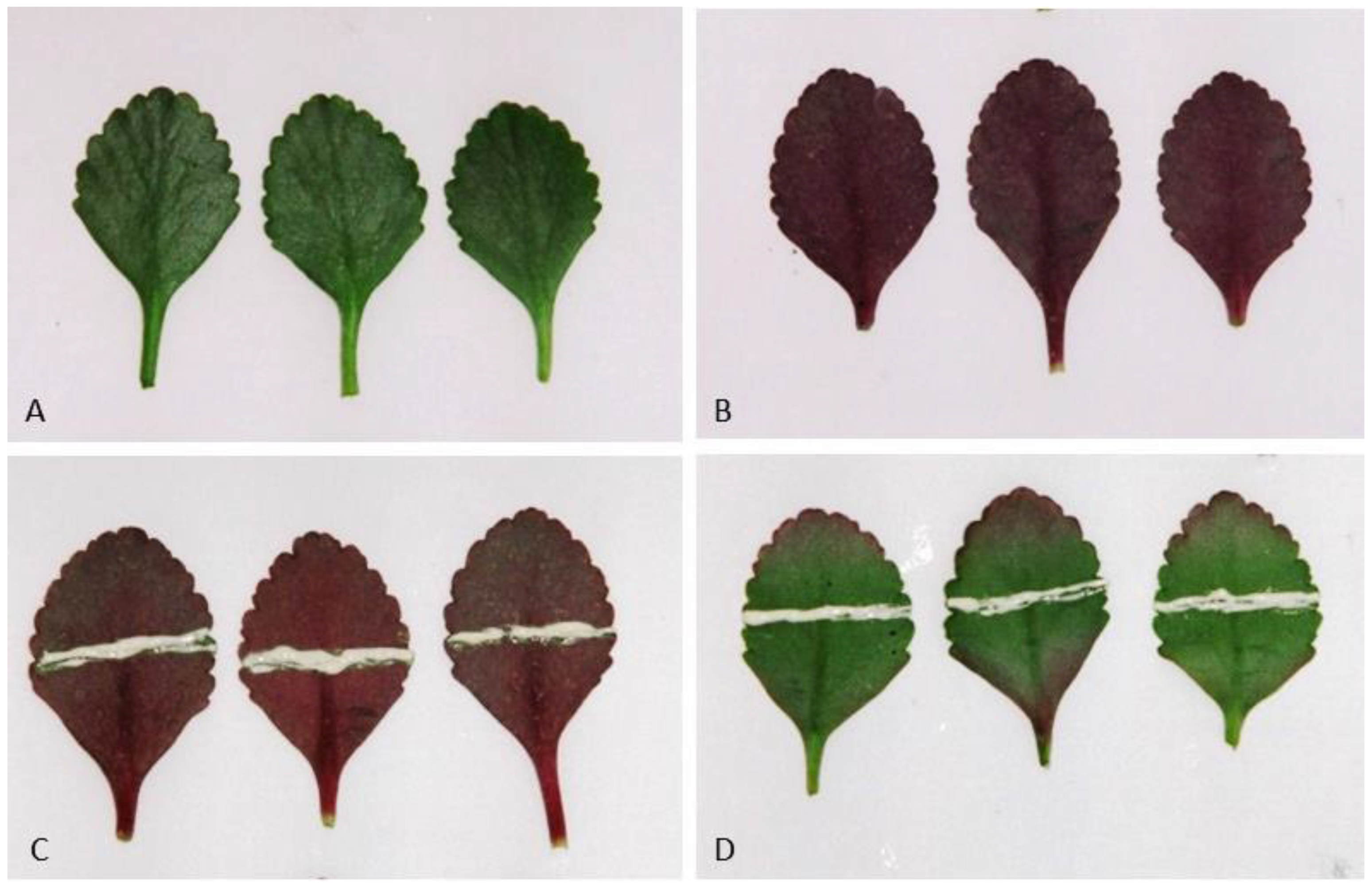
Plants of
Kalanchoë blossfeldiana grown in a greenhouse of the National Institute of Horticultural Research in Skierniewice, Poland (51°57′50.6″ N, 20°10′15.2″ E) were used. The shoot cuttings were taken in March for rooting in a mixture of soil, peat moss, and sand (2:1:1) in a greenhouse under natural conditions, and then replanted separately in pots in the same growing medium. Three- to five-month-old plants of
K. blossfeldiana having green leaves and stems were used for further experiments with excised leaves (large, older (40 mm width) and small, younger (25 mm width), and an excised stem after removal of leaves and cut at the level of medium (
Figure 6;
Supplementary Figure S2). All experiments were repeated three to five times, using 20 to 25 leaves per treatment and 20 stems.
Experiment A. Detached leaves of K. blossfeldiana were treated with JA-Me at concentrations of 0.1, 0.25, 0.5%, and 1.0% in lanolin paste. JA-Me was applied in the middle part of the leaf blade as a 1–2 mm-wide strip on its upper (adaxial) or lower (abaxial) side. The leaves were kept in a normal (abaxial side underside) or inverted position (adaxial side underside) under natural light conditions in the greenhouse at 20–24 °C under ambient light of 30–50 μmol/m2/s PPFD. Control leaves were not treated or treated with lanolin only and kept in the same conditions.
Experiment B. Detached leaves of K. blossfeldiana were soaked with water and JA-Me in water solutions at concentrations of 25, 50, 100, and 200 mg/L and in JA-Me at a concentration 10−3 M and 10−4 M in ethanol-water solutions for 1 h and then were kept in an inverted position in natural light conditions in a greenhouse.
Experiment C. Detached leaves of K. blossfeldiana were treated with JA-Me and ethephon dissolved in lanolin paste separately and simultaneously on the abaxial side of the leaves. The leaves were then kept for 4 days in an inverted position under natural light conditions without watering and/or on moistened filter paper.
Experiment D. Excised entire stems of K blossfeldiana, after removal of leaves and roots, were treated with JA-Me at a concentration of 0.05, 0.1, and 0.5% (w/w in lanolin) as a 2 mm-wide ring in the middle of the internode in the middle stem and kept in a natural light conditions in a greenhouse. Anthocyanin accumulation was observed morphologically only.
4.2. Analyses of Total Anthocyanins and Proanthocyanidins
Total anthocyanin content was measured as described by Mancinelli [
57]. Briefly, freeze-dried and pulverized tissues were extracted with acidified (1% HCl,
w/
v) methanol for 24 h at room temperature in darkness with occasional shaking. Then the extracts were carefully decanted, and their absorbance was measured at 530 nm and 657 nm. The formula A
530–0.25A
657 was used to compensate for the absorption of chlorophyll degradation. Anthocyanin content was calculated as cyanidin-3-glucoside using 29,600/M x cm as molecular extinction coefficient.
For the determination of total proanthocyanidins, a method based on their hydrolysis to anthocyanidins in HCl-butanol solution was used [
58,
59]. A freeze-dried and pulverized sample was vortexed with 3.8 mL mixture of n-butanol and concentrated HCl (95:5,
v/
v) and 0.2 mL of 2% (
w/
v) NH
4Fe(SO
4)
2 · 12 H
2O in 2 M HCl and centrifuged, and then absorbances were measured at 550 nm. The mixture was then heated in a heating block at 95 °C for 60 min. After cooling and centrifugation, absorbance at 550 nm was measured. The absorbance values of the reaction mixtures before hydrolysis were subtracted from the final absorbance after hydrolysis. Absorbance values were converted to proanthocyanidin equivalents using the molar absorption coefficient of cyanidin chloride in a 5% HCl-butanol solution.
Reagents including methanol, hydrochloric acid, n-butanol, and NH4Fe(SO4)2 · 12 H2O were purchased from POCH S.A. (Gliwice, Poland). The absorbance was measured using a Rayleigh type UV-1800 UV/Vis spectrophotometer (Beijing Beifen-Ruili Analytical Instrument Co., Ltd., Nanjing, China)
4.3. Polar Metabolite Analyses
Polar metabolites were extracted from freeze-dried and pulverized leaf tissues according to the method described earlier [
60]. Briefly, the polar metabolites were extracted from samples with a mixture of methanol:water (1:1,
v/
v, containing ribitol as an internal standard) at 70 °C for 30 min. Homogenates were centrifuged, and aliquots of the clear supernatant were extracted with chloroform to remove non-polar compounds. The polar fraction was concentrated to dryness in a speed vacuum rotary evaporator (JWElectronic, Warsaw, Poland). The metabolites were derivatized with
O-methoxamine hydrochloride and then with
N-methyl-
N-trimethylsilyl-trifluoroacetamide. The resulting trimethylsilyl derivatives were analyzed on a ZEBRON ZB-5MSi Guardian capillary column (Phenomenex, Torrance, CA, USA).
Metabolites were identified by comparison of their retention time, retention indices, and mass spectra of original standards purchased from Sigma-Aldrich (Saint Louis, MO, USA) and from the NIST 05 library (National Institute of Standards and Technology, NIST, Gaithersburg, MD, USA). Two gas chromatographs were applied: the GC-2030 Nexis (Shimadzu, Kyoto, Japan) equipped with a flame ionization detector and the GC-2010 coupled with a quadrupole mass spectrometry analyzer (GCMS-QP2010 Plus, Shimadzu, Kyoto, Japan). The details of chromatographic separations were described previously [
61]. For the quantitative analyses of identified polar metabolites, the data obtained for the peak areas from the GC-FID analyses were used.
4.4. Histological Analyses of the Local Occurrence of Anthocyanins in Leaves
Detached leaves were taken for histological observation at the beginning of the experiment (0 day after leaf detachment, control) and after 4 days of keeping them in an inverted position under natural light conditions. Cross-sections of detached leaves of K. blossfeldiana were prepared free-hand using a razor blade. Microscopic preparations were also made of the isolated adaxial and abaxial epidermis, which were observed unstained in a light microscope (Eclipse 80i, Nikon, Tokyo, Japan).
4.5. Statitistics
Analyses were performed in three replicates. An analysis of variance (one-way ANOVA) and Tukey’s post hoc test were used to check the significance of the differences. These calculations and the Pearson’s correlation coefficients were performed using Statistica 12PL software (StatSoft, Tulsa, OK, USA). The results are shown as means ± standard deviation.
Normalized data were considered using a multivariate statistical analysis (principal component analysis, PCA, and hierarchical cluster analysis, HCA), which was performed in three replicates using COVAIN, a MATLAB toolbox including a graphical user interface (MATLAB version 2013a; Math Works, Natick, MA, USA) [
61].

 ,
, 


