
Triple Genetic Diagnosis in a Patient with Late-Onset Leukodystrophy and Mild Intellectual Disability

 , , , , ,
Abstract
:1. Introduction
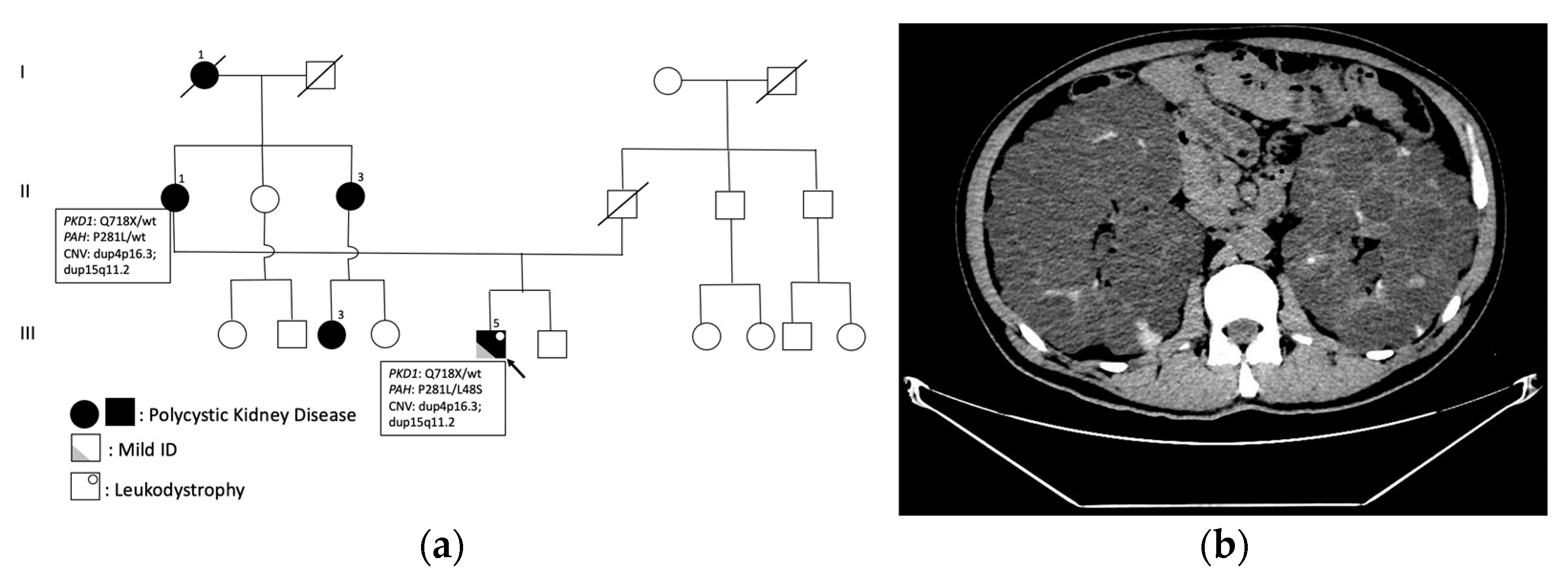
2. Results
3. Discussion
4. Materials and Methods
Author Contributions
Funding
Institutional Review Board Statement
Informed Consent Statement
Data Availability Statement
Conflicts of Interest
References
- Gilchrist, D.M. Medical Genetics: 3. An Approach to the Adult with a Genetic Disorder. CMAJ Can. Med. Assoc. J. 2002, 167, 1021–1029. [Google Scholar]
- Resende, L.L.; De Paiva, A.R.B.; Kok, F.; Da Costa Leite, C.; Lucato, L.T. Adult Leukodystrophies: A Step-by-Step Diagnostic Approach. RadioGraphics 2019, 39, 153–168. [Google Scholar] [CrossRef] [PubMed]
- Lynch, D.S.; Wade, C.; De Paiva, A.R.B.; John, N.; Kinsella, J.A.; Merwick, Á.; Ahmed, R.M.; Warren, J.D.; Mummery, C.J.; Schott, J.M.; et al. Practical approach to the diagnosis of adult-onset leukodystrophies: An updated guide in the genomic era. J. Neurol. Neurosurg. Psychiatry 2019, 90, 543–555. [Google Scholar] [CrossRef] [PubMed]
- Rossi, S.; Concolino, P.; Di Natale, D.; Pasquetti, D.; Di Lella, G.M.; Chiurazzi, P.; Silvestri, G. Clinical Reasoning: A Young Man With Subacute Onset of Spastic Paraparesis. Neurology 2023, 100, 199–205. [Google Scholar] [CrossRef] [PubMed]
- Kurolap, A.; Orenstein, N.; Kedar, I.; Weisz Hubshman, M.; Tiosano, D.; Mory, A.; Levi, Z.; Marom, D.; Cohen, L.; Ekhilevich, N.; et al. Is One Diagnosis the Whole Story? Patients with Double Diagnoses. Am. J. Med. Genet. A 2016, 170, 2338–2348. [Google Scholar] [CrossRef] [PubMed]
- Blau, N.; van Spronsen, F.J.; Levy, H.L. Phenylketonuria. Lancet Lond. Engl. 2010, 376, 1417–1427. [Google Scholar] [CrossRef] [PubMed]
- Chen, S.; Zhu, M.; Hao, Y.; Feng, J.; Zhang, Y. Effect of Delayed Diagnosis of Phenylketonuria With Imaging Findings of Bilateral Diffuse Symmetric White Matter Lesions: A Case Report and Literature Review. Front. Neurol. 2019, 10, 1040. [Google Scholar] [CrossRef] [PubMed]
- Tufekcioglu, Z.; Cakar, A.; Bilgic, B.; Hanagasi, H.; Gurvit, H.; Emre, M. Adult-Onset Phenylketonuria with Rapidly Progressive Dementia and Parkinsonism. Neurocase 2016, 22, 273–275. [Google Scholar] [CrossRef]
- Weglage, J.; Oberwittler, C.; Marquardt, T.; Schellscheidt, J.; von Teeffelen-Heithoff, A.; Koch, G.; Gerding, H. Neurological Deterioration in Adult Phenylketonuria. J. Inherit. Metab. Dis. 2000, 23, 83–84. [Google Scholar] [CrossRef]
- Dworniczak, B.; Grudda, K.; Stümper, J.; Bartholomé, K.; Aulehla-Scholz, C.; Horst, J. Phenylalanine Hydroxylase Gene: Novel Missense Mutation in Exon 7 Causing Severe Phenylketonuria. Genomics 1991, 9, 193–199. [Google Scholar] [CrossRef]
- Okano, Y.; Wang, T.; Eisensmith, R.C.; Longhi, R.; Riva, E.; Giovannini, M.; Cerone, R.; Romano, C.; Woo, S.L. Phenylketonuria Missense Mutations in the Mediterranean. Genomics 1991, 9, 96–103. [Google Scholar] [CrossRef] [PubMed]
- Danecka, M.K.; Woidy, M.; Zschocke, J.; Feillet, F.; Muntau, A.C.; Gersting, S.W. Mapping the Functional Landscape of Frequent Phenylalanine Hydroxylase (PAH) Genotypes Promotes Personalised Medicine in Phenylketonuria. J. Med. Genet. 2015, 52, 175–185. [Google Scholar] [CrossRef] [PubMed]
- BIOPKU: International Database of Patients and Mutations Causing BH4-Responsive HPA/PKU. Available online: http://www.biopku.org/authorisation/copyrightdisclaimer.asp?o=1 (accessed on 19 December 2022).
- Santos, L.L.; Fonseca, C.G.; Starling, A.L.P.; Januario, J.N.; Aguiar, M.J.B.; Peixoto, M.G.C.D.; Carvalho, M.R.S. Variations in Genotype-Phenotype Correlations in Phenylketonuria Patients. Genet. Mol. Res. 2010, 9, 1–8. [Google Scholar] [CrossRef] [PubMed]
- Blau, N. Genetics of Phenylketonuria: Then and Now. Hum. Mutat. 2016, 37, 508–515. [Google Scholar] [CrossRef] [PubMed]
- Zurflüh, M.R.; Zschocke, J.; Lindner, M.; Feillet, F.; Chery, C.; Burlina, A.; Stevens, R.C.; Thöny, B.; Blau, N. Molecular Genetics of Tetrahydrobiopterin-Responsive Phenylalanine Hydroxylase Deficiency. Hum. Mutat. 2008, 29, 167–175. [Google Scholar] [CrossRef] [PubMed]
- Burnside, R.D.; Pasion, R.; Mikhail, F.M.; Carroll, A.J.; Robin, N.H.; Youngs, E.L.; Gadi, I.K.; Keitges, E.; Jaswaney, V.L.; Papenhausen, P.R.; et al. Microdeletion/Microduplication of Proximal 15q11.2 between BP1 and BP2: A Susceptibility Region for Neurological Dysfunction Including Developmental and Language Delay. Hum. Genet. 2011, 130, 517–528. [Google Scholar] [CrossRef] [PubMed]
- Biembengut, Í.V.; Silva, I.L.Z.; Souza, T.d.A.C.B.d.; Shigunov, P. Cytoplasmic FMR1 Interacting Protein (CYFIP) Family Members and Their Function in Neural Development and Disorders. Mol. Biol. Rep. 2021, 48, 6131–6143. [Google Scholar] [CrossRef]
- Schenck, A.; Bardoni, B.; Moro, A.; Bagni, C.; Mandel, J.L. A Highly Conserved Protein Family Interacting with the Fragile X Mental Retardation Protein (FMRP) and Displaying Selective Interactions with FMRP-Related Proteins FXR1P and FXR2P. Proc. Natl. Acad. Sci. USA 2001, 98, 8844–8849. [Google Scholar] [CrossRef]
- De Rubeis, S.; Pasciuto, E.; Li, K.W.; Fernández, E.; Di Marino, D.; Buzzi, A.; Ostroff, L.E.; Klann, E.; Zwartkruis, F.J.T.; Komiyama, N.H.; et al. CYFIP1 Coordinates mRNA Translation and Cytoskeleton Remodeling to Ensure Proper Dendritic Spine Formation. Neuron 2013, 79, 1169–1182. [Google Scholar] [CrossRef]
- Abekhoukh, S.; Sahin, H.B.; Grossi, M.; Zongaro, S.; Maurin, T.; Madrigal, I.; Kazue-Sugioka, D.; Raas-Rothschild, A.; Doulazmi, M.; Carrera, P.; et al. New Insights into the Regulatory Function of CYFIP1 in the Context of WAVE- and FMRP-Containing Complexes. Dis. Model. Mech. 2017, 10, 463–474. [Google Scholar] [CrossRef]
- Benítez-Burraco, A.; Barcos-Martínez, M.; Espejo-Portero, I.; Jiménez-Romero, S. Variable Penetrance of the 15q11.2 BP1-BP2 Microduplication in a Family with Cognitive and Language Impairment. Mol. Syndromol. 2017, 8, 139–147. [Google Scholar] [CrossRef]


Disclaimer/Publisher’s Note: The statements, opinions and data contained in all publications are solely those of the individual author(s) and contributor(s) and not of MDPI and/or the editor(s). MDPI and/or the editor(s) disclaim responsibility for any injury to people or property resulting from any ideas, methods, instructions or products referred to in the content. |
© 2023 by the authors. Licensee MDPI, Basel, Switzerland. This article is an open access article distributed under the terms and conditions of the Creative Commons Attribution (CC BY) license (https://creativecommons.org/licenses/by/4.0/).
Share and Cite
Pasquetti, D.; Gazzellone, A.; Rossi, S.; Orteschi, D.; L’Erario, F.F.; Concolino, P.; Minucci, A.; Dionisi-Vici, C.; Genuardi, M.; Silvestri, G.; et al. Triple Genetic Diagnosis in a Patient with Late-Onset Leukodystrophy and Mild Intellectual Disability. Int. J. Mol. Sci. 2024, 25, 495. https://0-doi-org.brum.beds.ac.uk/10.3390/ijms25010495
Pasquetti D, Gazzellone A, Rossi S, Orteschi D, L’Erario FF, Concolino P, Minucci A, Dionisi-Vici C, Genuardi M, Silvestri G, et al. Triple Genetic Diagnosis in a Patient with Late-Onset Leukodystrophy and Mild Intellectual Disability. International Journal of Molecular Sciences. 2024; 25(1):495. https://0-doi-org.brum.beds.ac.uk/10.3390/ijms25010495
Chicago/Turabian StylePasquetti, Domizia, Annalisa Gazzellone, Salvatore Rossi, Daniela Orteschi, Federica Francesca L’Erario, Paola Concolino, Angelo Minucci, Carlo Dionisi-Vici, Maurizio Genuardi, Gabriella Silvestri, and et al. 2024. "Triple Genetic Diagnosis in a Patient with Late-Onset Leukodystrophy and Mild Intellectual Disability" International Journal of Molecular Sciences 25, no. 1: 495. https://0-doi-org.brum.beds.ac.uk/10.3390/ijms25010495