Article

Patterns of Distribution of Phoretic Deutonymphs of Uropodina on Longhorn Beetles in Białowieża Primeval Forest, Central Europe

by
Szymon Konwerski
1,*,
Jerzy M. Gutowski
2 and
Jerzy Błoszyk
1,3
1
Natural History Collections, Faculty of Biology, Adam Mickiewicz University, 61-614 Poznań, Poland
2
Forest Research Institute, Department of Natural Forests, 17-230 Białowieża, Poland
3
Department of General Zoology, Faculty of Biology, Adam Mickiewicz University, 61-614 Poznań, Poland
*
Author to whom correspondence should be addressed.
Submission received: 29 April 2020 / Revised: 4 June 2020 / Accepted: 10 June 2020 / Published: 12 June 2020
(This article belongs to the Special Issue Biodiversity of Mites)

Abstract

:
We studied the distributional patterns of phoretic deutonymphs of the genera Oodinychus and Trichouropoda (Uropodina) on the longhorn beetles Monochamus sartor urussovii, Plagionotus detritus, Tetropium castaneum, and Te. fuscum, based on large samples of specimens (992 beetles and 25,587 mites) collected in the Białowieża Primeval Forest in Central Europe in the years 2008 and 2012–2016. All the studied phoretic phoront-host associations are characterized by different patterns of the attachment sites of mites on beetle’s body. In the case of O. ovalis and M. sartor urussovii association, the deutonymphs were found mostly on the pronotum and dorsal surface of the elytra. This is the only instance in which phoronts were absent on the legs. Deutonymphs of T. sociata on P. detritus preferred the abdomnen (both tergites and ventrites) and the hindlegs. Only in this case the phoronts were attached inside the subelytral space. The highest number of deutonymphs of T. shcherbakae on both Tetropium species occured on the legs. In the case of Te. castaneum, similar proportions of mites were recorded on all pairs of legs, while the preferred location of mites phoretic on Te. fuscum were the forelegs. Both the preferences of phoretic deutonymphs for specific parts of the host’s body and the participation of carriers transporting deutonymphs on particular parts of their bodies were very consistent.

Graphical Abstract

1. Introduction

Mites, even those inhabiting well-studied systems are often overlooked or ignored, although they are strong interactors and major components of biological diversity—not passive inhabitants of ecosystems [1]. This statement is particularly relevant to Uropodina mites in natural forest ecosystems. In this group of mites, the unique morphological adaptations for phoretic purposes, namely the production of an anal pedicel [2,3], enabled them to use the saproxylic beetles as carriers (dispersants). This, in turn, allowed for the efficient use of unstable, temporary, cyclical, and patchy merocenoses in dying and dead trees, including beetle-generated microhabitats. Close and specific relationships characterize the pioneer species colonizing dying and dead trees in early stages of decay [4], among which the most important as dispersants of mites are Scolytinae and Cerambycidae [5]. While Uropodina and bark beetles relationships have been the subject of numerous studies, and their role is relatively well known, both in terms of phoresy and species communities in beetle galleries [6,7,8,9,10,11,12,13,14,15,16,17,18], little attention has been paid to uropodids and longhorn beetles, so the importance of their ecological relationships for the forest ecosystem is essentially unknown [19].
The phoretic relationships between uropodid mites and Cerambycidae in the natural forest ecosystem has been studied in recent years in Białowieża Primeval Forest, which is considered the best-preserved forest in the European lowlands. The studies have been focused on new associations of uropodids and longhorn beetle species [20], relationships between phoronts and their carriers [21,22], the contribution of beetle dispersants in increasing of alpha diversity of mites in dead wood merocenoses [23], as well as the stability and repeatability of phoretic relationships [24,25].
The aim of this research was to find the distributional patterns of phoretic deutonymphs for longhorn beetle species utilized as dispersants by uropodids in the natural forest ecosystem of Białowieża Primeval Forest in Central Europe, based on a large data set and complementing earlier studies, in order to clarify our understanding of relationships between the Uropodina mites and their saproxylic Cerambycidae carriers.
The Białowieża Primeval Forest is unique within Europe because of its natural structure [26,27,28], and plays a role of a living laboratory for ecological research [29]. It is also a European hotspot for Coleoptera [30,31,32,33,34,35]. Studies of primary forest provide reference data for ecological research in managed forests and improve the knowledge on conservation of saproxylic organisms [36].

2. Materials and Methods

The study concentrated on the Uropodina mites (Acari: Mesostigmata) phoretic on the most numerous (over 30 specimens with attached mites) longhorn beetle species: Monochamus sartor urussovii, Plagionotus detritus, Tetropium castaneum, and Tetropium fuscum.
The nomenclature of cerambycids follows Löbl and Smetana [37] and Plewa et al. [38], and the nomenclature of Uropodina follows Błoszyk [39]. The terminology of beetle morphological structures follows Lawrence et al. [40].
The research was conducted in the old growth (natural) Białowieża Primeval Forest, in natural stands rich in dead wood. The study sites were located in forest compartments 338A (N52º44’, E23º45’), near Teremiski, about 8 km west-north of the village of Białowieża, 448C (N52º42’, E23º46’) in the protected area “Professor Władysław Szafer’s Nature Reserve”, and 496C (N52º40’, E23º47’), about 4 km south west of the village of Białowieża. Detailed information about the study sites are in Błoszyk et al. [21,23] and Konwerski et al. [24,25].
Beetles were caught in 66 IPM-Intercept traps and 48 12-unit Lindgren multiple funnel traps, provided by the Canadian Forest Service. They were set in forest stands from May to July in 2008 and 2012–2016, and emptied every two weeks. Cerambycidae specimens were kept in 70% ethanol. In the laboratory they were individually (each specimen separately) examined under a stereomicroscope, to determine their species and to record the presence of mites. Beetles with attached mites (carriers) were selected for further study. Using a stereomicroscope (Olympus SZX12), the number of deutonymphs on beetles was counted and their location of attachment to beetles (hosts) was determined. The possible locations were the legs, head, scutellum, dorsal surface of the elytra, subelytra, membranous wings, pronotum, prosternum, mesoventrite, metaventrite, abdominal ventrites, and tergites.
All the uropodid mites found attached to longhorn beetles were identified using the comparative collection of J. Błoszyk, deposited in the Natural History Collections at the Faculty of Biology, Adam Mickiewicz University in Poznań (Poland), and the keys to mite species identification [41,42,43,44]. For biometrical analyses, selected mites were mounted on microscope slides using Hoyer’s medium. The remaining mites were identified from temporary preparations after clearing in the lactic acid using an open slide technique and later placed in 75% ethanol.
The voucher specimens of longhorn beetles and mites have been deposited in the Natural History Collections at the Faculty of Biology, Adam Mickiewicz University in Poznań, Poland.
Distribution of phoretic deutonymphs on various parts of host’s body was compared using multivariate exploratory techniques: cluster analysis (distance: Euclidean, method: ward). Frequency distribution tables were used for calculation of the phoretic load for each beetle species. Statistical analyses were performed using the software Statistica 10 [45].

3. Results

During the six years of the research project, 992 specimens of Cerambycidae with 25,587 transported deutonymphs of Uropodina were collected. On 54 specimens of M. sartor urussovii, there were 1088 phoretic deutonymphs belonging to a single species Oodinychus ovalis. On 494 specimens of P. detritus, there were 19,145 phoretic deutonymphs belonging to a single species Trichouropoda sociata. On 97 specimens of Te. castaneum and 347 specimens of Te. fuscum, there were 839 and 4515 phoretic deutonymphs, respectively, all belonging to Trichouropoda shcherbakae.
The mites were found on the following body parts of the beetles: all pairs of legs, head, scutellum, dorsal surface of the elytra, subelytra, membranous wings, pronotum, prosternum, mesoventrite, metaventrite, abdominal ventrites, and tergites (Table 1, Figure 1 and Figure 2).

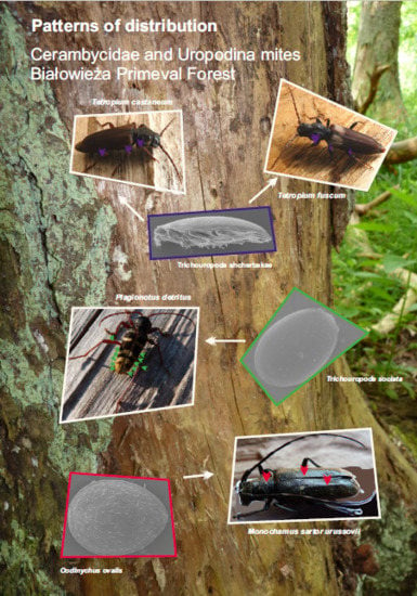
3.1. Distribution of Oodinychus ovalis on Monochamus sartor urussovii

The mites were found on dorsal surface of the elytra, pronotum, mesoventrite, and abdominal ventrites (Table 1). The number of deutonymphs attached to one beetle varied between one and 149.
The deutonymphs were found most frequently attached to the dorso-lateral side of the prothorax—the pronotum (85.4% of all carried deutonymphs, up to 141 individuals at one time). On the dorsal surface of the elytra, there were 12.9% of all deutonymphs (up to 122 individuals at one time), whereas on ventral side of the body the mites were attached only to the mesoventrite (1.2% of all carried deutonymphs, up to six individuals at one time) and abdominal ventrites (0.4%, four individuals—only once) (Figure 3).
The majority of collected beetles carried mites on the pronotum (92.6% of the carriers), whereas on the dorsal surface of the elytra the mites were found in 24.1% of the carriers. Only 5.5% of beetles carried deutonymphs on the mesoventrite, and 1.8% on the abdominal ventrites (Figure 4).
The cluster analysis of distribution of deutonymphs on different parts of host’s body shows that the pronotum and the surface of the elytra are similar (Figure 5A).
Analysis of the phoretic load shows that 81.5% of hosts carry 1–20 deutonymphs per beetle, whereas heavy phoretic load (over 100 deutonymphs per host) is observed in 7.4% of beetles.

3.2. Distribution of Trichouropoda sociata on Plagionotus detritus

The mites were found on all pairs of legs, head, scutellum, dorsal surface of the elytra, subelytra, membranous wings, pronotum, prosternum, mesoventrite, metaventrite, abdominal ventrites, and tergites (Table 1, Figure 1). The number of deutonymphs attached to one beetle varied between one and 502.
The deutonymphs were found most frequently attached to the abdominal tergites (39.6% of all carried deutonymphs) and ventrites (31.3%). These parts of the body were also characterized by the highest variation in the number of the attached mites (tergites 1–231 and ventrites 1–245 individuals). Most mites were attached to tergite III, followed by IV and II. Much fewer deutonymphs were carried on tergites I, VI, and V (Figure 6). In the case of the abdominal ventrites, the number decreased gradually from ventrite I to V (Figure 7).
Mites attached to the legs, showed a strong preference for the hindlegs. On the forelegs there were 0.2% of all deutonymphs (up to 14 individuals at one time); midlegs: 3.6% (up to 36 individuals at one time); and hindlegs: 8.6% (up to 67 individuals at one time). The preferred locations of mites attached to the legs were the femora and metacoxae (Table 2, Figure 1c).
On the dorsal surface of the elytra, there were 3.9% of all deutonymphs (up to 122 individuals at one time), whereas on the subelytra there were 4.7% (up to 101 individuals at one time).
On the ventral side of the thorax, the most common location of attachment was the mesoventrite (3.6% of all deutonymphs, up to 75 individuals at one time). The prosternum and metaventrite had a similar percentage of attached deutonymphs, i.e., 1.4% (up to 34 individuals at one time) and 1.3% (up to 11 individuals at one time). On the dorso-lateral side of the thorax (the pronotum), there were 1.4% of all deutonymphs (up to 45 individuals at one time). On the head, scutellum, and membranous wings, there were fewer than 1% of all deutonymphs (Figure 3). Moreover, in the case of the wings, only one out of 64 individuals was attached to the anal pedicel.
The majority of collected beetles carried mites on the abdominal tergites (78.9% of the carriers) and ventrites (53% of the carriers). On the hindlegs, the mites were found in 38.5% of the carriers, whereas they were on the midlegs and forelegs in 24.7% and 1.6% of the carriers, respectively. In the case of the elytra, the mites occurred more often on the subelytra (22.5% of carriers) than on the dorsal surface (10.1% of carriers). In the case of the ventral side of the thorax, more beetles carried deutonymphs on the metaventrite (19.8%) than on the mesoventrite (11.5%) and the prosternum (6.7% of the carriers) (Figure 4).
The cluster analysis of distribution of deutonymphs on different parts of host’s body shows that the legs, together with head, vary from all other parts. There is also a similarity between the subelytra and abdominal tergites as well as the metaventrite and abdominal ventrites (Figure 5B).
Analysis of the phoretic load shows that 76.1% of hosts carry 1–50 deutonymphs per beetle, whereas heavy phoretic load (over 200 deutonymphs per host) is observed in 1.4% of beetles.

3.3. Distribution of Trichouropoda shcherbakae on Tetropium castaneum

The mites were found on all pairs of legs, head, dorsal surface of the elytra, pronotum, prosternum, mesoventrite, metaventrite, abdominal ventrites, and tergites (Table 1). The number of deutonymphs attached to one beetle varied between one and 59.
The deutonymphs were found most frequently attached to the legs: on the forelegs there were 25.5% of all deutonymphs (up to 20 individuals at one time); midlegs: 31.6% (up to 18 individuals at one time); and hindlegs: 27.5% (up to 36 individuals at one time). The preferred locations of mites attached to the legs were the femora (Table 2). On the dorso-lateral side of the thorax (the pronotum), there were 4.8% of all deutonymphs (up to six individuals at one time). On the dorsal surface of the elytra and the abdominal ventrites, there were equally 2.5% of attached mites (up to three and five individuals at one time, respectively). On the prosternum, mesoventrite, and metaventrite, there were fewer than 2% of all deutonymphs, whereas on the head and abdominal tergites there were fewer than 1% (Figure 3).
Most beetles carried mites on all pairs of legs: on the midlegs: 58.8% of the carriers; on the forelegs and hindlegs: 55.7%. On the pronotum, the mites were found in 18.6% of the carriers. On the dorsal surface of the elytra deutonymphs occurred in 13.4% of beetles. All other locations were found in fewer than 10% of the carriers (Figure 4).
The cluster analysis of distribution of deutonymphs on different parts of host’s body shows that the legs vary from all other parts, and the forelegs differ from the midlegs and hindlegs. There is also a similarity between the pronotum and prosternum, as well as between the surface of the elytra and metaventrite (Figure 5C).
Analysis of the phoretic load shows that 86.7% of hosts carry 1–20 deutonymphs per beetle, whereas heavy phoretic load (over 40 deutonymphs per host) is observed in 6.1% of beetles.

3.4. Distribution of Trichouropoda shcherbakae on Tetropium fuscum

The mites were found on all pairs of legs, head, scutellum, dorsal surface of the elytra, pronotum, prosternum, mesoventrite, metaventrite, and abdominal ventrites (Table 1, Figure 2). The number of deutonymphs attached to one beetle varied between one and 126.
The deutonymphs were found most frequently attached to the legs, with a strong preference for the first pair: on the forelegs, there were 50.1% of all deutonymphs (up to 61 individuals at one time); midlegs: 28.1% (up to 49 individuals at one time); and hindlegs: 13.1% (up to 25 individuals at one time). The preferred locations of mites attached to the legs were the femora (Table 2, Figure 2a). On the dorso-lateral side of the thorax (the pronotum), there were 4.7% of all deutonymphs (up to 15 individuals at one time). On the dorsal surface of the elytra there were only 1.1% of attached mites (up to four individuals at one time). On the head, scutellum, prosternum, mesoventrite, metaventrite, and abdominal ventrites, there were fewer than 1% of all deutonymphs (Figure 3).
Most beetles carried mites on the forelegs (79.8% of the carriers), whereas on the midlegs and hindlegs the mites were found in 59.4% and 37.2%, respectively. Deutonymphs on the pronotum occurred in 21.6% of beetles. All other locations were found in fewer than 10% of the carriers (Figure 4).
The cluster analysis of distribution of deutonymphs on different parts of host’s body shows that the legs together with head vary from all other parts and the forelegs vary from other legs. There is also a similarity between the surface of the elytra and pronotum, as well as between the metaventrite and abdominal ventrites (Figure 5D).
Analysis of the phoretic load shows that 81.8% of hosts carry 1–20 deutonymphs per beetle, whereas heavy phoretic load (over 40 deutonymphs per host) is observed in 8.1% of beetles.

4. Discussion

The results of our studies indicates that every analyzed carrier species has different pattern of distribution of phoretic deutonymphs of Uropodina attached to its body. This also applies to morphologically similar species of the genus Tetropium, utilized as dispersants of the single Uropodina mite species T. shcherbakae.
In the case of O. ovalis and M. sartor urussovii association, the deutonymphs were found mostly on the pronotum and dorsal surface of the elytra. This is the only instance in which phoronts were absent on the legs (Table 1). Interestingly, also in the case of two North American representatives of the genus Monochamus no uropodid mites were found on the legs, despite heavy phoretic load [46] and large analyzed samples [47].
Deutonymphs of T. sociata on P. detritus preferred the abdomen (both tergites and ventrites) and the hindlegs. Only, in this case, the phoronts were attached inside the subelytral space (abdominal tergites covered by elytra, subelytra, and membranous wings) (Table 1).
The highest number of deutonymphs of T. shcherbakae on both Tetropium species occurred on the legs. In the case of Te. castaneum, similar proportions of mites were recorded on all pairs of legs, while the preferred location of mites phoretic on Te. fuscum were the forelegs (Table 1). Konwerski et al. [24] proposed that the different preferences for spatial attachment of T. shcherbakae, depending on the species of carriers, may be due to different preferences of the two Tetropium species for host trees and microhabitats. This, in turn, could influence the mites’ climbing behavior in various merocenoses.
Analysis of the presence of T. sociata on the abdominal tergites of P. detritus revealed the preferences for the middle part (tergites III, IV, and II) (Figure 6). We may suspect that mites use the relatively large subelytral space to avoid falling off from the carrier during the folding and unfolding of the membranous wings. The two last visible tergites (V and VI) are avoided, probably because of relatively high mobility of the end of abdomen, and the possibility of detaching of the mites by the ridges of the elytra.
The number of deutonymphs of T. sociata on the abdominal ventrites of P. detritus decreases gradually from ventrite I to V (Figure 7). The highest number of mites on the ventrite I is highly likely to be connected with the large number of phoronts attached to the metacoxal plates and metafemora.
The legs were the place of attachment for deutonymphs in the case of three out of four analyzed phoront-host associations, and the femora were the preferred location. The metacoxa of P. detritus also were utilized by a large number of mites. The trochanters, tibiae, and tarsi were avoided in all the analyzed species (Table 2). While it is easy to understand that the number of mites attached to the trochanters is limited by its size, the tibiae are similar in length to femora (Figure 2a). However, the tibiae are much thinner and more movable, so the risk of detachment during walking or cleaning is higher. High mobility and the segmented structure of tarsi can cause detachment of deutonymphs, thus, these parts of legs are avoided by the phoronts.
Our observations show that the deutonymphs attach to surfaces of various types: smooth (e.g., subelytra in P. detritus), with setae of low density (e.g., femora in Tetropium and P. detritus), and setae of high density (e.g., abdominal ventrites and surface of the elytra in P. detritus). This conforms to the results obtained by Bajerlein et al. [48], who analyzed carrier surface morphology and topography in the case of coprophilous beetles and deutonymphs of Uropoda orbicularis.
No phoronts were found on the antennae and protarsi of the carriers. It appears that avoidance of these body parts may be caused by the behavior of the host, that is, the ability to clean the protarsi with the mouthparts and the antennae (the most important sensory organ) with both, the mouthparts and protarsi. Also avoiding of the forelegs in P. detritus may stem from the cleaning behavior of the host [25].
Although the low number of mites attached to the beetle’s head is a rule, in the case of fairly large carriers, there is a lot of space for small deutonymphs to attach in places where they would not cause any hindrance, e.g., on the ventral side of the head—below the mouthparts [25]. We found the presence of mites in this location in P. detritus (Figure 1b). It is noteworthy that this attachment site of Uropodina has been found also in the case of Passalidae, which are large saproxylic beetles [49,50].
In our research the majority of deutonymphs were found attached to the heavy sclerotized regions of carrier’s body (e.g., elytra, pronotum, ventrites, legs). Only, in the case of T. sociata and P. detritus, the mites also preferred less sclerotized regions, namely abdominal tergites. In the abovementioned association one phoront was found attached to the membranous wing as well. Attaching to the heavy sclerotized parts of beetles’ body should be considered a rule in phoretic relationships between Uropodina and saproxylic Coleoptera, although attachment to the flexible membrane at the joint between the head and prosternum in the case of Lucanus cervus and the intersegmental membrane under elytra of Rhynchophorus ferrugineus were also reported [51,52]. Oodinychus ovalis, which, in our research, was utilizing M. sartor urussovii, has been also recorded from Chilopoda [53]. Interestingly, most deutonymphs found on centipedes were attached to the membranous and flexible cuticle of the pleurites [54,55]. This, in turn, means that the same species of Uropodina has the ability to utilize various types of surfaces for attachment, and their preferences differ, depending on the carrier.
The presence of unattached deutonymphs on the membranous wings of P. detritus can be explained by the “scooping” mechanism proposed by Konwerski et al. [25]. As a result of the folding and unfolding of the membranous wings, some mites previously attached to the subelytra and abdominal tergites may be detached, especially when there are many of them.
The cluster analysis of distribution of phoretic deutonymphs on various parts of beetles’ body shows some similarities and differences (Figure 5). However, its interpretation is only of a speculative nature, as no observations of mite attachment to the carrier were made. The legs differ from other body parts in all the cases when phoronts are attached to them (T. sociataP. detritus, T. shcherbakaeTe. castaneum, and Te. fuscum). This may be caused by the specific structure of these attachment sites, which are very long and narrow. The similarity between the pronotum and elytral surface (O. ovalisM. sartor urussovii), as well as the metaventrite and abdominal ventrites (T. sociataP. detritus, T. shcherbakaeTe. fuscum) can be explained by the fact that these adjacent parts create wide dorsal and ventral surfaces, suitable for mite movement. Climbing or descending of deutonymphs on lateral surfaces may explain the similarities between the pronotum and prosternum, as well as the metaventrite and elytral surface (T. shcherbakaeTe. castaneum). The subelytra and abdominal tergites (T. sociataP. detritus) together create specific closed subelytral space.
Small phoretic load should be considered a rule in all of the analyzed phoront–host associations. The data presented in this study show that the highest percentage of beetles (76.1–86.7%) transported relatively low number of deutonymphs per carrier, while a heavy phoretic load was observed in fairy low percentage of hosts (1.4–8.1%). Similar observations have been made in the case of Scolytinae [13,56,57,58].
It can be noticed that, in the case of all studied phoront–host associations, the strength of the relationship was also expressed by the fact that both the preferences of phoretic deutonymphs for specific parts of the host’s body, and the participation of carriers transporting deutonymphs on particular parts of their bodies were very consistent (Figure 3 and Figure 4).

5. Conclusions

Comparing the distribution of phoretic deutonymphs of Uropodina on the four longhorn beetle species, namely M. sartor urussovii, P. detritus, Te. castaneum, and Te. fuscum, it can be concluded that all the studied phoretic associations are characterized by different patterns of the attachment sites of mites on host’s body.
Both the preferences of phoretic deutonymphs for specific parts of the host’s body and the participation of carriers transporting deutonymphs on particular parts of their bodies were very consistent in the analyzed mite-longhorn beetle associations.
This is by far the most thorough analysis of patterns of phoretic Uropodina on Cerambycidae, based on the large data set collected in a natural forest over a long period of time. The study confirms the high stability and strength of the phoretic relationships between uropodid mites and their longhorn beetle dispersants.

Author Contributions

Conceptualization, S.K. and J.B.; methodology, S.K., J.M.G. and J.B.; software, S.K., J.M.G. and J.B.; validation, S.K., J.M.G. and J.B.; formal analysis, S.K., J.M.G. and J.B.; investigation, S.K., J.M.G. and J.B.; resources, S.K., J.M.G. and J.B.; data curation, S.K. and J.B.; writing—original draft preparation, S.K. and J.B.; writing—review and editing, S.K., J.M.G. and J.B.; visualization, S.K. and J.B.; supervision, S.K.; Project administration, J.M.G.; Funding acquisition, J.M.G.; All authors have read and agreed to the published version of the manuscript.

Funding

This work was partly funded by the project 680602 implemented under the agreement between the Canadian Forest Service in Canada and the Forest Research Institute in Poland.

Acknowledgments

We thank Jon Sweeney, the coordinator of entomological research, for his permission to use the samples in the acarological survey. We thank Krzysztof Sućko for his assistance with the trapping of beetles. We thank Zbigniew Adamski for taking the SEM images and Jerzy Kozak for valuable help in statistical analyses.

Conflicts of Interest

The authors declare no conflict of interest.

References

  1. Walter, D.E.; Proctor, H.C. Mites: Ecology, Evolution & Behaviour. Life at a Microscale, 2nd ed.; Springer: Berlin/Heidelberg, Germany, 2013; pp. 1–494. [Google Scholar]
  2. Bajerlein, D.; Witaliński, W. Anatomy and fine structure of pedicellar glands in phoretic deutonymphs of uropodid mites (Acari: Mesostigmata). Arthropod Struct. Dev. 2012, 41, 245–257. [Google Scholar] [CrossRef] [PubMed]
  3. Bajerlein, D.; Witaliński, W.; Adamski, Z. Morphological diversity of pedicels in phoretic deutonymphs of Uropodina mites (Acari: Mesostigmata). Arthropod Struct. Dev. 2013, 42, 185–196. [Google Scholar] [CrossRef] [PubMed]
  4. Athias-Binche, F. Dispersal in varying environments: The case of phoretic uropodid mites. Can. J. Zool. 1993, 71, 1793–1798. [Google Scholar] [CrossRef]
  5. Hunter, P.E.; Rosario, R.M.T. Associations of Mesostigmata with other arthropods. Annu. Rev. Entomol. 1988, 33, 393–417. [Google Scholar] [CrossRef]
  6. Vitzthum, H. Acari als Commensalen von Ipiden. (Der Acarologischen Beobachtungen 11. Reihe.). Zool. Jahrb. Abt. Syst. Geogr. Biol. Tiere 1926, 52, 407–503. [Google Scholar]
  7. Gwiazdowicz, D.J. Mesostigmatid mites (Acari) associated with Scolytidae in Poland. In Selected Problems of Acarological Research in Forests, 1st ed.; Gwiazdowicz, D.J., Ed.; Wydawnictwo Uniwersytetu Przyrodniczego w Poznaniu: Poznań, Poland, 2008; pp. 59–95. [Google Scholar]
  8. Moser, J.C.; Konrad, H.; Blomquist, S.R.; Kirisits, T. Do mites phoretic on elm bark beetles contribute to the transmission of Dutch elm disease? Naturwissenschaften 2009, 97, 219–227. [Google Scholar] [CrossRef] [Green Version]
  9. Gwiazdowicz, D.J.; Kamczyc, J.; Teodorowicz, E.; Błoszyk, J. Mite communities (Acari, Mesostigmata) associated with Ips typographus (Coleoptera, Scolytidae) in managed and natural Norway spruce stands in Central Europe. Open Life Sci. 2012, 7, 910–916. [Google Scholar] [CrossRef] [Green Version]
  10. Gwiazdowicz, D.J.; Błoszyk, J.; Gdula, A.K. Alpha diversity of mesostigmatid mites associated with the bark beetle Ips typographus (L.) in Poland. Insect Conserv. Divers. 2015, 8, 448–455. [Google Scholar] [CrossRef]
  11. Hofstetter, R.W.; Moser, J.C.; Blomquist, S.R. Mites associated with bark beetles and their hyperphoretic ophiostomatoid fungi. In The Ophiostomatoid Fungi: Expanding Frontiers, 1st ed.; Seifert, K.A., de Beer, Z.W., Wingfield, M.J., Eds.; CBS-KNAW Biodiversity Centre: Utrecht, The Netherlands, 2014; pp. 165–176. [Google Scholar]
  12. Chaires-Grijalva, M.P.; Estrada-Venegas, E.G.; Equihua-Martínez, A.; Moser, J.C.; Blomquist, S.R. Trophic habits of mesostigmatid mites associated with bark beetles in Mexico. J. Acarol. Soc. Jpn. 2016, 25, 161–167. [Google Scholar] [CrossRef]
  13. Khaustov, A.A.; Trach, V.; Bobylev, A.N. Mites (Acari) phoretic on six-toothed spruce bark beetle, Pityogenes chalcographus Linnaeus (Coleoptera: Curculionidae: Scolytinae), in Western Siberia, Russia. Acarina 2016, 24, 137–151. [Google Scholar] [CrossRef] [Green Version]
  14. Zach, P.; Kršiak, B.; Kulfan, J.; Parák, M.; Kontschán, J. Mites Trichouropoda and Uroobovella spp. (Uropodoidea) phoretic on bark beetles (Scolytinae): A comparison from a declining mountain spruce forest in Central Europe. Int. J. Acarol. 2016, 42, 212–217. [Google Scholar] [CrossRef]
  15. Khaustov, A.A.; Klimov, P.B.; Trach, V.; Bobylev, A.N.; Salavatulin, V.M.; Khaustov, V.A.; Tolstikov, A.V. Review of mites (Acari) associated with the European spruce bark beetle, Ips typographus (Coleoptera: Curculionidae: Scolytinae) in Asian Russia. Acarina 2018, 26, 3–79. [Google Scholar] [CrossRef]
  16. Paraschiv, M.; Martínez-Ruiz, C.; Fernández, M.M. Dynamic associations between Ips sexdentatus (Coleoptera: Scolytinae) and its phoretic mites in a Pinus pinaster forest in northwest Spain. Exp. Appl. Acarol. 2018, 75, 369–381. [Google Scholar] [CrossRef] [PubMed]
  17. Kontschán, J.; Szőcs, G.; Kiss, B.; Khaustov, A.A. Bark beetle associated trematurid mites (Acari: Uropodina: Trematuridae) from Asian Russia with description of a new species. Syst. Appl. Acarol. 2019, 24, 1592–1603. [Google Scholar] [CrossRef] [Green Version]
  18. Vissa, S.; Hofstetter, R.W.; Bonifácio, L.; Khaustov, A.; Knee, W.; Uhey, D.A. Phoretic mite communities associated with bark beetles in the maritime and stone pine forests of Setúbal, Portugal. Exp. Appl. Acarol. 2019, 77, 117–131. [Google Scholar] [CrossRef]
  19. Hofstetter, R.W.; Vissa, S.; Moser, J.; Bonifácio, L. Interactions among mites, host trees, fungi and beetles. In Interactions behind the Pine Wilt Disease (FCT NEMATRANSFER Project—PTDC/AGR-FOR/4391/2014), Proceedings of the Interactions behind the Pine Wilt Disease, Oeiras, Portugal, 21 June 2018; Bonifácio, L., Henriques, J., Eds.; Instituto Nacional de Investigação Agrária e Veterinária: Oeiras, Portugal, 2018; pp. 31–37. [Google Scholar]
  20. Gwiazdowicz, D.J.; Gutowski, J. Records of phoretic mesostigmatid mites (Acari: Mesostigmata) on beetles (Coleoptera: Carabidae, Cerambycidae, Elateridae, Erotylidae, Scolytinae) in the Białowieża Primeval Forest. Pol. J. Èntomol. 2012, 81, 305–310. [Google Scholar] [CrossRef]
  21. Błoszyk, J.; Gutowski, J.; Gwiazdowicz, D.J.; Mądra-Bielewicz, A. Phoresy of Trichouropoda shcherbakae Hirschmann, 1972 (Acari: Mesostigmata) on beetles of the genus Tetropium Kirby, 1837 (Coleoptera: Cerambycidae) in Białowieża Forest, Poland. Open Life Sci. 2013, 8, 986–992. [Google Scholar] [CrossRef]
  22. Gwiazdowicz, D.J.; Gutowski, J.M.; Kamczyc, J.; Teodorowicz, E. Phoretic relationships between Plagionotus detritus (Coleoptera: Cerambycidae) and Trichouropoda sociata (Acari: Mesostigmata). Èntomol. Fenn. 2013, 24, 59–64. [Google Scholar] [CrossRef] [Green Version]
  23. Błoszyk, J.; Gutowski, J.M.; Gwiazdowicz, D.J.; Mądra-Bielewicz, A.; Konwerski, S.; Książkiewicz, Z. Monochamus sartor (Coleoptera: Cerambycidae) contributes to alpha diversity of Uropodina mites (Acari: Mesostigmata) in first stage of wood decay in Białowieża Primeval Forest. Int. J. Acarol. 2016, 42, 218–223. [Google Scholar] [CrossRef]
  24. Konwerski, S.; Gutowski, J.M.; Książkiewicz-Parulska, Z.; Błoszyk, J. Repeatability of the phoretic relationships between mites of the genus Trichouropoda Berlese (Parasitiformes: Uropodina) and longhorn beetles of the genus Tetropium Kirby (Coleoptera: Cerambycidae) in Białowieża Primeval Forest, Central Europe. Int. J. Acarol. 2017, 43, 612–621. [Google Scholar] [CrossRef]
  25. Konwerski, S.; Gutowski, J.M.; Błoszyk, J. Analysis of the phoretic relationships between mites of the genus Trichouropoda Berlese (Parasitiformes: Uropodina) and the longhorn beetle Plagionotus detritus (Linnaeus) (Coleoptera: Cerambycidae) based on multiannual observations in Białowieża Primeval Forest, Central Europe. Int. J. Acarol. 2018, 45, 29–40. [Google Scholar] [CrossRef]
  26. Faliński, J.B. Vegetation Dynamics in Temperate Lowland Primeval Forest: Ecological Studies in Białowieża Forest, 1st ed.; Dr W. Junk Publishers: Dordrecht, The Netherlands, 1986; pp. 1–537. [Google Scholar]
  27. Bobiec, A.; Van Der Burgt, H.; Meijer, K.; Zuyderduyn, C.; Haga, J.; Vlaanderen, B. Rich deciduous forests in Białowieża as a dynamic mosaic of developmental phases: Premises for nature conservation and restoration management. For. Ecol. Manag. 2000, 130, 159–175. [Google Scholar] [CrossRef]
  28. Kujawa, A.; Orczewska, A.; Falkowski, M.; Blicharska, M.; Bohdan, A.; Buchholz, L.; Chylarecki, P.; Gutowski, J.M.; Latałowa, M.; Mysłajek, R.W.; et al. The Białowieża Forest—A UNESCO Natural Heritage Site—Protection priorities. For. Res. Pap. 2016, 77, 302–323. [Google Scholar] [CrossRef] [Green Version]
  29. Jaroszewicz, B.; Cholewińska, O.; Gutowski, J.M.; Samojlik, T.; Zimny, M.; Latałowa, M. Białowieża Forest —A relic of the high naturalness of European forests. Forests 2019, 10, 849. [Google Scholar] [CrossRef] [Green Version]
  30. Eckelt, A.; Müller, J.; Bense, U.; Brustel, H.; Bußler, H.; Chittaro, Y.; Cizek, L.; Frei, A.; Holzer, E.; Kadej, M.; et al. “Primeval forest relict beetles” of Central Europe: A set of 168 umbrella species for the protection of primeval forest remnants. J. Insect Conserv. 2017, 22, 15–28. [Google Scholar] [CrossRef]
  31. Gutowski, J.M.; Królik, R.; Ługowoj, J.; Sućko, K.; Sweeney, J. New data on the occurrence of buprestid beetles (Coleoptera: Buprestidae) in the Białowieża Primeval Forest. For. Res. Pap. 2019, 80, 167–176. [Google Scholar] [CrossRef] [Green Version]
  32. Gutowski, J.M.; Kurzawa, J. New data on the distribution, biology and morphology of Asemum tenuicorne Kraatz, 1879 (Coleoptera: Cerambycidae), with new records from Poland. Èntomol. Fenn. 2019, 30, 57–71. [Google Scholar] [CrossRef] [Green Version]
  33. Plewa, R.; Jaworski, T.; Tarwacki, G.; Sućko, K.; Konwerski, S.; Królik, R.; Lasoń, A.; Melke, A.; Przewoźny, M.; Ruta, R.; et al. Beetles (Coleoptera) new for the fauna of the Białowieża Forest including a species new for Poland. Èntomol. Fenn. 2019, 30, 114–125. [Google Scholar] [CrossRef] [Green Version]
  34. Gutowski, J.M.; Sućko, K.; Borowski, J.; Kubisz, D.; Mazur, M.A.; Melke, A.; Mokrzycki, T.; Plewa, R.; Żmihorski, M. Post-fire beetle succession in a biodiversity hotspot: Białowieża Primeval Forest. For. Ecol. Manag. 2020, 461, 117893. [Google Scholar] [CrossRef]
  35. Plewa, R.; Jaworski, T.; Tarwacki, G.; Sućko, K.; Konwerski, S.; Królik, R.; Lasoń, A.; Melke, A.; Przewoźny, M.; Ruta, R.; et al. New records of beetle species (Coleoptera) from the Polish part of Białowieża Forest with special emphasis on the genus Episernus, C.G. Thomson, 1863 (Ptinidae) in Central Europe. Pol. J. Èntomol. 2020, 1, 26–42. [Google Scholar] [CrossRef]
  36. Lachat, T.; Müller, J. Importance of Primary Forests for the Conservation of Saproxylic Insects. In Parasitic Crustacea; Springer Science and Business Media LLC.: Berlin/Heidelberg, Germany, 2018; pp. 581–605. [Google Scholar]
  37. Löbl, I.; Smetana, A. Catalogue of Palaearctic Coleoptera, Vol. 6—Chrysomeloidea; Apollo Books: Stenstrup, Denmark, 2010; pp. 1–924. [Google Scholar]
  38. Plewa, R.; Sikora, K.; Gutowski, J.M.; Jaworski, T.; Tarwacki, G.; Tkaczyk, M.; Rossa, R.; Hilszczański, J.; Magoga, G.; Kajtoch, Ł. Morphology, genetics and Wolbachia endosymbionts support distinctiveness of Monochamus sartor sartor and M. s. urussovii (Coleoptera: Cerambycidae). Arthropod Syst. Phylo. 2018, 76, 123–135. [Google Scholar]
  39. Błoszyk, J. Uropodina. In Fauna of Poland. Characteristics and Checklist of Species, 1st ed.; Bogdanowicz, W., Chudzicka, E., Pilipiuk, I., Skibińska, E., Eds.; Muzeum i Instytut Zoologii PAN: Warszawa, Poland, 2008; Volume 3, pp. 61–64, 76–78. [Google Scholar]
  40. Lawrence, J.F.; Ślipiński, A.; Seago, A.E.; Thayer, M.K.; Newton, A.F.; Marvaldi, A.E. Phylogeny of the Coleoptera Based on Morphological Characters of Adults and Larvae. In Annales zoologici; Museum and Institute of Zoology, Polish Academy of Sciences: Warszawa, Poland, 2011; Volume 61, pp. 1–217. [Google Scholar] [CrossRef]
  41. Hirschmann, W.; Wiśniewski, J. Gangsystematik der Parasitiformes Teil 507. Weltweite Revision der Ganggatung Trichouropoda Berlese 1916—Geschichte, Bestimmungstabellen, Artenverzeichnisse (Trichouropodini, Uropodinae). Acarologia 1989, 36, 1–65. [Google Scholar]
  42. Karg, W. Acari (Acarina), Milben Unterordnung Parasitiformes (Anactinochaeta). Uropodina Kramer, Schildkrötenmilben; VEB Gustav Fischer Verlag: Jena, Germany, 1989; Volume 67, pp. 1–203. [Google Scholar]
  43. Kinn, D.; Linit, M. A Key to Phoretic Mites Commonly Found on Long-Horned Beetles Emerging from Southern Pines; Southern Forest Experiment Station: Asheville, NC, USA, 1989; Volume 357, pp. 1–8. [Google Scholar] [CrossRef]
  44. Mašán, P. Mites of the kohort Uropodina (Acarina, Mesostigmata) in Slovakia. Annot. Zool. Bot. 2001, 223, 1–320. [Google Scholar]
  45. StatSoft Inc. Statistica (Data Analysis Software System), version 10; StatSoft Polska Sp. z o.o.: Kraków, Poland, 2011. [Google Scholar]
  46. Judd, W.W. Nymphal Mites of the Genus Uropoda Attached to a Cerambycid Beetle. Ann. Èntomol. Soc. Am. 1950, 43, 608–609. [Google Scholar] [CrossRef]
  47. Knee, W.; Hartzenberg, T.; Forbes, M.R.; Beaulieu, F. The natural history of mites (Acari: Mesostigmata) associated with the white-spotted sawyer beetle (Coleoptera: Cerambycidae): Diversity, phenology, host attachment, and sex bias. Can. Èntomol. 2012, 144, 711–719. [Google Scholar] [CrossRef] [Green Version]
  48. Bajerlein, D.; Adamski, Z.; Kacalak, W.; Tandecka, K.; Wiesner, M.; Jurga, S. To attach or not to attach? The effect of carrier surface morphology and topography on attachment of phoretic deutonymphs of Uropoda orbicularis (Acari). Naturwissenschaften 2016, 103, 61. [Google Scholar] [CrossRef] [Green Version]
  49. Hunter, P.E. Mites associated with New World passalid beetles (Coleoptera: Passalidae). Acta Zool. Mex. 1993, 58, 1–37. [Google Scholar] [CrossRef]
  50. Barrios-Torres, P.L.; Villegas-Guzmán, G.A. Ácaros asociados a dos species de Odontotaenius (Coleopetra: Passalidae) en México. Rev. Biol. Trop. 2015, 63, 659–671. [Google Scholar] [CrossRef]
  51. Hawes, C. Hitching a lift: Stag beetles as carriers of mites. White Admiral 2010, 77, 16–17. [Google Scholar]
  52. Mazza, G.; Cini, A.; Cervo, R.; Longo, S. Just phoresy? Reduced lifespan in red palm weevils Rhynchophorus ferrugineus (Coleoptera: Curculionidae) infested by the mite Centrouropoda almerodai (Uroactiniinae: Uropodina). Ital. J. Zool. 2011, 78, 101–105. [Google Scholar] [CrossRef] [Green Version]
  53. Bloszyk, J.; Klimczak, J.; Lesniewska, M. Phoretic relationships between Uropodina (Acari: Mesostigmata) and centipedes (Chilopoda) as an example of evolutionary adaptation of mites to temporary microhabitats. Eur. J. Èntomol. 2006, 103, 699–707. [Google Scholar] [CrossRef] [Green Version]
  54. Napierała, A.; Książkiewicz, Z.; Lesniewska, M.; Gwiazdowicz, D.J.; Mądra-Bielewicz, A.; Błoszyk, J. Phoretic relationships between uropodid mites (Acari: Mesostigmata) and centipedes (Chilopoda) in urban agglomeration areas. Int. J. Acarol. 2015, 41, 250–258. [Google Scholar] [CrossRef]
  55. Leśniewska, M.; (Adam Mickiewicz University, Poznań, Poland). Personal communication, 2020.
  56. Moser, J.C. Phoretic carrying capacity of flying southern pine beetles (Coleoptera: Scolytidae). Can. Èntomol. 1976, 108, 807–808. [Google Scholar] [CrossRef] [Green Version]
  57. Moser, J.C. Surveying mites (Acarina) phoretic on the southern pine beetle (Coleoptera: Scolytidae) with sticky traps. Can. Èntomol. 1976, 108, 809–813. [Google Scholar] [CrossRef] [Green Version]
  58. Knee, W.; Forbes, M.R.; Beaulieu, F. Diversity and Host Use of Mites (Acari: Mesostigmata, Oribatida) Phoretic on Bark Beetles (Coleoptera: Scolytinae): Global Generalists, Local Specialists? Ann. Èntomol. Soc. Am. 2013, 106, 339–350. [Google Scholar] [CrossRef]
Figure 1. Deutonymphs of Trichouropoda sociata attached to Plagionotus detritus: (a) dorsal side of the head; (b) below the mouthparts; (c) foreleg; (d) dorsal surface of the elytra; (e) abdominal ventrites.
Diversity 12 00239 g001
Figure 2. Deutonymphs of Trichouropoda shcherbakae attached to Tetropium fuscum: (a) foreleg; (b) pronotum.
Diversity 12 00239 g002
Figure 3. The proportion of phoretic deutonymphs on various parts of beetles’ body: (PD) Plagionotus detritus; (TC) Tetropium castaneum; (TF) Tetropium fuscum; (MU) Monochamus sartor urussovii; (L1) forelegs; (L2) midlegs; (L3) hindlegs; (H) head; (Sc) scutellum; (Es) surface of the elytra; (Eu) subelytra; (W) membranous wings; (Pn) pronotum; (Ps) prosternum; (Me) mesoventrite; (Mt) metaventrite; (St) abdominal ventrites; (T) abdominal tergites. The colored lines serve as a guide to the eye.
Diversity 12 00239 g003
Figure 4. Frequency of individuals of beetles carrying the phoretic deutonymphs on various parts of their bodies: (PD) Plagionotus detritus; (TC) Tetropium castaneum; (TF) Tetropium fuscum; (MU) Monochamus sartor urussovii; (L1) forelegs; (L2) midlegs; (L3) hindlegs; (H) head; (Sc) scutellum; (Es) surface of the elytra; (Eu) subelytra; (W) membranous wings; (Pn) pronotum; (Ps) prosternum; (Me) mesoventrite; (Mt) metaventrite; (St) abdominal ventrites; (T) abdominal tergites. The colored lines serve as a guide to the eye.
Diversity 12 00239 g004
Figure 5. Cluster analysis of distribution of phoretic deutonymphs on various parts of beetles’ body: (A) Monochamus sartor urussovii; (B) Plagionotus detritus; (C) Tetropium castaneum, (D) Tetropium fuscum; Distance: Euclidean, cluster method: ward; (Es) surface of the elytra; (Eu) subelytra; (H) head; (L1) forelegs; (L2) midlegs; (L3) hindlegs; (Me) mesoventrite; (Mt) metaventrite; (W) membranous wings; (Pn) pronotum; (Ps) prosternum; (Sc) scutellum; (St) abdominal ventrites; (T) abdominal tergites.
Diversity 12 00239 g005
Figure 6. Number of phoretic deutonymphs of Trichouropoda sociata attached to the abdominal tergites of Plagionotus detritus.
Diversity 12 00239 g006
Figure 7. Number of phoretic deutonymphs of Trichouropoda sociata attached to the abdominal ventrites of Plagionotus detritus.
Diversity 12 00239 g007
Table 1. Number of deutonymphs attached to various parts of beetles’ body: (L1) forelegs; (L2) midlegs; (L3) hindlegs; (H) head; (Sc) scutellum; (Es) surface of the elytra; (Eu) subelytra; (W) membranous wings; (Pn) pronotum; (Ps) prosternum; (Me) mesoventrite; (Mt) metaventrite; (St) abdominal ventrites; (T) abdominal tergites.
Beetle SpeciesL1L2L3HScEsEuWPnPsMeMtStT
M. s. urussovii0000014100930013040
P. detritus3768816454197458396426126668125159897545
Te. Castaneum21426523150210040131216211
Te. Fuscum226212705912134900213322120330
Table 2. Number of deutonymphs attached to various parts of beetles’ legs: (1-3) pairs of legs; (Co) coxae; (Tr) trochanters; (Fe) femora; (Ti) tibiae; (Ta) tarsi.
Beetle SpeciesCo1Tr1Fe1Ti1Ta1Co2Tr2Fe2Ti2Ta2Co3Tr3Fe3Ti3Ta3
Plagionotus detritus70300025066120791584360
Tetropium castaneum802042051251803122241
Tetropium fuscum5002209304012651010058010

