Article

Serosurvey of Selected Zoonotic Pathogens in Polar Bears (Ursus maritimus Phipps, 1774) in the Russian Arctic

by
Alexander Yu. Alekseev
1,2,*,
Andrei N. Boltunov
3,
Anastasiya A. Derko
1,
Kirill A. Sharshov
1,
Lyubov S. Adamenko
1 and
Alexander M. Shestopalov
1,2
1
Research Institute of Virology, Federal Research Center of Fundamental and Translational Medicine, 630117 Novosibirsk, Russia
2
Institute of Ecology and Sustainable Development, Dagestan State University, 367000 Makhachkala, Russia
3
Marine Mammals Research and Expedition Center, LLC, 117997 Moscow, Russia
*
Author to whom correspondence should be addressed.
Submission received: 21 March 2022 / Revised: 23 April 2022 / Accepted: 2 May 2022 / Published: 5 May 2022
(This article belongs to the Special Issue Viral Diversity in Marine and Freshwater)

Abstract

:
Antibodies to several pathogens were detected in the serum samples of nine polar bears (Ursus maritimus, Phipps, 1774) from areas of the Russian Arctic. Plasma was studied for antibodies to sixteen infectious and parasitic diseases using indirect Protein-A ELISA. It is known that when using ELISA, the interaction of antibodies with a heterologous antigen is possible due to immunological crossings between antigens. We investigated the plasma for the presence of antibodies to the major pathogens and for the presence of antibodies to pathogens, for which the cross-immunological reactions to these pathogens are described. For example, antibodies to the pathogens of opisthorchiasis, clonorchiasis, and ascariasis were found simultaneously in four polar bears. Antibodies to both anisakidosis and trichinellosis pathogens were found in six animals. The data obtained may also indicate a joint invasion by these pathogens. Unfortunately, due to the small number of animals sampled, it is impossible to carry out statistical processing of the data.

1. Introduction

The polar bear (Ursus maritimus, Phipps, 1774) is an iconic Arctic predator and the object of international and national conservation, research, and management programs. One of the recent cornerstone international documents is a coordinated plan for polar bear conservation and management—the Circumpolar Action Plan: Conservation Strategy for Polar Bear [1]. According to the Plan, diseases and parasites are among major potential threats to the species. Such are presently rare, but ongoing warming Arctic temperatures may lead to widespread disease outbreaks due to the increasing exposure and susceptibility of polar bears to existing and new pathogens. That is why in the Plan, the consideration of the current and future impacts of disease and parasites is one of the four adaptive management actions, and “disease research” is listed as one of five monitoring and research actions. One of the “disease research” aims (in the Plan) is to develop baseline occurrence estimates of identified diseases/parasites in each of the 19 subpopulations of polar bears (as of July 2015 [1]).
Crowning the Arctic marine and coastal food webs, the polar bear is presumably exposed to a vast variety of pathogens circulating in these ecosystems. Although the ringed seal (Phoca hispida) is the primary prey for polar bears, seasonally, other food sources can comprise a majority of the diet in some regions. Hence, the diet of the polar bear varies considerably in different parts of its range, conditioning differences of pathogenic background. Proximity to human (towns, settlements, shift camps, etc., especially those with healthcare facilities) can also be a factor influencing the diversity of pathogens in polar bears. Throughout the range, their population density also varies considerably, from extremely sparse dispersal to tight aggregations of tens and even hundreds of animals. This factor directly affects the possibility of bears infecting each other.
Host immunosuppression, coinfections, nutrition stresses and shifts, accumulation of different anthropogenic pollutants, and appearance of new carriers of infectious agents can lead to circulation and development not only of well-known, formerly recorded pathogens, but also of new pathogens for the polar bear [2,3].
According to a number of studies, the polar bear population can be a natural reservoir for brucella (Brucellaceae, Brucella [4,5]), toxoplasma (Sarcocystidae, Toxoplasma [6,7]), trichinella (Trichinellidae, Trichinella [6,7,8]), and morbilliviruses (Paramyxoviridae, Morbillivirus [9,10,11,12]). Additionally, mycobacteria (Mycobacteriaceae, Mycobacterium) were isolated from polar bears in captivity [13].
The aim of our work is to study the serological indications of some pathogens of parasitic and infectious diseases in the blood serum of polar bears of the Russian Arctic.

2. Materials and Methods

The presence of IgG class antibodies to 16 pathogens was examined in blood serum sampled from 9 polar bears (Kara Sea and Laptev Sea polar bear subpopulations [1]). The bears were captured on islands and the mainland coast of the Kara and Laptev seas (Figure 1 and Table 1) during the vessel-based complex research expedition “Kara-Summer 2016” organized by the LLC “Arctic Research Center” by demand of the Rosneft Oil Company in August–October 2016.
A vessel-based Ka-32 helicopter was used to find and capture polar bears. The animals were immobilized by remote injection of a combination of medetomidine (Apicenna LCC, Moscow, Russia) with zoletil (Virbac Sante Animale, Val de Reuil, France). The drug was delivered from a 5–7 m distance using a CO2 Injection Rifle (Dan-Inject, Kolding, Denmark). Tranquilized bears were measured, and samples of hair, skin/blubber biopsy, and blood were collected. Antimedin (Atitamizole, Apicenna LCC, Moscow, Russia) was applied as an antidote to medetomidine after all procedures were completed.
In large mature males, blood samples were taken with a vacutainer from the tongue vein; in all other animals, they were taken from the forearm vein. No later than 12 h after the blood was taken, it was centrifuged for 15 min at 3000 rpm. Serum in plastic cryogenic tubes (2 cc) was stored in a refrigerator (−18 °C), and further transported to the laboratory in styrofoam isothermal containers with refrigerants. In the laboratory, the samples were stored in a refrigerator (−24 °C) before the start of the study. For use in the experiment, serum was defrosted and diluted in a ratio of 1:40 with phosphate-salt buffer solution (pH = 7.4).
Blood serum samples were tested using an enzyme-linked immunosorbent assay (ELISA). The horseradish peroxidase (HRP) conjugate of protein A [14,15] or a conjugate from the “VectoToxo-antibodies” test system (CJSC Vector-BEST, Novosibirsk, Russia) (to Toxoplasma gondii) was used to detect the antigen–antibody complex.
Adsorbed antigen from a diagnostic test system was used as the basis to detect the antibodies (see Supplementary Materials).
Tetrametylbenzidine was used as a chromogen in all blood serum samples. The staining intensity is pro rata to the concentration of specific antibodies in the tested samples. Optical density was measured using a spectrophotometer with a 450 nm main filter. In accordance with the instructions for the test systems, the test time varied from 100 to 140 min. The volume of the blood serum dilutions placed into a well was 100 μL. A sample was considered positive if all repetitions at a ratio of 1:40 dilution were positive.
To estimate the diversity of detected antibodies in a bear, we introduced the coefficient of antibody variety — the percentage of tested pathogen-specific antibodies found in a certain animal.

3. Results

Thirteen of sixteen ELISA test kits obtained positive results in the polar bear blood serum samples. The encounter rate (percentage of seropositive bears in the sample) of studied pathogens varied from 89% (8 bears) for Ascaris lumbricoides and Anisakis sp. to 11% (1 bear) for Taenia solium, Mycobacterium tuberculosis complex, and Varicella Zoster virus (Figure 2). Antibodies to coronaviruses, Mycoplasma hominis, M. pneumoniae, and Chlamydia trachomatis, were not detected in the samples.
Of the four polar bears whose blood serum tested as positive for anti-Brucella antibodies, that of only one was found to cross-react with antibodies to Yersinia sp. present.
A 100% cross-reaction of antibodies to the helminths Opisthorchis and Clonorchis present was confirmed. A high percentage of serological cross-reactions was revealed among the helminths identified by us.
The number of positive results to the applied kits also varied in the bears studied (Figure 2). The lowest variety (7%) was found in a mature male from Severnaya Zemlya Archipelago (ID 683; Figure 1, Table 1), and the highest (60%) was in a 2.5-year-old female cub (ID 667) captured near Dikson settlement on the mainland coast (Figure 1).
The territorial distribution is indicated in the Supplementary Materials (Figures S1 and S2).

4. Discussion

There are species-specific viruses in the herpesvirus family that cause diseases with certain clinical performance characteristics. These viruses, such as Phocid alphaherpesvirus 1 (PhHV-1) and Phocid gammaherpesvirus 2 (PhHV-2), were found in seals and other marine mammals [16]. The seals have also been found with Bovine alphaherpesvirus 1, Equid alphaherpesvirus 1, and Felid alphaherpesvirus 1 [17,18,19]. Since polar bears eat seals and ringed seals, we checked the presence of some HSV antibodies. We selected test systems with adsorbed antigens of two viruses: VZV (Varicella Zoster virus) and EBV (Epstein-Barr virus). VZV belongs to the Varicellovirus genus of the Alphaherpesvirinae subfamily. The Varicellovirus genus includes PhHV-1. EBV belongs to the Lymphocryptovirus genus of the Gammaherpesvirinae subfamily, which includes PhHV-2.
The use of human HSV antigens for the diagnosis of specific antibodies in polar bears is explained by the presence of common antigenic properties of the viruses within the same genus or subfamily, which determines the high similarity of the serological response of the infected body [18,20,21]. For example, Phocid alphaherpesvirus 1, which killed eleven seals in 1985, showed similar results to the feline and canine HSV through the use of serological methods [22]. The subsequent sequencing confirmed that these viruses are very similar. There are also many known cases of pinnipeds shedding HSV of horses and cattle. Thus, the positive results obtained by the VZV and EBV antigen test systems may indicate that bears have had contact with several viruses within the same subfamily.
It is known that HSVs are transmitted during sex or through contact. Contact transmission in the case of polar bears is extremely unlikely, although it may happen during fights, or when the mother licks a cub. Thus, the main mode of transmission is a sexual one. Consequently, HSVs, having entered the population of polar bears and not causing any significant symptoms of the disease, should gradually accumulate in the population and spread during the period of mating.
The SARS-CoV-2 pandemic has aroused intense interest in the study of the circulation of coronaviruses in new reservoirs. Coronaviruses are widespread among mammals and birds. In humans, the viruses cause respiratory infections. Their effects vary in other species: in chickens, they cause an upper respiratory disease, while in bovines and pigs coronaviruses cause diarrhea. The spectrum of coronaviruses and their symptoms in many other animals are poorly understood. Polar bears can be infected by coronaviruses from animals and humans.
The use of N protein (nucleocapsid) SARS-CoV-2 antigens for the diagnosis of specific antibodies in polar bears is explained by the presence of common antigenic properties of the viruses within the same family, which determines the high similarity of the serological response of the infected body [23,24].
The absence of antibodies to coronaviruses suggests that the viruses were not transmitted regardless of the contact between polar bears and other animals and humans.
Brucellosis is spread throughout the world. It is known that it affects humans, cattle, goats, and pigs, but in recent decades, it has been found in other domestic and wild mammals, including marine mammals [25,26,27,28]. It was reported that anti-Brucella sp. antibodies have been found in the bodies of dolphins and seals along the coast of Peru [29,30]. During the period 2002–2007, the research team found that 10 out of 147 belugas (Delphinapterus leucas) in the Sea of Okhotsk had antibodies to Brucella [14]. Later on (2013–2014), it was detected that 30 belugas out of 78 appeared to have antibodies [31]. There are many reports of finding anti-Brucella sp. antibodies among the main prey of the polar bear. Tryland et al. [28] reported that in the Barents Sea and in the northern part of the Atlantic Ocean, the ringed seals (Phoca hispida), hooded seals (Cystophora cristata), harp seals (Phoca groenlandica), common minke whales (Balaenoptera acutorostrata), fin whales (Balaenoptera physalus), and sei whales (Balaenoptera borealis) had been found with Brucella sp. In particular, in the vicinities of the Svalbard archipelago, the seroprevalence of ringed seals and harp seals was 10% (n = 49) and 2% (n = 811), respectively. In 2001, Tryland et al. [4] studied the blood plasma of 297 polar bears in the areas of Svalbard and the Barents Sea. The result of the study was that 5.4% of animals were found with anti-Brucella spp. Studies conducted during the period of 2003–2006 showed that Alaska’s polar bears had a Brucella antibody seroprevalence of 10.2% [5].
We have shown that four out of nine tested blood sera contain antibodies to the Brucella complex. Brucellae retain their infectious ability in the environment for a long time [32]. We believe that polar bears may have contracted brucellosis from both land and marine mammals. Reindeer and domestic dogs used by the indigenous population, such as sheep dogs, can act as a land source of the infection. Studies conducted in the Yamalo-Nenets Autonomous Okrug showed that 13 out of 84 sheep dogs had long-term bacteremia without any symptoms of brucellosis. The possibility of reindeer brucellosis persistence was also found in domestic dogs [33]. Serological studies of the Taimyr wild deer conducted throughout different years showed the presence of antibodies ranging from 13.3% to 35.9% [34]. Besides mammals, 18 species of blood-sucking arthropods are also considered Brucella carriers [35]. In the Far North, Brucella suis cultures of wolves, polar foxes, wolverines, and ermines were detected [35].
Additional research is needed to obtain a broad picture of the prevalence of brucellosis in polar bears, since the usage of methods aimed at determining specific antibodies implies the necessity of taking into account the possibility of having cross-reactions with Yersinia spp. [5].
The causative agents of yersiniosis—Yersinia enterocolitica and Y. pseudotuberculosis—are widespread in nature. They exist in the organs and feces of many species of mammals, birds, amphibians, reptiles, fish, and arthropods, as well as in vegetables, roots, soil, dust, and water. Y. enterocolitica can persist in the intestinal mucosa and lymphatic tissue for years. The main source of the pathogen is rodents that infect food, water, and soil with secretions, in which the pathogen not only persists for a long time, but also multiplies under certain conditions. Climate change causes mouse-like rodents to expand further in a northern direction [36]. As previously noted, polar bears can eat lemmings as they move further into the tundra in search of food [37].
An antibody test for virulence factors—Yersinia outer membrane proteins (Yop) or release proteins (RP)—is the informative way of confirming a lab test for yersiniosis. These proteins are specific to Yersinia and are not found in other bacteria. After testing antibodies to Yersinia and Brucella at the same time, we found the intersection of positive results in only one case, which may indicate the cross-reactivity of the results of one animal. Thus, three polar bears have antibodies to Brucella or Brucella-like microorganisms.
Tuberculosis is a human and animal disease that severely affects various organs and systems through pathogens which are transmitted through airborne droplets. Une and Mori, in 2007 [13], reported several cases of M. tuberculosis infection of polar bears at a zoo in Japan. During the period of 2013–2014, 24 out of 78 belugas of the Sea of Okhotsk were found with antibodies to Mycobacterium tuberculosis complex [31]. According to the conduction of our study, the absence of a significant number of animals that have antibodies to mycobacteria indicates insignificant, long-term contacts between polar bears and sick animals, such as deer.
Mycoplasma (Mycoplasma hominis and Mycoplasma pneumoniae) and chlamydia (Chlamydia trachomatis) are causative agents of human diseases. Mycoplasma are transmitted through airborne droplets when contact is close and lengthy [38]. They are vulnerable to changes in osmotic pressure and to the environment, as they live for no longer than 30 min in aerosol form. The absence of antibodies to mycoplasmas and chlamydia suggests that the pathogen was not transmitted regardless of the contact between polar bears and human waste products.
Toxoplasma gondii is the only member of its genus that is widespread among mammals, birds, and reptiles. Its primary hosts are felines (Felidae). Toxoplasma oocysts come out with feces and are able to maintain their viability in the external environment for more than a year. Any animal eating oocysts or tissue cysts contained in the meat of a prey or a dead animal becomes infected.
There are several ways in which polar bears can be infected with T. gondii: alimentary (through contaminated water and the meat of sick animals), percutaneous (when the skin is damaged because of ectoparasite activity, and because of contact with seal claws), and transplacental. We found that the blood plasma of seven bears contained antibodies to T. gondii: three bears of the Kara-Barents Sea population and four bears of the Laptev population. We believe that marine animals living in all bodies of water in the northern part of Russia are at enormous risk of being contaminated with Toxoplasma gondii. This is because huge rivers with polluted water (by cat feces and dead animals) drain into the northern seas and ocean from a vast territory in the south of Western and Eastern Siberia, which has a high human population density, as well as a large number of domestic and farm animals. Earlier, antibodies to Toxoplasma were diagnosed in the blood serum of sea lions [39] and belugas of the Sea of Okhotsk [14,31]. Thus, polar bears can be infected with oocysts through water or by eating infected animals.
There are two known cases of fungal diseases being found in polar bears: a bear in Tennessee with blastomycosis [40] and Candida albicans detected in the stomach and the mouth of a bear with gastritis [41]. Both cases described bears kept in captivity.
In nature, Conidia aspergillus is found in decaying plants, in soil, water, and air. The source of the pathogen is most often forage crops (hay, straw, grain) affected by fungi. Sick animals are also a source of the causative agent of infection. They infect forage crops, equipment, and litter with their secretions. Fungal spores can enter the body by inhalation, as well as by contact, but normally they do not cause diseases. Disease develops only in animals that have reduced resistance. We found that four out of nine animals had antibodies to Aspergillus (Aspergillus fumigatus). We assume that these animals had a weak immune system for various reasons, and they also frequently contacted with fungal conidia because of eating carrion.
Cases of wild animals with ascariasis are rare. In our study, tests for Ascaris lumbricoides and Anisakis sp. have afforded the highest percentages of positive results. However, Ascaris lumbricoides is not common among wild mammals and only infected humans can be a source of the parasite. Moreover, the intersection of immunological reactions on antibodies to Opisthorchis spp., Trichinella spp., Anisakis sp., Toxocariasis, and Echinococcosis is possible.
More than 100 species of carnivores and omnivores, including marine mammals, can be the carriers of Trichinella [42]. Regarding infected animals with which humans have contact, they include pigs, dogs, cats, and house rodents. In nature, the source of infection is wild boars, badgers, raccoon dogs, brown and polar bears, and foxes. It is known that some Trichinella species, including Trichinella spiralis nativa, are extremely resistant to low temperatures [8]. One known non-genomic change for T. nativa enables larvae to lose or acquire frost resistance, depending on the type of trophic relations of the host [3]. It is regularly reported that the indigenous people of the North who eat the meat of the bearded seal, ringed seal, and walrus have trichenellosis [43]. Six animals were found with antibodies to Trichinella, taken from all blood collection points. Only one individual from Bolshevik Island had no antibodies in the serum. While diagnosing, there is a possibility of an overlap in the serological response with opisthorchiasis, anisakids, and ascaris. We found a coincidence of positive results in six out of six cases (concerning ascaris, anisakid, and trichinella) and in two cases out of six (concerning trichinella and opisthorchus).
Opisthorchiasis is a natural-focal disease that affects the liver, gallbladder, and ducts, as well as the pancreas. Closely related genera of the same family, opisthorchis and clonorchis, have a serological overlap in the process of antibody formation. The Chinese liver fluke (Opisthorchidae, Clonorchis) is widespread in the Far East, mostly in the basin of the Amur river. Opistorchus is widespread in the Siberian part of Russia, mostly in Western Siberia (the Ob-Irtysh river basin). Both parasites have the same first intermediate host, which is the freshwater mollusk (Bithynia leachi). The second hosts are various fish species (mainly freshwater fish of the cyprinid family (Cypriniformes: Cyprinidae)). The metacercariae contained in the body of the Cyprinids causes the infection of the final host when eating fish. In places where large rivers flow into the sea, polar bears eat fish, including freshwater species.
Anisakidosis is a fairly recent issue in parasitology. The main hosts are marine mammals (dolphins, whales, seals, and walruses). The first intermediate hosts are usually crustaceans of the Euphausiacea order, and the second are marine and anadromous fish (salmon, cod, sardines, herring, etc.), squid, and cuttlefish. Humans and predatory land animals that eat raw fish can be occasional hosts. The polar bear eats not only sea fish, but also other marine mammals. Thus, a significant number of animals with antibodies to nematodes of the genus Anisakis is quite predictable (eight out of nine examined). In addition, when infected with trichinosis, echinococcosis, and ascariasis, there is the possibility of an overlap of immunological reactions in the process of diagnosis.
Contrary to the name of the parasite (pork tapeworm), rabbits, dogs, camels, cats, and other mammals can also act as an intermediate host, which makes their meat a source of infection. In the lifecycle of a pork tapeworm, humans are the only final hosts. Eggs, or eggs and mature proglottids, are excreted in the feces. Cysticercus can remain viable in the body of an animal for several years. Reindeer were infected with cysticercosis up to 52.1% [34,44]. When infected with echinococcosis, there is a possibility of an overlap of immunological reactions in the process of diagnosing antibodies to Taenia solium. It can be connected with both the co-invasion and with the interaction of antibodies with a heterologous antigen through immunological crossing between antigens. One animal in the area of the village of Dikson was diagnosed with antibodies to T. solium.
It should be noted that all sampling sites have the influence of continental runoff, which has an effect on both the temperature regime of the Kara and Laptev Seas, as well as on the geochemical and biological parameters of water [45]. This fact may explain the presence of blood sera with positive results of the helminth diseases group.
Due to melting and ice formation, polar bears perform seasonal migrations. The increased number of cases when bears are at the boundaries of anthropoecosystems reflects the untimely formation of the ice cover, which may be caused by climate changes in the Arctic. Such contacts of polar bears with the human world represent a potential risk zone for the emergence of new pathogens and adaptation of the existing human pathogens to the body of a polar bear, and vice versa.
The polar bear mainly eats the fat and skin of its prey. A bear cub at the age of six months eats about 2.5 kg of fat at a time, and an adult bear eats 6–8 kg [46]. Many substances with immunosuppressive properties accumulate in the adipose tissue (for example, polychlorinated biphenyls and organochlorine pesticides) [47,48]. Such substances are released (enter the bloodstream) during the sudden weight loss that occurs as a result of starvation during years unfavorable for the polar bear. When analyzing the data obtained, the fact that the immune function of the studied animals weakens should be taken into account.
We tested the sera of nine polar bears of the Russian Arctic. Studies revealed that 13 out of 16 blood sera had been found with antibodies to pathogens. Seasonal migrations, peculiarities of food, potential immunosuppression, and increased contacts with anthropoecosystems in the aggregate can be the reason for the presence of antibodies to such a wide range of pathogens. Additional microbiological studies of a larger number of bears are needed to determine the influence of infectious diseases on the health of the polar bear population.

Supplementary Materials

The following are available online at https://0-www-mdpi-com.brum.beds.ac.uk/article/10.3390/d14050365/s1, Table S1: Using adsorbed antigens from diagnostic test systems to determine the presence of antibodies to selected zoonotic pathogens, Figure S1: Locations of sampling, ID numbers of immobilized polar bears, percentage of positive pathogen-specific ELISA-tests for each of the bears, positive antibody results in the polar bear blood serum samples (to viruses, bacterium, protozoa), Figure S2: Locations of sampling, ID numbers of immobilized polar bears, percentage of positive pathogen-specific ELISA-tests for each of the bears, positive antibody results in the polar bear blood serum samples (to fungi, parasites).

Author Contributions

A.Y.A. and A.M.S. conceived and designed the research; A.Y.A., A.A.D., K.A.S. and L.S.A. conducted the experiments; A.N.B. contributed samples; A.Y.A., A.A.D. and A.N.B. analyzed the data; A.A.D., A.N.B., A.Y.A. and A.M.S. wrote the manuscript. All authors have read and agreed to the published version of the manuscript.

Funding

The bears were captured during the vessel-based complex research expedition “Kara-Summer 2016” organized by the LLC “Arctic Research Center” on demand of Rosneft Oil Company. Virus diagnostics in this study was funded by the Russian Foundation for Basic Research according to research project No. 20-04-60010. Laboratory diagnostics and analysis in this study were supported by the Ministry of Science and Higher Education of the Russian Federation (State Assignment 122012400086-2).

Institutional Review Board Statement

The animal study protocol was approved by the Ethics Committee of the LLC “Arctic Research Center” (protocol No. 1 of 16 March 2016).

Informed Consent Statement

Not applicable.

Data Availability Statement

Not applicable.

Conflicts of Interest

The authors declare no conflict of interest.

References

  1. Hale, P.; Aars, J.; Bakhtov, A.; Belikov, S.; DeBruyn, T.; Colligan, M.; Ekker, M.; Erbs-Maibing, S.; Gordon, A.; Hansen, H.; et al. Circumpolar Action Plan: Conservation Strategy for Polar Bears. A product of the representatives of the parties to the 1973 Agreement on the Conservation of Polar Bears; Ilulissat, Greenland, 2015; 80p, Available online: https://www.polarbearscanada.ca/en/management/international-initiatives/circumpolar-action-plan-polar-bears (accessed on 4 May 2022).
  2. Harvell, C.D.; Mitchell, C.E.; Ward, J.R.; Altizer, S.; Dobson, A.P.; Ostfeld, R.S.; Samuel, M.D. Climate Warming and Disease Risks for Terrestrial and Marine Biota. Science 2002, 296, 2158–2162. [Google Scholar] [CrossRef] [Green Version]
  3. Hueffer, K.; O’Hara, T.M.; Follmann, E.H. Adaptation of mammalian host-pathogen interactions in a changing arctic environment. Acta Vet. Scand. 2011, 53, 17. [Google Scholar] [CrossRef] [PubMed] [Green Version]
  4. Tryland, M.; Derocher, A.E.; Wiig, O.; Godfroid, J. Brucella sp. antibodies in polar bears from Svalbard and the Barents sea. J. Wildl. Dis. 2001, 37, 523–531. [Google Scholar] [CrossRef] [PubMed] [Green Version]
  5. O’Hara, T.M.; Holcomb, D.; Elzer, P.; Estepp, J.; Perry, Q.; Hagius, S.; Kirk, C. Brucella Species Survey in polar bears (Ursus maritimus) of Northern Alaska. J. Wildl. Dis. 2010, 46, 687–694. [Google Scholar] [CrossRef] [PubMed] [Green Version]
  6. Rah, H.; Chomel, B.B.; Kasten, R.; Hew, C.H.; Farver, T.B.; Follmann, E.H.; Garner, G.; Amstrup, S.C. Serosurvey of selected zoonotic agents in polar bears (Ursus maritimus). Vet. Rec. 2005, 156, 7–13. [Google Scholar] [CrossRef]
  7. Pilfold, N.W.; Richardson, E.S.; Ellis, J.; Jenkins, E.; Scandrett, W.B.; Hernández-Ortiz, A.; Buhler, K.; McGeachy, D.; Al-Adhami, B.; Konecsni, K.; et al. Long-term increases in pathogen seroprevalence in polar bears (Ursus maritimus) influenced by climate change. Glob. Chang. Biol. 2021, 27, 4481–4497. [Google Scholar] [CrossRef]
  8. Seymour, J.; Horstmann-Dehn, L.; Rosa, C.; Lopez, J. Occurrence and genotypic analysis of Trichinella species in Alaska marine-associated mammals of the Bering and Chukchi seas. Vet. Parasitol. 2014, 200, 153–164. [Google Scholar] [CrossRef]
  9. Follmann, E.H.; Garner, G.W.; Evermann, J.F.; McKeirnan, A.J. Serological evidence of morbillivirus infection in polar bears (Ursus maritimus) from Alaska and Russia. Vet. Rec. 1996, 138, 615–618. [Google Scholar] [CrossRef]
  10. Philippa, J.D.W.; Martina, B.E.E.; Kuiken, T.; Van de Bildt, M.W.G.; Osterhaus, A.D.M.E.; Leighton, F.A.; Daoust, P.Y.; Nielsen, O.; Pagliarulo, M.; Schwantje, H.; et al. Antibodies to selected pathogens in free-ranging terrestrial carnivores and marine mammals in Canada. Vet. Rec. 2004, 155, 135–140. [Google Scholar] [CrossRef]
  11. Tryland, M.; Neuvonen, E.; Huovilainen, A.; Tapiovaara, H.; Osterhaus, A.; Wiig, Ø.; Derocher, A.E. Serologic survey for selected virus infections in polar bears at Svalbard. J. Wildl. Dis. 2005, 41, 310–316. [Google Scholar] [CrossRef] [Green Version]
  12. Kirk, C.M.; Amstrup, S.; Swor, R.; Holcomb, D.; O’Hara, T.M. Morbillivirus and Toxoplasma Exposure and Association with Hematological Parameters for Southern Beaufort Sea Polar Bears: Potential Response to Infectious Agents in a Sentinel Species. EcoHealth 2010, 7, 321–331. [Google Scholar] [CrossRef]
  13. Une, Y.; Mori, T. Tuberculosis as a zoonosis from a veterinary perspective. Comp. Immunol. Microbiol. Infect. Dis. 2007, 30, 415–425. [Google Scholar] [CrossRef]
  14. Alekseev, A.Y.; Reguzova, A.Y.; Rozanova, E.I.; Abramov, A.V.; Tumanov, Y.V.; Kuvshinova, I.N.; Shestopalov, A. Detection of specific antibodies to morbilliviruses, Brucella and Toxoplasma in the Black Sea dolphin Tursiops truncatus ponticus and the beluga whale Delphinapterus leucas from the Sea of Okhotsk in 2002–2007. Russ. J. Mar. Biol. 2009, 35, 494–497. [Google Scholar] [CrossRef]
  15. Nymo, I.H.; Godfroid, J.; Åsbakk, K.; Larsen, A.K.; das Neves, C.G.; Rødven, R.; Tryland, M. A protein A/G indirect enzyme-linked immunosorbent assay for the detection of anti-Brucella antibodies in Arctic wildlife. J. Vet. Diagn. Investig. 2013, 25, 369–375. [Google Scholar] [CrossRef] [PubMed] [Green Version]
  16. Zarnke, R.L.; Harder, T.C.; Vos, H.W.; Ver Hoef, J.M. Osterhaus, A.D. Serologic survey for phocid herpesvirus-1 and -2 in marine mammals from Alaska and Russia. J. Wildl. Dis. 1997, 33, 459–465. [Google Scholar] [CrossRef] [Green Version]
  17. Frey, H.-R.; Liess, B.; Haas, L.; Lehmann, H.; Marschall, H.-J. Herpesvirus in Harbour Seals (Phoca vitulina): Isolation, Partial Characterization and Distribution4. J. Vet. Med. Ser. B 1989, 36, 699–708. [Google Scholar] [CrossRef] [PubMed]
  18. Stenvers, O.; Zhang, X.M.; Bries, E.T.; Ludwig, H. Characterization of viruses involved in the seal mass mortality. In Proceedings of the 6th International Conference on Wildlife Diseases, Berlin, Germany, 6–11 August 1990. [Google Scholar]
  19. Stenvers, O.; Zhang, X.; Ludwig, H. Herpesvirus infection in seals: A summary of present knowledge. Rev. Sci. Et Tech. 1992, 11, 1151–1154. [Google Scholar] [CrossRef] [PubMed] [Green Version]
  20. Maness, H.T.; Nollens, H.H.; Jensen, E.D.; Goldstein, T.; LaMere, S.; Childress, A.; Sykes, J.; Leger, J.S.; Lacave, G.; Latson, F.E. Phylogenetic analysis of marine mammal herpesviruses. Vet. Microbiol. 2011, 149, 23–29. [Google Scholar] [CrossRef]
  21. Derko, A.A.; Alekseev, A.Y.; Sharshov, K.A.; Burkanov, V.N.; Jamalutdinov, J.M.; Abdulgalimova, G.N.; Ibnumaskhudova, P.M. Modern views on the circulation of herpes viruses in the Okhotsk sea Steller sea lion (Eumetopias jubatus, Schreber, 1776). South Russ. Ecol. Dev. 2019, 14, 35–47. [Google Scholar] [CrossRef]
  22. Borst, G.H.A.; Walvoort, H.C.; Reijnders, P.J.H.; van der Kamp, J.S.; Osterhaus, A.D. An outbreak of a herpesvirus infection in harbor seals (Phoca vitulina). J. Wildl. Dis. 1986, 22, 1–6. [Google Scholar] [CrossRef]
  23. Dea, S.; Verbeek, A.J.; Tijssen, P. Antigenic and genomic relationships among turkey and bovine enteric coronaviruses. J. Virol. 1990, 64, 3112–3118. [Google Scholar] [CrossRef] [PubMed] [Green Version]
  24. Agnihothram, S.; Gopal, R.; Yount, B.L., Jr.; Donaldson, E.F.; Menachery, V.D.; Graham, R.L.; Scobey, T.D.; Gralinski, L.E.; Denison, M.R.; Zambon, M.; et al. Evaluation of Serologic and Antigenic Relationships Between Middle Eastern Respiratory Syndrome Coronavirus and Other Coronaviruses to Develop Vaccine Platforms for the Rapid Response to Emerging Coronaviruses. J. Infect. Dis. 2014, 209, 995–1006. [Google Scholar] [CrossRef] [PubMed] [Green Version]
  25. Foster, G.; Jahans, K.L.; Reid, R.J.; Ross, H.M. Isolation of Brucella species from cetaceans, seals and an otter. Vet. Rec. 1996, 138, 583–586. [Google Scholar] [CrossRef]
  26. Nielsen, O.; Nielsen, K.; Stewart, R.E. Serologic Evidence of Brucella Spp. Exposure in Atlantic Walruses (Odobenus Rosmarus Rosmarus) and Ringed Seals (Phoca Hispida) of Arctic Canada. Arctic 1996, 49, 383–386. [Google Scholar] [CrossRef] [Green Version]
  27. Jepson, P.D.; Brew, S.; MacMillan, A.P.; Baker, J.R.; Barnett, J.; Kirkwood, J.K.; Kuiken, T.; Robinson, I.R.; Simpson, V.R. Antibodies to Brucella in marine mammals around the coast of England and Wales. Vet. Rec. 1997, 141, 513–515. [Google Scholar] [CrossRef]
  28. Tryland, M.; Kleivane, L.; Alfredsson, A.; Kjeld, M.; Arnason, A.; Stuen, S.; Godfroid, J. Evidence of Brucella infection in marine mammals in the North Atlantic Ocean. Vet. Rec. 1999, 144, 588–592. [Google Scholar] [CrossRef]
  29. Retamal, P.; Abalos, P.; Blank, O.; Torres, D. Detection of anti-Brucella antibodies in pinnipeds from the Antarctic territory. Vet. Rec. 2000, 146, 166–167. [Google Scholar] [CrossRef]
  30. Van Bressem, M.F.; Van Waerebeek, K.; Raga, J.A.; Godfroid, J.; Brew, S.D.; MacMillan, A.P. Serological evidence of Brucella species infection in odontocetes from the South Pacific and the Mediterranean. Vet. Rec. 2001, 148, 657–661. [Google Scholar] [CrossRef]
  31. Alekseev, A.Y.; Shpak, O.V.; Adamenko, L.S.; Glazov, D.M.; Galkina, I.V.; Schelkanov, M.Y.; Shestopalov, A.M. Serological Detection of Causative Agents of Infectious and Invasive Diseases in the Beluga Whale Delphinapterus leucas (Pallas, 1776) (Cetacea: Monodontidae) from Sakhalinsky Bay. Russ. J. Mar. Biol. 2017, 43, 485–490. [Google Scholar] [CrossRef]
  32. Akhmedov, D.R. Brucellosis. In Infectious Diseases: National Recommendations, 2nd ed.; Yushchuk, N.D., Vengerov, Y.Y., Eds.; Series of National Guidelines; GEOTAR-Media: Moscow, Russia, 2019; pp. 309–317. ISBN 978-5-9704-4912-7. [Google Scholar]
  33. Gordienko, L.N.; Kulikova, E.V.; Anisimova, A.A. The domestic dog (Canis familiaris) is a reservoir of the causative agent of brucellosis of reindeer in natural foci of infection. Vet. Pathol. 2007, 3, 41–46. (In Russian) [Google Scholar]
  34. Layshev, K.A.; Zabrodin, V.A.; Prokudin, A.V.; Samandas, A.M. The Evaluation of the Epizootic Situation in the Populations of Wild Reindeer of the Arctic Zone of the Russian Federation (Literature Review). Top. Issues Vet. Biol. 2015, 4, 38–46. Available online: http://www.invetbio.spb.ru/avvb/AVVB_2015_04.pdf (accessed on 4 May 2022). (In Russian).
  35. Zheludkov, M.M.; Tsirelson, L.E. Reservoirs of Brucella infection in nature. Zool. Zhurnal. 2010, 89, 53–60. ISSN 00445134(In Russian) [Google Scholar] [CrossRef]
  36. Kerschengoltz, B.M.; Chernyavsky, V.F.; Repin, V.E.; Nikiforov, O.I.; Sofronova, O.N. Influence of global climatic changes on realization of infectious potential diseases of the population in the Russian arctic regions (on an example of Yakutia) Review. Ekol. Cheloveka (Hum. Ecol.) 2009, 9, 34–39. Available online: https://www.elibrary.ru/download/elibrary_12962854_38039529.pdf (accessed on 4 May 2022). (In Russian).
  37. Ovsyanikov, N.G. Polar bears on New Siberian Islands in ice free seasons. In Marine Mammals of the Holarctic, Collection of Scientific Papers; RPO “Marine Mammal Council”: Moscow, Russia, 2015; Volume 2, pp. 45–52. [Google Scholar]
  38. Yushchuk, N.D.; Ogienko, O.L. Mycoplasmosis. In Infectious Diseases: National Recommendations, 2nd ed.; Yushchuk, N.D., Vengerov, Y.Y., Eds.; Series of National Guidelines; GEOTAR-Media: Moscow, Russia, 2019; pp. 478–484. ISBN 978-5-9704-4912-7. [Google Scholar]
  39. Alekseev, A.I.; Ustinova, E.N.; Tumanov, I.V.; Zasypkin, M.I.; Burkanov, V.N.; Kuvshinova, I.N.; Shestopalov, A.M. Detection of specific antibodies to morbilliviruses, Toxoplasma, and Brucella species in eared seals in North-West of Pacific Ocean. J. Microbiol. Epidemiol. Immunobiol. 2008, 1, 63–65. [Google Scholar]
  40. Morris, P.J.; Legendre, A.M.; Bowersock, T.L.; Brooks, D.E.; Krahwinkel, D.J.; Shires, G.M.H.; Walker, M.A. Diagnosis and Treatment of Systemic Blastomycosis in a Polar Bear (Ursus maritimus) with Itraconazole. J. Zoo Wildl. Med. 1989, 20, 336–345. [Google Scholar]
  41. Finn, J.P. Pyocephalus and gastritis in a polar bear (Thalarctus maritimus). J. Am. Vet. Med. Assoc. 1969, 155, 1086–1089. [Google Scholar]
  42. Pozio, E.; Murrell, K.D. Systematics and epidemiology of Trichinella. Adv. Parasitol. 2006, 63, 367–439. [Google Scholar] [CrossRef]
  43. Uspensky, A.; Bukina, L.; Odoevskaya, I.; Movsesyan, S.; Voronin, M. The epidemiology of trichinellosis in the Arctic territories of a Far Eastern District of the Russian Federation. J. Helminthol. 2018, 93, 42–49. [Google Scholar] [CrossRef]
  44. Bolshakova, V.A.; Grigoriev, I.I. Helminthiasis of young domestic reindeer in the mountain taiga zone of Yakutia. Hippology Vet. Med. [Ippologiay i veterinariya] 2019, 1, 87–90. [Google Scholar] [CrossRef]
  45. Spiridonov, V.; Solovyev, B.; Chuprina, E.; Pantyulin, A.; Sazonov, A.; Nedospasov, A.; Stepanova, S.; Belikov, S.; Chernova, N.; Gavrilo, M.; et al. Importance of oceanographical background for a conservation priority areas network planned using MARXAN decision support tool in the Russian Arctic seas. Aquat. Conserv. Mar. Freshw. Ecosyst. 2017, 27, 52–64. [Google Scholar] [CrossRef] [Green Version]
  46. Polar Bear Diet. Available online: https://arcticwwf.org/species/polar-bear/diet/ (accessed on 4 May 2022).
  47. Andersen, M.; Lie, E.; Derocher, A.; Belikov, S.; Bernhoft, A.; Boltunov, A.; Garner, G.; Skaare, J.; Wiig, O. Geographic variation of PCB congeners in polar bears (Ursus maritimus) from Svalbard east to the Chukchi Sea. Polar Biol. 2001, 24, 231–238. [Google Scholar] [CrossRef]
  48. Lie, E.; Bernhoft, A.; Riget, F.; Belikov, S.; Boltunov, A.; Derocher, A.; Garner, G.; Wiig, Ø.; Skaare, J. Geographical distribution of organochlorine pesticides (OCPs) in polar bears (Ursus maritimus) in the Norwegian and Russian Arctic. Sci. Total Environ. 2003, 306, 159–170. [Google Scholar] [CrossRef]
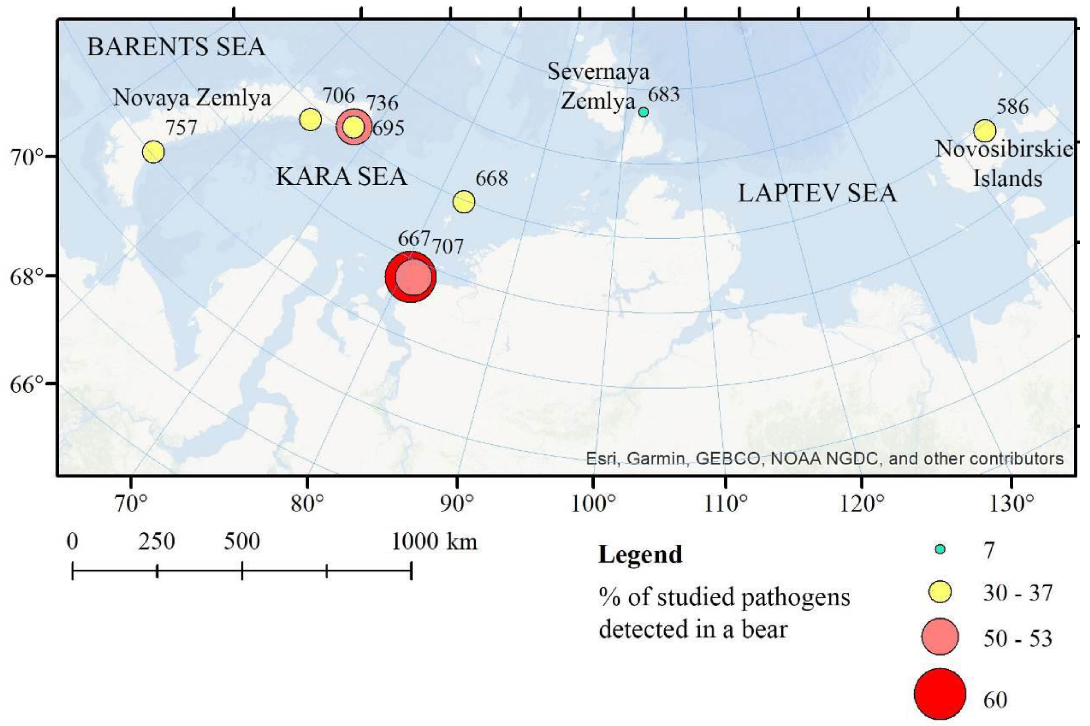
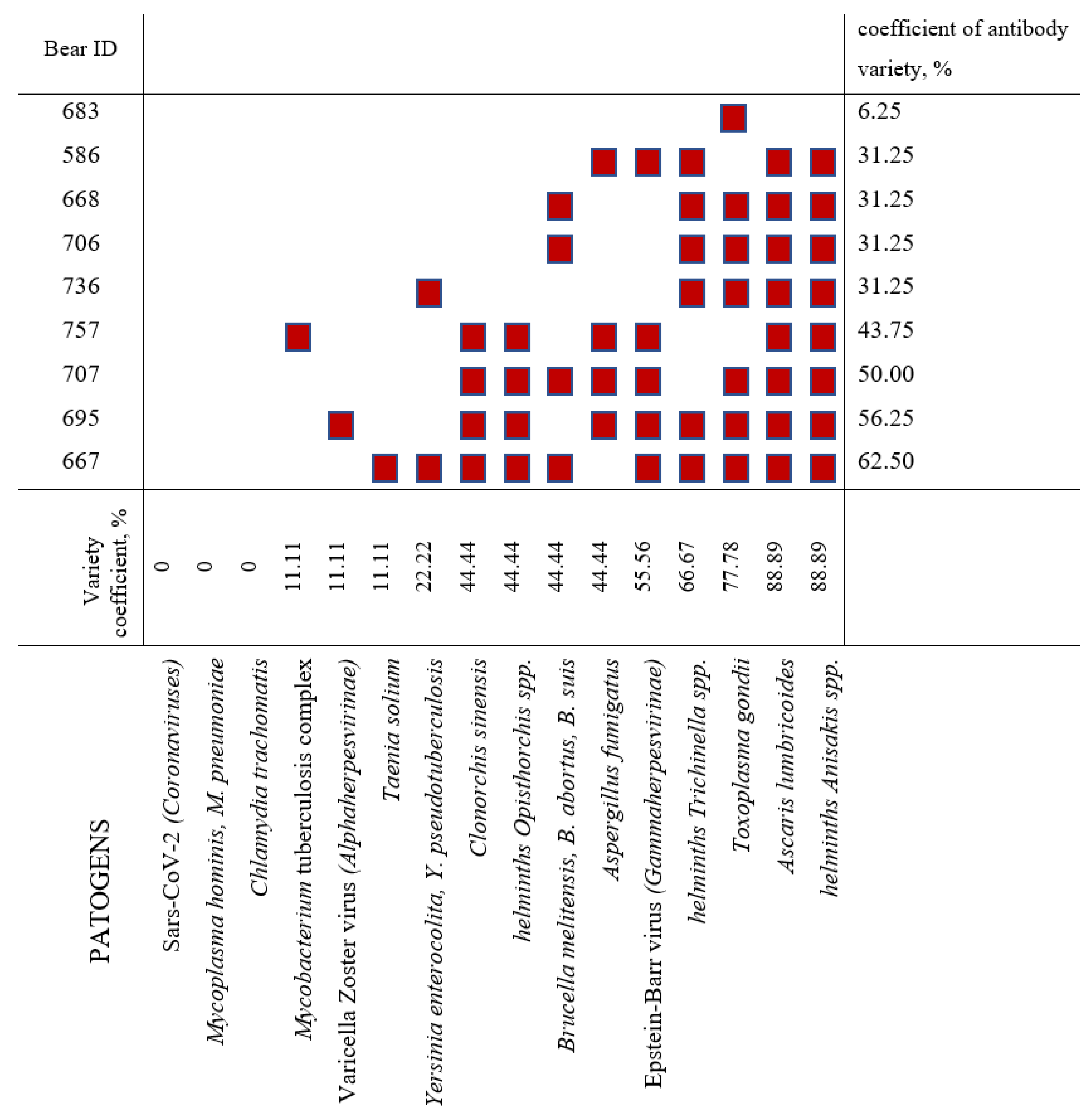
Figure 1. Locations of sampling, ID numbers of immobilized polar bears (see Table 1), and percentage of positive pathogen-specific ELISA tests for each of the bears (see Figure 2).
Diversity 14 00365 g001
Figure 2. Presence of antibodies to analyzed pathogens in polar bear serum.
Diversity 14 00365 g002
Table 1. General information on polar bears sampled in 2016 (ID numbers correspond to Figure 1).
Animal IDSexAge CategorySampling DatePlace of Sampling
586FemaleMature30 August 2016Kotelny Island (Novosibirskie Islands)
667Female2+ years old13 September 2016near Dikson (urban-type settlement, Kara Sea)
668FemaleMature14 September 2016Troynoy Island (Izvestiy TSIK Islands, Kara Sea)
683MaleMature22 September 2016Bolshevik Island (Severnaya Zemlya)
695MaleMature26 September 2016archipelago Novaya Zemlya
706MaleMature27 September 2016archipelago Novaya Zemlya
707Female1+ year old28 September 2016near Dikson (urban-type settlement, Kara Sea)
736FemaleMature05 November 2016archipelago Novaya Zemlya
757FemaleMature06 November 2016archipelago Novaya Zemlya
Publisher’s Note: MDPI stays neutral with regard to jurisdictional claims in published maps and institutional affiliations.

