Article

Morphology of Streptocara crassicauda and S. recta, with a Review of the Genus Streptocara (Nematoda: Acuariidae) and an Identification Key to Its Species

by
Yasen Mutafchiev
* and
Boyko B. Georgiev
Institute of Biodiversity and Ecosystem Research, Bulgarian Academy of Sciences, 2 Gagarin Street, 1113 Sofia, Bulgaria
*
Author to whom correspondence should be addressed.
Submission received: 15 December 2022 / Revised: 2 January 2023 / Accepted: 3 January 2023 / Published: 5 January 2023
(This article belongs to the Special Issue Biodiversity Research in Bulgaria)

Abstract

:
The cosmopolitan genus Streptocara Railliet, Henry and Sisoff, 1912 (Nematoda: Acuariidae) comprises parasites of the upper alimentary tract of aquatic and, rarely, terrestrial birds. Two species of the genus Streptocara are redescribed from Bulgaria by means of light and scanning electron microscopy: Streptocara crassicauda (Creplin, 1829), based on specimens from Larus genei, Larus minutus (Laridae) and Aythya ferina (Anatidae), and Streptocara recta (von Linstow, 1879) from Podiceps nigricollis (Podicipedidae). This is the first record of S. recta from Bulgaria. The host and distribution records of the species of the genus Streptocara are reviewed. Six species are recognised as valid. Streptocara crassicauda (Creplin, 1829), S. californica (Gedoelst, 1919), S. formosensis Sugimoto, 1930 and S. incognita Gibson, 1968, seem to be parasites well-adapted to birds of the family Anatidae (Anseriformes) and occasionally found in other birds, whereas S. recta (von Linstow, 1879) and S. longispiculata Gibson, 1968, are specialists of Podicipediformes and Gaviiformes, respectively. Streptocara indica Fotedar and Chishti, 1974, is recognised as a junior synonym of S. crassicauda, and Schistogendra oligopapillata Zhang and An, 2002, is considered as a junior synonym of Streptocara formosensis Sugimoto, 1930. An identification key to the species of the genus Streptocara is proposed.

1. Introduction

The family Acuariidae (Nematoda: Spiruridae) is a homogenous group of 45 genera and more than 300 species parasitizing almost exclusively birds; just a few taxa are known from mammals [1]. Traditionally, the family has been split into three subfamilies based on the anterior cuticular ornamentation in the form of cordons, collarette or ptilina [2,3]. A recent molecular study [1] demonstrated that the members of the subfamily Seuratiinae Chitwood and Wehr, 1934, do not form a monophyletic group and that the cuticular ornamentation in the form of collarette is a convergent character. As a result, it was suggested that the genera previously classified in Seuratiinae are placed in the subfamily Acuariinae, together with the acuariids characterized by cordons.
Acuariids of the genus Streptocara Railliet, Henry and Sisoff, 1912 (Acuariinae) are parasites in the upper alimentary tract of a diverse range of aquatic birds worldwide [3]. On some occasions, they may cause severe and even lethal infections in birds [4]. Amphipods are known as suitable intermediate hosts for Streptocara crassicauda (Creplin, 1829), while fish may serve as paratenic hosts [5]. Gibson [6] demonstrated the importance of the morphology of the cephalic region and the deirids for the taxonomy of the species of Streptocara, and recognised the following six taxa as valid within the genus: S. crassicauda crassicauda (Creplin, 1829), S. crassicauda longispiculata Gibson, 1968, S. recta (von Linstow, 1879), S. californica (Gedoelst, 1919), S. formosensis Sugimoto, 1930 and S. incognita Gibson, 1968. Subsequently, Streptocara indica Fotedar and Chishti, 1974, was described [7] and Streptocara longispiculata Gibson, 1968, was elevated to full species rank [8]. In this article, we provide detailed morphological descriptions of S. crassicauda and S. recta based on specimens from Bulgaria by means of light and scanning electron microscopy. We also review the host and distribution records of the members of the genus Streptocara and propose a key for their identification.

2. Materials and Methods

All materials belonging to the genus Streptocara deposited in the Helminthological Collection of the Institute of Biodiversity and Ecosystem Research, Bulgarian Academy of Sciences (IBER) were studied. The following two species were identified:
Streptocara crassicauda ex Larus genei Breme (Charadriiformes: Laridae), a single bird collected at lake Pomorie, Bulgarian Black Sea coast, 13 October 1988, 3 male and 12 female nematodes collected from the stomach and oesophagus: IBER N000.486 (1 male and 10 females), N000.842 (1 male nematode), IBER N001.040 (SEM stub with 2 female nematodes) and IBER N001.041 (SEM stub with 1 male nematode); ex Larus minutus Pallas, one bird collected at lake Durankulak, Bulgarian Black Sea coast, 5 October 1988, with one female nematode, coll. number N000.492 (found in the caecum?); ex Aythya ferina (L.) (Anseriformes: Anatidae), 3 birds at lake Durankulak, Bulgarian Black Sea coast: one bird on 21 January 1993, 6 female nematodes from the stomach, coll. number IBER N000.862; on 24 January 1993, 4 male and 2 female nematodes from the stomach, coll. number N000.249; 24 January 1993, 1 male and 1 female nematodes from the proventriculus, coll. number IBER N000.865.
Streptocara recta ex Podiceps nigricollis Brehm (Podicipediformes: Podicipedidae): at lake Durankulak, Bulgarian Black Sea coast—from one bird, 8 October 1989—3 female nematodes, coll. number N000.659 (1 female) and N001.042 (SEM stub 2 females); from a single bird, 21 January 1991, 1 male and 1 female nematodes, coll. number N000.867; in the vicinity of the village of Elena, SW Bulgaria, one bird, 25 August 2005, 1 male and 1 female nematode, coll. number N000.895. All collected under the lining of the gizzard.
Nematodes were preserved in 70% ethanol. For light microscopy, specimens were cleared and examined as temporary mounts in glycerine. Specimens used for scanning electron microscopy (SEM) were transferred from 70% ethanol to 40% ethanol for 10 min, rinsed twice in 0.1 M cacodylate buffer (10 min each time), post-fixed in 1% OsO4 for 2 h, dehydrated through a graded ethanol series and critical-point dried with CO2 by BAL-TEC CPD 030. Specimens were coated with gold-palladium in a Polaron SC7640 sputter coater and examined using a LEO microscope at an accelerating voltage of 10 kV.
Metrical data are presented as the range, with the mean in parentheses.

3. Results

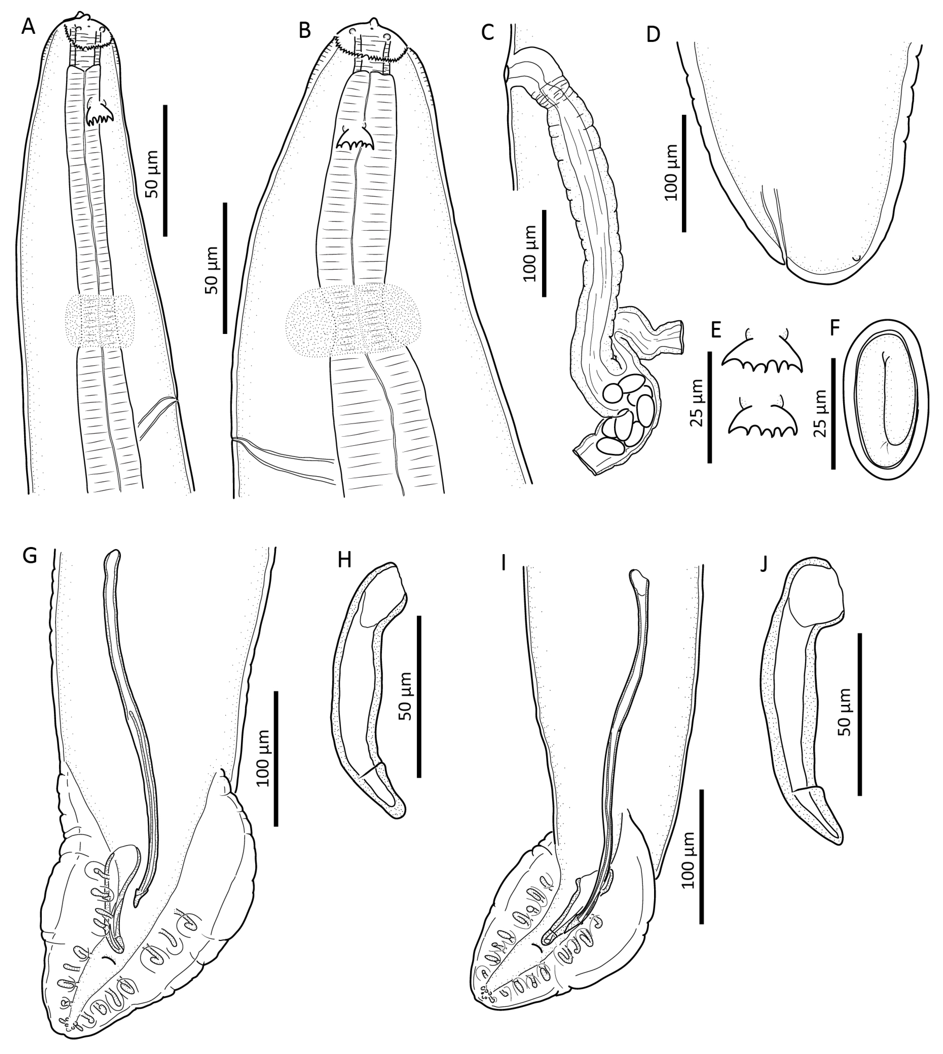
3.1. Streptocara crassicauda (Creplin, 1829) Railliet, Henry and Sisoff, 1912

Redescription based on specimens from L. genei

General: Anterior end with 2 triangular pseudolabia, each bearing single amphid and one pair of papillae (Figure 1A,B and Figure 2A–C). Sublabia absent. Collarette, delicate, reaching posteriorly to level about midlength of buccal cavity; collarette bearing about 80 small teeth on its posterior rim. Deirids 1.5–1.7 times as wide as long with five to eight cusps (Figure 1E and Figure 2A), situated about two buccal lengths from anterior extremity. Postdeirids c. 2 µm in diameter (Figure 2F). Buccal cavity short. Nerve ring distinct; excretory pore situated posterior to it. Posterior part of muscular oesophagus similar in diameter to anterior part of glandular oesophagus. Cuticle distinctly thicker anteriorly of deirids (cuticular inflation) (Figure 1B) as well as in posterior part of body. Transverse cuticular striations interrupted along lateral sides of body (Figure 2E).
Male (n = 2, see Table 1 for other measurements): Small-sized acuariid nematodes with maximum width in posterior half of body. Body at level of cloaca 39 and 41 µm wide. Deirids 7–8 µm long, 11–12 µm wide. Buccal cavity 10 and 11 µm wide. Muscular oesophagus 26 and 32 µm wide. Glandular oesophagus 57 and 64 µm (60 µm) wide. Relative length between muscular and glandular oesophagus 1:2.9. Total length of oesophagus represented as portion of body length 1:2.4 and 1:2.6. Cuticle 3–4 µm thick; distance between striations 2 µm. Caudal region slightly ventrally and laterally curved. Caudal papillae: single median precloacal papillae and ten pairs, including four precloacal (one specimen with three precloacal papillae on left) and five postcloacal pedunculate papillae and one pair of postcloacal sessile papillae situated between bases of last pedunculate papillae (Figure 1G and Figure 2D). Phasmids situated posterior to last pair of caudal papillae. Left spicule with projection near its distal end (Figure 1G). Right spicule robust, with retrograde projection near apex (Figure 1H). Relative length between right and left spicule 1:3.1.
Female (n = 10, except otherwise indicated; see Table 1 for other measurements): Small-sized nematode with maximum width at level of vulva; body width at level of anus 63–81 µm (73 µm). Tail short, blunt with subterminal phasmids (Figure 1D and Figure 2H). Left postdeirid and right postdeirid at 5.3 and 4.9 mm, respectively from anterior body end (measured from specimen with body length 8.7 mm). Buccal cavity 11–14 µm (13 µm) wide. Muscular and glandular oesophagus 39–45 µm (41 µm) and 72–95 µm (87 µm) wide, respectively. Relative length between muscular and glandular oesophagus 1:2.6–3.6 (1:3.1). Total length of oesophagus represented as portion of body length 1:3.4–4.2 (1:3.9). Cuticle 6–11 µm thick; distance between transverse striations 3–4 µm. Vulva at 4.5–6.0 mm (5.0 mm) from anterior extremity (Figure 2G). Uterus didelphic (Figure 1C); vagina vera turn into posterior direction, with well-developed circular musculature in walls; vagina uterina longer. Eggs oval, 37–39 × 19–21 µm (38 µm× 20 µm, n = 10), containing first stage larva (Figure 1F).
Remarks. Nematodes from L. genei and A. ferina studied by us correspond morphologically to one another, although the latter have slightly smaller body dimensions (see Table 1 and Figure 1I,J). The material from Bulgaria falls within the known range of morphological variation of S. crassicauda (Table 1).
The morphology of Streptocara crassicauda from Bulgaria was studied by Vassilev [9]. In Bulgaria this nematode has been known to parasitize domestic and wild Anas platyrhynchos (L.), domestic chickens Gallus gallus (L.), and domestic turkey, Meleagris gallopavo L. summarised in [9] and Alectoris chukar (J. E. Gray) (Galliformes) [10].
Table 1. Metrical data of Streptocara crassicauda and Streptocara recta from various hosts and localities (in micrometres except otherwise indicated).
SpeciesS. crassicaudaS. recta
SourceGibson [6]Present StudyPresent StudyGibson [6]Smogor-
zhevskaya [11]
Present Study
HostHost range *Larus geneiAythya ferinaPodiceps auritusPodiceps
cristatus
Podiceps nigricollis
LocalityCanadaBulgariaBulgariaCanadaUkraineBulgaria
Malen = 36n = 2n = 4n = 4n = 2
Body length (mm)3.4–5.1 (4.1)4.3; 4.63.5–4.1 (3.8)4.2–5.3 (4.7)4.5–5.05.9; 6.0
Maximum body width92–132 (106)143; 16193; 120 **145–185 (168)170; 183
Tail, length59–73 (64)63; 6650; 63 **74–86 (80)90–11372; 75
Collarette, length12–15 (14)10; 1112–14 (13)27–29 (28)25; 26
Collarette, width21–26 (23)22; 2320–23 (22)45–46 (45)37; 41
Deirids from anterior end, distance28–46 (35)34; 4332–45 (36)30–45 (39)43–5042; 47
Buccal cavity, length17–21 (20)20; 2418–21 (20)21–25 (23)21–2328; 29
Muscular oesophagus, length336–468 (378)420; 491407–438 (425)443–492 (476)450–510617; 635
Glandular oesophagus, length940–1230 (1100)1230; 14301180–1380 (1260)1720–2070 (1900)1800–23401970; 2350
Nerve ring from anterior end, distance107–128 (177)100; 113106–108 (107)129–132(130)128–130136; 143
Excretory pore from anterior end, distance157–193 (177)158; 182149–175 (161)174150–170188; 206
Caudal alae, length129–234 (175)161; 221161; 217 **197–276 (225)220–260295; 303
Left spicule, length224–302 (257)260; 269271; 321 **280–314 (293)287–325292; 313
Right spicule, length67–84 (73)83; 8881–90 (86) ***86–98 (92)75–9899; 103
Femalen = 46n = 10n = 8n = 4n = 3
Body length (mm)5.5–12.9 (9.7)8.1–10.4 (8.9)7.5–8.6 (7.9)10.36.2–11.411.3–14.5 (13.2)
Maximum body width160–246 (200)233–291 (263)163–234 (182)350241–358 (314)
Tail, length43–58 (53)13–20 (17)13–22 (19)626550–69 (67)
Collarette, length13–17 (5)11–14 (13)13–15 (14)3623–3830–36 (34)
Collarette, width24–30 (27)24–27 (25)23–25 (24)4539–44 (41)
Deirids from anterior end, distance28–43 (36)36–50 (43)32–41 (36)6225–6038–51 (46)
Buccal cavity, length17–20 (19)22–26 (24)18–21 (20)2223–2829–36 (32)
Muscular oesophagus, length330–603 (454)505–631 (564)425–568 (488)600460–581469–814 (669)
Glandular oesophagus, length1040–1760 (1420)1720–2010 (1850)1340–1970 (1540)26102460–24902630–3130 (2920)
Nerve ring from anterior end, distance95–142 (128)90–132 (116)90–115 (105)15460–105141–188 (163)
Excretory pore from anterior end, distance157–203 (192)125–191 (171)145–192 (167)199113–155191–295 (247)
Vulva from anterior end, distance (%)55–62 (59)57–61 (59)57–62 (59)573.7–4.856–64 (0.59)
* Anas platyrhynchos, A. clypeata (=Spatula clypeata), Aythya valisineria, A. marila, Bucephala clangula, B. islandica, Clangula hyemalis, Melanitta perspicillata, M. nigra (=Oidemia nigra), Oxyura jamaicensis, Lophodytes cucullatus (L.), Mergus merganser, M. serrator, Tympanuchus phasianellus (L.) (=Pedioecetes phasianellus), Actitis macularia (L.) and Cepphus columba Pallas; ** n = 3; *** n = 2.

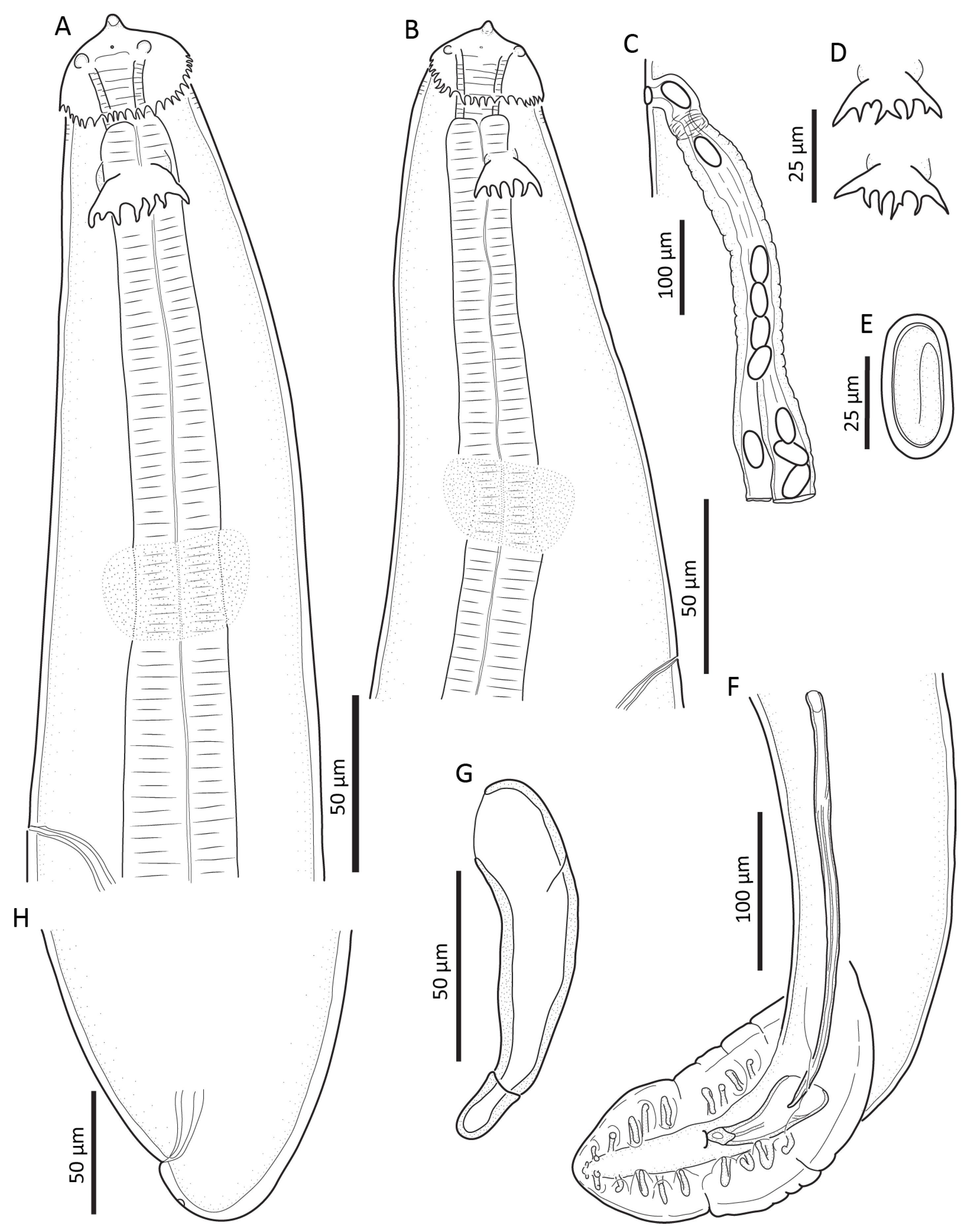
3.2. Streptocara recta (von Linstow, 1879) Skrjabin, 1916

General: Anterior end with 2 triangular pseudolabia, each bearing single amphid and pair of papillae; anteriorly to amphids numerous small pores present (Figure 3A,B and Figure 4A–D). Sublabia absent. Collarette, well-developed reaching posterior end of buccal cavity; collarette bearing about 60 small teeth on posterior rim. Deirids almost twice as wide as long with six or seven cusps (Figure 3D and Figure 4A), situated behind buccal cavity. Postdeirids c. 2 µm in diameter, bifurcate (Figure 4F). Buccal cavity short. Nerve ring well-distinct; excretory pore situated posterior to it. Posterior part of muscular oesophagus similar in diameter to anterior part of glandular oesophagus. Cuticle thicker anteriorly of deirids as well as in posterior part of the body. Transverse cuticular striations interrupted along lateral sides of body (Figure 4E).
Male (n = 2, see Table 1 for other measurements): Small-sized nematode with maximum width in posterior half of body. Body width at level of cloaca 45 and 51 (48). Deirids 12–14 long, 20–21 wide. Buccal cavity 13–14 wide. Muscular and glandular oesophagus 21–29 (25) and 90–93 (92) wide, respectively. Relative length between muscular and glandular oesophagus 1:3.1 and 1:3.8. Total length of oesophagus represented as portion of body length 1:2 and 1:2.3. Cuticle 4–6 µm thick, distance between transverse striations 4–6 µm. Caudal region slightly ventrally and laterally curved. Caudal papillae: single median precloacal papillae and ten pairs, including 4 precloacal and 5 postcloacal pedunculated papillae and one pair of postcloacal sessile papillae situated between bases of last pedunculate papillae (Figure 3F). Phasmids situated posterior to last pair of caudal papillae. Left spicule with simple tip. Right spicule robust (Figure 3G). Relative length between right and left spicule 1:3.
Female (n = 3, see Table 1 for other measurements): Middle-sized nematode with maximum width in posterior part of body. Body width at vulva 240–300 µm (271 µm) and at level of anus 50–69 µm (57 µm). Tail short, blunt with subterminal phasmids (Figure 3H and Figure 4H). Left postdeirid and right postdeirid at 9.3 mm and 8.7 mm, respectively from anterior body end (measured from specimen with body length 14.5 mm). Buccal cavity 15–20 µm (17 µm) wide. Muscular and glandular oesophagus 27–43 µm (38 µm) and 93–118 µm (106 µm) wide, respectively. Relative length between muscular and glandular oesophagus 1:3.8–5.6 (1:4.4). Total length of oesophagus represented as portion of body length 1:3.5–3.9 (1:3.7). Cuticle 9–13 µm thick, distance between striations 3–5 µm. Vulva at 7.2–8.3 mm (7.7 mm) from anterior extremity (Figure 3G). Uterus didelphic (Figure 3C); vagina vera turn into posterior direction, with well-developed circular musculature in walls; vagina uterina longer. Eggs oval, 34–39 × 17–19 µm (37 µm× 18 µm, n = 10), containing first stage larva (Figure 3E).
Remarks. The material of S. recta from grebes in Bulgaria differs from the samples from Canada and Ukraine with its slightly longer body, longer muscular and glandular oesophagus and longer caudal alae (Table 1); however, it corresponds to those samples in the size and shape of the deirids and the size of spicules. This is the first record of S. recta from Bulgaria.

4. Discussion

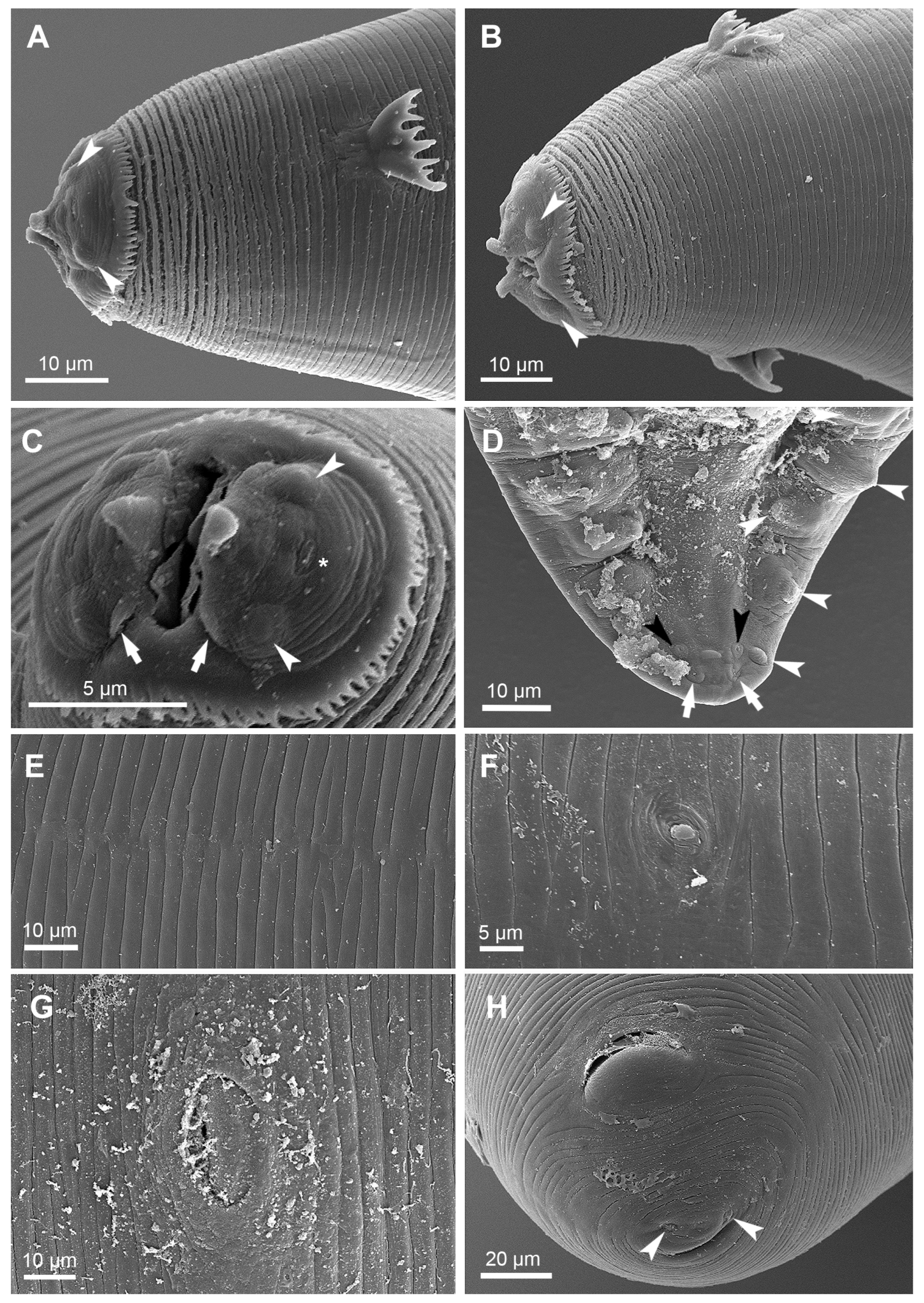
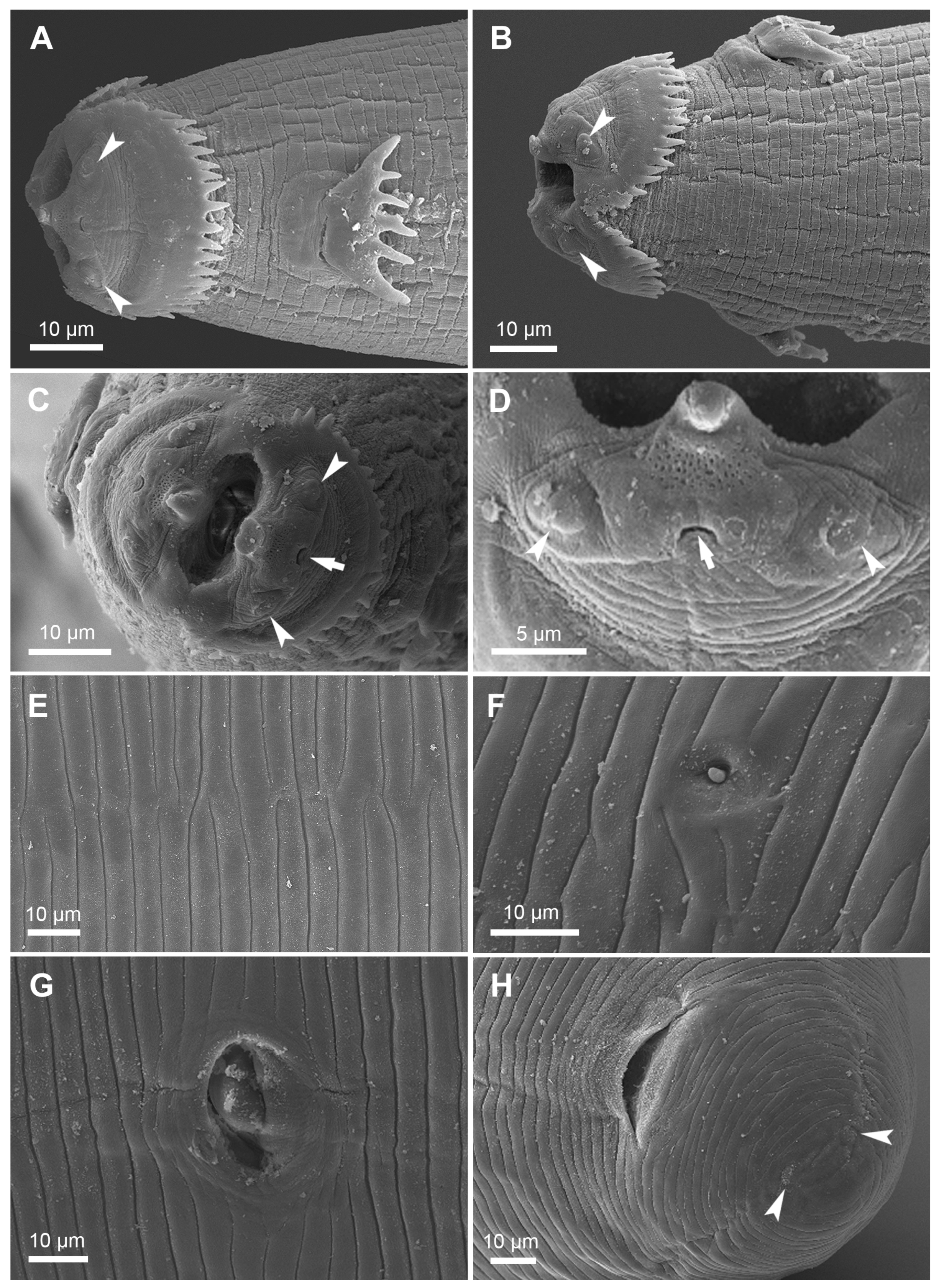
4.1. Ultrastructural Details Revealed by the SEM

The scanning electron microscopy revealed a field of numerous small pores situated between the apex of each pseudolabium and the amphid of S. recta. A similar field of pores was documented by means of the SEM in S. longispiculata [8]: Figure 10. We could not distinguish such pores in S. crassicauda; however, the quality of the studied material did not allow us to obtain a high-resolution image. Chen and Yu [12] studied S. crassicauda by SEM; however, the quality of the images does not allow any detailed comparisons of the SEM images provided. We cannot distinguish pores anterior to amphids on the SEM images of S. formosensis Sugimoto, 1930 provided by Agüero and Diaz [13] and Zhang and An [14]; the material studied by the latter authors was described as Schistogendra oligopapillata Zhang and An, 2002 (recognised here as a junior synonym of Streptocara formosensis Sugimoto, 1930, see discussion below). Well-observed pores situated between the amphids and the apexes of the pseudolabia, have been documented also in Decorataria decorata (Cram, 1927) by Mutafchiev and Georgiev [15]: Figure 11. The function of the minute pores on the pseudolabia is currently unknown; further studies are also needed for understanding of their value for the taxonomy and systematics of the acuariid nematodes. The SEM study documented a pair of small lappet-like lobes at the lateral sides of each pseudolabium of S. crassicauda (Figure 2C), such structures are missing in the S. recta studied here or in S. longispiculata studied by Diaz and Kinsella [8]. The taxonomical value of this character needs further evaluation.

4.2. Review of the Genus Streptocara

The generic diagnosis of Streptocara as formulated by Gibson [6] is still valid. We considered that the most distinct characters for the genus, beside the specific collarette or the lack of cuticular ornamentation in form of cordons, collarette or ptilina, are: the lack of sublabia; the relatively short for acuariid nematodes buccal cavity; the relatively large deirids with usually three or more cusps; males with broad caudal alae bearing nine pairs of pedunculate caudal papillae, four precloacal and five postcloacal; as well as one pair of sessile papillae situated between the most posterior pair of pedunculated papillae, the vulva is situated slightly behind the mid-body and the female tail is short and rounded.
We recognise as valid the following six species in the genus Streptocara.

4.2.1. S. crassicauda (Creplin, 1829) Railliet, Henry and Sisoff, 1912

Synonyms: Spiroptera crassicauda Creplin, 1829; Spiroptera pectinifera Neumann, 1900; Streptocara pectinifera (Neumann, 1900) Railliet, Henry and Sisoff, 1912; Streptocara indica Fotedar and Chishti, 1980, new synonymy.
Streptocara crassicauda is one of the most commonly reported acuariid species and has a cosmopolitan distribution. It is known as a parasite of domestic chickens and ducks as well as of a variety of wild birds belonging to the families Anatidae (Anseriformes), Alcidae, Charadriidae, Haematopodidae, Laridae, Recurvirostridae, Scolopacidae, Sternidae (Charadriiformes) but also from Gaviidae (Gaviiformes), Ardeidae (Ciconiiformes), Podicipedidae (Podicipediformes), Phalacrocoracidae (Pelecaniformes), Meleagrididae, Numididae, Phasianidae (Galliformes) and Sturnidae (Passeriformes) [6,11,16]. Streptocara indica Fotedar et Chishti, 1974, was described as a parasite of domestic chickens in Kashmir, India [7]. Although the description of S. indica was not very detailed and accurate, the provided measurements and drawings reveal that this species morphologically corresponds well to S. crassicauda and the two species can be distinguished only by the number of the caudal papillae of males. Unlike the other members of the genus Streptocara characterised by four pairs of precloacal papillae and six pairs of postcloacal papillae, S. indica was described as possessing five pairs of precloacal papillae and seven pairs of postcloacal papillae. We consider the genus Streptocara as a member of the subfamily Acuariinae (see Mutafchiev et al. [1]). The genera within Acuariinae possess four pairs of precloacal papillae whereas the members of the subfamily Schistorophinae Travassos, 1918, are characterised with a multiplication of the pairs of precloacal papillae [1,3]. Thus, it is more likely that the male described as S. indica was an abnormally developed specimen. The most posterior pair of postcloacal papillae in the description of S. indica is likely the pair of phasmids, which in appearance looks like a pair of small sessile papillae (see Figure 2D). Therefore, we consider S. indica conspecific with S. crassicauda and propose it as its junior synonym.

4.2.2. S. recta (von Linstow, 1879) Skrjabin, 1916

Synonyms: Filaria recta von Linstow, 1879; Spiroptera recta (von Linstow, 1879) Mueller, 1897.
Several authors considered S. recta as a grebe specialist [6,11,17]. This species has been reported from Podiceps cristatus (L.) (type-host), Podiceps auritus (L.), P. nigricollis, Tachybaptus novaehollandie (Stephens), T. ruficollis (Pallas), Poliocephalus poliocephalus (Jardine and Selby) and has European (probably Palaearctic), North American and Australian distribution [6,11,18]. Baruš et al. [16] recognised all records of S. recta from the former USSR and that from Japan by Yamaguti [19] as belonging to S. crassicauda. Considering the cosmopolitan distribution of the appropriate hosts of S. recta, it cannot be excluded that this species has a wider distribution than is currently known.

4.2.3. S. californica (Gedoelst, 1919) Gedoelst and Liégeois, 1922

Synonyms: Yseria californica Gedoelst, 1919; Korjakinema gusi Oshmarin, 1950; Skrjabinobronema californicum (Gedoelst, 1919) Yamaguti, 1961; Streptocara dogieli Belopol’skaya, 1952.
Streptocara californica has been reported mainly from anatid birds of the tribe Mergini (Anatinae): Melanitta deglandi (Bonaparte) (type-host), Melanitta perspicillata (L.), Melanitta americana (Swainson), Melanitta nigra (L.), Melanitta fusca (L.), Bucephala islandica (Gmelin), Clangula hyemalis (L.), Mergus merganser L., Mergus serrator L., Somateria mollisima (L.), Somateria spectabilis L. and, with a few exceptions, of hosts of other anatids, i.e., Anas platyrhynchos (Anatinae, Anatini), Aythya valisineria (Wilson) and Aythya marila (L.) (Aythyinae: Aythyini). The geographic range of this species is California, USA (type-locality) (Gedoelst 1919); British Columbia, Canada [6,20], Russian territories—Murmansk [17,20], Kamchatka [21,22], Chukotka [23], lower Ob River [24] and Yakutsk [25] as well as the Netherlands [26].

4.2.4. S. formosensis Sugimoto, 1930

Synonyms: Streptocara somateriae Rhyzhikov, 1960; Paracuaria somateriae (Rhyzhikov, 1960) Leonov, Tsimbaliuk and Belogurov, 1963; Schistogendra oligopapillata Zhang and An, 2002 new synonymy.
Streptocara formosensis is the only known species of Streptocara without a collarette. However, it resembles the other members of the genus by the structure of the pseudolabia, the large deirids bearing several cusps, the shape of the caudal alae and the arrangement of the caudal papillae in males as well as the rounded female tail and the position of the vulva.
Streptocara formosensis was described from domestic A. platyrhynchos. Subsequently, it has been reported from various anatid birds, mainly of the tribe Mergini: Bucephala albeola (L.), Bucephala clangula (L.), B. islandica, C. hyemalis, M. deglandi, M. perspicillata, M. americana, M. fusca, M. merganser, M. serrator, S. mollisima, S. spectabilis, and less often from the tribe Aythyini: A. marila, Aythya nyroca (Güldenstädt), Aythya fuligula (L.) in Japan (type locality), Canada, Russia (from Kaliningradskaya Oblast’ to the Russian Far East), Estonia [6,16,27], and more recently it was reported from Tachyeres leucocephalus Humphrey and Thompson in Argentina [13] and A. marila, B. clangula, C. hyemalis, M. nigra, M. fusca, M. merganser, Mergellus albellus (L.) in Poland [28].
Schistogendra oligopapillata Zhang and An, 2002 was described from a domestic duck, A. platyrhynchos, from Nanjing, China [14]. This species differs from the other members of the genus Schistogendra Chabaud and Rousselot, 1956, recognised as a junior synonym of Quasithelazia Maplestone, 1932 [29,30] and was treated a species incertae sedis by Mutafchiev et al. [30]. Schistogendra oligopapillata resembles S. formosensis in the morphology of the anterior end and the deirids, the morphology of the posterior end of males and the tail of females, as well as, in most morphometrics (Table 2), except the position of the vulva, which was described as positioned at level of the second and third fifth of the body length by Zhang and An [14]. All acuariid genera are characterised by an equatorial or post-equatorial position of the vulva [3] and it is likely that the position of the vulva was incorrectly provided. Based on the above discussion we recognise Schistogendra oligopapillata as a junior synonym of S. formosensis.

4.2.5. S. incognita Gibson, 1968

This species was described from Oxyura jamaicensis (Gmelin) (Anatidae) in Canada and further reported as a parasite of A. platyrhynchos in the USA and Canada, B. islandica in Canada, and Anser cygnoides (L.) in former Yugoslavia [6]. Streptocara incognita was also reported as a highly pathogenic parasite causing death in captive Phoenicopterus chilensis (Phoenicopteriformes: Phoenicopteridae) in the San Francisco Zoological Gardens, USA [31] and the anatids A. platyrhynchos in Montana, USA [32], domestic Muscovy duck Carina moschata (L.) in Italy [33] and Cygnus olor (Gmelin) in Bosnia and Herzegovina [34]. A 28S rDNA sequence of Streptocara incognita Gibson, 1968 from Tadorna tadorna (L.) in France was published [1].

4.2.6. S. longispiculata Gibson, 1968

This species was initially described as a subspecies of S. crassicauda from Gavia immer (Brunnich) (Gaviiformes) from Chesapeake Bay, USA [6], and subsequently elevated to a full species rank by Diaz and Kinsella [8]. Streptocara longispiculata is known from a few records from loons in North America: G. immer in Florida, USA [35] and Gavia stellata (Pontoppidan) in the Northwest Territories, Canada [8].

4.3. Identification Key to the Species of the Genus Streptocara

We propose the following identification key to the species of Streptocara which takes into account the morphological characters for species of the genus provided above:
1a. Without cuticular collarette surrounding the cephalic region … S. formosensis
1b. Cuticular collarette surrounding the cephalic region present … 2
2a. Buccal cavity longer than 90 µm; deirids with three approximately equal in size cusps; … S. californica
2b. Buccal cavity shorter than 30 µm. Deirids with more than three cusps … 3
3a. Cuticular collarette reaches the posterior third of the buccal cavity; deirids about twice as wide as long, with 6–7 cusps. Left spicule with simple bluntly pointed distal end … S. recta
3b. Cuticular collarette does not exceed the midlength of the buccal cavity. Left spicule with distal tip that is not simple and bluntly pointed … 4
4a. Deirids as long as wide, deeply cleft medially, with well-developed most outer cusps and one, two or three smaller cusps in-between … S. incognita
4b. Deirids wider than long with 5–8 similar in size cusps … 5
5a. Cuticular inflation at the anteriorly extremity present. Left spicule shorter than 320 µm … S. crassicauda
5b. Without prominent cuticular inflation at the anterior extremity. Left spicule longer than 380 µm … S. longispiculata

5. Conclusions

The species of the genus Streptocara are well-known parasites of aquatic birds. We recognise six species as valid within the genus. Out of these, S. crassicauda, S. californica, S. formosensis and S. incognita are common parasites of birds of the family Anatidae (Anseriformes), although some of these species have also been recorded from other aquatic birds and occasionally terrestrial birds of the orders Galliformes and Passeriformes. Streptocara californica and S. formosensis are more often reported from hosts of the tribe Mergini; however, the reasons for this affinity are not yet understood but could be due to some particular features of the life cycles of the nematodes, which are still unknown. Streptocara recta is a parasite of Podicipediformes, while S. longispiculata is a parasite of Gaviiformes. Our review of the genus Streptocara revealed that there have not been new species described within the genus for more than 50 years, suggesting that the species diversity is well-studied. This is likely due to the extensive helminthological studies carried out on waterfowls.

Author Contributions

Conceptualization, Y.M. and B.B.G.; investigation, Y.M.; resources, B.B.G.; writing—original draft preparation, Y.M.; writing—review and editing, B.B.G. All authors have read and agreed to the published version of the manuscript.

Funding

This research received no external funding.

Institutional Review Board Statement

Not applicable.

Informed Consent Statement

Not applicable.

Data Availability Statement

Not applicable.

Acknowledgments

This study was based on specimens deposited in the Helminthological Collection of the Institute of Biodiversity and Ecosystem Research—Bulgarian Academy of Sciences upgraded in the frames of the project DiSSCo-BG (Upgrade of the Research Infrastructure “Distributed System of Scientific Collections—Bulgaria”) funded by the National Roadmap for Research Infrastructures, Ministry of Education and Science of the Republic of Bulgaria. SEM observations were carried out using facilities of the Museum für Naturkunde, Berlin in the course of a research visit of the senior author granted by Synthesys DE-TAF-3124 (EC FP6) and kindly hosted by B. Neuhaus.

Conflicts of Interest

The authors declare no conflict of interest.

References

  1. Mutafchiev, Y.; Georgiev, B.B.; Mariaux, J. A 28S RDNA-Based Phylogeny of the Nematode Family Acuariidae (Spirurida) Parasitic in Vertebrates. Zool. Scr. 2020, 49, 641–657. [Google Scholar] [CrossRef]
  2. Chabaud, A.G. Keys to Genera of the Order Spirurida. Part 2. Spiruroidea, Habronematoidea and Acuarioidea. In CIH Keys to the Nematode Parasites of Vertebrates; Commonwealth Agricultural Bureaux: Wallingford, UK, 1975; Volume 3, pp. 28–58. [Google Scholar]
  3. Bain, O.; Mutafchiev, Y.; Junker, K. Order Spirurida. In Handbook of Zoology: Gastrotricha, Cycloneuralia and Gnathifera; Schmidt-Rhaesa, A., Ed.; De Gruyter: Boston, MA, USA, 2014; Volume 2, pp. 661–732. [Google Scholar]
  4. Carreno, R.A. Dispharynx, Echinuria, and Streptocara. In Parasitic Diseases of Wild Birds; Atkinson, C.T., Thomas, N.J., Hunter, D.B., Eds.; Wiley-Blackwell: Ames, IA, USA, 2008; pp. 326–341. [Google Scholar]
  5. Anderson, R.C. Nematode Parasites of Vertebrates: Their Development and Transmission; CABI: Wallingford, UK, 2000. [Google Scholar]
  6. Gibson, G.G. Species Composition of the Genus Streptocara Railliet et al., 1912 and the Occurrence of These Avian Nematodes (Acuariidae) on the Canadian Pacific Coast. Can. J. Zool. 1968, 46, 629–645. [Google Scholar] [CrossRef]
  7. Fotedar, D.N.; Chishti, M.Z. Streptocara indica n. sp. (Acuarioidea Sobolev, 1949: Nematoda) from Gallus domesticus in Kashmir, India. Indian J. Helminthol. 1980, 32, 4–8. [Google Scholar]
  8. Diaz, J.I.; Kinsella, J.M. Streptocara longispiculata Gibson, 1968 (Nematoda: Acuariidae) from Gavia immer and Gavia stellata from North America: Redescription and Taxonomic Position. Comp. Parasitol. 2006, 73, 285–288. [Google Scholar] [CrossRef]
  9. Vassilev, I. Regarding the Specific Appurtenance of Streptocarae among Domestic Fowls in Bulgaria. Bull. Cent. Helminthol. Lab. 1968, 12, 5–21. (In Bulgarian) [Google Scholar]
  10. Vassilev, I. Helminths in Thracian Rock Partridge (Alectoris chukar Kleini) in Bulgaria. Helminthologia 1992, 29, 117–120. [Google Scholar]
  11. Smogorzhevskaya, L.A. Nematodes. Part 3. Acuarioidea. In Fauna Ukrainy; Naukova Dumka: Kiev, Ukraine, 1990; Volume 32. [Google Scholar]
  12. Chen, K.; Li, X. Observation of Streptocara crassicauda and Amidostomum anseris by Electron Microscope. Chin. J. Vet. Sci. Technol. 2002, 32, 42–44. [Google Scholar]
  13. Agüero, M.L.; Diaz, J.I. First Record of Streptocara formosensis (Nematoda: Acuariidae) from the Chubut Steamerduck, Tachyeres leucocephalus, Endemic to the Patagonian Coast, Southwest Atlantic. Comp. Parasitol. 2013, 80, 304–307. [Google Scholar] [CrossRef]
  14. Zhang, L.; An, R. A New Species of Genus Schistogendra Parasitizing the Domestic Duck, Anas platyrhynchos domesticus from Nanjing, China (Nematoda: Acuariidae). Wuyi Sci. J. 2002, 27, 42–45. [Google Scholar]
  15. Mutafchiev, Y.; Georgiev, B.B. Redescription of Decorataria decorata (Spirurida, Acuariidae) Based on Nematodes from Podiceps cristatus and P. grisegena (Aves, Podicipediformes) from Bulgaria. Acta Parasitol. 2008, 53, 158–164. [Google Scholar] [CrossRef]
  16. Baruš, V.; Sergeeva, T.P.; Sonin, M.D.; Ryzhikov, K.M. Helminths of Fish-Eating Birds of the Palaearctic Region. 1. Nematoda; Czechoslovak Academy of Sciences: Prague, Czech Republic, 1978. [Google Scholar]
  17. Ryzhikov, K.M. [Species Composition of the Genus Streptocara (Nematoda, Spirurata)]; Material’ k Nauchnoi Koferencii Vsesoiuznoi Obshtestva Gel’mitologov: Moscow, Russia, 1966; Volume 3. [Google Scholar]
  18. Storer, R.W. The Metazoan Parasite Fauna of Grebes (Aves: Podicipediformes), and Its Relationship to the Birds’ Biology; University of Michigan: Ann Arbor, MI, USA, 2000; Volume 188. [Google Scholar]
  19. Yamaguti, S. Studies on the Helminth Fauna of Japan. Part. 12 Avian Nematodes, I. Jpn. J. Zool. 1935, 6, 403–431. [Google Scholar]
  20. Gibson, G.G. Taxonomic and Biological Observations on Streptocara californica (Gedoelst, 1919) Gedoelst and Liégeois, 1922 and the Genus Streptocara (Nematoda: Acuariidae). J. Helminthol. 1964, 3, 773–783. [Google Scholar] [CrossRef]
  21. Oshmarin, P.G. Helminth Fauna of Birds of the Far East (Kamchatka, Zemlya Koryakov and the Kuril Island). Tr. GELAN 1950, 3, 167–179. (In Russian) [Google Scholar]
  22. Ryzhikov, K.M. [Nematodes of Anseriformes from Kamchatka]. Tr. GELAN 1963, 13, 133–143. (In Russian) [Google Scholar]
  23. Ryzhikov, K.M. Nematodes from Anseriform Birds in Chukotka. Helminthologia 1963, 4, 413–423. [Google Scholar]
  24. Daiya, G.G. Nematodes of Fish-Eating Birds of the Lower Ob. In Sbornik Rabot po Gel’mintofaune Ryb i Ptits.; Akademii Nauk SSSR: Moscow, Russia, 1967; pp. 133–153. [Google Scholar]
  25. Ryzhikov, K.M.; Gubanov, N.M.; Tolkacheva, L.M.; Khokhlova, I.G.; Zinoveva, E.N.; Sergeeva, T.P. Helminths of Birds in Yakutia and Cross-Border Regions. Nematodes and Thorn-Headed Worms; Nauka: Moscow, Russia, 1973. [Google Scholar]
  26. van den Broek, E.; Jansen, J. Parasites of Animals in the Netherlands. Supplement V: Parasites of Animal in the Netherlands. Ardea 1971, 59, 28–33. [Google Scholar]
  27. Kurochkin, Y.V.; Ryzhikov, K.M. About Nematode Species in the Genus Paracuaria Rao, 1951 (Nematoda: Spirurata). Sb. Parazitol. Rab. Tr. Astrah. Zapov. 1964, 39, 182–191. (In Russian) [Google Scholar]
  28. Królaczyk, K.; Kavetska, K.M.; Stapf, A.; Kalisińska, E. Streptocara formosensis Sugimoto, 1930 (Nematoda: Acuariidae) in Wild Ducks from the Southern Coast of the Baltic Sea. Helminthologia 2012, 49, 247–252. [Google Scholar] [CrossRef] [Green Version]
  29. Wong, P.L.; Lankester, M.W. Revision of the Genus Viktorocara Guschanskaya, 1950 (Nematoda, Acuarioidea). Can. J. Zool. 1984, 62, 2541–2549. [Google Scholar] [CrossRef]
  30. Mutafchiev, Y.; Mariaux, J.; Georgiev, B.B. Two New Species of Quasithelazia Maplestone, 1932 (Nematoda: Acuariidae) from Malaysia, with an Amended Diagnosis and Review of the Genus. Syst. Parasitol. 2014, 88, 103–117. [Google Scholar] [CrossRef]
  31. Fox, J.G.; Snyder, S.B.; Schmidt, G.D.; Campbell, L.H. Infection with the Nematode Streptocara incognita in the Chilean Flamingo. J. Wildl. Dis. 1974, 10, 66–69. [Google Scholar] [CrossRef]
  32. Sterner, M.C.; Stackhouse, L. Parasitic Ulcerative Ventriculitis in Mallards (Anas platyrhynchos). J. Wildl. Dis. 1987, 23, 680–682. [Google Scholar] [CrossRef] [PubMed] [Green Version]
  33. Bano, L.; Natale, A.; Vascellari, M.; Comin, D.; Mutinelli, F.; Agnoletti, F. First Report of Parasitic Esophagitis by Streptocara incognita in Muscovy Ducks (Cairina moschata domesticus) in Italy. Avian Dis. 2005, 49, 298–300. [Google Scholar] [CrossRef] [PubMed]
  34. Alić, A.; Prašović, S.; Hodžić, A.; Beširović, H.; Rešidbegović, E.; Omeragić, J. Fatal Verminous Pharyngitis and Esophagitis Caused by Streptocara incognita in Mute Swans (Cygnus olor). Avian Dis. 2013, 57, 147–151. [Google Scholar] [CrossRef]
  35. Kinsella, J.M.; Forrester, D.J. Parasitic Helminths of the Common Loon, Gavia immer, on Its Wintering Grounds in Florida. J. Helminthol. Soc. Wash. 1999, 66, 1–6. [Google Scholar]
Figure 1. Streptocara crassicauda ex Larus genei, unless indicated otherwise. (A) Anterior end, male, lateral view; (B) anterior end, female, lateral view; (C) vagina; (D) posterior end, female, lateral view; (E) deirids; (F) egg; (G) posterior end, male, ventrolateral view; (H) right spicule, dextral view; (I) posterior end, male, ventrolateral view, ex Aythya ferina; and (J) right spicule, dextral view, ex A. ferina.
Diversity 15 00067 g001
Figure 2. Streptocara crassicauda, female, unless indicated otherwise, SEM. (A) Anterior end, lateral view; note the cephalic papillae (arrowheads); (B) anterior end, ventral view; note the cephalic papillae (arrowheads); (C) anterior end, apical view note the cephalic papillae (arrowheads), the amphid (asterisk) and the lappet-like lobes (arrows); (D) posterior end, male, note the pedunculated papillae (white arrowheads), the sessile pair of papillae (black arrowheads) and the phasmids (arrows); (E) lateral field, note the cuticular striation; (F) postdeirid; (G) vulva; and (H) tail, note the phasmids (arrowheads).
Diversity 15 00067 g002
Figure 3. Streptocara recta. (A) Anterior end, female, lateral view; (B) anterior end, male, lateral view; (C) vulva; (D) deirids; (E) egg; (F) posterior end, male, ventrolateral view; (G) right spicule, sinistral view; and (H) posterior end, female, lateral view. Scale-bars a, b = 100 µm; c = 50 µm.
Diversity 15 00067 g003
Figure 4. Streptocara recta, female, SEM. (A) Anterior end, lateral view; note the cephalic papillae (arrowheads); (B) anterior end, lateral view; note the cephalic papillae (arrowheads); (C) anterior end, apical view, note the cephalic papillae (arrowheads), the amphid (arrow); (D) pseudolabium, apical view, note the cephalic papillae (arrowheads), the amphid (arrow) and the field with pores anterior to the amphid; (E) lateral field, note the cuticular striation; (F) postdeirid; (G) vulva; and (H) posterior end, note the phasmids (arrowheads).
Diversity 15 00067 g004
Table 2. Metrical data of Streptocara formosensis from various hosts and localities (in micrometres except otherwise indicated).
SourceGibson [6]Kurochkin and Ryzhikov [27]Zhang and An [14]
HostHost range * Anas platyrhynchos
LocalityCanadaRussiaChina
Malen = 8n = 10n = 8
Body length (mm)5.1–7.85.7–7.25.9–7.0
Maximum body width117–160140–200161–200
Tail, length73–8052–81
Deirids from anterior end, distance143–231110–240109–169
Buccal cavity, length41–4930–6026–31
Muscular oesophagus, length584–1010310–900693–872
Glandular oesophagus, length1560–25502000–37001890–2730
Nerve ring from anterior end, distance135–172135–169
Excretory pore from anterior end, distance188–224185–234
Caudal alae, length331–424
Left spicule, length314–364280–460234–273
Right spicule, length86–12062–9662–86
Femalen = 10n = 20n = 13
Body length (mm)8.2–15.79.4–18.011.3–15.9
Maximum body width172–240170–520210–336
Tail, length49–6039–52
Deirids from anterior end, distance166–222109–200153–195
Buccal cavity, length41–5023–31
Muscular oesophagus, length756–1144660–1200735–882
Glandular oesophagus, length1840–34602160–31002100–3570
Nerve ring from anterior end, distance98–185156–208
Excretory pore from anterior end, distance224–270203–289
Vulva from anterior end, distance (%)52–64 (61)40
Eggs35 × 1930–40 × 18–3234–38 × 20–23
* Anas platyrhynchos, Bucephala clangula, B. islandica, B. albeola, Clangula hyemails, Melanitta deglandi, M. perspicillata, Mergus merganser and M. serrator.
Disclaimer/Publisher’s Note: The statements, opinions and data contained in all publications are solely those of the individual author(s) and contributor(s) and not of MDPI and/or the editor(s). MDPI and/or the editor(s) disclaim responsibility for any injury to people or property resulting from any ideas, methods, instructions or products referred to in the content.

