Article

Simplified Attachable EEG Revealed Child Development Dependent Neurofeedback Brain Acute Activities in Comparison with Visual Numerical Discrimination Task and Resting

1
Engineering Department, Graduate School of Sciences and Technology for Innovation Yamaguchi University, Yamaguchi 755-8611, Japan
2
Department of Cell and Regenerative Biology, University of Wisconsin, Madison, Madison, WI 53706, USA
3
Department of Pediatrics, Saitama Medical University, Saitama 350-0495, Japan
4
Graduate School of Information Sciences, Tohoku University, Sendai 980-8579, Japan
*
Author to whom correspondence should be addressed.
Submission received: 6 August 2022 / Revised: 16 September 2022 / Accepted: 19 September 2022 / Published: 23 September 2022
(This article belongs to the Special Issue Sensor Technologies for Human Health Monitoring)

Abstract

:
The development of an easy-to-attach electroencephalograph (EEG) would enable its frequent use for the assessment of neurodevelopment and clinical monitoring. In this study, we designed a two-channel EEG headband measurement device that could be used safely and was easily attachable and removable without the need for restraint or electrode paste or gel. Next, we explored the use of this device for neurofeedback applications relevant to education or neurocognitive development. We developed a prototype visual neurofeedback game in which the size of a familiar local mascot changes in the PC display depending on the user’s brain wave activity. We tested this application at a local children’s play event. Children at the event were invited to experience the game and, upon agreement, were provided with an explanation of the game and support in attaching the EEG device. The game began with a consecutive number visual discrimination task which was followed by an open-eye resting condition and then a neurofeedback task. Preliminary linear regression analyses by the least-squares method of the acquired EEG and age data in 30 participants from 5 to 20 years old suggested an age-dependent left brain lateralization of beta waves at the neurofeedback stage (p = 0.052) and of alpha waves at the open-eye resting stage (p = 0.044) with potential involvement of other wave bands. These results require further validation.

1. Introduction

Brain growth is a central issue in child development. Assessment of electroencephalograph (EEG) readings during specific tasks can serve as a method for quantitatively determining the physiological growth state of the brain [1]. Changes in basic EEG rhythms with age have been identified. In an eyes-closed resting condition, age-dependent decreases in delta and theta waves and increases in alpha and beta waves, with decreases in the theta/alpha and theta/beta ratios have previously been reported [2]. The use of EEG to visualize brain activity may prove to be an innovative education and rehabilitation tool during specific developmental periods [3]. Cognitive advancement in children inevitably requires further complex learning. Novel education technologies such as those we have developed that support neurodevelopmental visualization of self-regulating functions in cognition and emotion, such as concentration and relaxation, may play an important role [4].
Neurofeedback technology (NFT) that actively activates brain self-regulating functions by visualizing their own brain activity utilizing EEG [5] has emerged in recent years. NFT has been applied less often in typically developing children, but [6] it has been used clinically in children to both diagnose and provide therapy [7] for ADHD [8], using NFT theta, alpha, sensorimotor (SMR), and beta wave training [9] and for children on the autism spectrum [10] and with learning disorders [11] to improve cognitive function. The beneficial neuronal plasticity effects of NFT education and therapy likely require repetition [12], which means that the NFT system needs to be something easy that children will willingly participate in and can be easily worn at home or school. A simple attachable EEG would enable frequent use for neurodevelopmental support applications in daily education settings and clinical monitoring. Recordings from sphenoidal electrodes have been suggested to improve the localization precision of EEG recordings in patients with temporal lobe epilepsy. However, the usage was limited [13].
To meet these requirements, we designed a two-channel wireless EEG headband device set with electrodes for Sp1 and Sp2 of the 10–20 system [14]. This device can be safely and easily placed on the forehead without the need for restraint or electrode paste and can be easily removed.
We explored the utility of our device for NFT methods that could be applied to education and therapy for neural development by having children wear the device while playing games that should appeal to variously aged children. We developed a visual NFT game prototype in which the local mascot of Hagi city in Yamaguchi prefecture, Japan, called “Haginyan”, changes size in the PC display depending on the user’s beta/theta wave ratios. These ratios were chosen based on their potential as biomarkers of development (healthy children [15], young adults [16]), developmental disorders (e.g., autism spectrum disorders (ASD) [17], attention-deficit/hyperactivity disorders (ADHD) [18]), and intellectual disabilities [19]. To verify the applicability of the device and to visualize any unique NFT function, we looked for age-dependent changes in EEG signals in visitors at a local children’s play event, the “Wakuwaku Kid’s Festa”, where children were invited to experience the game.
The NFT game was made available in a small indoor space during the play event. An adult male instructor was present to help with the attachment of the EEG device and to explain its use. The game included three stages of visual discrimination tasks: Stage 1, a consecutive number task that induced neurocognitive activation; Stage 2, an open-eye resting condition; and Stage 3, the NFT task. Stages 1 and 2 were used as controls for comparison to Stage 3. To visualize relative changes in function-dependent NFT activity, Stage 1 was designed as the previously well-reported visual cognition but EEG-independent task. In contrast, Stage 2 was defined as another control task, “resting”, with active visual function (opening the eyes) but no cognitive function. Acquired EEG data were analyzed to determine any age-related differences using linear regression analysis [20] in 2 dimension plots of each EEG brain wave marker versus age.

2. Materials and Methods

2.1. Participant Age and Sex

Several hundred children and adults attended the “Wakuwaku Kids Festa” in Hagi city on 20 March 2021. Over a 3-h period, 36 people played the NFT game. The age and gender of each player were recorded. Of the 36 players, 6 were removed from the analysis; 2 because they were older than 30 years, and 4 because their EEG data were particularly noisy. The children who were removed were one 3-year-old, one 4-year-old, and two 7-year-olds. Table 1 shows the age and sex of the 30 remaining participants.

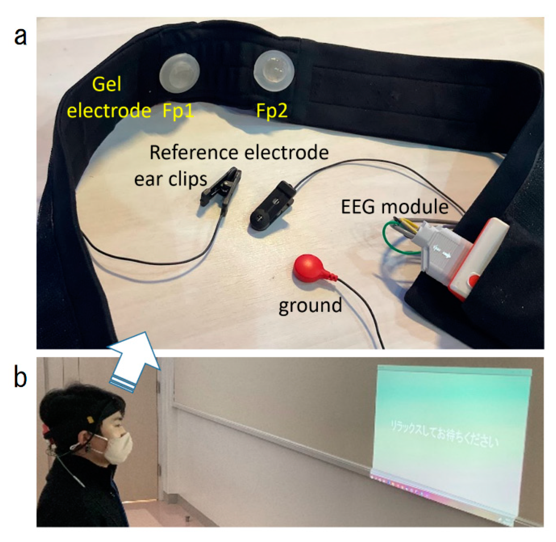
2.2. EEG Measurement System

2.2.1. Hardware

A prototype EEG electrode headset was fabricated for this study (Figure 1). Two EEG scalp electrodes (approved medical devices, Japanese notification number 13B2 × 10278000001) were placed in a headband to be worn on the forehead. The headband allowed quick (10–15 s) and easy placement of Fp1 and Fp2 of the international 10–20 system [14] by the subject. Occasionally minor placement adjustments were required by the instructor. The electrodes were attached with disposable solid gel electrode patches (intercross-810: Intercross Corporation, Tokyo, Japan) to stabilize electrode-to-electrode resistance [21]. Electrophysiological reference and ground electrodes were clip-on silver chloride electrodes placed on the left and right ears, respectively. The headset was equipped with a commercially available 38 g small EEG module (intercross-413 Intercross Co., Japan) connecting all the electrodes (Figure 1).
To play, the participant sits looking toward a small projector displaying PC images sized 75 × 50 cm on the wall approximately 1 m away. Instructions for the game are displayed on the screen at the beginning of each stage.

2.2.2. Signal Pre-Processing

EEG signals obtained from the GEL electrodes were input to the monitoring module (Figure 1) for 12-fold amplification, converted to digital data by 500 Hz, 24-bit AD conversion (integrated with time data by the electroencephalograph’s internal clock), and transferred to a Windows PC via Bluetooth. The transferred EEG signals were first processed with digital filtering High-Pass above 2 Hz with the Butterworth second-order filter to exclude body motion errors. The Low-Pass filter was digitally operated under 40 Hz by a Butterworth 8th order filter.

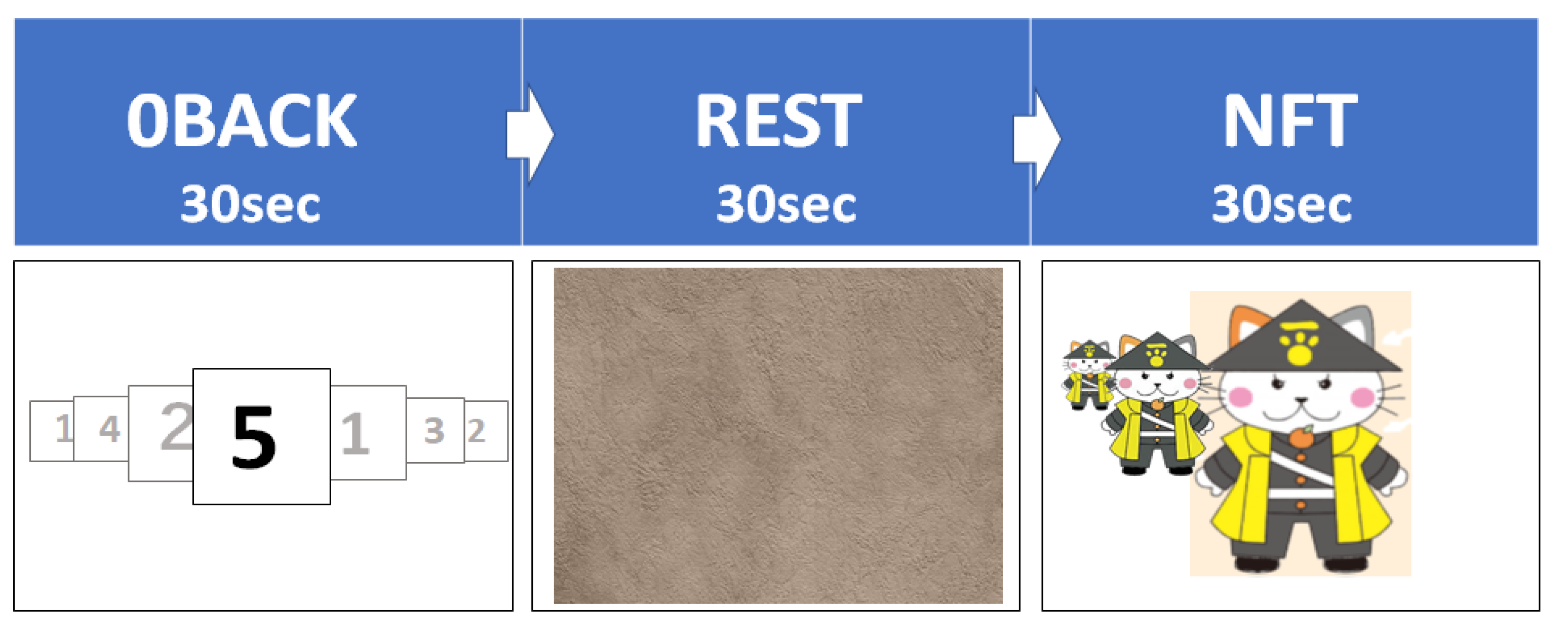
2.3. Three Stages of Visual Tasks, “0back”, “Rest”, and “NFT”

We designed a visual task with three stages (Figure 2). The first: 0BACK; the second: REST; the third: NFT sequentially, without any intervals. Each stage lasted 30 s with a 5 s interval between stages. The first two stages were reference tasks, and the third stage was the principal task, NFT. Using customized software (C#, Microsoft, Redmond, DC, USA), the tasks were synchronized to regulate the EEG monitoring software by trigger signals at the beginning and the end of each stage.
The first reference stage, “0back”, was designed as a neurocognitive activation task requiring sustained attention and concentration. Numerical values from 1 to 5 were randomly displayed and replaced every 0.6 s in the 15 × 15 cm area of the wall screen. The player began the task with their hands open and was instructed to close both hands as soon as the “5” was displayed. Given the simplicity of this task for all players, the accuracy evaluation was omitted.
In the second reference stage, “REST”, in a relaxed open-eye condition, the player passively viewed a non-arousing display image, Wall 2.jpg, from the Open Affective Standardized Image Set (OASIS; the minimum score for arousal index, 1.693069307) [22].
The last stage was the principal NFT task. In this task, the player was asked to interactively change the size of a familiar illustrated character (Haginyan, the Hagi City mascot) that was shown on the screen. In this prototype, the size of Haginyan increased or decreased based on the player’s beta/theta ratios relative to their calculated reference value (average of the beta/theta ratios from each player’s two reference stages) [23].

2.4. Analyses of Age-Dependency in Relative Power of Four EEG Wave Bands

The acquired EEG data were divided into frames per second. Frames with waveforms of 80 μV or higher were regarded as noise and were excluded. Each remaining frame underwent frequency analysis by Discrete Fourier Transform (DFT) [24]. The power values and content ratios per stage were calculated for each frequency band, delta, theta, alpha, and beta (Table 2) from the obtained DFT results. The trend graph of the four EEG bands and the beta/theta ratios were seen only in real-time by the instructor to confirm the success of the trial. To assure equality of data across subjects and to avoid missing data, a 10-s period (from 20–30 s of each Stage, 1 and 2) of the “0back” and “Rest” stages were analyzed, and a 25-s period (from 5–30 s) of the “NFT” stage was analyzed.
To compare between stages or SP1, left (L) and Sp2, right (R), the following equation with median values of each wave band power was used for the ratio variables to be plotted.
[Comparison in Stages (0BACK, REST, NFT)]
A
Ratio (0BACK versus REST) = (0BACK − REST)/(0BACK + REST)
B
Ratio (NFT versus REST) = (NFT − REST)/(NFT + REST)
C
Ratio (NFT versus 0BACK) = (NFT − 0BACK)/(NFT + 0BACK)
[Comparison in Left and Right brain sides (Sp1, Sp2)]
LR difference Ratio: LRD (Sp1 versus Sp2) = (Sp1 − Sp2)/(Sp1 + Sp2)
After visualizing each scatter plot with age as the independent variable on the horizontal axis and the average of each band index as the dependent variable on the vertical axis, a linear single regression analysis was performed to evaluate if the age-dependent model was significant using statistical software R, ggplot [25]. The significance was indicated as * (p < 0.05) and (*) (nearly p = 0.05).

3. Results

Of the 36 visitors (including 33 children) to play our NFT game, all could mount the headset easily within ten seconds and appeared to enjoy the game. Even though none of the participants had prior experience with the game, all completed all three stages without stopping. EEG signal acquisition was successful for all participants, but data from four children were omitted from the analysis because of noise in the post-analysis.

3.1. Trends of Four EEG Wave Band Ratios during Three Stages of the Game

Figure 3 shows two 10-s trend examples per stage of two subjects, an 8-year-old male and a 12-year-old female. The upper panel shows the power value from the DFT results for each frequency band, delta, theta, alpha, and beta, integrated from the bottom to the top, and the lower panel shows the change in the beta/theta for each left (Sp1) or right (Sp2) forehead electrode record. Results from Sp1 and Sp2 were generally synchronized within individuals but varied by individual, age, stage, and wave bands.

3.2. Age-Dependent Linear Regression Analysis

3.2.1. Four Wave Band Power Content and Beta/Theta Ratios

In the age-dependent linear regression analysis of EEG wave bands and beta/theta ratios (Figure 4 and Table 3), no results were statistically significant, though an age-dependent decrease in delta at Sp1 approached significance (p = 0.052, enclosed by a red square line in Figure 4). In general, though not significant, the age-dependent linear regression model patterns in Sp1 looked weakly similar across the three stages, whereas Sp2 was more diverse.
The horizontal axis; age [y; year], the vertical axis; each wave ratio in four wave bands (delta, theta, alpha, beta) [%] and the beta/theta ratio. A pentagram or a parenthetic pentagram shows p < 0.05 or nearly p = 0.05 by linear regression analysis (blue lines), respectively.

3.2.2. Median Value Comparison between Stages

When comparing median values between stages, delta band power was significantly higher in NFT compared to REST at Sp2 (Figure 5B: NFT versus REST, enclosed by a red box, and Table 4, p = 0.030 *) with other delta and theta lines showing weakly similar trends.
A pentagram or a parenthetic pentagram shows p < 0.05 or nearly p = 0.05 by linear regression analysis (blue lines), respectively.
There was a trend towards an age-dependent increase in Sp1 alpha in NFT compared to 0BACK (Figure 5C: NFT versus 0BACK, enclosed by a black box, and Table 4, p = 0.085 (*)). In addition, the age-dependent linear regression lines of 0BACK versus REST (Figure 5A) showed a general trend of OBACK exceeding REST, whereas no trend was seen in fast waves, beta, and alpha, of NFT versus REST (Figure 5B).
The horizontal axis; age [y; year], the vertical axis; each wave ratio between each pair of stages. An asterisk or a parenthetic asterisk shows p < 0.05 or nearly p = 0.05 by linear regression analysis, respectively.

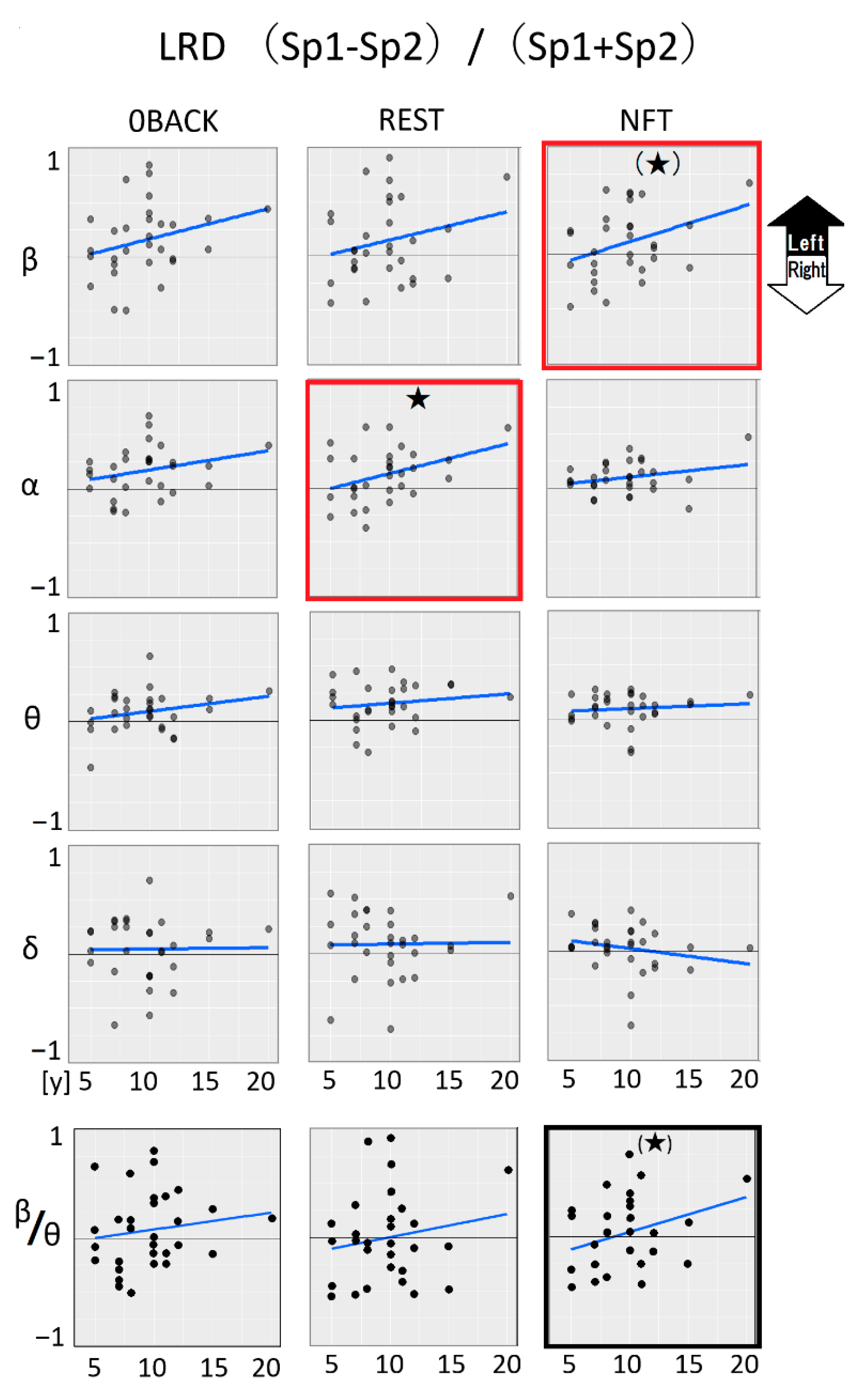
3.2.3. Median Value Comparison between Left (Sp1) and Right (Sp2)

Finally, we explored the lateralization of brain functional dominance. We found an age-dependent trend toward left-brain dominance in beta waves during the NFT stage (Figure 6 beta and NFT, the graph enclosed by a red square, Table 5, p = 0.052 (*)). The NFT trigger signals, beta/theta ratios at NFT also showed a similar weak trend toward left dominance (p = 0.087 (*), enclosed by a black square in Figure 6).
A pentagram or a parenthetic pentagram shows p < 0.05 or nearly p = 0.05 by linear regression analysis (blue lines), respectively.
Another fast wave band, alpha, exhibited significant age-dependent left lateralization at the REST stage (Figure 6 alpha REST, Table 5. p = 0.044).
The horizontal axis; age (y; year), the vertical axis; each wave ratio between Left (Sp1) and Right (Sp2) electrodes. An asterisk or a parenthetic asterisk shows p < 0.05 or nearly p = 0.05 by linear regression analysis, respectively.

4. Discussion

A home-based, easy-to-use EEG system to monitor brain development and assist with clinical diagnosis and treatment in children has long been promised [26]. Various EEG devices and systems have been recently released [27], though the widespread utility of these devices suffers due to the complicated requirements associated with mounting and wearing a delicate EEG device on the head and difficulty in performing meaningful analysis of the obtained data. This study addressed these challenges by developing an easy-to-deploy headband-mounted EEG device designed for children with gel electrode patches, Bluetooth EEG, and an NFT-based interactive PC game and associated analysis software. We evaluated whether children would readily use this system at a one-day local children’s event. We further explored whether there were any age-dependent EEG signal differences that would reproduce findings of previous reports [7,17,19,28].
Thirty-six individuals completed the experiment. Of these, a 3-year-old male and a 4-year-old female’s EEG data were too noisy to be analyzed. If this availability might be for 5 years and older, 32 were 5 to 15 years old. Of the 32, EEG data from 30 subjects were deemed appropriately consistent for analysis, indicating the success of this preliminary prototype design. The experiment consisted of three stages. The first two stages, 0BACK and REST (which were known to change in an age-dependent manner [2,4]), were used for reference and did not involve any feedback from the subjects. These stages were designed with an aspect of comparative baselines. In the final stage, NFT, the size of the projected image changed in response to the subject’s own beta/theta ratios relative to their value during the reference stages. This was the first experience with such technology for all subjects. If the function could be arbitrarily controlled, it might lead to the development of novel neurological function, much like a magic ability to change images with “telekinesis”. This may be one potential core technology that could be applied to future work on brain–computer interfaces [29]. Because the NFT stage was limited to 30 s in this study, we did not evaluate learning and neuronal plasticity [30,31], but rather, we focused on acute and real-time responses. For this reason, we did not evaluate the control status of the NFT but plan to do so in future studies.
In our age-dependent linear regression models, we found some significance in delta, alpha, and beta waves and left-right or stage differences. In general, there was more age dependence in NFT and REST stages, close-eyes [2] and open-eyes [32], and left lateralization than in the visual cognition task, 0BACK [33]. We found an age-dependent decrease in Sp1 delta at stage REST along with other band trends that were consistent with previous reports, indicating that our device functioned correctly and that the children were similar to the previously reported population. We also found age-dependent left-dominance lateralization [34], age-dependent NFT dominance versus REST at Sp2 delta, and a weak age-dependent 0BACK dominance versus NFT at Sp1 alpha. Our study population had a strong female bias; thus, the study population should be enhanced in both age and sex to allow determination regarding the generalizability of our models.
Although this study for child use selected a simplified strategy of forehead Sp1 and Sp2 channels in the 10–20 method, the collectible neuronal information was restricted. Multiple electrodes or other placement extensions should be designed to reach more complex functions such as neuropsychological or cognitive development with another supporting headset such as a cap or headphone style [35].
This child-friendly system holds promise for helping to address the recent increase in neurodevelopmental disorders. Regarding diagnostics, EEG-determined brain waves and theta/beta ratio have potential benefits as a biomarker for ADHD and ASD [36]. Children with ADHD have decreased beta waves and increased theta waves, which is believed to be correlated with poor attention [37]. Furthermore, this fun-to-use system has the potential to assist in development through its longitudinal use in educational settings [38,39].
As with other aspects of therapeutic and rehabilitative education for developmental disorders, the combination of EEG and digital tools, such as virtual and augmented reality devices and game software, have been recently suggested to lead the evolution of treatment [40].
Clinical use of NFT in children was reported in ADHD [41], ASD [17], anxiety disorders, and depression [28]. In addition to neurofeedback applications, EEG itself may enable predictive diagnosis of neurodevelopmental disorders in early ages [42], given that resting EEG correlates with neurochemical imaging MRS [43], the default mode network [44], visual [45,46], auditory [47], tactile [48], and subcortical networks [42] in autism [49].
Since the enactment of the Developmental Disabilities Support Law in Japan in 2005, there has been increased attention paid to early diagnosis of developmental disabilities, developmental support within schools, and building support centers [50]. In 2012, the “Survey on Students with Special Educational Needs with Developmental Disabilities in Regular School Classes” conducted by the Ministry of Education, Culture, Sports, Science, and Technology, reported that 6.5% of students are estimated to have developmental disabilities, and the number has increased significantly over the past decade (https://www.soumu.go.jp/menu_news/s-news/110614.html, accessed on 31 July 2022). Finding simple and scalable neurodevelopmental biomarkers for early detection of developmental disabilities is needed [51] because neurodevelopmental symptoms may precede behavioral symptoms by months to years, allowing for early intervention to perhaps prevent negative outcomes [52], such as withdrawal from school. Thus, our approach to develop an EEG system for children involving interactive games that can be implemented in a home or child-friendly social environment indoors and outdoors would be an important contribution to current education practices [53]. We believe that the easy-to-use design of our prototype is more important in NFT than the additional information that could be obtained using more restrictive techniques. Extension of novel designs of NFT would be expected by this simplified attachable EEG.

5. Conclusions

The Sp1 and Sp2 channel EEG headsets we designed to support children’s general and clinical education took less than 10 s to be properly placed by the individuals and were successfully worn by 32 children aged 5 to 15 years. All subjects appeared to enjoy the three stages of the task, identifying the numbers on the display, open-eyes resting, and using neurofeedback to adjust the size of the local mascot image according to the beta/theta ratio. Post analysis of the recorded EEG data suggested a moderate age-dependent left dominance lateralization in the beta wave band or relatively age-dependent dominance versus open-eyes resting at Sp2. These models need further testing.

Author Contributions

Conceptualization, all; methodology and software, K.O.; validation, formal analysis, investigation, resources, K.O. and M.K.; data curation, all; writing—original draft preparation, writing K.O. and M.K.; writing—review and editing, R.C.; visualization, K.O. and M.K.; supervision, M.K. and R.C.; project administration, M.K.; funding acquisition, M.K. All authors have read and agreed to the published version of the manuscript.

Funding

This research was funded by the Female researcher and AI research design project (2001000010) of Yamaguchi University.

Institutional Review Board Statement

The study was conducted in accordance with the Declaration of Helsinki. The Institute Review Board for human research of Yamaguchi University reviewed and approved this study (2020-047). All study members complied with the approved protocol.

Informed Consent Statement

Not applicable since no personal information is collected.

Data Availability Statement

The datasets generated for this study are available on request to the corresponding authors.

Acknowledgments

We acknowledge all the visitors of children and adult citizens, staff, and students. We appreciate Yamaguchi University.

Conflicts of Interest

The first author discloses the chief executive officer of the EEG device company, Intercross Co. This scientific study report is of the first author’s thesis supervised by the corresponding author in Yamaguchi University research supports. Consequently, we declare no conflicts of interest.

References

  1. Sitaram, R.; Ros, T.; Stoeckel, L.; Haller, S.; Scharnowski, F.; Lewis-Peacock, J.; Weiskopf, N.; Blefari, M.L.; Rana, M.; Oblak, E.; et al. Closed-loop brain training: The science of neurofeedback. Nat. Rev. Neurosci. 2017, 18, 86–100. [Google Scholar] [CrossRef] [PubMed]
  2. Clarke, A.R.; Barry, R.J.; McCarthy, R.; Selikowitz, M. Age and sex effects in the EEG: Differences in two subtypes of attention-deficit/hyperactivity disorder. Clin. Neurophysiol. 2001, 112, 815–826. [Google Scholar] [CrossRef]
  3. Enriquez-Geppert, S.; Huster, R.J.; Herrmann, C.S. EEG-neurofeedback as a tool to modulate cognition and behavior: A review tutorial. Front. Hum. Neurosci. 2017, 11, 51. [Google Scholar] [CrossRef] [PubMed]
  4. Tanaka, M.; Shigihara, Y.; Ishii, A.; Funakura, M.; Kanai, E.; Watanabe, Y. Effect of mental fatigue on the central nervous system: An electroencephalography study. Behav. Brain Funct. 2012, 8, 48. [Google Scholar] [CrossRef] [PubMed]
  5. Papo, D. Neurofeedback: Principles, appraisal, and outstanding issues. Eur. J. Neurosci. 2019, 49, 1454–1469. [Google Scholar] [CrossRef]
  6. Nan, W.; Wan, M.; Jiang, Y.; Shi, X.; Wan, F.; Cai, D. Alpha/Theta Ratio Neurofeedback Training for Attention Enhancement in Normal Developing Children: A Brief Report. Appl. Psychophysiol. Biofeedback 2022, 47, 223–229. [Google Scholar] [CrossRef]
  7. Shereena, E.A.; Gupta, R.K.; Bennett, C.N.; Sagar, K.J.V.; Rajeswaran, J. EEG Neurofeedback Training in Children With Attention Deficit/Hyperactivity Disorder: A Cognitive and Behavioral Outcome Study. Clin. EEG Neurosci. 2019, 50, 242–255. [Google Scholar] [CrossRef]
  8. Lenartowicz, A.; Loo, S.K. Use of EEG to Diagnose ADHD. Curr. Psychiatry Rep. 2014, 16, 498. [Google Scholar] [CrossRef]
  9. Moriyama, T.S.; Polanczyk, G.; Caye, A.; Banaschewski, T.; Brandeis, D.; Rohde, L.A. Evidence-Based Information on the Clinical Use of Neurofeedback for ADHD. Neurotherapeutics 2012, 9, 588–598. [Google Scholar] [CrossRef]
  10. Mekkawy, L. Efficacy of neurofeedback as a treatment modality for children in the autistic spectrum. Bull. Natl. Res. Cent. 2021, 45, 45. [Google Scholar] [CrossRef]
  11. Martínez-Briones, B.J.; Bosch-Bayard, J.; Biscay-Lirio, R.J.; Silva-Pereyra, J.; Albarrán-Cárdenas, L.; Fernández, T. Effects of neurofeedback on the working memory of children with learning disorders—An EEG power-spectrum analysis. Brain Sci. 2021, 11, 957. [Google Scholar] [CrossRef]
  12. Loriette, C.; Ziane, C.; Ben Hamed, S. Neurofeedback for cognitive enhancement and intervention and brain plasticity. Rev. Neurol. 2021, 177, 1133–1144. [Google Scholar] [CrossRef] [PubMed]
  13. Hamaneh, M.B.; Limotai, C.; Lüders, H.O. Sphenoidal electrodes significantly change the results of source localization of interictal spikes for a large percentage of patients with temporal lobe epilepsy. J. Clin. Neurophysiol. 2011, 28, 373–379. [Google Scholar] [CrossRef] [PubMed]
  14. Homan, R.W.; Herman, J.; Purdy, P. Cerebral location of international 10–20 system electrode placement. Electroencephalogr. Clin. Neurophysiol. 1987, 66, 376–382. [Google Scholar] [CrossRef]
  15. Lutsyuk, N.V.; Éismont, E.V.; Pavlenko, V.B. Modulation of attention in healthy children using a course of EEG-feedback sessions. Neurophysiology 2006, 38, 389–395. [Google Scholar] [CrossRef]
  16. Nan, W.; Wan, F.; Vai, M.I.; Da Rosa, A.C. Resting and initial beta amplitudes predict learning ability in beta/theta ratio neurofeedback training in healthy young adults. Front. Hum. Neurosci. 2015, 9, 677. [Google Scholar] [CrossRef]
  17. Wang, Y.; Sokhadze, E.M.; El-Baz, A.S.; Li, X.; Sears, L.; Casanova, M.F.; Tasman, A. Relative power of specific eeg bands and their ratios during neurofeedback training in children with autism spectrum disorder. Front. Hum. Neurosci. 2016, 9, 723. [Google Scholar] [CrossRef]
  18. Bazanova, O.M.; Auer, T.; Sapina, E.A. On the Efficiency of Individualized Theta/Beta Ratio Neurofeedback Combined with Forehead EMG Training in ADHD Children. Front. Hum. Neurosci. 2018, 12, 3. [Google Scholar] [CrossRef]
  19. Hong, C.; Lee, I. Effects of Neurofeedback Training on Attention in Children with Intellectual Disability. J. Neurother. 2012, 16, 110–122. [Google Scholar] [CrossRef]
  20. Koshiba, M.; Shirakawa, Y.; Mimura, K.; Senoo, A.; Karino, G.; Nakamura, S. Familiarity Perception Call Elicited under Restricted Sensory Cues in Peer-Social Interactions of the Domestic Chick. PLoS ONE 2013, 8, e58847. [Google Scholar] [CrossRef] [Green Version]
  21. Toyama, S.; Takano, K.; Kansaku, K. A non-adhesive solid-gel electrode for a non-invasive brain-machine interface. Front. Neurol. 2012, 3, 114. [Google Scholar] [CrossRef] [PubMed]
  22. Kurdi, B.; Lozano, S.; Banaji, M.R. Introducing the Open Affective Standardized Image Set (OASIS). Behav. Res. Methods 2017, 49, 457–470. [Google Scholar] [CrossRef] [PubMed]
  23. Van Doren, J.; Heinrich, H.; Bezold, M.; Reuter, N.; Kratz, O.; Horndasch, S.; Berking, M.; Ros, T.; Gevensleben, H.; Moll, G.H.; et al. Theta/beta neurofeedback in children with ADHD: Feasibility of a short-term setting and plasticity effects. Int. J. Psychophysiol. 2017, 112, 80–88. [Google Scholar] [CrossRef]
  24. Penttilä, M.; Partanen, J.V.; Soininen, H.; Riekkinen, P.J. Quantitative analysis of occipital EEG in different stages of Alzheimer’s disease. Electroencephalogr. Clin. Neurophysiol. 1985, 60, 1–6. [Google Scholar] [CrossRef]
  25. T Package. Package ‘Cosinor’; R Foundation for statistical Computing: Vienna, Austria, 2022.
  26. Marzbani, H.; Marateb, H.R.; Mansourian, M. Methodological note: Neurofeedback: A comprehensive review on system design, methodology and clinical applications. Basic Clin. Neurosci. J. 2016, 7, 143–158. [Google Scholar] [CrossRef]
  27. Batail, J.M.; Bioulac, S.; Cabestaing, F.; Daudet, C.; Drapier, D.; Fouillen, M.; Fovet, T.; Hakoun, A.; Jardri, R.; Jeunet, C.; et al. EEG neurofeedback research: A fertile ground for psychiatry? Encephale 2019, 45, 245–255. [Google Scholar] [CrossRef]
  28. Simkin, D.R.; Thatcher, R.W.; Lubar, J. Quantitative EEG and Neurofeedback in Children and Adolescents. Child Adolesc. Psychiatr. Clin. N. Am. 2014, 23, 427–464. [Google Scholar] [CrossRef]
  29. Sun, J.; He, J.; Gao, X. Neurofeedback Training of the Control Network Improves Children’s Performance with an SSVEP-based BCI. Neuroscience 2021, 478, 24–38. [Google Scholar] [CrossRef]
  30. Marins, T.; Rodrigues, E.C.; Bortolini, T.; Melo, B.; Moll, J.; Tovar-Moll, F. Structural and functional connectivity changes in response to short-term neurofeedback training with motor imagery. Neuroimage 2019, 194, 283–290. [Google Scholar] [CrossRef]
  31. Engelbregt, H.J.; Keeser, D.; van Eijk, L.; Suiker, E.M.; Eichhorn, D.; Karch, S.; Deijen, J.B.; Pogarell, O. Short and long-term effects of sham-controlled prefrontal EEG-neurofeedback training in healthy subjects. Clin. Neurophysiol. 2016, 127, 1931–1937. [Google Scholar] [CrossRef]
  32. Perone, S.; Palanisamy, J.; Carlson, S.M. Age-related change in brain rhythms from early to middle childhood: Links to executive function. Dev. Sci. 2018, 21, e12691. [Google Scholar] [CrossRef] [PubMed]
  33. Krause, C.M.; Sillanmäki, L.; Koivisto, M.; Saarela, C.; Häggqvist, A.; Laine, M.; Hämäläinen, H. The effects of memory load on event-related EEG desynchronization and synchronization. Clin. Neurophysiol. 2000, 111, 2071–2078. [Google Scholar] [CrossRef]
  34. Koshiba, M.; Nakamura, S.; Deng, C.; Rogers, L.J. Light-dependent development of asymmetry in the ipsilateral and contralateral thalamofugal visual projections of the chick. Neurosci. Lett. 2003, 336, 81–84. [Google Scholar] [CrossRef]
  35. Badcock, N.A.; Mousikou, P.; Mahajan, Y.; De Lissa, P.; Thie, J.; McArthur, G. Validation of the Emotiv EPOC® EEG gaming systemfor measuring research quality auditory ERPs. PeerJ 2013, 1, e38. [Google Scholar] [CrossRef]
  36. Jeste, S.S.; Frohlich, J.; Loo, S.K. Electrophysiological biomarkers of diagnosis and outcome in neurodevelopmental disorders. Curr. Opin. Neurol. 2015, 28, 110–116. [Google Scholar] [CrossRef] [PubMed]
  37. Arns, M.; Clark, C.R.; Trullinger, M.; deBeus, R.; Mack, M.; Aniftos, M. Neurofeedback and Attention-Deficit/Hyperactivity-Disorder (ADHD) in Children: Rating the Evidence and Proposed Guidelines. Appl. Psychophysiol. Biofeedback 2020, 45, 39–48. [Google Scholar] [CrossRef] [PubMed]
  38. Janssen, T.W.P.; Bink, M.; Weeda, W.D.; Geladé, K.; van Mourik, R.; Maras, A.; Oosterlaan, J. Learning curves of theta/beta neurofeedback in children with ADHD. Eur. Child Adolesc. Psychiatry 2017, 26, 573–582. [Google Scholar] [CrossRef]
  39. Yang, Z.; An, P.; Yang, J.; Strojny, S.; Zhang, Z.; Sun, D.; Zhao, J. Designing Mobile EEG Neurofeedback Games for Children with Autism: Implications from Industry Practice. In Adjunct Publication of the 23rd International Conference on Mobile Human-Computer Interaction; Association for Computing Machinery: New York, NY, USA, 2021; pp. 1–6. [Google Scholar] [CrossRef]
  40. Barba, M.C.; Covino, A.; De Luca, V.; De Paolis, L.T.; D’Errico, G.; Di Bitonto, P.; Di Gestore, S.; Magliaro, S.; Nunnari, F.; Paladini, G.I.; et al. BRAVO: A Gaming Environment for the Treatment of ADHD BT—Augmented Reality, Virtual Reality, and Computer Graphics; De Paolis, L.T., Bourdot, P., Eds.; Springer: Cham, Switzerland, 2019; pp. 394–407. [Google Scholar]
  41. Zamora Blanón, D.; Muñoz, J.E.; Lopez, D.S.; Henao Gallo, O. Processing Dedicated Toolbox; MindReflector Technol. LLC.: Harrisburg, PA, USA, 2016; Volume 3, pp. 1–8. [Google Scholar]
  42. Bosl, W.J.; Tager-Flusberg, H.; Nelson, C.A. EEG Analytics for Early Detection of Autism Spectrum Disorder: A data-driven approach. Sci. Rep. 2018, 8, 6828. [Google Scholar] [CrossRef]
  43. Pierce, S.; Kadlaskar, G.; Edmondson, D.A.; McNally Keehn, R.; Dydak, U.; Keehn, B. Associations between sensory processing and electrophysiological and neurochemical measures in children with ASD: An EEG-MRS study. J. Neurodev. Disord. 2021, 13, 5. [Google Scholar] [CrossRef]
  44. Padmanabhan, A.; Lynch, C.J.; Schaer, M.; Menon, V. The Default Mode Network in Autism. Biol. Psychiatry Cogn. Neurosci. Neuroimaging 2017, 2, 476–486. [Google Scholar] [CrossRef]
  45. Trachtman, J.N. Background and history of autism in relation to vision care. Optom.-J. Am. Optom. Assoc. 2008, 79, 391–396. [Google Scholar] [CrossRef] [PubMed]
  46. Sheela, P.; Puthankattil, S.D. A hybrid method for artifact removal of visual evoked eeg. J. Neurosci. Methods 2020, 336, 108638. [Google Scholar] [CrossRef] [PubMed]
  47. De Stefano, L.A.; Schmitt, L.M.; White, S.P.; Mosconi, M.W.; Sweeney, J.A.; Ethridge, L.E. Developmental Effects on Auditory Neural Oscillatory Synchronization Abnormalities in Autism Spectrum Disorder. Front. Integr. Neurosci. 2019, 13, 34. [Google Scholar] [CrossRef] [PubMed]
  48. Portnova, G.V.; Ivanova, O.; Proskurnina, E.V. Effects of EEG examination and ABA-therapy on resting-state EEG in children with low-functioning autism. AIMS Neurosci. 2020, 7, 153–167. [Google Scholar] [CrossRef]
  49. Chen, C.P.; Keown, C.L.; Jahedi, A.; Nair, A.; Pflieger, M.E.; Bailey, B.A.; Müller, R.A. Diagnostic classification of intrinsic functional connectivity highlights somatosensory, default mode, and visual regions in autism. NeuroImage Clin. 2015, 8, 238–245. [Google Scholar] [CrossRef]
  50. Branjerdporn, G.; Meredith, P.; Wilson, T.; Strong, J. Infant Developmental Outcomes: Influence of Prenatal Maternal–Fetal Attachment, Adult Attachment, Maternal Well-Being, and Perinatal Loss. Int. J. Environ. Res. Public Health 2022, 19, 2433. [Google Scholar] [CrossRef]
  51. Kana, R.K.; Libero, L.E.; Moore, M.S. Disrupted cortical connectivity theory as an explanatory model for autism spectrum disorders. Phys. Life Rev. 2011, 8, 410–437. [Google Scholar] [CrossRef]
  52. Hernandez, L.M.; Rudie, J.D.; Green, S.A.; Bookheimer, S.; Dapretto, M. Neural signatures of autism spectrum disorders: Insights into brain network dynamics. Neuropsychopharmacology 2015, 40, 171–189. [Google Scholar] [CrossRef]
  53. Fjørtoft, I. The natural environment as a playground for children: The impact of outdoor play activities in pre-primary school children. Early Child. Educ. J. 2001, 29, 111–117. [Google Scholar] [CrossRef]
Figure 1. Headset of EEG device (a), a player wearing the headset and looking at the game display (b).
Sensors 22 07207 g001
Figure 2. The three stages of the game protocol.
Sensors 22 07207 g002
Figure 3. Examples of the EEG trend of four band and beta/theta ratios per stage, 0BACK, REST, and NFT in younger and older subjects left (Sp1) or right (Sp2) brain.
Sensors 22 07207 g003
Figure 4. Age-dependent comparison of wave bands and beta/theta in stages and channels.
Sensors 22 07207 g004
Figure 5. Each median of channel and wave band comparisons between stages.
Sensors 22 07207 g005
Figure 6. Left or right brain dominance: age-dependent lateralization.
Sensors 22 07207 g006
Table 1. Number and ages of participants.
Age [year]n (Female)n (Male)n (Total)
5134
75-5
8-44
10718
113-3
123-3
152-2
201-1
Total [n]22830
Average [year]10.47.19.5
SD [year]3.21.83.3
Table 2. The wave bands of EEG.
BandRange [Hz]
delta2~4
theta4~8
alpha8~13
beta13~30
Table 3. p-values associated with age-dependent EEG wave linear regression shown in Figure 4.
Sp1 Sp2
0BACKRESTNFT0BACKRESTNFT
beta0.2090.1210.3590.7580.8050.407
alpha0.8420.4050.9210.2160.6620.675
theta0.2300.2280.4230.9110.7320.643
delta0.1690.052 (*)0.1870.9800.4390.515
beta/theta0.3510.1320.3010.7220.3860.710
(*), bold: nearly p = 0.05.
Table 4. p-values associated with age-dependent EEG wave linear regression shown in Figure 5.
A: 0BACK Versus RESTB: NFT Versus RESTC: NFT Versus 0BACK
Sp1Sp2Sp1Sp2Sp1Sp2
beta0.5570.4060.7180.3330.4150.194
alpha0.5080.2210.330.5910.095 (*)0.279
theta0.2370.4590.3180.2990.4950.993
delta0.170.210.1960.030 *0.7090.575
*, bold: p < 0.05, (*), bold: nearly p = 0.05 (p < 0.1).
Table 5. p-values associated with age-dependent EEG wave linear regression shown in Figure 6.
LRD(Sp1 − Sp2)/(Sp1 + Sp2)
0BACKRESTNFT
beta0.1360.2140.052 (*)
alpha0.1580.044 *0.157
theta0.1830.4230.59
delta0.9410.9240.246
beta/theta0.4330.3560.087 (*)
*, bold: p < 0.05, (*), bold: nearly p = 0.05 (p < 0.1).
Publisher’s Note: MDPI stays neutral with regard to jurisdictional claims in published maps and institutional affiliations.

