Article

Effect of Ducrosia flabellifolia and Savignya parviflora Extracts on Inhibition of Human Colon and Prostate Cancer Cell Lines

by
Youssef Saeed Alghamdi
1,
Osama Moseilhy Saleh
2,3,
Nada Alqadri
1,
Mutaib Mosaued Mashraqi
4,
Omar Bahattab
5 and
Nabil Saied Awad
3,6,*
1
Department of Biology, Turabah University College, Taif University, Taif 21995, Saudi Arabia
2
Natural Products Research Department, National Centre for Radiation Research and Technology (NCRRT), Egyptian Atomic Energy Authority, Cairo 9621, Egypt
3
College of Biotechnology, Misr University for Science and Technology, Giza 12563, Egypt
4
Department of Clinical Laboratory Sciences, College of Applied Medical Sciences, Najran University, Najran 61441, Saudi Arabia
5
Department of Biology, Faculty of Science, University of Tabuk, Tabuk 71497, Saudi Arabia
6
Department of Genetics, Faculty of Agriculture and Natural Resources, Aswan University, Aswan 81528, Egypt
*
Author to whom correspondence should be addressed.
Curr. Issues Mol. Biol. 2021, 43(3), 1518-1528; https://0-doi-org.brum.beds.ac.uk/10.3390/cimb43030107
Submission received: 1 September 2021 / Revised: 4 October 2021 / Accepted: 5 October 2021 / Published: 10 October 2021

Abstract

:
The goal of this study was to investigate whether Ducrosia flabellifolia and Savignya parviflora methanol extract the have effect on colon and prostate cancer cell lines. Analysis of total content of phenolics and flavonoids of each plant extract was carried out. Cytotoxic effect, cell cycle analysis, induction of apoptosis and gene expression of Bcl-2 and Bax genes were studied. Obtained results indicated that, the plant extracts exhibit growth inhibition of used cancer cell lines and induced apoptosis as well as arresting of cell cycle. At the molecular level, changes in gene expression were detected via qPCR and confirmed by western blotting. The exhibited anticancer potentialities of plant extracts against utilized cancer cell lines are due to its containing bioactive compounds. Further detailed isolation, fractionation and characterization of bioactive compounds are needed.

1. Introduction

Worldwide, cancer is one of the most serious health problems [1]. Overcoming this threat requires many health challenges worldwide. Colorectal, lung and breast cancers are the most common types of cancer worldwide [2]. Young males with colorectal cancer have an increased risk of cancer prostate [3]. Colon cancer increasing within a group of less than 50 years prostatic cancer patients [4].
The ineffectiveness of clinical cancer rehabilitation approaches, such as chemotherapy, radiotherapy, surgery, and immunosuppression are manifested with high mortality and morbidity. This has created a necessary for promising, effective and safe compounds for cancer prevention and treatment [5]. Thus, herbal remedies have been reborn as nutritional and botanical preparations. Many medicinal plants are used directly as botanicals of medical importance. Medicinal plants are considered as one of a great resource of chemicals with potential therapeutic effects [6].
Effects of plants such as the active components of antitumor compounds have been shown to increase the effects of drugs, where drugs such as vinblastine, vincristine, toxol and camptothecin have improved the chemotherapy of some cancers [7]. Some species, such as Convolvulus sp. and Astragalus sp. have the potential to induce apoptotic cell death against lung and colon cancer cell lines [8].
The Arabian Gulf region have several species of Ducrosia [9]. Coumarin is one of the various phytochemicals found in it [10]. Active components of Ducrosia flabellifolia have been collected and used as an anticancer for various cell lines and have been demonstrated to reduce the likelihood of tumor burden [11]. D. flabellifolia contains many active ingredients such as Ducrosin B, which shows excellent cytotoxicity against human ovary; SKOV-3 and colon; HCT-116 cell lines [12]. Another active ingredient though; furocoumarin has activity against human hepatoma; HepG2 cell lines [13].
The ethanolic extract of S. Parviflora has abundant useful active ingredients, including anti-inflammatory, anti-ulcerogenic and antioxidant agents [14]. Ethanol extract of S. Parviflora at concentrations of 25 and 50 µg/mL inhibits RAW264.7 cell line growth and is might be promising for antitumor use as reported [15]. Inhibition of metalloproteinase 2 activity, cell growth inhibition, induction of apoptosis, cell migration inhibition, signal pathway of survival suppression and invasion of the HeLa cervical cancer cell lineage have been documented after treatment with Kaempferia parviflora extract [16]. They suggest that the herb contains active compounds that suppress EGF-based signal transduction pathways and then inhibit tumor progression and induce cancer cell death. Coniferia parviflora extract is a new candidate for an ovarian cancer chemotherapeutic agent in response to cell proliferation, cell migration and support apoptotic cell death induction in SKOV-3 cells [17].
This study aimed to in vitro assessment of the effectiveness of D. flabellifolia and S. parviflora methanolic extracts as natural herbs against prostate and colon cancer cell lines. Moreover, the molecular mechanism behind antitumor activity through these two extracts was investigated by cell cycle analysis, apoptosis analysis, gene expression analysis using qPCR and Western blotting assays.

2. Materials and Methods

2.1. Research Design and Statistical Analysis

To examine the antiprolifrative effect of each plant extract, the cancer cells treated with different concentrations of plant extract to identify the IC50 in comparison with un-treated cells.
IC50 had been used to explore the ability of plant extract on the induction of apoptosis, cell cycle arrest, gene expression among treated and untreated cells.
Students’ t-test was performed to assess the significant differences between treated and untreated cells. Statistical analysis was performed using SPSS version 22.0 software (IBM, Chicago, IL, USA). The p-value < 0.05 is statistically significant and the p-value < 0.01 is highly significant.

2.2. Plant Materials

The plant samples were collected and dried at Taif, West Region, Saudi Arabia. Samples of dried plants were ground into powder. Plant species have been identified and certified as D. flabellifolia and S. parviflora, which belong to the families Apiaceae and Brassicaceae, respectively.

2.3. Preparation of Methanolic Extract

Dry plant materials were used to prepare the methanolic plant extract via the Soxhlet extraction procedure. The methanolic extracts were evaporated to dryness and concentrated under pressure at temperatures; 40 to 50 °C in a rotary evaporator. The extracts have been collected and stored in airtight and dark bottles until use.
All the following experimental analyses were carried out in three replicates

2.4. Analysis of the Total Content of Phenolics, Flavonoids and Antioxidant Effect of Plant Extracts

Total content of phenolics (TPC) and flavonoids (TFC) were estimated in D. flabellifolia and S. parviflora plant extracts using a colorimetric assay according to [18] based on procedures described by [19] and [20], respectively. The total phenolic compounds were illustrated as mg/g gallic acid equivalent (GAE). Total flavonoid content (mg/g) was measured using a calibration curve of quercetin and illustrated as mg quercetin equivalents. The antioxidant activity of the extracts collected has been studied according to the methods described by [21] with minor modifications as applied by [22]. Briefly, methanolic extracts of D. flabellifolia and S. parviflora in various concentrations (100–500 μL) were mixed separately with 2.5 mL of 0.2 mM PBS (pH 7.4) and 2.5 mL of potassium ferricyanide (1% w/v). This mixture was incubated at 500 °C for 20 min. Then, 2.5 mL of trichloroacetic acid (10% w/v) was added and centrifuged at 3500 rpm for 8 min, followed by 2.5 mL of distilled water and later 0.5 mL of ferrous chloride (0.1% w/v). The absorbance at 700 nm was estimated. As a positive reference standard, ascorbic acid was utilized.

2.5. Cell Lines and Cell Cultures

The HCT 116 and PC3 cancer cell lines were used. The used cell lines were purchased from VACSERA, Egypt. The RPMI 1640 medium (Gibco-BRL, Carlsbad, CA, USA) was used to culture the cells. Medium was enhanced with FBS (Sijixin Inc., Dalian, China) and 1% penicillin–streptomycin mixture (Invitrogen, Grand Island, NY, USA) and incubated at 37 °C in CO2 incubator with 5% CO2.

2.6. In Vitro Cytotoxicity Assay

For the cytotoxicity assay, 1 × 10⁵ cells/mL (100 μg/well) were seeded in 96-well tissue culture plates and incubated at 37 °C for 24 h to produce a complete monolayer sheet. Using methanol extracts at different concentrations; 4–100 ug /mL, each plant was incubated at 37 °C with and without extract. After 72 h of incubation, cytotoxicity was assessed using MTT assay as reported by [23]. The cells were washed with 1 × phosphate buffer saline and incubated for 2–3 h with MTT (3-[4,5-dimethylthiazol-2-yl]-2,5-diphenyltetrazolium bromide) solution (50 μL of 0.5 mg/mL in RPMI 1640 without phenol red; Sigma). Then, MTT solution was removed and formazan crystals were dissolved in 75 μL of isopropanol: HCl (v/v 1:0.04) mixture. The viable cells were detected spectrophotometrically by the absorption at 570 nm for both treated and untreated cells in addition of wells without sample containing cells as blanks. Measurements were carried out and the IC50 was graphically determined. The percentages of viable cells were calculated as follows: % cell viability = A570 of treated cells/A570 of control cells × 100.

2.7. Apoptosis Detection

Annexin V-fluorescein isothiocyanate (FITC)/propidium iodide (PI) staining was used to estimate apoptotic cells after treatment of HCT and PC3 cells with IC50 concentration of each plant extract in comparison with untreated cells.

2.8. Cell Cycle Analysis

Cell cycle analysis of plant extracts treated, and untreated cells was carried out using flow cytometry (BD FACS Array Bioanalyzer) to detect the cellular phase in which the cell cycle has been arrested.

2.9. Detection of Expression Levels of Bax and Bcl-2 Genes

Among treated with IC50 and untreated HCT and PC3 cells, changes in mRNA levels of apoptotic-related genes Bax and Bcl-2 were analyzed using qPCR as previously described [24]. HCT-116 and PC3 cells were treated with IC50 concentrations for 48 h. Total RNA was carried out according to triazole reagent manufacturer’s instructions.

2.10. Real-Time Quantitative PCR (qPCR)

One μg of RNA was reverse transcribed to first-strand cDNA. The obtained cDNA was amplified to examine the expressions of Bcl-2 and Bax genes. An internal control β-actin was utilized as a standard for the real-time PCR reaction. Sequences of forward and reverse primers for genes used in this study are shown in Table 1.
The thermocycle program for cDNA amplification was carried out as follow: 95 °C for 35 s followed by 40 cycles of denaturation (95 °C for 5 s), annealing at 58 °C for 10 s (β-actin) and 55 °C for 10 s (Bax and Bcl-2), and extension at 72 °C for 30 s. A non-template control had been done using water instead of the cDNA template. All PCR amplified products were electrophoresized with DNA ladder (100 bp) (Fermen-tas, USA) in 2.5% agarose gel. Ethidium bromide was used to visualize the gels using a UV trans-illuminator. Quantitative real-time (qRT-PCR) was used to quantify Bcl-2, Bax and β-actin mRNA levels. A real-time PCR kit (BIORAD iScriptTM One-Step RT-PCR Kit) using SYBR Green used to perform the quantitative RT-PCR. Diethyl pyro carbonate (DEPC) water was used as a negative control instead of the cDNA template. The results for Bcl-2 and Bax mRNA expression exhibited in relation to the expression of β-actin. The PCR products were determined by melting curve analysis for each primer pairs to specify the amplification. The 2ΔΔCT method was used to analyze the obtained data.
Target mRNA level results were generalized against β-actin mRNA. Results were presented as fold change (RFC) relative to negative control.

2.11. Western Blotting Analysis

Western blotting method has been used to assess the effect of tested plant extracts on the translation level of the Bax and Bcl-2 genes as described previously by [25,26].

3. Results

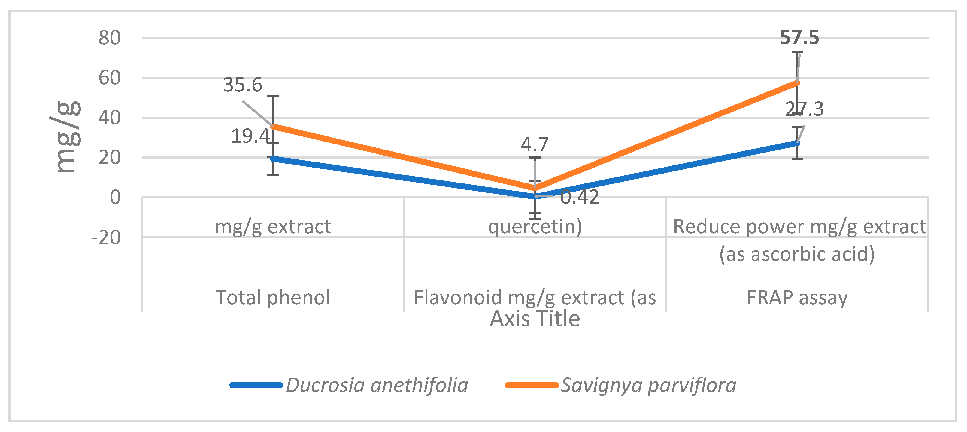
3.1. Analysis of the Total Content of Phenolics, Flavonoids and Antioxidant Effect of Plant Extracts

Among studied plant extracts, the total phenolic content (TPC) and total flavonoid content (TFC) were measured. Obtained results showed that, S. parviflora extract had higher amounts of TPC and TFC than D. flabellifolia as in the case of antioxidant power (Figure 1).

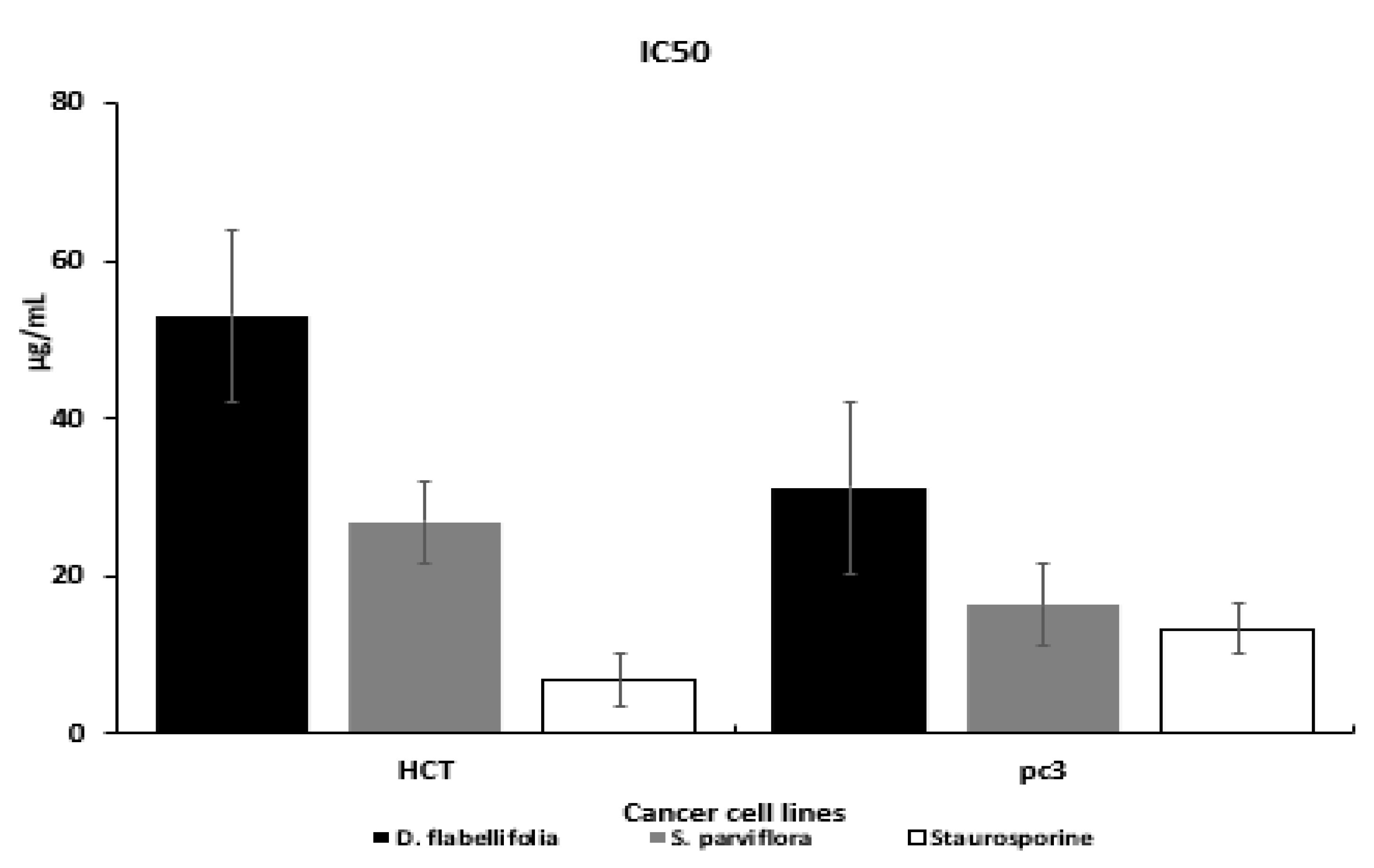
3.2. Cytotoxic Effects

The cytotoxic effect of methanol extract of S. parviflora and D. flabellifolia on the growth of HCT-116 and PC3 cell lines were separately examined by MTT assay. Dose dependent response was obtained between the range 0.4 and 100 µg/mL for plant extracts and Staurosporine (standard control) decreasing number of viable cells with an increasing concentration of plant extracts as well as Staurosporine were noted. Calculation of IC50 value was carried out (Table 2).
The susceptibility of cells to the examined plant extracts was characterized by IC50 (Table 3) and (Figure 2). The IC50 values of S. parviflora with HCT-116 and PC3 cell lines were (26.9 and 16.4 ug/mL respectively), which lowers than the IC50 values of D. flabelli-folia against the two cell lines.

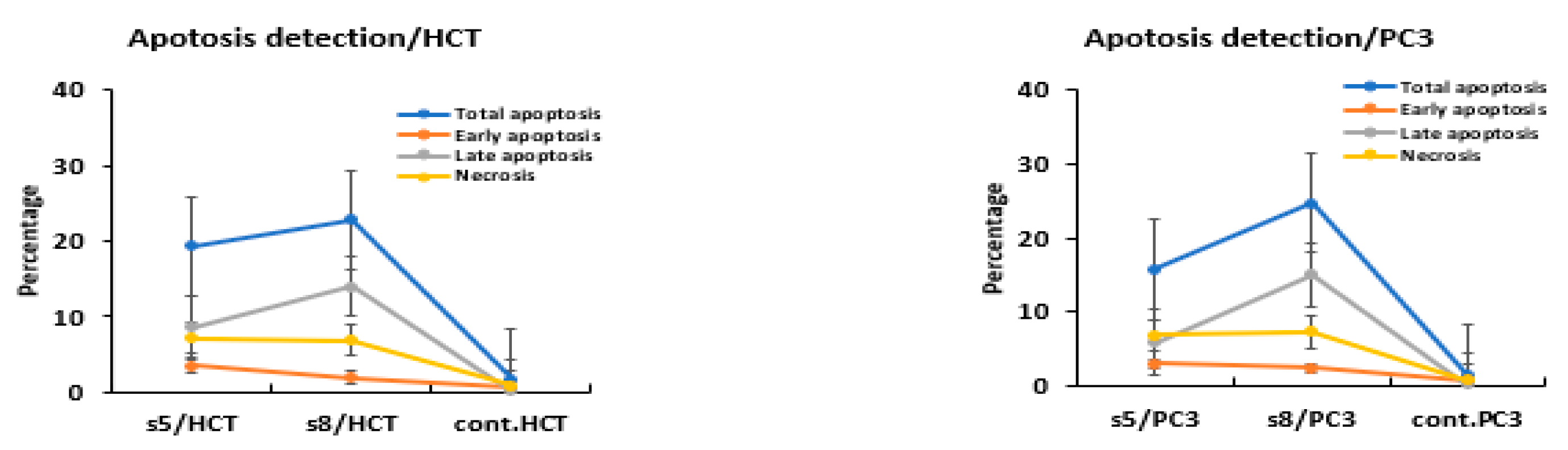
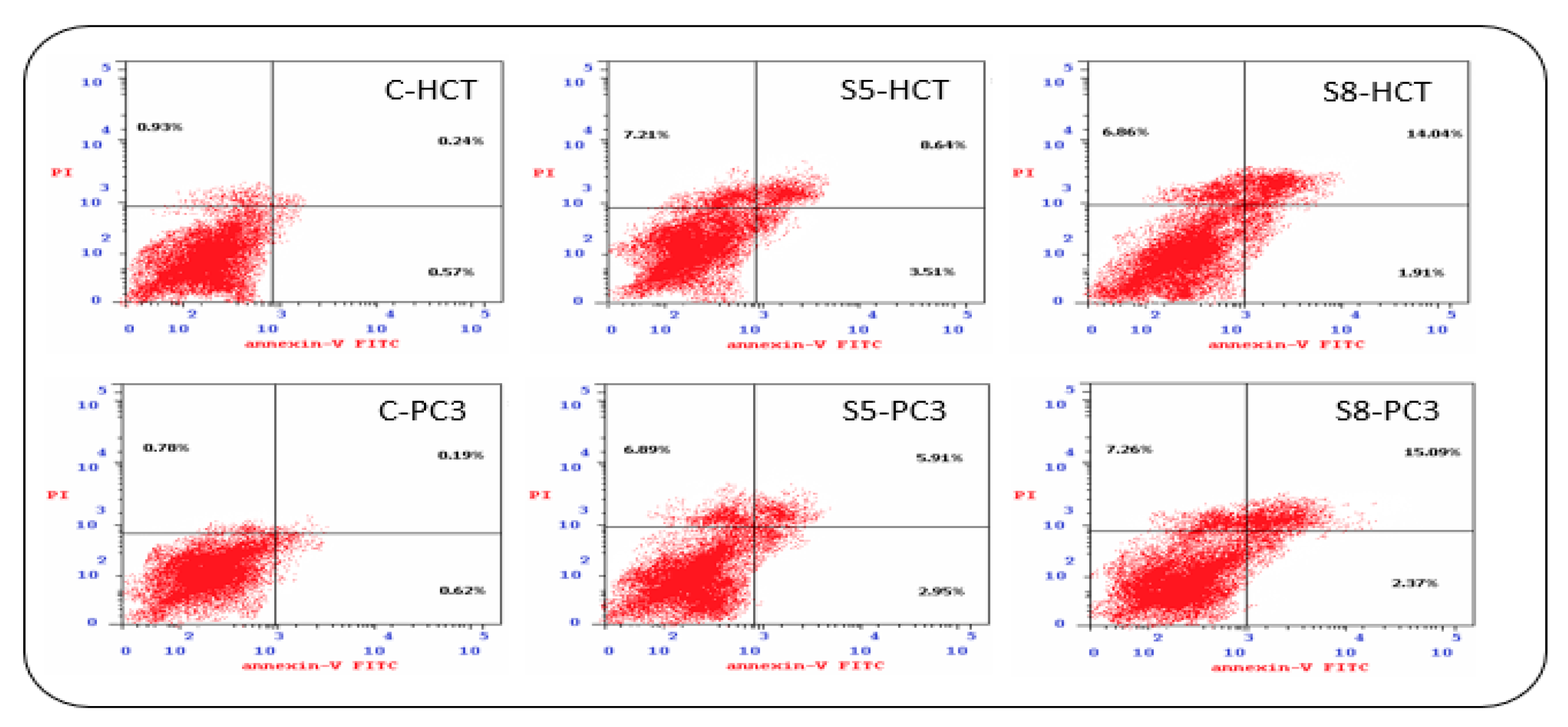
3.3. Apoptosis Detection

The extracts’ potentially to induce apoptosis was examined in HCT-116 and PC3 cancer cells using annexin-FITC/propidium iodide double staining cytometric assay. The cells were treated with IC50 of each extract for 48 h (Figure 3 and Figure 4). At IC50 concentrations, HCT-116 and PC3 cells underwent late apoptosis and induced mild necrosis.

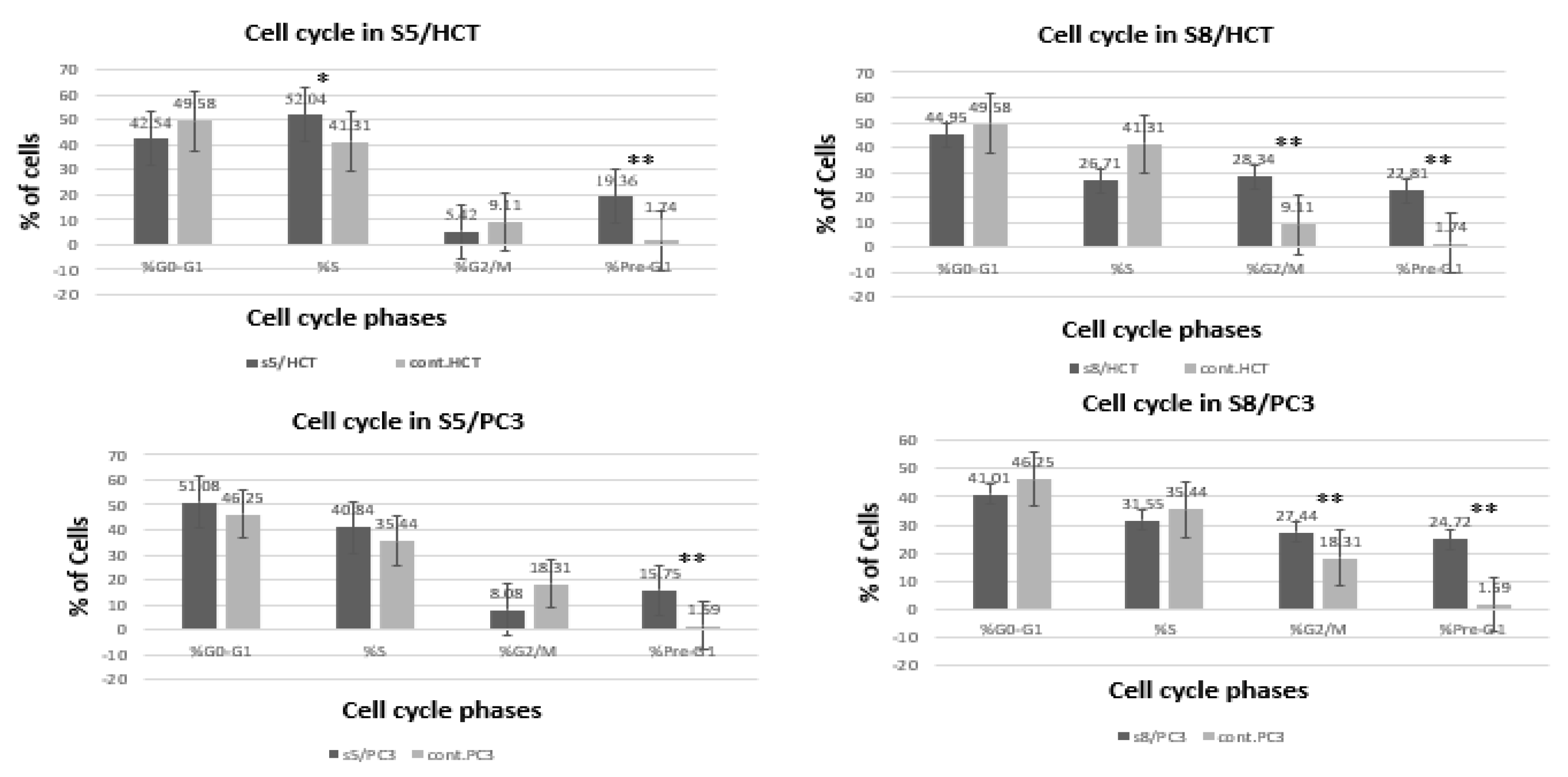
3.4. Cell Cycle Analysis

Cell cycle analysis was carried out in treated and untreated cancer cell lines. In response to treatment of the HCT-116 cell line with IC50 of D. flabellifolia extract, cells arrested significantly in the S and pre-G1 phases. In addition, PC3 cells were significantly arrested in the pre-G1 phase. On the other hand, treatment of HCT-116 and PC3 with IC50 of S. parviflora extract induced significantly arresting of the cell cycle at G2/M and pre-G1 phases (Figure 5).

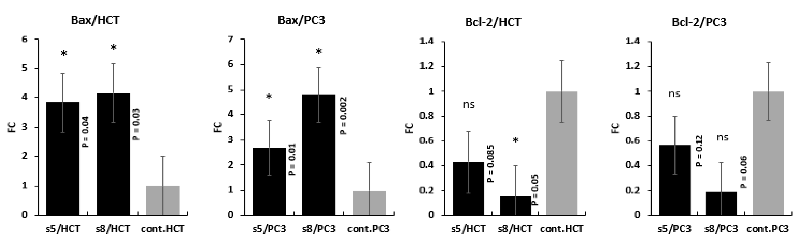
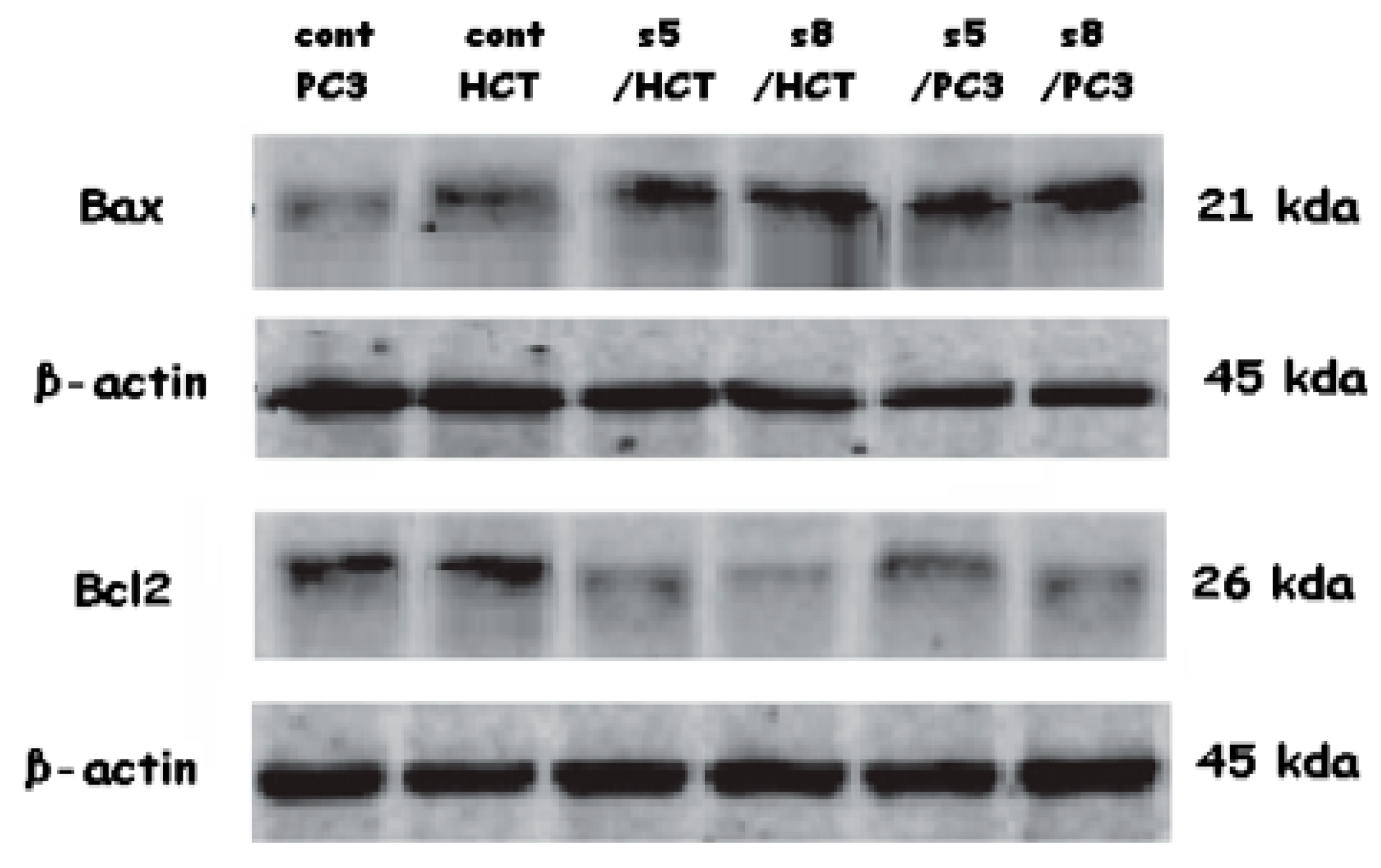
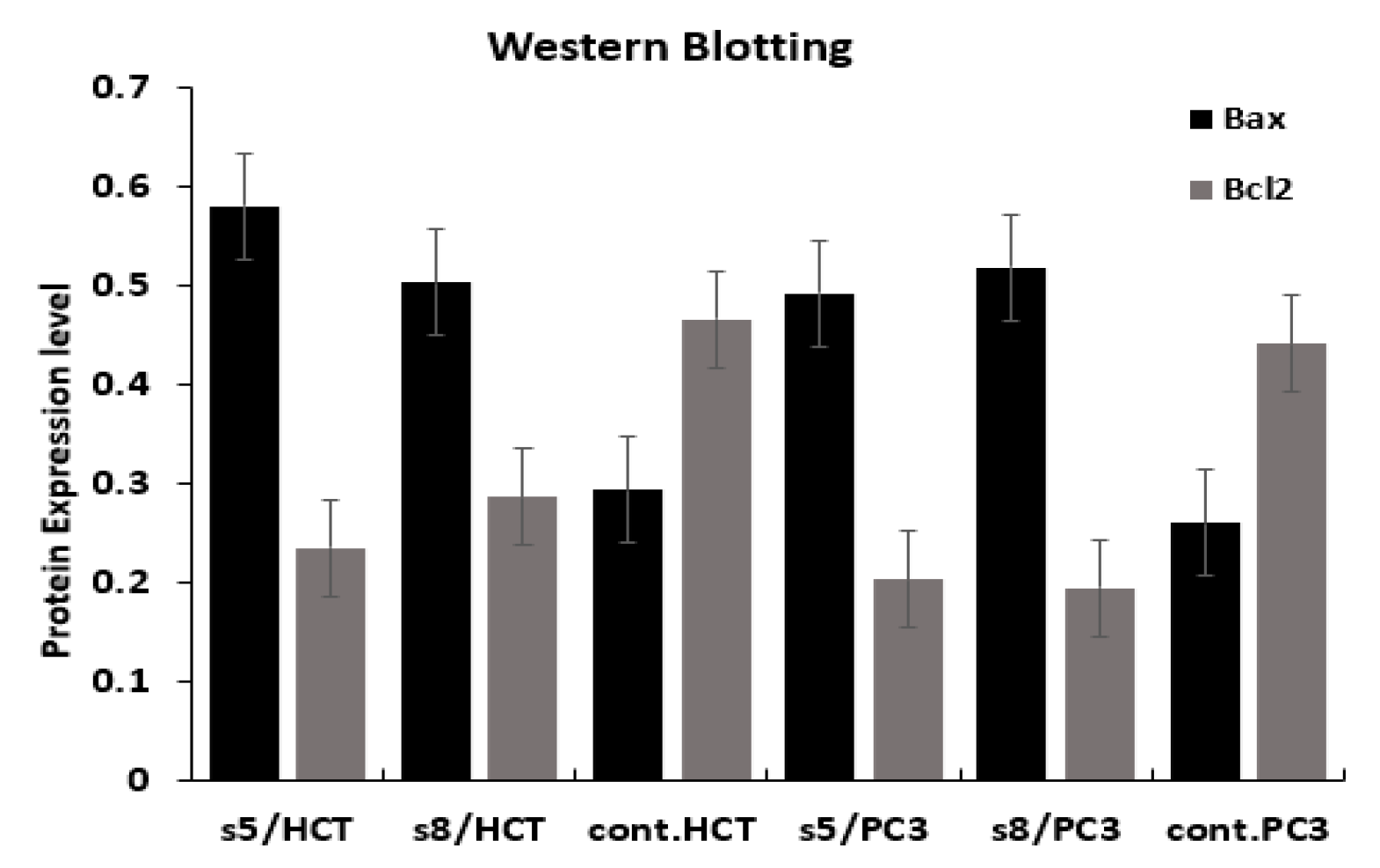
3.5. Gene Expression Analysis

The gene expression level changes of Bax and Bcl-2 genes in response to treatment with IC50 of D. flabellifolia and S. parviflora extracts were assessed by two different methodologies (qPCR and Western blotting).
Results of qPCR indicated that, treatment of HCT-116 and PC3 with IC50 of D. flabellifolia and S. parviflora extracts leads to upregulation of Bax gene and downregulation of Bcl-2 gene (Figure 6). Expression level of Bax gene in both plant extract with the two utilized cell lines was upregulated significantly. Bcl-2 gene expression level was downregulated significantly due to treatment of HCT with S. parviflora and not significant in treatment of HCT with D. flabellifolia. In the PC3 cell line, no significant effects were detected of both S. parviflora and D. flabellifolia extracts on Bcl-2 gene expression. Regards to confirmation of the changes occurred in gene expression levels in response to treatments, the protein level of Bax and Bcl-2 genes were measured via western blotting technique. The obtained results were consistent with Real time PCR data (Figure 7 and Figure 8).

4. Discussion

Cancer is considered as one of the first deadly diseases. There are many cancer treatments that depend on different methods of treatment. But it is established that all these treatments face many challenges such as adverse effects and side effects [27]. Hence, there has been a wide-ranging interest in discovering effective anticancer substances that avoid many of the challenges. Wild plants are considered an influential source in the discovery of many natural substances with anticancer abilities. Therefore, there is an interest in researching and discovering more wild plants that may have natural substances with which it is possible to develop new safe and high quality treatments against cancer [28].
The obtained results of our study showed that the methanolic extracts of both plants have anticancer effect against both colon and prostate cancer cell lines. This is what was shown by the results for cytotoxic effects and IC50 values (Table 2 and Table 3, Figure 2). From these values, the S. parviflora has a higher anticancer capacity than the D. flabellifolia. When the IC50 value is less 20 μg/mL, 20 to 100 μg/mL or higher 100 μg/mL, the US National Cancer Institute considers the cytotoxicity standard for raw extract to be active, moderately active or inactive respectively [29]. These results can be interpreted based on the analysis of the total content of phenolics, flavonoids and antioxidant activity (Figure 1) which showed that the total content of phenolics and flavonoids of S. parviflora, as well as antioxidative power, was higher than that of the D. flabellifolia [30,31,32,33]. It showed that the cytotoxic effect of the extracts of these plants was higher on prostate cancer cell (PC3) than on colon cancer cell (HCT-116). The antiproliferative effects observed for both plant extracts against the two cancer cells used may be due to the oxidant effect of their phytochemical components. Plant substances containing phenolic compounds exert their antiproliferative effects through increase oxidant stress in cancer cells by inhibiting ROS-scavenging systems, inactivating pro-survival signals, activating apoptosis related signals, inducing DNA damage and inhibiting signaling favorable pathways to cancer cell growth [34]. To investigate the mechanism behind the cytotoxic effects of these plant extracts against colon and prostate cancer cell lines, flow cytometry was used to test the apoptosis events and cell cycle analysis. Our results indicated both plant extracts were able to arrest the cell cycle at different phases (Figure 5). It is reported that flavonoids and phenols can arrest the cell cycle in different cancer cells [35,36,37]. The results indicated that both plant extracts under study can induce apoptosis as late apoptosis and arresting the cells in different cell cycle phases (Figure 3 and Figure 4). Similarly, many natural agents exhibit anticancer or cancer protection properties by inducing apoptotic pathways in transformed cells in the process of carcinogenesis. [38,39]. In the present study, the expression of two mitochondrial apoptosis related genes (Bax and Bcl-2) was measured at transcription and translation levels. Gene expression study indicates upregulation of Bax and downregulation of Bcl-2 (Figure 6, Figure 7 and Figure 8). These results suggest that S. parviflora and D. flabellifolia bioactive phytoconstituents induce apoptosis in HCT-116 and PC3 cells by mechanisms related to the mitochondria-based pathway. This is consistent with the reported results [40].
Results showed that S. parviflora and D. flabellifolia extracts have a significant anti-proliferative effect on HCT-116 and PC3 cells by inducing apoptosis in a significant way. The obtained results showed that the S. parviflora and D. flabellifolia extracts exerted a significant anti proliferative effect on the HCT-116 and PC3 cells by inducing apoptosis. The obtained results showed that the S. parviflora and D. flabellifolia promotes apoptosis which was achieved by the pro-apoptotic and anti-apoptotic members Bax and Bcl-2 genes, respectively [41]. Although the antioxidant activity of phytochemicals is well known, they exhibit pro-oxidant activities under certain conditions at high doses, they produce ROS, with the oxidation of all hydroxyl-forming groups increasing radical production occurring the presence of metal ions [42]. cl-2 protein, as the main regulator of apoptosis, promotes cell survival by inhibiting factors that activate caspases [43], or by controling apoptosis, despite its active functional antagonism through the formation of heterodimers with other Bcl-2 family members. Bax, a pro-apoptotic member, also binds to the anti-apoptotic protein Bcl-2 and thus acts by inhibiting the action of Bcl-2 to terminate apoptosis. Moreover, induction of Bax was also observed to enhance the release of cytochrome c from mitochondria, which ultimately leads to apoptosis [44]. It was suggested that both Bcl-2 family of proteins (Bax and Bcl-2) play pivotal roles in S. parviflora and D. flabellifolia induced apoptosis. Thus, the results indicated that the observed upregulation of Bax and corresponding downregulation of Bcl-2 genes might be one of the crucial mechanisms that induce which S. parviflora and D. flabellifolia apoptosis in HCT-116 and PC3 cells. S. parviflora and D. flabellifolia extracts permanently interfered with the cell cycle in vitro (Figure 5), significantly arresting cells in different phases indicating the pro-apoptotic activity of the extracts. The results of this research and others, which show that Bcl-2 family members have cell cycle inhibitory functions, now provide an explanation for how Bcl-2 downregulation and of Bax upregulation may play a role in tumor progression [45,46].

5. Conclusions

Antiproliferative potentiality of S. parviflora and D. flabellifolia methanolic extract against HCT-116 and PC3 cancer cells was detected. Both plant extracts exhibited proliferation inhibition of HCT-116 and PC3 cancer cells. Regards to the IC50, the S. parviflora was lower than the IC50 of D. flabellifolia. Results of cell cycle analysis and induction of apoptosis pointed that the antiproliferative potentiality of studied extracts was due to the induction of late apoptosis and arresting cells at different cell cycle phases. The level of gene expression was changed due to treatments with each plant extract IC50. The Bax gene expression was upregulated while Bcl-2 gene expression was downregulated. Results showed that the extracts contained antiproliferative bioactive compounds that require further larger and detailed studies focused on fractionation and characterization of these bioactive compounds, as well as study the mode of action of such compounds at different levels.

Author Contributions

Y.S.A., N.A., M.M.M., O.B.: samples collection, designing the study, drafting the final version of the article. N.S.A.: conception, design, lab work, analysis, and interpretation of data, writing of the paper, approval of the final version of the article. O.M.S.: conception, design, writing of the paper, approval of the final version of the article. All authors have read and agreed to the published version of the manuscript.

Funding

The Taif university funded this research project through Taif University Researchers Sup-porting Project number (TURSP-2020/258), Taif University, Taif, Saudi Arabia.

Institutional Review Board Statement

Not applicable.

Informed Consent Statement

Not applicable.

Data Availability Statement

Not applicable.

Conflicts of Interest

The authors declare that there are no conflict of interest.

References

  1. Twilley, D.; Rademan, S.; Lall, N. A review on traditionally used South African medicinal plants, their secondary metabolites and their potential development into anticancer agents. J. Ethnopharmacol. 2020, 261, 113101. [Google Scholar] [CrossRef]
  2. Bray, F.; Ferlay, J.; Soerjomataram, I.; Siegel, R.L.; Torre, L.A.; Jemal, A. Global cancer statistics 2018: GLOBOCAN estimates of incidence and mortality worldwide for 36 cancers in 185 countries. CA Cancer J. Clin. 2018, 68, 394–424. [Google Scholar] [CrossRef] [PubMed] [Green Version]
  3. Moot, A.; Polglase, A.; Giles, G.; Garson, M.; Thursfield, V.; Gunter, D. Men with colorectal cancer are predisposed to prostate cancer. ANZ J. Surg. 2003, 73, 289–293. [Google Scholar] [CrossRef] [PubMed]
  4. Hugh, D.; Hetzel, J.T.; Roy, H.; Rubin, D.T. Association of colorectal cancer and prostate cancer and impact of radiation therapy. Cancer Epidemiol. Biomark. Prev. 2009, 18, 1979–1985. [Google Scholar]
  5. Dhanasekaran, S. Phytochemical characteristic s of aerial part of Cissus quadrangularis (L.) and it’s in vitro inhibitory activity against leukemic cells and antioxidant properties. Saudi J. Biol. Sci. 2020, 27, 1302–1309. [Google Scholar] [CrossRef] [PubMed]
  6. Akhtar, M.F.; Saleem, A.; Rasul, A.; Baig, M.M.F.A.; Bin-Jumah, M.; AbdelDaim, M.M. Anticancer natural medicines: An overview of cell signaling and other targets of anticancer phytochemicals. Eur. J. Pharmacol. 2020, 888, 173488. [Google Scholar] [CrossRef] [PubMed]
  7. Newman, D.J.; Cragg, G.M.; Snader, K.M. Natural Products as Sources of New Drugs over the Period 1981–2002. J. Nat. Prod. 2003, 66, 1022–1037. [Google Scholar] [CrossRef]
  8. Al-Harbi, N.A.; Awad, N.S.; Alsberi, H.M.; Abdein, M.A. Apoptosis Induction, Cell Cycle Arrest and in vitro Anticancer Potentiality of Convolvulus spicatus and Astragalus vogelii. World J. Environ. Biosci. 2014, 8, 69–75. [Google Scholar]
  9. Hajhashemi, V.; Rabbani, M.; Ghanadi, A.; Davari, E. Evaluation of anti-anxiaty and sedative effects of essential oil of Ducrossia anethifolia in mice. Clinics 2010, 10, 1037–1042. [Google Scholar] [CrossRef] [Green Version]
  10. Karimi, M.; Ebrahimi, A.; Sahraroo, A.; Moosavi, S.A.; Moosavi, F.; Bihamta, M.R. Callus formation and shoot organogenesis in Moshgak (Ducrosia flabellifolia Boiss.) from cotyledon. J. Food. Agric. Environ. 2009, 7, 441–445. [Google Scholar]
  11. Talib, W.H.; Issa, R.A.; Kherissat, F.; Mahasneh, A.M. Jordanian Ducrosia flabellifolia Inhibits Proliferation of Breast Cancer Cells by Inducing Apoptosis. Br. J. Med. Med. Res. 2013, 3, 771–783. [Google Scholar] [CrossRef]
  12. Queslati, M.H.; Bouajila, J.; Belkacem, M.A.; Harrath, A.H.; Alwasel, S.H.; Ben Jannet, H. Cytotoxicity of new secondary metabolites, fatty acids and tocols composition of seeds of Ducrosia anethifolia (DC.) Boiss. Nat. Prod. Res. 2017, 6419, 1–7. [Google Scholar]
  13. Luo, K.W.; Sun, J.G.; Chan, J.Y.W.; Yang, L.; Wu, S.H.; Fung, K.P.; Liu, F.Y. Anticancer effects of imperatorin isolated from Angelica dahurica: Induction of apoptosis in HepG2 cells through both death-receptor- and mitochondria-mediated pathways. Chemotherapy 2011, 57, 449–459. [Google Scholar] [CrossRef]
  14. Nermien, Z.; Ahmed, A.; Farid, O.A. Assessment of In vitro and In vivo Bioactivities Potential of Some Egyptian Wild Plants as Anti-Ulcerogenic Effect on male albino rats. Pharm. Chem. J. 2017, 4, 68–86. [Google Scholar]
  15. Manshood, M.A.; Al-Halbosiy, M.F.M.; Radhwan, M.M. In Vitro Cytotoxic Activity of Some Wild Plants Extracts Against Raw264.7 Cell Line. Plant Arch. 2019, 19, 3983–3986. [Google Scholar]
  16. Potikanond, S.; Sookkhee, S.; Na Takuathung, M.; Mungkomasawakul, P.; Wikan, N.; Smith, D.R.; Nimlamool, W. Kaempferia parviflora Extract Exhibits Anti-Cancer Activity against HeLa Cervical Cancer Cells. Front. Pharmacol. 2017, 8, 630. [Google Scholar] [CrossRef] [Green Version]
  17. Paramee, S.; Sookkhee, S.; Sakonwasun, C.; Na Takuathung, M.; Mungkomasawakul, P.; Nimlamool, W.; Potikanond, S. Anti-cancer effects of Kaempferia parviflora on ovarian cancer SKOV3 cells. BMC Complement. Altern. Med. 2018, 18, 178–190. [Google Scholar] [CrossRef] [PubMed] [Green Version]
  18. Koldas, S.; Demirtas, I.; Ozen, T.; Demircia, M.; Behçet, L. Phytochemical screening, anticancer and antioxidant activities of Origanum vulgare L. ssp. Viride (Boiss.) Hayek, a plant of traditional usage. J. Sci. Food Agric. 2015, 95, 786–798. [Google Scholar] [CrossRef]
  19. Singleton, L.; Rossi, A. Colorimetry of total phenolics with phosphomolybdic–phosphotungstic acid reagents. Am. J. Enol. Vitic. 1965, 16, 144–158. [Google Scholar]
  20. Chang, C.; Yang, H.; Wen, M.; Chern, C. Estimation of total flavonoid content in propolis by two complementary colorimetric methods. J. Food Drug Anal. 2002, 10, 178–182. [Google Scholar]
  21. Oyaizu, M. Antioxidative Activities of Products of Browning Reaction Prepared from Glucose amine. Jpn. J. Nutr. Diet. 1986, 44, 307–315. [Google Scholar] [CrossRef] [Green Version]
  22. Puranik, S.; Murigendra, B.; Hiremath, H.; Nerli, R.; Ghagane, S. Evaluation of in vitro Antioxidant and Anticancer Activity of Tabernaemontana divaricata Leaf Extracts Against T-24 Human Bladder Cancer Cell Lines. Int. J. Cancer Res. 2018, 14, 100–108. [Google Scholar] [CrossRef] [Green Version]
  23. Mielanczyk, A.; Mrowiecb, K.; Kupczaka, M.; Mielanczykc, Ł.; Scieglinskab, D.; Gogler-Piglowskab, A.; Michalskic, M.; Gabrielc, A.; Neugebauera, D.; Skonieczna, M. Synthesis and in vitro cytotoxicity evaluation of star-shaped polymethacrylic conjugates with methotrexate or acitretin as potential antipsoriatic prodrugs. Eur. J. Pharmacol. 2020, 866, 172804. [Google Scholar] [CrossRef]
  24. Suzuki, K.; Kazui, T.; Yoshida, M.; Uno, T.; Kobayashi, T. Drug-induced apoptosis and p53, bcl2 and bax expression in breast cancer tissues in vivo and in fibroblast cells in vitro. Jpn. J. Clin. Oncol. 1999, 29, 323–331. [Google Scholar] [CrossRef] [Green Version]
  25. Burnette, W.N. Western Blotting: Electrophoretic Transfer of Proteins from Sodium Dodecyl Sulfate Polyacrylamide Gels to Unmodified Nitrocellulose and Radiographic Detection with Antibody and Radioiodinated Protein A. Annal. Biochem. 1981, 112, 195–203. [Google Scholar] [CrossRef]
  26. Sambrook, J.; Fritsch, E.F.; Maniatis, T. Molecular Cloning: A Laboratory Manual, 2nd ed.; Cold Spring Harbor Laboratory Press: Plainview, NY, USA, 1989. [Google Scholar]
  27. Liu, S.; Khan, A.; Yang, X.; Dong, B.; Ji, J.; Zhai, G. The reversal of chemotherapy-induced multidrug resistance by nanomedicine for cancer therapy. J. Control. Release 2021, 335, 1–2. [Google Scholar] [CrossRef]
  28. Khan, T.; Ali, M.; Khan, A.; Nisar, P.; Jan, S.; Afridi, S.; Khan Shinwari, Z. Anticancer Plants: A Review of the Active Phytochemicals, Applications in Animal Models, and Regulatory Aspects. Biomolecules 2020, 10, 47. [Google Scholar] [CrossRef] [PubMed] [Green Version]
  29. Ramos-Silva, A.; Tavares-Carreón, F.; Figueroa, M.; De la Torre-Zavala, S.; Gastelum-Arellanez, A.; Rodríguez-García, A.; Galán-Wong, L.; Avilés-Arnaut, H. Anticancer potential of the vetiaperuviana fruit methanolic extract. BMC Complement. Altern. Med. 2017, 17, 241. [Google Scholar] [CrossRef]
  30. Tungmunnithum, D.; Thongboonyou, A.; Pholboon, A.; Yangsabai, A. Flavonoids and other phenolic compounds from medicinal plants for pharmaceutical and medical aspects: An overview. Medicines 2018, 5, 93. [Google Scholar] [CrossRef] [PubMed]
  31. Bendary, E.; Francis, R.R.; Ali, H.M.G.; Sarwat, M.I.; El Hady, S. Antioxidant and structure–activity relationships (SARs) of some phenolic and anilines compounds. Ann. Agric. Sci. 2013, 58, 173–181. [Google Scholar] [CrossRef] [Green Version]
  32. Côté, J.; Caillet, S.; Doyon, G.; Sylvain, J.F.; Lacroix, M. Bioactive compounds in cranberries and their biological properties. Crit. Rev. Food Sci. Nutr. 2010, 50, 666–679. [Google Scholar] [CrossRef]
  33. Oberoi, H.S.; Sandhu, S.K. Therapeutic and Nutraceutical Potential of Bioactive Compounds Extracted from Fruit Residues AU—Babbar, Neha. Crit. Rev. Food Sci. Nutr. 2015, 55, 319–337. [Google Scholar]
  34. Chikara, S.; Nagaprashantha, L.; Singhal, J.; Horne, D.; Awasthi, S.; Singhal, S. Oxidative stress and dietary phytochemicals: Role in cancer chemoprevention and treatment. Cancer Lett. 2018, 413, 122–134. [Google Scholar] [CrossRef]
  35. Auyeung, B.; Taylor, K.; Hackett, G.; Baron-Cohen, S. Foetal testosterone and autistic traits in 18 to 24-month-old children. Mol. Autism 2010, 1, 11. [Google Scholar] [CrossRef] [Green Version]
  36. Priyadarsini, R.V.; Murugan, R.S.; Maitreyi, S.; Ramalingam, K.; Karunagaran, D.; Nagini, S. The flavonoid quercetin induces cell cycle arrest and mitochondria-mediated apoptosis in human cervical cancer (HeLa) cells through p53 induction and NF-κB inhibition. Eur. J. Pharmacol. 2010, 649, 84–91. [Google Scholar] [CrossRef]
  37. Anwar, A.; Uddin, N.; Siddiqui, B.S.; Siddiqui, R.A.; Begum, S.; Choudhary, M.I. A natural flavonoid lawsonaringenin induces cell cycle arrest and apoptosis in HT-29 colorectal cancer cells by targeting multiple signalling pathways. Mol. Biol. Rep. 2018, 45, 1339–1348. [Google Scholar] [CrossRef]
  38. Bishayee, A.; Darvesh, S. Pomegranate-derived constituents as inducers of cell death: Implications in cancer prevention and therapy. In Natural Compounds as Inducers of Cell Death; Diederich, M., Noworyta, K., Eds.; Springer: Berlin/Heidelberg, Germany; Amsterdam, The Netherlands, 2012; Volume 1, pp. 33–47. [Google Scholar]
  39. Mukhtar, E.; Adhami, V.M.; Khan, N.; Mukhtar, H. Apoptosis, and autophagy induction as mechanism of cancer prevention by naturally occurring dietary agents. Curr. Drug Targets 2012, 13, 1831–1841. [Google Scholar] [CrossRef] [Green Version]
  40. Bhatia, D.; Mandal, A.; Nevo, E.; Bishayee, A. Apoptosis-inducing effects of extracts from desert plants in HepG2 human hepatocarcinoma cells. Asian Pac. J. Trop. Biomed. 2015, 5, 87–92. [Google Scholar] [CrossRef] [Green Version]
  41. Miyoshi, N.; Nakamura, Y.; Ueda, Y.; Abe, M.; Ozawa, Y.; Uchida, K.; Osawa, T. Dietary Ginger constituents galangals A and B, are potent apoptosis inducers in human T lymphoma Jurkat T cells. Cancer Lett. 2003, 199, 113–119. [Google Scholar] [CrossRef]
  42. Kocyigit, A.; Guler, E.M.; Dikilitas, M. Role of antioxidant phytochemicals in prevention, formation, and treatment of cancer in Reactive Oxygen Species (ROS) in Living Cells. In Reactive Oxygen Species (ROS) in Living Cells; Cristiana, F., Elena, A., Eds.; InTech Open: Rijeka, Croatia, 2018. [Google Scholar] [CrossRef] [Green Version]
  43. Ling, H.; Liebes, L.; Ng, B.; Buckley, M.; Elliott, J.; Adams, J.; Jiang, D.; Muggia, M.; Perez-Soler, R. PS-341, a novel proteasome inhibitor, induces Bcl-2 phosphorylation and cleavage in association with G2-M phase arrest and apoptosis. Mol. Cancer Ther. 2002, 1, 841–849. [Google Scholar] [PubMed]
  44. Thomas, A.; Giesler, T.; White, E. p53 mediates Bcl-2 phosphorylation and apoptosis via activation of the Cdc42/JNK1 pathway. Oncogene 2000, 19, 5259–5269. [Google Scholar] [CrossRef] [PubMed] [Green Version]
  45. Crescenzi, E.; Palumbo, G. Bcl-2 exerts a pRb-mediated cell cycle inhibitory function in HEC1B endometrial carcinoma cells. Gynecol. Oncol. 2001, 81, 184–192. [Google Scholar] [CrossRef] [PubMed]
  46. Crescenzi, E.; Palumbo, G.; Brady, H.J. Bcl-2 activates a program of premature senescence in human carcinoma cells. Biochem. J. 2003, 15, 263–274. [Google Scholar] [CrossRef] [PubMed] [Green Version]
Figure 1. Analysis of total content of phenolics and flavonoids and their antioxidant power.
Cimb 43 00107 g001
Figure 2. Inhibition concentration of D. flabellifolia and S. parviflora against HCT-116 and PC3 cell lines.
Cimb 43 00107 g002
Figure 3. Effects of IC50 concentrations of D. flabellifolia (S5) and S. parviflora (S8) on the induction of apoptosis among HCT-116 and PC3 cell lines.
Cimb 43 00107 g003
Figure 4. Induction of apoptosis among HCT-116 and PC3 cell lines in response to treatment with IC50 of D. flabellifolia (S5) and S. parviflora (S8) extracts in comparison with controls (C).
Cimb 43 00107 g004
Figure 5. Cell cycle analysis of HCT-116 and PC3 cell lines in response to treatment with IC50 of D. flabellifolia (S5) and S. parviflora (S8) extracts in comparison with controls (C). Statistically significant indicated by * p-value < 0.05 and highly significant indicated by ** p-value < 0.01.
Cimb 43 00107 g005
Figure 6. Changes in mRNA levels of Bax and Bcl-2 genes among HCT-116 and PC3 cell lines in response to treatment with IC50 of D. flabellifolia (S5) and S. parviflora (S8) extracts in comparison with controls (C). * p-value < 0.05; ns = non significant.
Cimb 43 00107 g006
Figure 7. Changes in protein levels of Bax and Bcl-2 genes among HCT-116 and PC3 cell lines in response to treatment with IC50 of D. flabellifolia (S5) and S. parviflora (S8) extracts in comparison with controls (C).
Cimb 43 00107 g007
Figure 8. Western blotting analysis showed changes in protein levels of Bax and Bcl-2 genes among HCT-116 and PC3 cell lines in response to treatment with IC50 of D. flabellifolia (S5) and S. parviflora (S8) extracts in comparison with controls (C).
Cimb 43 00107 g008
Table 1. The sequences of the primers used were as follows.
GenePrimer SequenceAnnealing Temperature
ForwardReverse
1Bcl-25′-CCTGTGGATGACTGAGTACC-3′5′-GAGACAGCCAGGAGAAATCA-3′55 °C
2Bax5′-TTCCGAGTGGCAGCTGAGATGTTT-3′5′-TGCTGGCAAAGTAGAAGAGGGCAA-3′55 °C
3β-actin5′-GTGACATCCACACCCAGAGG-3′5′-ACAGGATGTCAAAACTGCCC-3′58 °C
Table 2. In vitro cytotoxic activity of D. flabellifolia and S. parviflora against HCT-116 and PC3 cell lines.
Concentration (µg/mL)% of Viability
D. flabellifoliaS. parviflora
HCT-116PC3HCT-116PC3
10045434040
2557525245
6.364605956
1.673697168
0.484787771
Table 3. Inhibition concentration IC50 of D. flabellifolia and S. parviflora against HCT-116 and PC3 cell lines.
No.SampleCytotoxicity
CodeIC50 µg/mL
CodeHCTPC3
1D. flabellifolia53.1 ± 2.9831.3 ± 1.7
2S. parviflora26.9 ± 1.5116.4 ± 0.89
3Staurosporine6.9 ± 0.3913.4 ± 0.73
Publisher’s Note: MDPI stays neutral with regard to jurisdictional claims in published maps and institutional affiliations.

