Review

Head-to-Head Comparison between Peptide-Based Radiopharmaceutical for PET and SPECT in the Evaluation of Neuroendocrine Tumors: A Systematic Review

1
Nuclear Medicine Unit, Department of Medicine DIMED, University of Padua, 35128 Padua, Italy
2
Department of Nuclear Medicine, Santa Maria Goretti Hospital, 04100 Latina, Italy
3
Department of Pharmaceutical and Pharmacological Sciences, University of Padua, 35131 Padua, Italy
*
Author to whom correspondence should be addressed.
Curr. Issues Mol. Biol. 2022, 44(11), 5516-5530; https://0-doi-org.brum.beds.ac.uk/10.3390/cimb44110373
Submission received: 4 August 2022 / Revised: 3 November 2022 / Accepted: 4 November 2022 / Published: 7 November 2022

Abstract

:
We compared head-to-head the most used radiolabeled peptides for single photon computed emission tomography (SPECT) and positron emission tomography (PET) imaging of neuroendocrine tumors (NETs). A comprehensive literature search was performed in PubMed, Web of Science, and Scopus databases. The following words, coupled two by two, were used: 68Ga-DOTATOC; 68Ga-DOTATATE; 68Ga-DOTANOC; 99mTc-EDDA/HYNIC-TOC; 64Cu-DOTATATE; and 111In-DTPA-octreotide. Moreover, a second-step search strategy was adopted by using the following combined terms: “Somatostatin receptor imaging,”; “Somatostatin receptor imaging” and “Functional,”; “Somatostatin receptor imaging” and “SPECT,”; and “Somatostatin receptor imaging” and “PET”. Eligible criteria were: (1) original articles focusing on the clinical application of the radiopharmaceutical agents in NETs; (2) original articles in the English language; (3) comparative studies (head-to-head comparative or matched-paired studies). Editorials, letters to the editor, reviews, pictorial essays, clinical cases, or opinions were excluded. A total of 1077 articles were found in the three electronic databases. The full texts of 104 articles were assessed for eligibility. Nineteen articles were finally included. Most articles focused on the comparison between 111In-DTPA-Octreotide and 68Ga-DOTATOC/TATE. Few papers compared 64Cu-DOTATATE and 68Ga-DOTATOC/TATE, or SPECT tracers. The rates of true positivity were 63.7%, 58.5%, 78.4% and 82.4%, respectively, for 111In-DTPA-Octreotide, 99mTc-EDDA/HYNIC-TOC, 68Ga-DOTATATE/TOC and 64Cu-DOTATATE. In conclusion, as highly expected, PET tracers are more suitable for the in vivo identification of NETs. Indeed, in comparative studies, they demonstrated a higher true positive rate than SPECT agents.

1. Introduction

Neuroendocrine tumors (NETs) are a heterogeneous family of relatively uncommon neoplasms originating from endocrine cells. They can originate from the lung, thymus, gastrointestinal tract, or pancreas, sharing some morphological and immunohistochemical characteristics. In recent years the incidence of NETs has increased, although it is still considered a rare neoplasm [1,2].
More than 80–90% of NETs express somatostatin receptors (SSTR), which are integral membrane glycoproteins that can be physiologically found in different tissues throughout the body, such as the spleen, kidneys, liver, pituitary, thyroid, and adrenal glands. Five different types of SSTR (sst1–sst5) have been identified with different tissue distributions [1,2]. The somatostatin receptor type 2 (sst2) is the one expressed more frequently by NETs, but also, sst3 and sst5 can be significantly found. The expression of SSTR by NETs offers a very specific target for diagnostic imaging and therapy. Thus, techniques such as single photon emission tomography (SPECT) and positron emission tomography (PET) can be used for the detection of NETs combined with conventional imaging modalities such as computed tomography (CT), magnetic resonance (MR), and ultrasound [2].
In 1989, the first successful visualization of SSTR expression in NETs was obtained thanks to 123I-tyr-3-octreotide. However, due to the short half-life of 123I (13.2 h) and its high cost, this tracer was soon replaced by 111In-DTPA-octreotide, which is still commercially available (Octreoscan, distributed by Curium US LCC). The presence of octreotide allows this tracer to bind mainly to sst2 and sst5, and the presence of 111In gives it a half-life of 2.8 days. [2] [https://www.accessdata.fda.gov/drugsatfda_docs/label/2021/020314S014lbl.pdf accessed on 13 July 2022].
Another alternative for SPECT imaging of NETs is represented by 99mTc-EDDA/HYNIC-TOC. This tracer has a high affinity for sst2 and a half-life of 6.02 h due to the presence of 99mTc. Even this SPECT tracer is commercially available in a lyophilized form (Tektrotyd, distributed by ROTOP Pharma GmbH). [https://file.wuxuwang.com/hma/DE_H_3726_001_FinalSPC.pdf accessed on 13 July 2022].
Nowadays, PET investigations are increasingly arising in the nuclear medicine field, and this involves NETs’ analysis too. Therefore radioisotopes such as 68Ga and 64Cu have been used even for the radiolabeling of the SSTR’s agonists DOTATOC, DOTATATE, and DOTANOC [3]. The related tracers all show a high affinity for sst2, whereas derivatives radiolabeled with 64Cu have a longer half-life (12.7 h) and a lower positron range (thus an improved spatial resolution) compared to 68Ga ones. [https://go.drugbank.com/drugs/DB15494; https://go.drugbank.com/drugs/DB13925 accessed on 13 July 2022].
DOTATOC is commercially available as a lyophilized kit (Somakit TOC, distributed by Advanced Accelerator Applications), whereas DOTATATE and DOTANOC are not. Moreover, an injectable solution of 64Cu-DOTATATE is commercially available only in the USA (Detecnet, distributed by Curium US LCC) [https://go.drugbank.com/drugs/DB15494; https://go.drugbank.com/drugs/DB15873, accessed on 13 July 2022].
The labeling of 64Cu was even tested on the somatostatin analog SARTATE (octreotate with MeCOSar as chelant). SARTATE can bind even 67Cu, making this somatostatin analog suitable for theragnostic purposes. Indeed, 64Cu/67Cu-SARTATE combines the imaging properties of 64Cu with the therapeutic ones of 67Cu. Despite its high potential, studies on this new theragnostic agent are still in the preclinical stage [4,5] or limited to a small number of patients [6].
Other PET radioisotopes have been tested for NETs analysis, such as 11C or 18F, but they have shown a number of limitations [3].
Recently, a large development has been reported even for the treatment of NETs with peptide receptor radionuclide therapy (PRRT) [2]. The imaging-based diagnosis here is associated with molecular-targeted therapy, giving birth to the so-called theragnostic, which paves the way to personalized therapy, reducing the side effects associated with the treatment and maximizing the therapeutic efficacy [2].
Early clinical trials of PRRT tested octreotide radiolabeled with high doses of 111In for therapy. 111In, indeed, besides gamma particles, also emits Auger electrons with a medium-to-short tissue penetration, thus making it a suitable radionuclide for large tumor treatments. However, partial remission of the tumor mass was seen only exceptionally, and 111In was soon replaced in therapy by beta-emitters 90Y and 177Lu [7,8].
90Y-DOTATOC was first administered in patients affected by NETs in 1996, whereas 177Lu-DOTATATE was introduced in 2000 and received FDA approval as Luthathera in January 2018, becoming the first radiopharmaceutical approved for the therapy of gastroenteropancreatic neuroendocrine tumors (GEP-NETs) [8].
In the present systematic review, we aimed to analyze papers comparing head-to-head radiolabeled peptides for SPECT and PET imaging of NET in order to answer the following question: “Is there still a role for SPECT agents in the management (from diagnosis to therapy) of NETs in the PET era?”

2. Materials and Methods

This review was conducted using the Preferred Reporting Items for the Systematic Review and Meta-Analysis (PRISMA) approach. A comprehensive literature search was separately performed by G.P. and L.E in two steps. Initially, the three databases, PubMed, Web of Science, and Scopus, were searched. The following words were used to search the three databases: “68Ga-DOTATOC” and “68Ga-DOTATATE,”; “68Ga-DOTATOC” and “68Ga-DOTANOC,”; “68Ga-DOTATOC” and “99mTc-EDDA/HYNIC-TOC,”; “68Ga-DOTATOC” and “64Cu-DOTATATE,”; “68Ga-DOTATOC” and “111In-DTPA-octreotide,”; “68Ga-DOTATATE” and “68Ga-DOTANOC,”; “68Ga-DOTATATE” and “99mTc-EDDA/HYNIC-TOC,”; “68Ga-DOTATATE” and “64Cu-DOTATATE,”; “68Ga-DOTATATE” and “111In-DTPA-octreotide,”; “68Ga-DOTANOC” and “99mTc-EDDA/HYNIC-TOC,”; “68Ga-DOTANOC” and “64Cu-DOTATATE,”; “68Ga-DOTANOC” and “111In-DTPA-octreotide,”; “99mTc-EDDA/HYNIC-TOC” and “64Cu-DOTATATE,”; “99mTc-EDDA/HYNIC-TOC” and “111In-DTPA-octreotide,”; “64Cu-DOTATATE” and “111In-DTPA-octreotide,”; “68Ga TOC” and “68Ga TATE,”; “68Ga TOC” and “68Ga NOC,”; “68Ga TOC” and “99mTc TOC,”; “68Ga TOC” and “64Cu TATE,”; “68Ga TOC” and “111In octreotide,”; “68Ga TATE” and “68Ga NOC,”; “68Ga TATE” and “99mTc TOC,”; “68Ga TATE” and “64Cu TATE,”; “68Ga TATE” and “111In octreotide,”; “68Ga NOC” and “99mTc TOC,”; “68Ga NOC” and “64Cu TATE,”; “68Ga NOC” and “111In octreotide,”; “99mTc TOC” and “64Cu TATE,”; “99mTc TOC” and “111In octreotide,”; “64Cu TATE” and “111In octreotide”. No filters were applied.
Subsequently, the three databases were searched again with the following words: “Somatostatin receptor imaging”, “Somatostatin receptor imaging” and “Functional”, “Somatostatin receptor imaging” and “SPECT”, “Somatostatin receptor imaging” and “PET”. For this second round, we filtered only papers based on comparative studies.
Among the collected papers were selected the ones that meet these criteria: (1) original articles in the English language; (2) clinical application of the radiopharmaceutical agents in NETs, and (3) head-to-head comparative studies of SPECT and/or PET radiotracers in NETs imaging. Conversely, editorials, letters to the editor, reviews, pictorial essays, clinical cases, or opinions were excluded.
After the recovery of the PDF files, a new search across the reference lists in the selected studies was conducted by G.P. and L.E.
The quality of clinical papers was assessed with a modified version of the Critical Appraisal Skills Program (CASP) checklist for diagnostic studies [https://casp-uk.b-cdn.net/wp-content/uploads/2018/03/CASP-Diagnostic-Checklist-2018_fillable_form.pdf] (access on 26 July 2022). This critical appraisal was done by two reviewers (G.P. and L.E.), and any divergence in opinion was resolved by discussion with a third author (D.C.).

3. Results and Discussion

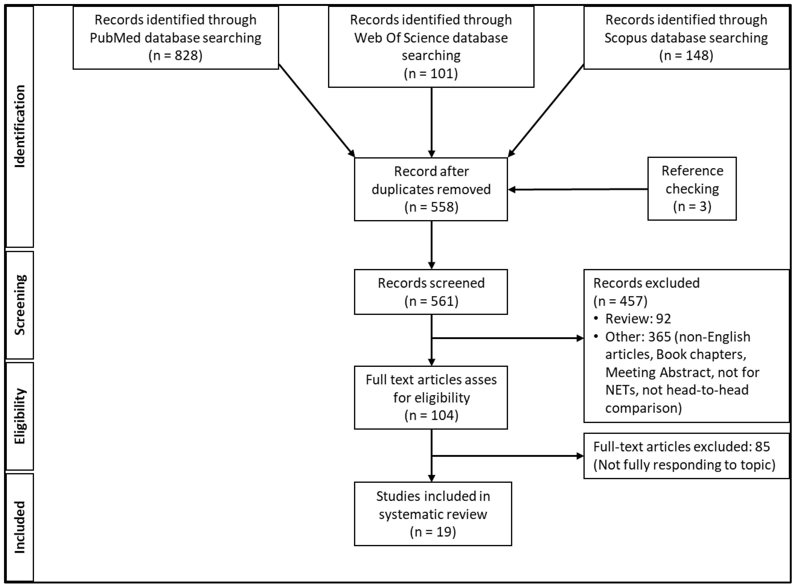
3.1. Literature Search Analysis

A total of 1077 articles were found. All duplicates were removed, leaving 558 records. Then, all reviews and all articles not entirely consistent with the inclusion criteria were excluded. The full texts of 104 articles were assessed for eligibility, and a further three articles emerged upon checking the reference lists. Finally, 19 articles were included (Figure 1). The quality of the selected articles, based on the CAPS for diagnostic studies, is reported in Supplementary Table S1.
As illustrated in the supplementary material, in many cases, the studies have not included a standard of reference, or different types of analyses were used (i.e., lesion-based, region-based, or patient-based), thus rendering difficult the comparison between or among the radiopharmaceutical agents. Moreover, in many cases, the impact of the imaging on the selected population was not clearly stated.
Table 1 reports the main characteristics of 19 selected articles.
Three papers aimed to assess the comparison between 111In-DTPA-Octreotide and 99mTc-EDDA/HYNIC-TOC [9,10,13]. In the study by Bangard et al. [9], the authors compared nine patients who underwent a scintigraph examination with both tracers and described different biokinetics between them. The uptake of 99mTc-EDDA-HYNIC-TOC was lower in the spleen and kidney than 111In-DTPA-Octreotide. However, lesion-based analysis, 111In-DTPA-Octreotide, detected more lesions, mainly in the liver, while 99mTc-EDDA-HYNIC-TOC identified more abdominal lesions. Conversely, in a head-to-head comparison, Decristoforo et al. [10] demonstrated, in 10 patients, that 99mTc-EDDA-HYNIC-TOC was simpler to produce and more detectable of lesions than 111In-DTPA-Octreotide, thus, opening the way for new alternative SPECT agents for the NET detection. Three years later, Gabriel et al. [13] concluded that 99mTc-EDDA-HYNIC-TOC scintigraphy is more performant than 111In-DTPA-Octreotide, mainly if the acquisition is made by an early and late acquisition (after 1–2 h from the tracer injection), in order to improve the tumor/background ratio.
Eleven out of 19 papers, including 498 patients, focused on the comparison between 111In-DTPA-Octreotide SPECT or SPECT/CT and 68Ga-DOTATOC PET or PET/CT. [11,12,14,15,16,17,18,19,20,22,26] Hofmann et al. [11] enrolled eight patients with metastatic carcinoid who underwent 68Ga-DOTATOC SPECT, CT, MRI, and 111In-DTPA-Octreotide, showing the power of PET tracer in detecting the lesions (100% vs. 85%, respectively for 68Ga-DOTATOC and 111In-DTPA-Octreotide). A similar and limited experience was reported by Kowalski et al. in four patients [12]. In this small patient population, the researchers found that PET was able to better detect small lesions with low-density SSTR expression. The studies by Buchmann et al. [14], Gabriel et al. [15], Srirajaskanthan et al. [17], Krausz et al. [18], Hofman et al. [19] demonstrated that 68Ga-DOTATOC/TATE PET/CT was able to detect more NET lesions than 111In-DTPA-Octreotide SPECT or SPECT/CT during patient-based and lesion-based analysis. In particular, 68Ga-DOTATOC/TATE PET/CT was able to better define the extension of metastatic disease in the liver, skeleton, and thoracic/abdominal lymph nodes. Moreover, based on the study by Krausz et al. [18], primary NET in the pancreas was more often detected by 68Ga-DOTATOC/TATE PET/CT than 111In-DTPA-Octreotide SPECT/CT, thus increasing its performance also in primary tumors and not only for metastatic disease. Additionally, in the papers by Buchmann et al. [14], Gabriel et al. [15], Srirajaskanthan et al. [17], and Hofman et al. [19], 68Ga-DOTATOC/TATE PET/CT was able to improve the clinical management in comparison with 111In-DTPA-Octreotide SPECT/CT. Indeed, based on the study by Buchmann et al. [14], surgical intervention was extended in seven patients owing to PET findings. Similarly, PET was able to change the therapeutic approach from a surgical to a systemic one after identifying more distant NET lesions (12/51 patients; 24%), in accordance with Gabriel et al. [15]. In the study by Srirajaskanthan et al. [17], the change of management with PET imaging was reported in 36/51 (70.6%) patients, mainly by providing the opportunity to undergo PRRT with 90Y/177Lu-DOTATATE/TOC. Finally, Hofman et al. [19] reported that PET imaging had a high management impact in 28/58 (47%) patients. Indeed, 68Ga-DOTATOC/TATE PET/CT increased the number of lesions detected; thus, many patients received systemic therapy rather than undergoing surgery.
Sadowski et al. [22] assessed the comparison between 68Ga-DOTATATE PET/CT and 111In-DTPA-Octreotide SPECT/CT in a cohort of patients affected by MEN1, demonstrating that 68Ga-DOTATATE PET/CT is more sensitive in detecting MEN1 lesions than SPECT imaging and it could also alter the management; therefore the authors strongly recommend to introduce this imaging modality in the diagnostic flow-chart of patients affected by MEN1 syndrome.
Finally, in the study by Van Binnebeek et al. [20] and Hope et al. [26] appeared, the term “tumor burden” relative to the extension of SSTR-positive disease. In both the studies, 68Ga-DOTATOC or 68Ga-DOTATATE was superior to SPECT imaging with 111In in assessing the tumor burden, mainly for the identification of small lesions detected by the PET scanner rather than by the SPECT one.
The articles focused on the comparison between 68Ga-DOTATOC/68Ga-DOTATATE and 99mTc-EDDA/HYNIC-TOC were 2 [23,25]. In the study by Madrzak et al. [23], 24 patients underwent both images with PET/CT and SPECT/CT. The authors found that 68Ga-DOTATOC PET/CT altered the treatment procedures in only 8.4% of patients (2 persons). However, due to the limited patient enrolment, the authors suggested additional studies to confirm this assumption. Therefore, one year later, Kunikowska et al. [25] enrolled 68 patients showing the advantages of 68Ga-DOTATATE PET/CT over 99mTc-EDDA/HYNIC-TOC in detecting NET lesions and underlined that 68Ga-DOTATATE PET/CT was able to change the clinical decision in one-third of patients.
Two articles (n = 64 patients) [24,27] assessed the comparison between 64Cu-DOTATATE and 68Ga-DOTATOC/TATE. Johnbeck et al. [24] enrolled 59 patients who underwent PET imaging with both tracers within 1 week. Through an intra-patient analysis, it emerged that PET images were concordant in 37 patients and discordant in 22 patients. Among this later subset of patients, most additional lesions were found by 64Cu-DOTATATE vs. 68Ga-DOTATOC/TATE (14 vs. eight patients, respectively). Although 64Cu-DOTATATE seems more performant in this study, in a very recent pilot analysis performed on five patients, Jha et al. [27] concluded that the data currently available was not conclusive about the superiority of one over the other.
Finally, the residual paper was a comparative analysis between 111In-DTPA-Octreotide and 64Cu-DOTATATE [21]. This was a large experience in 112 patients demonstrating that, similar to 68Ga radiolabeled peptides, 64Cu-DOTATATE PET is superior to 111In-DTPA-Octreotide SPECT.

3.2. Comparative Perfomances

For SPECT radiopharmaceuticals, the rate of true positive was 63.7% and 58.5%, respectively, for 111In-DTPA-Octreotide and 99mTc-EDDA/HYNIC-TOC as expressed in studies by Gabriel et al. [13] Srirajaskanthan et al. [17], Krausz et al. [18], Pfeifer et al. [21] and Madrzak et al. [23] and illustrated in Table 2.
Conversely, for PET radiopharmaceuticals, the rate of true positivity was 78.4% and 82.4%, respectively, for 68Ga-DOTATOC and 64Cu-DOTATATE., as demonstrated by Gabriel et al. [15], Srirajaskanthan et al. [17], Krausz et al. [18], Madrzak et al. [23], and Johnbeck et al. [24] (Table 2). As clearly shown in Table 2, the number of patients with false negative results was higher for SPECT radiopharmaceuticals rather than for PET ones.
Indeed, as is visible from Table 3, the sensitivity was, as expected, higher for PET radiopharmaceuticals either with 68Ga and 64Cu than for SPECT agents.
However, by comparing 111In-DTPA-Octreotide and 99mTc-EDDA/HYNIC-TOC, the latter has a higher sensitivity, although a lower specificity. Until now, only limited data are available on the comparison between 68Ga-DOTATOC/TATE and 64Cu-DOTATATE. The study by Johnbeck et al. [24] found a slightly higher sensitivity and specificity for 64Cu-DOTATATE when compared to 68Ga-DOTATOC. However, the data are still limited for final evidence.
For lesion-based and site-based analyses, radiopharmaceuticals for PET imaging were more performant than SPECT agents in identifying the number and the presence of lesions in the musculoskeletal system, bone, liver, and lymph nodes (mainly in the abdominal region).
The paper by Mussig et al. [16] analyzed the association between the expression of sst2 and the uptake of 68Ga-DOTATOC and 111In-DTPA-Octreotide. The authors found that a positive scan with both the tracers was associated with a high expression of sst2; however, tumors without immunohistochemical sst2 expression could show 68Ga-DOTATOC tracer uptake, probably due to the expression of sst3 or sst5, or simply because of the tumor heterogeneity.

3.3. The Theragnostic Role of Radiopharmaceuticals for NET

The theragnostic role of the abovementioned radiopharmaceuticals in the different settings of disease (detection, staging, status of SSTR, and follow-up) has scarcely been reported in comparative studies.
From a careful analysis of the available data, 56 (7.8%) patients were enrolled for the detection of NET, 120 (16.9%) for staging, and 535 (75.3%) for the assessment of SSTR expression and follow-up. As expected, follow-ups for the evaluation of SSTR expression were the most common indication in many selected studies. Higher diagnostic performances have been reported for the assessment of SSTR status in the follow-up settings for PET tracers as compared to SPECT tracers, for patient-based analysis but also regional- and lesion-based ones. After different previous treatments, the assessment of SSTR expression is essential in planning PRRT, and the PET tracer results were more accurate in these settings at any level of analysis. However, no information has been found about PET and SPECT tracers in monitoring the response to PRRT in NETs, although it would be an interesting and important topic from a diversified point of view.

4. Discussion and Conclusions

Scintigraphy with radiolabeled SSTR has gained widespread acceptance as the imaging method of choice in NET patients, showing high sensitivity and good specificity, as emerged from the previous study and from this systematic review. From planar imaging with 111In-DTPA-Octreotide to SPECT with 99mTc-HYNIC/EDDA-TOC, a gain in terms of detection has been obtained. However, due to the limited spatial resolution of SPECT imaging, PET tracers radiolabeled with either 64Cu or 68Ga have been introduced in clinical practice, thus increasing the detection of NET lesions.
From the present comparative review, it emerged that for patient-based analysis, the rate of true positive and the diagnostic performance is as expected, higher for PET tracers when compared to SPECT tracers, mainly when 111In-DTPA-Octreotide was compared to 68Ga-DOTATOC/TATE/NOC. Moreover, when analyzing the available data for lesion- and region-based analyses, we found that PET radiopharmaceutical agents were more performant in detecting bone, lymph nodes, and liver metastases than SPECT agents. This advantage was mainly due to the PET scanner technology rather than the radiopharmaceutical itself. It would be interesting to understand if new technological achievements in SPECT technology, such as solid-state detectors and 360° detector coverage, could fill this gap. Indeed, PET tomographic images can significantly improve the detection of deep lesions or visceral metastases when compared with planar or SPECT images.
By a comparative analysis between 64Cu-DOTATATE and 68Ga-DOTATOC emerged that the performances were quite similar; however, the number of true positive lesions was slightly higher for 64Cu-DOTATATE than 68Ga-DOTATOC (33 vs. 7), as reported by Johnbeck et al. [24] The affinity for SSTR was quite similar for all the imaging agents, particularly for those used in PET, as recently reported by some authors [28,29,30]. Nevertheless, the intrinsic physical characteristics of radioisotopes can have an important effect on lesion detectability. Indeed, 64Cu has a shorter positron range than 68Ga, thus possibly improving the detection rate of small lesions. Moreover, the radiation burden is different between 64Cu and 68Ga. Similarly, 99mTc has the advantage of a lower radiation dose than 111In. This latter physical characteristic can be translated into the advantageous use of 99mTc for repeated investigations, for example, in monitoring the response to PRRT or in children. However, to date, no information about the cost-saving, other than the radioprotection information, is available for 99mTc-EDDA/HYNIC-TOC SPECT in comparison to 68Ga-DOTATOC/TATE PET. Conversely, Schreiter et al. [31] found that 68Ga-DOTATOC PET/CT was considerably cheaper than 111In-DTPA-octreotide with respect to both material and personnel costs. Therefore, additional cost analyses are welcome also for the other agents.
It should be noted, however, that comparisons between PET and SPECT agents were made considering different acquisition protocols. Table 4 reports some of the pros and cons of SPECT and PET imaging for detecting NETs.
The question that arises from the above considerations is, “Can the improved detection rate affect clinical management?”
In NETs, changes in treatment strategy are nearly always based on clinical or imaging-based signs of progression. Thus, high performance in the detection of any new lesions is of great value in patients affected by these rare diseases. For example, the additional evidence of bone metastases can have either an important effect on the therapeutic intervention or a prognostic implication because unknown distant metastases are considered a negative prognostic factor, possibly requiring a more aggressive treatment regimen [32,33]. Based on the available data, the inclusion of PET imaging in clinical practice impacted the change of management from 3.7% to 70.6% (Table S2) of patients. Therefore, PET imaging should be preferred to SPECT imaging when available.
From a careful analysis of the selected studies, no comparative data were available about the role of PET and SPECT imaging in monitoring the response to PRRT. The recent introduction of PRRT in clinical practice (Netter 1 trial) and the opportunity of monitoring the response to therapy, both in the interim and at the end, is essential for testing the efficacy of therapy. To date, some studies have been published about the role of 68Ga-DOTATOC/TATE/NOC in monitoring the response to PRRT in comparison to morphological criteria without reporting conclusions [34,35]. Indeed, functional imaging is not yet accepted as a substitute for morphological imaging as a means to assess tumor response to treatment [36]. However, the opportunity to use both SPECT (especially with new scanners) and PET tracers during and after PRRT would also be an advantage in the case of retreatment or an early treatment interruption. Future studies should be conducted to test these hypotheses.
In this systematic review, we focused our attention on SPECT/PET radiotracers based on SSTR analogs, though theoretically, other PET tracers can be used for NETs imaging.
One alternative is represented by 11C-hydroxytriptophan (11C-5-HTP), a serotonin precursor that allows the evaluation of the serotonin pathway, which is one of the active metabolic pathways in NETs [37]. This tracer has a high sensitivity, especially for pancreatic NETS, but its use is limited by the half-life of the radionuclide (20 min), which requires the presence of an on-site cyclotron [3,37].
18F-DOPA (18F-L-dihydroxyphenylalanine) is another PET tracer that finds a high application in NETs [37]. Indeed, NETs cells can often take up decarboxylate monoamine precursors, such as DOPA. 18F-DOPA seems to be useful for imaging well-differentiated midgut tumors, though they often overexpress sst2 [38].
In a recent meta-analysis of head-to-head comparative studies emerged that at patient-based and region-based analysis, 68Ga-DOTA-peptides performed better than 18F-DOPA PET in detecting intestinal NETs, but at lesion-based analysis, 18F-DOPA PET was more accurate [38].
Another alternative to peptide analogs is represented by 18F-FDG (18F-fluorodeoxyglucose), which is recently becoming the PET tracer of choice in many cancer forms. 18F-FDG exploits cancer cells’ preferential utilization of aerobic glycolysis. Similarly, to glucose, it enters cells via glucose transporters GLUT-1 and GLUT-3, but it doesn’t follow the same metabolic pathway of glucose due to a lack of an oxygen atom in its C2 position. Thus, it accumulates in cells proportional to their glucose consumption. [37] However, for many years it was not used in NETs due to its low sensitivity in the detection of these tumors, but more recently, the utility of FDG-PET scans has been reassessed [1,3]. NETs with poor differentiation, a high grade, and rapid proliferation have a decreased expression of SSTR expression; thus, scans with peptide analogs may be negative, while 18F-FDG imaging may be positive [1,39]. Liu et al. [39] analyzed 30 studies focused on 68Ga-radiolabelled agonist SSTR and FDG PET/CT in NET patients. From the meta-analysis emerged that 18F-FDG PET/CT has the lowest sensitivity in detecting NET lesions. However, it has a complementary role in the case of moderately or scarcely differentiated NETs.
The abovementioned radiopharmaceuticals have shown promising results, but these substances do not provide any theragnostic options, unlike somatostatin analogs.
Lastly, new interest is increasing in the use of SSTR antagonists. Compared to agonists, they showed better pharmacokinetics and image contrast, a higher tumor uptake, and a better residence time [37], [40]. Among them, 68Ga-NOGADA-JR11 (or 68Ga-OPS202) and 68Ga-DOTA-JR11 have also demonstrated advantages for potential theragnostic application [40,41,42]. In particular, the study by Zhu et al. [40] showed that in 12 patients undergoing imaging with both radiopharmaceutical agents on two consecutive days, 68Ga-DOTA-JR11 outperformed better that 68Ga-DOTATATE in detecting liver metastases, while 68Ga-DOTATATE was better for the identification of bone lesions. However, to date, little clinical evidence is still available.
The present systematic review has limitations. The limited number of studies comparing 64Cu-DOTATATE vs. 68Ga-DOTATATE. Moreover, in the study by Pfeifer et al. [21], 64Cu-DOTATATE PET/CT was compared with 68Ga-DOTATOC PET, therefore by using a hybrid vs. non-hybrid system, thus reducing the detection power of the second imaging modality. In the study by Buchman et al. [14], the authors reported that the region-based analysis could have overestimated the sensitivity of 111In-DTPA-octreotide. Few data about the standard of reference, as also emerged by the CAPS evaluation; indeed, it missed 10/19 (53%) of papers, thus reducing the opportunity to perform an adequate comparison in terms of diagnostic performances.
In conclusion, PET imaging, as expected, is more suitable for the identification of NET. Indeed, they demonstrated a higher true positive rate than SPECT imaging. However, the availability of new SPECT scanners, more favorable radioprotection and synthetical characteristics (mainly for 99mTc), and the consolidated experiences for conventional scintigraphy examination should be considered in the diagnostic and therapeutic path, also for health equity.

Supplementary Materials

The following supporting information can be downloaded at: https://www.mdpi.com/article/10.3390/cimb44110373/s1, Table S1: Clinical studies assessment using Critical Appraisal Skill Progam (CASP) checklist for diagnostic studies; Table S2: Change of management with PET imaging on patients affected by the neuroendocrine tumor.

Author Contributions

Conceptualization, L.E. and G.P.; methodology, L.E.; validation, D.C., L.F., S.S. and N.R.; formal analysis, L.E.; investigation, L.E.: and G.P.; resources, L.E. and G.P.; data curation, L.E., and L.F.; writing—original draft preparation, L.E., and G.P.; writing—review and editing, L.F. and D.C.; visualization, N.R.; supervision, S.S. All authors have read and agreed to the published version of the manuscript.

Funding

This research received no external funding.

Institutional Review Board Statement

Not applicable.

Informed Consent Statement

Not applicable.

Data Availability Statement

Not applicable.

Conflicts of Interest

D.C. received research and travel grants from Life Molecular Imaging and General Electric and travel grants from Curium, but they were unrelated to the content of the present review. The other authors do not declare any conflict of interest.

References

  1. Desai, H.; Borges-Neto, S.; Wong, T.Z. Molecular Imaging and Therapy for Neuroendocrine Tumors. Curr. Treat. Options Oncol. 2019, 20, 78. [Google Scholar] [CrossRef] [PubMed]
  2. Werner, R.; Bluemel, C.; Lassmann, M.; Kudlich, T.; Higuchi, T.; Lopci, E.; Allen-Auerbach, M.; Colletti, P.M.; Rubello, D.; Zatelli, M.C.; et al. SPECT- and PET-Based Patient-Tailored Treatment in Neuroendocrine Tumors. Clin. Nucl. Med. 2015, 40, e271–e277. [Google Scholar] [CrossRef] [PubMed]
  3. Kjaer, A.; Knigge, U. Use of radioactive substances in diagnosis and treatment of neuroendocrine tumors. Scand. J. Gastroenterol. 2015, 50, 740–747. [Google Scholar] [CrossRef] [PubMed] [Green Version]
  4. Dearling, J.L.J.; van Dam, E.M.; Harris, M.J.; Packard, A.B. Detection and therapy of neuroblastoma minimal residual disease using [64/67Cu]Cu-SARTATE in a preclinical model of hepatic metastases. EJNMMI Res. 2021, 11, 20. [Google Scholar] [CrossRef] [PubMed]
  5. Cullinane, C.; Jeffery, C.M.; Roselt, P.D.; van Dam, E.M.; Jackson, S.; Kuan, K.; Jackson, P.; Binns, D.; van Zuylekom, J.; Harris, M.J.; et al. Peptide receptor radionuclide therapy with 67Cu-CuSarTATE is highly efficacious against a somatostatin-positive neuroendocrine tumor model. J. Nucl. Med. 2020, 61, 1800–1805. [Google Scholar] [CrossRef] [PubMed]
  6. Hicks, R.J.; Jackson, P.; Kong, G.; Ware, R.E.; Hofman, M.S.; Pattison, D.A.; Akhurst, T.A.; Drummond, E.; Roselt, P.; Callahan, J.; et al. 64Cu-sartaTE PET imaging of patients with neuroendocrine tumors demonstrates high tumor uptake and retention, potentially allowing prospective dosimetry for peptide receptor radionuclide therapy. J. Nucl. Med. 2019, 60, 777–785. [Google Scholar] [CrossRef] [Green Version]
  7. Cives, M.; Strosberg, J. Radionuclide Therapy for Neuroendocrine Tumors. Curr. Oncol. Rep. 2017, 19, 9. [Google Scholar] [CrossRef]
  8. Bodei, L.; Cremonesi, M.; Paganelli, G. Yttrium-based therapy for neuroendocrine tumors. PET Clin. 2014, 9, 71–82. [Google Scholar] [CrossRef]
  9. Bangard, M.; Behe, M.; Guhlke, S.; Otte, R.; Bender, H.; Maecke, H.R.; Biersack, H.J. Detection of somatostatin receptor-positive tumours first results in patients and comparison with 111In-DTPA-D-Phe1-octreotide. Eur. J. Nucl. Med. 2000, 27, 628–637. [Google Scholar] [CrossRef]
  10. Decristoforo, C.; Mather, S.J.; Cholewinski, W.; Donnemiller, E.; Riccabona, G.; Moncayo, R. Original article a new 99m Tc-labelled radiopharmaceutical for imaging somatostatin receptor-positive tumours: First clinical results and intra-patient comparison with 111In-labelled octreotide derivatives. Eur. J. Nucl. Med. 2000, 27, 1318–1325. [Google Scholar] [CrossRef]
  11. Hofmann, M.; Maecke, H.; Börner, A.; Weckesser, E.; Schöffski, P.; Oei, M.; Schumacher, J.; Henze, M.; Heppeler, A.; Meyer, J.; et al. Biokinetics and imaging with the somatostatin receptor PET radioligand 68Ga-DOTATOC: Preliminary data. Eur. J. Nucl. Med. 2001, 28, 1751–1757. [Google Scholar] [CrossRef] [PubMed]
  12. Kowalski, J.; Henze, M.; Schuhmacher, J.; Mäcke, H.R.; Hofmann, M.; Haberkorn, U. Evaluation of positron emission tomography imaging using [68Ga]-DOTA-D Phe1-Tyr3- octreotidein comparison to [111In]-DTPAOC SPECT. First results in patients with neuroendocrine tumors. Mol. Imaging Biol. 2003, 5, 42–48. [Google Scholar] [CrossRef]
  13. Gabriel, M.; Decristoforo, C.; Donnemiller, E.; Ulmer, H.; Rychlinski, C.W.; Mather, S.J.; Moncayo, R. An intrapatient comparison of 99mTc-EDDA/HYNIC-TOC with 111In-DTPA-octreotide for diagnosis of somatostatin receptor-expressing tumors. J. Nucl. Med. 2003, 44, 708–716. [Google Scholar]
  14. Buchmann, I.; Henze, M.; Engelbrecht, S.; Eisenhut, M.; Runz, A.; Schäfer, M.; Schilling, T.; Haufe, S.; Herrmann, T.; Haberkorn, U. Comparison of 68Ga-DOTATOC PET and 111In-DTPAOC (Octreoscan) SPECT in patients with neuroendocrine tumours. Eur. J. Nucl. Med. Mol. Imaging 2007, 34, 1617–1626. [Google Scholar] [CrossRef] [PubMed]
  15. Gabriel, M.; Decristoforo, C.; Kendler, D.; Dobrozemsky, G.; Heute, D.; Uprimny, C.; Kovacs, P.; von Guggenberg, E.; Bale, R.; Virgolini, I.J. 68Ga-DOTA-Tyr3-octreotide PET in neuroendocrine tumors: Comparison with somatostatin receptor scintigraphy and CT. J. Nucl. Med. 2007, 48, 508–518. [Google Scholar] [CrossRef] [PubMed] [Green Version]
  16. Müssig, K.; Öksüz, M.; Dudziak, K.; Ueberberg, B.; Wehrmann, M.; Horger, M.; Schulz, S.; Häring, H.; Pfannenberg, C.; Bares, R.; et al. Association of somatostatin receptor 2 immunohistochemical expression with [111In]-DTPA octreotide scintigraphy and [68Ga]-DOTATOC PET/CT in neuroendocrine tumors. Horm. Metab. Res. 2010, 42, 599–606. [Google Scholar] [CrossRef] [PubMed]
  17. Srirajaskanthan, R.; Kayani, I.; Quigley, A.M.; Soh, J.; Caplin, M.E.; Bomanji, J. The role of 68Ga-DOTATATE PET in patients with neuroendocrine tumors and negative or equivocal findings on 111In-DTPA-octreotide scintigraphy. J. Nucl. Med. 2010, 51, 875–882. [Google Scholar] [CrossRef] [Green Version]
  18. Krausz, Y.; Freedman, N.; Rubinstein, R.; Lavie, E.; Orevi, M.; Tshori, S.; Salmon, A.; Glaser, B.; Chisin, R.; Mishani, E.; et al. 68Ga-DOTA-NOC PET/CT imaging of neuroendocrine tumors: Comparison with 111In-DTPA-octreotide (OctreoScan®). Mol. Imaging Biol. 2011, 13, 583–593. [Google Scholar] [CrossRef]
  19. Hofman, M.S.; Kong, G.; Neels, O.C.; Eu, P.; Hong, E.; Hicks, R.J. High management impact of Ga-68 DOTATATE (GaTate) PET/CT for imaging neuroendocrine and other somatostatin expressing tumours. J. Med. Imaging Radiat. Oncol. 2012, 56, 40–47. [Google Scholar] [CrossRef]
  20. Van Binnebeek, S.; Vanbilloen, B.; Baete, K.; Terwinghe, C.; Koole, M.; Mottaghy, F.M.; Clement, P.M.; Mortelmans, L.; Bogaerts, K.; Haustermans, K.; et al. Comparison of diagnostic accuracy of 111In-pentetreotide SPECT and 68Ga-DOTATOC PET/CT: A lesion-by-lesion analysis in patients with metastatic neuroendocrine tumours. Eur. Radiol. 2016, 26, 900–909. [Google Scholar] [CrossRef]
  21. Pfeifer, A.; Knigge, U.; Binderup, T.; Mortensen, J.; Oturai, P.; Loft, A.; Berthelsen, A.K.; Langer, S.W.; Rasmussen, P.; Elema, D.; et al. 64Cu-DOTATATE PET for neuroendocrine tumors: A prospective head-to-head comparison with 111In-DTPA-octreotide in 112 patients. J. Nucl. Med. 2015, 56, 847–854. [Google Scholar] [CrossRef] [PubMed]
  22. Sadowski, S.M.; Millo, C.; Cottle-Delisle, C.; Merkel, R.; Yang, L.A.; Herscovitch, P.; Pacak, K.; Simonds, W.F.; Marx, S.J.; Kebebew, E. Results of 68Gallium-DOTATATE PET/CT Scanning in Patients with Multiple Endocrine Neoplasia Type 1. J. Am. Coll. Surg. 2015, 221, 509–517. [Google Scholar] [CrossRef] [PubMed] [Green Version]
  23. Madrzak, D.; Mikołajczak, R.; Kaminski, G. Influence of PET/CT 68Ga somatostatin receptor imaging on proceeding with patients, who were previously diagnosed with 99mTc-EDDA/HYNIC-TOC SPECT. Nucl. Med. Rev. 2016, 19, 88–92. [Google Scholar] [CrossRef] [PubMed] [Green Version]
  24. Johnbeck, C.B.; Knigge, U.; Loft, A.; Berthelsen, A.K.; Mortensen, J.; Oturai, P.; Langer, S.W.; Elema, D.R.; Kjaer, A. Head-to-Head Comparison of 64Cu-DOTATATE and 68Ga-DOTATOC PET/CT: A prospective study of 59 patients with neuroendocrine tumors. J. Nucl. Med. 2017, 58, 451–457. [Google Scholar] [CrossRef] [Green Version]
  25. Kunikowska, J.; Lewington, V.; Krolicki, L. Optimizing somatostatin receptor imaging in patients with neuroendocrine tumors: The impact of 99mTc-HYNICTOC SPECT/SPECT/CT versus 68Ga-DOTATATE PET/CT upon clinical management. Clin. Nucl. Med. 2017, 42, 905–911. [Google Scholar] [CrossRef]
  26. Hope, T.A.; Calais, J.; Zhang, L.; Dieckmann, W.; Millo, C. 111In-pentetreotide scintigraphy versus 68Ga-DOTATATE PET: Impact on krenning scores and effect of tumor burden. J. Nucl. Med. 2019, 60, 1266–1269. [Google Scholar] [CrossRef] [Green Version]
  27. Jha, A.; Patel, M.; Carrasquillo, J.A.; Chen, C.C.; Millo, C.; Maass-Moreno, R.; Ling, A.; Lin, F.I.; Lechan, R.M.; Hope, T.A.; et al. Chice is a ggod time: The emergence of [64Cu]Cu-DOTATATE based somatostatin receptor imaging in the era of [68Ga]Ga-DOTATATE. J. Nucl. Med. 2022, 63, 1300–1301. [Google Scholar] [CrossRef]
  28. Johnbeck, C.B.; Knigge, U.; Kjær, A. PET tracers for somatostatin receptor imaging of neuroendocrine tumors: Current status and review of the literature. Futur. Oncol. 2014, 10, 2259–2277. [Google Scholar] [CrossRef] [Green Version]
  29. Treglia, G.; Castaldi, P.; Rindi, G.; Giordano, A.; Rufini, V. Diagnostic performance of Gallium-68 somatostatin receptor PET and PET / CT in patients with thoracic and gastroenteropancreatic neuroendocrine tumours: A meta-analysis. Endocrine 2012, 42, 80–87. [Google Scholar] [CrossRef]
  30. Wild, D.; Bomanji, J.B.; Benkert, P.; Maecke, H.; Ell, P.J.; Reubi, J.C.; Caplin, M.E. Comparison of 68Ga-DOTANOC and 68Ga-DOTATATE PET/CT within patients with gastroenteropancreatic neuroendocrine tumors. J. Nucl. Med. 2013, 54, 364–372. [Google Scholar] [CrossRef] [Green Version]
  31. Schreiter, N.F.; Brenner, W.; Nogami, M.; Buchert, R.; Huppertz, A.; Pape, U.-F.; Prasad, V.; Hamm, B.; Maurer, M.H. Cost comparison of 111In-DTPA-octreotide scintigraphy and 68Ga-DOTATOC PET/CT for staging enteropancreatic neuroendocrine tumours. Eur. J. Nucl. Med. Mol. Imaging 2012, 39, 72–82. [Google Scholar] [CrossRef] [PubMed]
  32. Panzuto, F.; Nasoni, S.; Falconi, M.; Corleto, V.D.; Capurso, G.; Cassetta, S.; Di Fonzo, M.; Tornatore, V.; Milione, M.; Angeletti, S.; et al. Prognostic factors and survival in endocrine tumor patients: Comparison between gastrointestinal and pancreatic localization. Endocr. Relat. Cancer 2005, 12, 1083–1092. [Google Scholar] [CrossRef] [PubMed] [Green Version]
  33. Gupta, S.; Johnson, M.M.; Murthy, R.; Ahrar, K.; Wallace, M.J.; Madoff, D.C.; McRae, S.E.; Hicks, M.E.; Rao, S.; Vauthey, J.-N.; et al. Hepatic Arterial Embolization and Chemoembolization for the Treatment of Patients with Metastatic. Cancer 2005, 104, 1590–1602. [Google Scholar] [CrossRef] [PubMed]
  34. Vandecaveye, V.; van Binnebeek, S.; Mottaghy, F.M.; van Cutsem, E. Early Whole-Body Diffusion-weighted MRI Helps Predict Long-term Outcome Following Peptide Receptor Radionuclide Therapy for Metastatic Neuroendocrine. Radiol. Imaging Cancer 2022, 1, 1–12. [Google Scholar] [CrossRef]
  35. Pauwels, E.; Van Binnebeek, S.; Vandecaveye, V.; Baete, K.; Vanbilloen, H.; Koole, M.; Mottaghy, F.M.; Haustermans, K.; Clement, P.M.; Nackaerts, K.; et al. Inflammation-Base Index and 68Ga-DOTATOC PET-Derived Uptake and Volumetric Parameters Predict Outcome in Neuroendocrine Tumor Patients Treated with 90Y-DOTATOC. J. Nucl. Med. 2020, 61, 1014–1020. [Google Scholar] [CrossRef]
  36. Zaknun, J.J.; Bodei, L. The joint IAEA, EANM, and SNMMI practical guidance on peptide receptor radionuclide therapy (PRRNT) in neuroendocrine tumours. Eur. J. Nucl. Med. Mol. Imaging 2013, 40, 800–816. [Google Scholar] [CrossRef] [Green Version]
  37. Abrantes, A.M.; Pires, A.S.; Monteiro, L.; Teixo, R.; Neves, A.R.; Tavares, N.T.; Marques, I.A.; Botelho, M.F. Tumour functional imaging by PET. Biochim. Biophys. Acta-Mol. Basis Dis. 2020, 1866, 165717. [Google Scholar] [CrossRef]
  38. Piccardo, A.; Fiz, F.; Bottoni, G.; Ugolini, M.; Noordzij, W.; Trimboli, P. Head-to-head comparison between 18F-DOPA PET/CT and 68Ga-DOTA peptides PET/CT in detecting intestinal neuroendocrine tumours: A systematic review and meta-analysis. Clin. Endocrinol. 2021, 95, 595–605. [Google Scholar] [CrossRef]
  39. Liu, X.; Li, N.; Jiang, T.; Xu, H.; Ran, Q.; Shu, Z.; Wu, J.; Li, Y.; Zhou, S.; Zhang, B. Comparison of gallium-68 somatostatin receptor and 18 F-fluorodeoxyglucose positron emission tomography in the diagnosis of neuroendocrine tumours: A systematic review and meta-analysis. Hell. J. Nucl. Med. 2020, 23, 188–200. [Google Scholar]
  40. Zhu, W.; Cheng, Y.; Wang, X.; Yao, S.; Bai, C.; Zhao, H.; Jia, R.; Xu, J.; Huo, L. Head-to-head comparison of 68Ga-DOTA-JR11 and 68Ga-DOTATATE PET/CT in patients with metastatic, well-differentiated neuroendocrine tumors: A prospective study. J. Nucl. Med. 2020, 61, 897–903. [Google Scholar] [CrossRef]
  41. Nicolas, G.P.; Schreiter, N.; Kaul, F.; Uiters, J.; Bouterfa, H.; Kaufmann, J.; Erlanger, T.E.; Cathomas, R.; Christ, E.; Fani, M.; et al. Sensitivity comparison of 68Ga-OPS202 and 68Ga-DOTATOC PET/CT in patients with gastroenteropancreatic neuroendocrine tumors: A prospective phase II imaging study. J. Nucl. Med. 2018, 59, 915–921. [Google Scholar] [CrossRef] [PubMed] [Green Version]
  42. Mansi, R.; Fani, M. Design and development of the theranostic pair 177Lu-OPS201/68Ga-OPS202 for targeting somatostatin receptor expressing tumors. J. Label. Compd. Radiopharm. 2019, 62, 635–645. [Google Scholar] [CrossRef] [PubMed]
Figure 1. Scheme of record selection.
Cimb 44 00373 g001
Table 1. Characteristics of the selected studies.
Author, RefYear of PubCountryN ptsComparative RFSORInterpretationOutcome
1Bangard et al. [9]2000Germany9111In-DTPA-OC vs.
99mTc-EDDA/HYNIC-TOC
NoneVisual ans semiquantitative analysisBoth tracers outperform similarly for the detection of tumors, although 99mTc-EDDA/HYNIC-TOC detects more abdominal lesions and 111In-DTPA-OC more liver metastases
2Decristoforo et al. [10]2000Austria10111In-DTPA-Octreotide/111In-DTPA-TOC vs.
99mTc-EDDA/HYNIC-TOC
NoneVisual ans semiquantitative analysisBoth tracers outperform similarly for the detection of tumor
3Hofmann et al.
[11]
2001Germany8111In-DTPA-Octreotide vs.
68Ga-DOTATOC
NoneVisual analysis by three observers68Ga-DOTATOC outperformed 111In-DTPA-Octretide detecting more lesions
4Kowalski et al.
[12]
2003Germany4111In-DTPA-Octreotide vs.
68Ga-DOTATOC
NoneVisual analysis68Ga-DOTATOC outperformed 111In-DTPA-Octretide detecting more lesions
5Gabriel et al.
[13]
2003Austria41111In-DTPA-Octreotide vs.
99mTc-EDDA/HYNIC-TOC.
ImagingVisual analysis by two observers99mTc- EDDA/HYNIC- TOC outperformed 111In-DTPA-Octretide detecting more lesions
6Buchmann et al.
[14]
2007Germany27111In-DTPA-Octreotide vs.
68Ga-DOTATOC
Histology and imagingVisual analysis by two observers68Ga-DOTATOC outperformed 111In-DTPA-Octretide detecting more lesions and changing the management
7Gabriel et al.
[15]
2007Austria84111In-DTPA-Octreotide vs.
68Ga-DOTATOC
Histology and imagingVisual analysis by two observers (third in discordant case)68Ga-DOTATOC outperformed 111In-DTPA-Octretide detecting more lesions and changing the management
8Mussig K et al.
[16]
2010USA36111In-DTPA-Octreotide vs.
68Ga-DOTATOC
Histology Visual analysis by two observers111In-DTPA-Octreotide and
68Ga-DOTATOC both correlate with SSTR expression
9Srirajaskanthan et al. [17]2010UK51111In-DTPA-Octreotide vs.
68Ga-DOTATATE
NoneVisual analysis68Ga-DOTATATE outperformed 111In-DTPA-Octretide detecting more lesions and changing the management
10Krausz et al.
[18]
2011Israel19111In-DTPA-Octreotide vs.
68Ga-DOTANOC
NoneVisual analysis68Ga-DOTANOC outperformed 111In-DTPA-Octretide detecting more lesions and changing the management
11Hofman et al. [19]2012Australia40111In-DTPA-Octretide vs.
68Ga-DOTATATE
NoneVisual analysis68Ga-DOTATATE outperformed 111In-DTPA-Octretide detecting more lesions
12Van Binnebeek et al. [20]2016Belgium53111In-DTPA-Octreotide vs.
68Ga-DOTATOC
ImagingVisual analysis68Ga-DOTATOC outperformed 111In-DTPA-Octretide detecting more lesions
13Pfeifer et al.
[21]
2015Denmark112111In-DTPA-Octretide vs.
64Cu-DOTATATE
Histology and imagingVisual analysis by two observers64Cu-DOTATATE outperformed 111In-DTPA-Octretide detecting more lesions
14Sadowski et al. [22]2015Austria26111In-DTPA-Octreotide vs.
68Ga-DOTATOC
Histology and imagingVisual analysis68Ga-DOTATOC outperformed 111In-DTPA-Octretide detecting more lesions without changing the management
15Madrzak et al.
[23]
2016Poland2499mTc-EDDA/HYNIC-TOC vs.
68Ga-DOTATOC/TATE
Not clearNo data68Ga-DOTATOC outperformed 99mTc-EDDA/HYNIC-TOC by detecting more lesions and by changing the management
16Johnbeck et al.
[24]
2017Denmark5964Cu-DOTATATE vs.
68Ga-DOTATOC
Clinical follow-upVisual analysis by one observer64Cu-DOTATATE outperformed 68Ga-DOTATOC detecting more lesions
17Kunikowska et al. [25]2017Poland6899mTc-HYNIC-TOC vs.
68Ga-DOTATATE
ImagingVisual analysis68Ga-DOTATATE outperformed 99Tc-HYNIC-TOC detecting more lesions, and by changing the management
18Hope et al. [26]2019USA150111In-DTPA-Octreotide vs.
68Ga-DOTATOC
NoneVisual and semiquantitative analysis68Ga-DOTATOC outperformed 111In-DTPA-Octreotide in higher Krenning score lesions (mainly for size < 2 cm
19Jha et al.
[27]
2022USA564Cu-DOTATATE vs.
68Ga-DOTATATE
NoneVisual analysisBoth 64Cu-DOTATATE and
68Ga-DOTATATE can be interchangeably
SOR = standard of reference; SSA: somatostatin analogs
Table 2. Patient-based performances of the diverse radiopharmaceutical agents.
Author, RefN pts111In-DTPA
Octreotide
99mTc-EDDA/HYNIC-TOC68Ga-DOTATATE/NOC/TOC64Cu-DOTATATE
TPTNFPFNTPTNFPFNTPTNFPFNTPTNFPFN
1Gabriel et al. [13]4121411527239--------
2Gabriel et al. [15]843712134********691212----
3Srirajaskanthan et al. [17]51153132----42315----
4Krausz et al. [18]1919000----19000----
5Pfeifer et al. [21]1128712013--------971203
6Madrzak et al.
[23]
24----111111121200----
7Johnbeck et al. * [24]59--------43915431410
* Follow-up was unverified in 1 patient; ** Both 111In-DTPA-Octreotide and 99mTc-HYNIC-TOC for SPECT imaging.
Table 3. Diagnostic accuracies of some selected papers.
Author, Ref111In-DTPA-Octreotide99mTc-EDDA/HYNIC-TOC68Ga-DOTATATE/NOC/TOC64Cu-DOTATATE
Sens
(95% CI)
Spec (95% CI)Acc (95% CI)Sens
(95% CI)
Spec (95% CI)Acc (95% CI)Sens
(95% CI)
Spec (95% CI)Acc (95% CI)Sens
(95% CI)
Spec (95% CI)Acc (95% CI)
1Gabriel et al.
[13]
58.3 (42–73)80
(64–90)
6175 (58–87)40
(25–56)
70.7------
2Gabriel et al.
[15]
52.1 (41–63)92.3 (84–97)58.3******97.2 (90–99)92.3 (84–97)96.4---
3Srirajaskanthan et al. [17]31.9 (20–46)75
(61–86)
35---89.4 (77–96)75
(61–86)
88---
4Pfeifer et al.
[21]
87 (79–92)100 (96–100)88.4------97
(91–99)
100 (96–100)97.3
5Madrzak et al.
[23]
---91.7 (72–99)91.7 (72–99)91.7100 (83–100)100 (83–100)100---
6Johnbeck et al. *
[24]
------89.6 (78–96)90 (79–96)89.7100 (92–100)93
(83–98)
98.3
* Follow-up was unverified in 1 patient; ** Both 111In-DTPA-Octreotide and 99mTc-Hynic-TOC for SPECT imaging.
Table 4. Advantages and disadvantages for PET and SPECT imaging in NET.
PROSCONS
SPECTPETSPECTPET
  • High availability of gamma-camera scanners
  • High clinical experience, mainly with 111In-DTPA-octreotide
  • High availability of 99mTc
  • Higher spatial resolution [15,17]
  • Improved target-to-background contrast [18]
  • Broader affinity of PET tracers for SSTR [18]
  • High detection rate for PET tracers [15,18]
  • Improved PK [18]
  • Less time between tracer injection and scan acquisition [14], [18]
  • Fast acquisition protocols [18]
  • Lower effective dose equivalent [18]
  • Lower unspecific radiation exposure of medical personnel [14]
  • Possibility to quantify findings [14]
  • Potential use for monitoring therapy response [14]
  • Limitation on liver metastases detection [15]
  • 2 days protocol [14,18]
  • Whole-body SPECT is uncomfortable for patients [14]
  • Hardly associated with CT [18]
  • Low availability of 111In-DTPA-octreotide
  • Limited availability of PET tracers [14]
  • Limited availability of PET scanners
PK = pharmacokinetic, CT = computed tomography.
Publisher’s Note: MDPI stays neutral with regard to jurisdictional claims in published maps and institutional affiliations.

