Article

Early-Stage Ovarian Malignancy Score versus Risk of Malignancy Indices: Accuracy and Clinical Utility for Preoperative Diagnosis of Women with Adnexal Masses

by
Phichayut Phinyo
1,
Jayanton Patumanond
2,
Panprapha Saenrungmuaeng
3,
Watcharin Chirdchim
4,
Tanyong Pipanmekaporn
5,
Apichat Tantraworasin
6,
Theera Tongsong
7 and
Charuwan Tantipalakorn
7,*
1
Department of Family Medicine, Faculty of Medicine, Chiang Mai University, Chiang Mai 50200, Thailand
2
Center for Clinical Epidemiology and Clinical Statistics, Faculty of Medicine, Chiang Mai University, Chiang Mai 50200, Thailand
3
Department of Obstetrics and Gynecology, Faculty of Medicine, Mahasarakham University, Maha Sarakham 44150, Thailand
4
Department of Obstetrics and Gynecology, Phrapokklao Hospital, Chanthaburi 22000, Thailand
5
Department of Anesthesiology, Faculty of Medicine, Chiang Mai University, Chiang Mai 50200, Thailand
6
Department of Surgery, Faculty of Medicine, Chiang Mai University, Chiang Mai 50200, Thailand
7
Department of Obstetrics and Gynecology, Faculty of Medicine, Chiang Mai University, Chiang Mai 50200, Thailand
*
Author to whom correspondence should be addressed.
Submission received: 10 November 2020 / Revised: 11 December 2020 / Accepted: 14 December 2020 / Published: 16 December 2020
(This article belongs to the Special Issue Diagnosis, Prevention and Treatment for Diseases Specific to Women)

Abstract

:
Background and objectives: To compare the diagnostic accuracy and clinical utility of the Early-stage Ovarian Malignancy (EOM) score with the Risk of Malignancy Index (RMI) in the presurgical assessment of women presenting with adnexal masses. Materials and Methods: A secondary analysis was carried out in a retrospective cohort of women who presented with an adnexal mass and were scheduled for surgery at Phrapokklao Hospital between September 2013 and December 2017. The clinical characteristics, ultrasonographic features of the masses, and preoperative CA-125 levels were recorded. The EOM and the RMI score were calculated and compared in terms of accuracy and clinical utility. Decision curve analysis (DCA), which examined the net benefit (NB) of applying the EOM and the RMI in practice at a range of threshold probabilities, was presented. Results: In this study, data from 270 patients were analyzed. Fifty-four (20.0%) women in the sample had early-stage ovarian cancer. All four RMI versions demonstrated a lower sensitivity for the detection of patients with early-stage ovarian cancer compared to an EOM score ≥ 15. An EOM ≥ 15 resulted in a higher proportion of net true positive or NB than all versions of the RMIs from a threshold probability of 5% to 30%. Conclusions: It also showed a higher capability to reduce the number of inappropriate referrals than the RMIs at a threshold probability between 5% and 30%. The EOM score showed higher diagnostic sensitivity and has the potential to be clinically more useful than the RMIs to triage women who present with adnexal masses for referral to oncologic gynecologists. Further external validation is required to support our findings.

1. Introduction

An accurate preoperative diagnosis of women who present with adnexal masses is essential for general gynecologists to arrive at the appropriate management decisions [1]. Generally, all women with ovarian tumors should be referred for subjective assessment by experienced sonographers, which is currently accepted as the most accurate and effective decision-making tool for women with ovarian tumors [2]. However, expert examiners are not widely available, especially in resource-limited settings. Thus, the clinical burden of a preoperative diagnosis of adnexal masses usually falls in the hand of general gynecologists. Women with a high probability of malignant ovarian tumors should then be referred to gynecologic oncologists for specialized oncological managements, as it is evident that this approach leads to a more favorable survival outcome for the patients [3,4]. In contrast, women with benign ovarian tumors should not be referred to avoid excessive stress for patients, creation of long waiting times, unnecessary healthcare costs, and, at the worst, overly radical surgical interventions [5]. Expectant and conservative surgical management by general gynecologists may be more appropriate in this domain of patients to reduce morbidity and preserve fertility [6].
Over the last three decades, several diagnostic algorithms and multimodal tests have been developed and recommended for use in practice to assist the differentiation of ovarian tumors. These include the International Ovarian Tumor Analysis (IOTA) systems (e.g., IOTA Simple Rules and IOTA logistic regression model (LR2)), the Risk of Ovarian Malignancy Algorithm (ROMA), and the Risk of Malignancy Index (RMI) [7,8]. Each diagnostic tool uses different predictive parameters for ovarian malignancy. The IOTA systems rely mainly on specific ultrasonographic features, whereas the ROMA focuses on serum biomarkers. The classic RMI combines multiple parameters, including menopausal status, simple ultrasonographic patterns, and serum CA-125, a widely recognized ovarian malignancy marker. A recent meta-analysis in 2014 concluded that the IOTA Simple Rules and the IOTA LR2 were the diagnostic rules with the highest accuracy level [9]. Even though the IOTA Simple Rules carried high diagnostic performance, up to about 10–20% of the examinations were inconclusive and required subsequent specialist consultations [10,11]. Due to this disadvantage of the IOTA Simple Rules, the RMI is still the most widely used and the most frequently validated decision tool for presurgical differentiation of ovarian pathology because of its simplicity and conclusive results [1,9].
The main disadvantage of the RMI is that it is heavily influenced by serum CA-125 levels, which is not a sensitive marker in the case of type I epithelial tumors (i.e., low grade serous, endometrioid, mucinous, and clear cell carcinoma) and early-stage ovarian cancer (i.e., stage I-IIA and IIIA1 according to the Fédération Internationale de Gynécologie et d’Obstétrique (FIGO) staging) [12,13,14]. Besides, high levels of serum CA-125 are not solely specific to ovarian cancer as it can be elevated in other benign conditions, for example, endometriosis [15]. In Thailand and some other Asian countries, there is a high prevalence of endometriosis and type I epithelial tumors; therefore, the application of RMI might not be appropriate and may lead to patient misclassification [16,17]. A validation study of all four RMI versions was done in Thailand in 2013 [18]. It was found that the diagnostic accuracy and discriminative ability of the RMI were not as high as previously claimed [19,20,21,22].
In 2019, the Early-stage Ovarian Malignancy (EOM) score was developed from a study in a cohort of women with benign ovarian tumors and early-stage ovarian cancer [23]. The EOM score was calculated based on the same parameters as the RMI. It demonstrated excellent discriminative ability with an area under the receiver operating characteristic curve of 0.88 and with good calibration. More evidence is needed to confirm the diagnostic ability and clinical usefulness of the EOM score as a proper triage tool for patient referral to specialized oncology care. This study aimed to evaluate the diagnostic accuracy and the clinical utility of the recently developed EOM score against all four versions of the RMI scoring in the presurgical assessment of women presenting with adnexal masses.

2. Materials and Methods
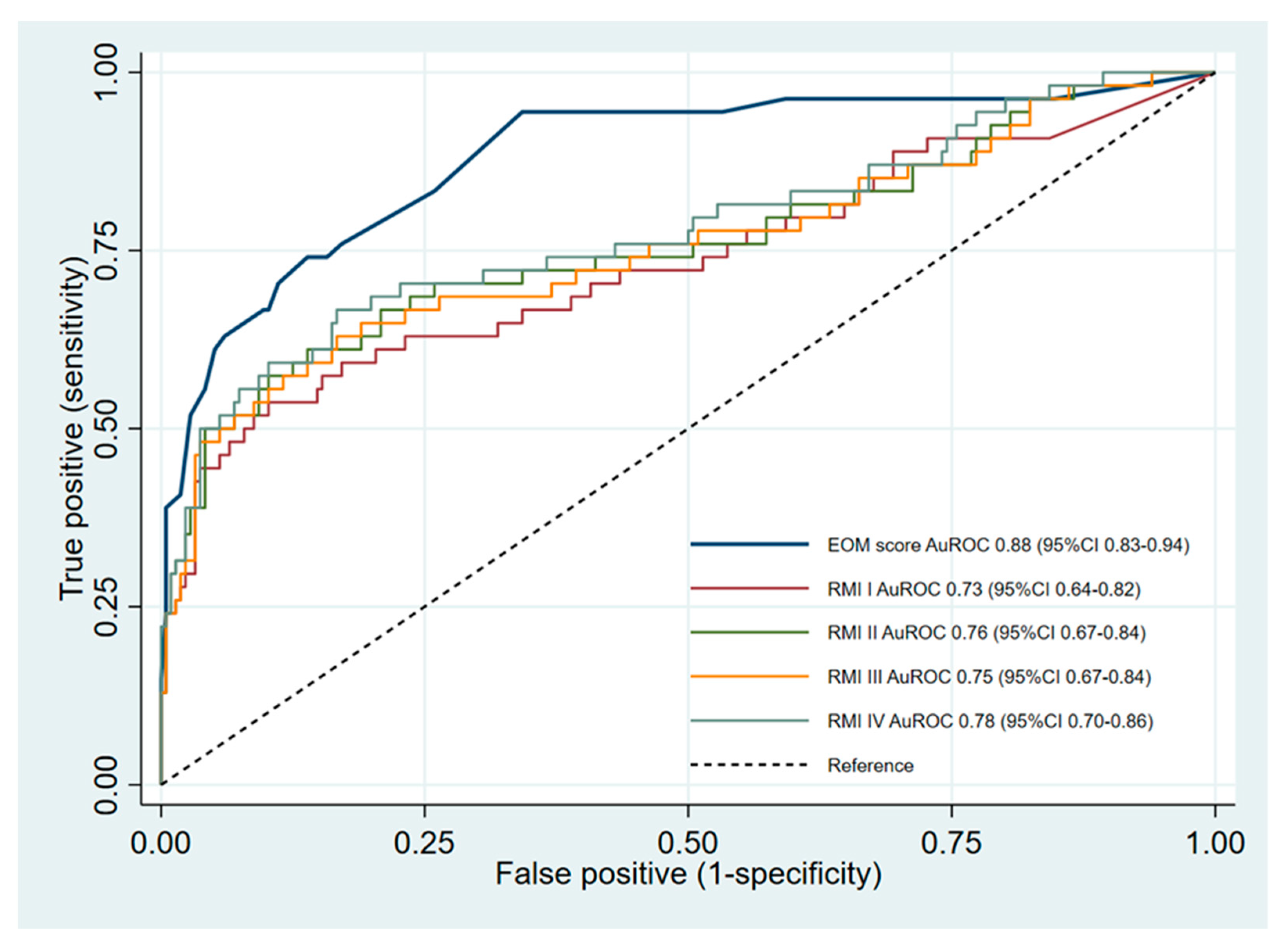
A secondary analysis was conducted using a retrospective cohort of women that presented with an adnexal mass at Phrapokklao Hospital from September 2013 to December 2017. Phrapokklao Hospital is a university-affiliated tertiary care hospital located in Chanthaburi Province located in the eastern seaboard of Thailand. The Research in Human Ethical Committee of Chanthaburi Province (CTIREC 046) and the Institutional Ethics Committee of the Faculty of Medicine, Chiang Mai University, approved the study protocol (Research ID: 7254 No. 179/2020). Informed consent was waived as data were collected retrospectively and anonymously. Patient data were kept confidential and in compliance with the Declaration of Helsinki.
The study included all women with an adnexal mass who presented consecutively at the hospital and were scheduled for surgery during the study period. Patients were eligible regardless of the type of ovarian operation. Key data collection comprised the preoperative clinical characteristics, the ultrasonographic features of the mass, and the tumor marker (CA-125), which were recorded during routine case management. The routine ultrasonographic assessment was generally performed by non-expert sonographers (including residents in training and general gynecologists). All ultrasonographic images were reevaluated and verified by a single gynecologic oncologist (WC). Ultrasound examination had been carried out by connecting a 3.5–5 MHz transabdominal or a 5–7.5 MHz transvaginal transducer to a sonoaceX7 (Samsung, South Korea). If multiple masses were present in a single patient, aspects of the mass with the most complex ultrasonographic features or the mass with the largest maximal diameter were collected.
The histopathologic reports of the resected masses were reviewed. Only patients with the diagnoses of benign ovarian tumors and early-stage ovarian cancer were included. In this study, early-stage ovarian cancer was defined as FIGO stage I, II, and IIIA1 (microscopic) [24,25]. Data from patients with advanced-stage ovarian cancer, recurrent ovarian cancer, metastatic cancer to the ovary, patients with a history of neoadjuvant chemotherapy, and patients with incomplete data were excluded. Patients with a pathological diagnosis of borderline ovarian tumors were included in the early-stage ovarian cancer group.
In this study, we planned to evaluate the diagnostic accuracy and the clinical utility of 5 diagnostic risk models for preoperative discrimination of benign and malignant adnexal tumors: EOM, RMI-I, RMI-II, RMI-III, and RMI-IV. Table 1 illustrates the components and score rating of each risk model.
The Early-stage Ovarian Malignancy score or the EOM score was recently developed from the dataset used in the primary data analysis prior to this study [23]. The EOM score is a combination of five simple predictors: menopausal status, tumor size, the presence of a solid component, the presence of ascites, and serum CA-125. Each component of the EOM score was rated with a different scoring regime, which was based on the logit coefficients of the prediction model. The total EOM score, ranging from 0 to 51, was derived from the summation of the score from each component. In the case of the EOM score, two cutoff points were proposed to categorize patients into low, moderate, and high-risk groups. The score cutoff point at ≥15 was intended to be used by general gynecologists to make a referral decision, whereas the score cutoff point at ≥30 was for oncologic specialists to prioritize patients for surgery or further investigation.
The Risk of Malignancy Index (RMI) is the classic diagnostic tool for preoperative diagnosis of ovarian cancer, which was developed in 1990 [19]. The RMI incorporates three main components, which are menopausal status, the ultrasound score, and the serum CA-125 level. The total score is the multiplicative product of all three components. The cutoff point for suggesting a high risk of a malignant tumor was originally set at an RMI ≥200 [26]. This cutoff point was generally used as the criteria for referring patients to a specialist. Currently, there are four versions of the RMI: RMI I [19], RMI II [20], RMI III [21], and RMI IV [22]. The differences in each version of the RMI give different score ratings for each component in RMI I to RMI III. In the fourth version of the RMI, tumor size is included in the score calculation, and a different cutoff point of >450 had been proposed.
All statistical analyses were performed using Stata version 16 (StataCorp, College Station, TX, USA). Frequency and percentage were used to describe the categorical data. The mean and standard deviation or median (interquartile range) were used to describe the continuous data, as appropriate. The comparison of categorical data was made using the exact probability test. The comparison of continuous data was made using an independent t-test or Mann–Whitney U test according to the data distribution. Two-tailed p-values < 0.05 were considered as statistically significant.
Diagnostic accuracy indices, sensitivity, specificity, positive predictive values, negative predictive values, and positive likelihood ratios were calculated for each risk model. The 95% confidence intervals were estimated using the Clopper Pearson’s binomial exact method. The comparison of sensitivity and specificity between the EOM score and each version of the RMI was carried out using an exact McNemar’s test. The area under the receiver operating characteristic curve (AuROC) was used to represent the discriminative ability of the risk models. We compared the equality of the AuROC of each RMI model against the EOM score and used Sidak’s adjustment for multiple comparisons.
To evaluate the clinical utility of the risk models, we employed the concept of decision curve analysis (DCA), which was introduced in 2006 by Vickers et al. [27]. DCA focuses on the net benefit (NB) gained from the application of risk models in practice. NB indicates the proportion of the net true positives, which remain after considering the presence of false positives. In a clinical context, the benefits of true positives and the harm of false positives are generally on different scales. Weighting factors must be used to convert false positives to the same scale as the true positives [28]. In this study, we focused on the appropriate referrals of patients to oncologic specialists. Thus, the weighting factor corresponds to the odds of the chosen threshold probability (T) to refer the patients to specialists (odds = T/1 − T). For instance, a threshold probability of 10% for patient referral implies that up to nine false positives are acceptable per one true positive (odds or benefit-to-harm ratio = 1/9). In summary, the NB is calculated as follows:
Net   Befit = ( Number   of   True   positives ( Number   of   False   positives   ×   ( T ÷ ( 1 T ) ) ( Total   sample   size )
where T is the threshold probability.
However, in actual practice, the choice of threshold probability varies largely depending on the discretion of each physician, as the reasonable threshold involves holistic incorporation of all potential disease outcomes. It is essential to evaluate the NB over a range of reasonable threshold probabilities within the clinical context of interest, as there is no single acceptable threshold probability [29]. In this study, we estimated the NB of all the risk models (at pre-specified cutoff points) from the risk threshold probability range of 5% to 50%, which was based on a report from a recent study on the clinical utility of the International Ovarian Tumor Analysis (IOTA) risk models [5]. We focused primarily on two important threshold probabilities, 10% and 25%, as these points were generally used as cutoff points for malignancy in the IOTA logistic regression model [30].
The decision curves were plotted to visualize the trend in NB for each model across the range of threshold probabilities. The NBs of two default strategies were also included in the graph for comparison: referring all patients or referring none. The NB of any risk model should be higher than the NB of the default strategies; otherwise, the model would be considered clinically harmful in its application. As the statistical significance and confidence intervals are not important in classical decision theory [31], we did not present the confidence intervals and p-values for the decision curve analysis.
We also presented an alternative expression of NB by calculating the net proportion of true negatives, which can be interpreted as the number of inappropriate referrals avoided per 100 patients. The equation used is as follows:
Net   proportion   of   True   negatives =   NB NB ReferAll Odds ( T )
where NB is the net benefit; and T is the threshold probability.

3. Results

During the study period, there were 640 women with an adnexal mass scheduled for surgery and who were assessed for eligibility. Of this number, 370 women were excluded (Figure 1). Two hundred and seventy patients were therefore included in the analysis. The mean age of the patients was 44.5 ± 13.0 years. Only 25.6% of the patients (n = 69) were of postmenopausal age. One-third of the patients (n = 90, 33.3%) were nulliparous. Of the 270 patients included in the analysis, 216 (80.0%) were diagnosed with benign ovarian tumors, and 54 (20.0%) with early-stage ovarian cancer. In this dataset, there were eight patients with borderline ovarian tumors. All eight patients were included in the early-stage ovarian cancer group. The histopathological classification of all ovarian tumors included in the study is presented in Table 2. The comparison of the clinical characteristics, ultrasonographic features, and tumor marker levels between patients with ovarian cancer and patients with benign ovarian tumors are shown in Table 3.
The sensitivity of the EOM score at the cutoff point of ≥15 was significantly higher than all versions of the RMI (p < 0.001), while the specificity was significantly lower (p < 0.001 for RMI I, RMI III, and RMI IV, and p = 0.002 for RMI II). In contrast, the EOM score at the higher cutoff point of ≥30 showed a significantly higher specificity than that of all RMI versions (p < 0.001). However, at this EOM cutoff, the sensitivity was significantly lower than all RMI versions (p < 0.001). The other diagnostic indices (positive predictive values, negative predictive values, and positive likelihood ratios) are illustrated in Table 4. With regard to the discriminative ability via the AuROC, the EOM score was significantly more superior than RMI-I (0.88 vs. 0.73, p < 0.001), RMI-II (0.88 vs. 0.76, p < 0.001), RMI-III (0.88 vs. 0.75, p < 0.001), and RMI-IV (0.88 vs. 0.78, p = 0.002) (Figure 2).
Regarding clinical utility, the EOM score at the cutoff point of ≥15 demonstrated a higher proportion of net true positive or NB than all versions of the RMIs at the pre-specified cutoffs from the threshold probabilities of 5% to 30% (Table 5). At the threshold probabilities of 10% and 25%, a NB of 0.158 and 0.098 by the EOM score means that there are 15.8 and 9.8 net-detected early-stage ovarian cancers per 100 patients without inappropriate referrals, respectively. At the threshold probability beyond 30%, the NB of the EOM score decreased. Using all versions of the RMI for guiding referral of patients with adnexal masses was clinically harmful at a threshold probability lower than 10%, as an approach of referral of all patients to oncologic specialists shows a higher NB. If the threshold probability for referral was between 10% and 15%, the RMIs were not considered harmful, as the NB of all the RMIs were higher than both the default strategies (refer all and refer none). However, at this risk threshold, the NB of the EOM score was higher than that of the RMIs (Figure 3a).
The EOM score also showed a higher capability to reduce inappropriate referrals than the RMIs from the threshold probabilities of 5% to 30% (Table 5) (Figure 3b). In comparison to an approach of referring all patients to oncologists, using the EOM score could reduce the number of inappropriate referrals by up to 43 per 100 patients at the risk threshold of 10% and 48 per 100 patients at the threshold probability of 20%. In contrast, at the threshold probability of 10%, only the RMI-II at the cutoff of 200 could reduce the number of inappropriate referrals by about 3 per 100 patients, whereas the other RMIs increased the number of inappropriate referrals. At a risk threshold of 25%, the EOM score reduced inappropriate referrals by between 5 and 8 (per 100 patients) more than when the RMI was used.

4. Discussion

In this study, all four RMI versions at their pre-specified cutoff points demonstrated a lower sensitivity for detecting patients with early-stage ovarian cancer when compared to the EOM score ≥15. Although the specificity of the EOM score at this cutoff was significantly lower than the RMI, its negative predictive value was the highest and may be more effective as a triage tool for patient referrals. An EOM score ≥15 also demonstrated clinical utility over a wide range of threshold probabilities, from as low as 5% up to 30%. The RMI had less clinical utility and was considered harmful at a low threshold probability but was beneficial at a higher threshold probability (>30%). Our findings suggest that, in some specific situations, the EOM score at the cutoff point of ≥15 should be used instead of the RMI to increase the detection of ovarian cancer cases while minimizing the number of inappropriate patient referrals.
In a recent meta-analysis, the pooled estimates of the sensitivity and specificity of the RMI I were 72.0% (95% CI: 67.0, 76.0%) and 92.0% (95% CI: 89.0, 93.0%), respectively [9]. The pooled sensitivity and specificity of the other RMI versions were comparable to the first version. The original publication of the RMI in 1990 reported the sensitivity and specificity of the RMI as 85.4% and 95.9% [19], respectively, which was significantly higher than the number reported in the meta-analysis and also from our findings. Out of all patients with malignant diseases included in the original study, around two-thirds (28/42, 66.7%) of the patients were at advanced stages of ovarian cancer or metastatic disease. As the RMI scoring is largely dependent on the serum CA-125 levels, a biomarker that is more specific to an advanced stage or type II ovarian cancer [14], the diagnostic performance of the RMI is undoubtedly disturbed in studies that include a lower proportion of these patients [18,32,33,34,35]. Our study only included patients with early-stage ovarian cancer, which probably explains the lower sensitivity of all the RMIs.
The EOM score at the cutoff point of ≥15 outperformed all four RMI versions in terms of diagnostic sensitivity in the detection of patients with early-stage ovarian cancer. The ability to discriminate between benign and early malignant conditions of the EOM score was also significantly higher than in all the RMIs. The derivation of the EOM score from a cohort of patients with benign ovarian tumors and early-stage ovarian cancer improves the ability to distinguish both conditions. Moreover, the EOM scoring system appropriately adjusts the influence of the serum CA-125 level into the model by categorizing this classic marker into three categories, assigning a weighted score to each. In our prior work, we proposed two cutoff points, at ≥15 and ≥30, for the implementation of the EOM score in practice [23]. The finding from this study adds weight to the existing evidence that the lower cutoff (≥15) is suitable for triage purposes, while the higher cutoff (≥30) might be more appropriate for diagnosis and giving guidance for the prioritization of patients for surgery.
Even though the EOM score exhibited good discrimination and calibration properties, this does not justify and establish its usefulness in making appropriate decisions for the referral of patients with a high probability of ovarian cancer to gynecologic oncologists [28]. In this study, we examined the clinical utility of the EOM score and the RMI in supporting decision-making by applying a novel decision-analytic approach, or DCA [27]. One of the most troublesome challenges to this approach lies within the definition of a reasonable threshold probability of patient referral [36], as the threshold generally depends on multiple factors, such as the judgement of the physician, patient preferences, local referral guidelines, and availability of healthcare resources [5]. As early detection of ovarian cancer would result in early treatment, appropriate staging, and survival improvements [6,37], it is more important to detect one case of ovarian cancer than to avoid referrals to oncologists (i.e., sensitivity is more important than specificity [1]). In our setting, patients with benign ovarian tumors who were misclassified and referred to specialists, or false-positive cases, usually undergo further non-invasive investigation before being scheduled for invasive surgery. In contrast, patients with ovarian cancer who otherwise would not be referred, or false-negative cases, might be subject to more unfavorable outcomes in this context, such as inadequate staging of the cancer [38,39] or suboptimal surgical management [40].
The NB of the EOM score at the cutoff point of ≥15 was higher than the approach of referring all patients, not referring any patients, and all the RMI versions from a threshold probability of 5% up to 30%. The application of the EOM score would also result in a lower number of inappropriate referrals. This was due to the higher sensitivity of the EOM score compared to the RMI. At a lower threshold probability, the detection of one ovarian cancer case weighs substantially more than one inappropriate referral. Thus, when false-positive cases are acceptable, a diagnostic rule with higher sensitivity would gain a higher NB. However, when false-positive cases are unacceptable, a prediction model with high specificity is warranted. In practice, a decision to refer patients only when the threshold probability was higher than 50% was not sufficiently reasonable, as it implies that referring false-positive cases is more harmful than not referring any patients [5]. We focused on the clinical utility of the EOM score and the RMI at two clinically relevant thresholds, 10% and 25%. We concluded that the EOM score ≥15 was clinically more useful in informing general gynecologists regarding the preoperative assessment of patients with adnexal masses than the RMI. The RMI would be useful only when the threshold probability was higher than 30%.
There were some limitations to be addressed. First, this study was a secondary analysis of a retrospective cohort study used to develop the EOM score. Thus, the accuracy and clinical utility of the EOM score might be overestimated. Further external validation studies on both aspects should be performed and are currently in process to support the findings in this study. Second, only patients with adnexal masses who were scheduled for surgery were included in this study. Patients with benign conditions who were conservatively managed were excluded, which might account for a large number of patients. However, this enables us to base all the diagnoses on the more reliable reference standard, histopathologic examinations. Third, the routine ultrasonographic evaluations were performed by non-expert sonographers, which limits the generalizability of our results to the setting where ultrasound examination was performed by an experienced sonographer. Nonetheless, by using data from a routine examination, our results could be considered as pragmatic regarding the accuracy and utility of both models. Fourth, only early-stage ovarian cancer cases were included. At this point, the transferability of our results could not be extended to patients with advanced-stage cancer or metastatic diseases. Fifth, our study included patients with FIGO stage IIIA as early-stage ovarian cancer following a previous study [25], which might not be conventional and could overestimate the discriminative ability of both the EOM score and the RMIs. However, as the overestimation occurs equally in both groups, differential bias during a comparative validation is unlikely. Finally, we did not examine the effect modification of menopausal status on the diagnostic accuracy and clinical utility as our study size in terms of menopausal women was limited and might not provide adequate statistical power. Therefore, subgroup effects might be present and should not be overlooked until further evidence is available.

5. Conclusions

In conclusion, the EOM score provides high diagnostic sensitivity, making it more suitable to use as a triage tool for referring women with an adnexal mass to specialized oncologic care than all versions of the RMI. It also demonstrates potential clinical utility by increasing the net detection of early-stage ovarian cancer cases without raising the number of inappropriate referrals. The results are promising but an external validation study would be required to confirm the diagnostic performance and clinical utility of the EOM score before being endorsed for clinical use.

Author Contributions

Conceptualization, P.P., J.P. and C.T.; methodology, P.P. and J.P.; software, P.P. and A.T.; validation, J.P., P.S., W.C., T.P., A.T., T.T., and C.T.; formal analysis, P.P.; investigation, P.P.; resources, P.S., W.C., T.T., and C.T.; data curation, P.S. and W.C.; writing—original draft preparation, P.P.; writing—review and editing, J.P., P.S., W.C., T.P., A.T., T.T., and C.T.; visualization, P.P.; supervision, J.P. and C.T.; project administration, W.C. and C.T. All authors have read and agreed to the published version of the manuscript.

Funding

This research work was partially supported by Chiang Mai University.

Conflicts of Interest

The authors declare no conflict of interest.

References

  1. Kaijser, J.; Bourne, T.; Valentin, L.; Sayasneh, A.; Van Holsbeke, C.; Vergote, I.; Testa, A.C.; Franchi, D.; Van Calster, B.; Timmerman, D. Improving strategies for diagnosing ovarian cancer: A summary of the International Ovarian Tumor Analysis (IOTA) studies. Ultrasound Obs. Gynecol. 2013, 41, 9–20. [Google Scholar] [CrossRef]
  2. Valentin, L.; Hagen, B.; Tingulstad, S.; Eik-Nes, S. Comparison of ‘pattern recognition’ and logistic regression models for discrimination between benign and malignant pelvic masses: A prospective cross validation. Ultrasound Obs. Gynecol. 2001, 18, 357–365. [Google Scholar] [CrossRef] [PubMed]
  3. Vernooij, F.; Heintz, P.; Witteveen, E.; Van Der Graaf, Y. The outcomes of ovarian cancer treatment are better when provided by gynecologic oncologists and in specialized hospitals: A systematic review. Gynecol. Oncol. 2007, 105, 801–812. [Google Scholar] [CrossRef] [PubMed]
  4. Elattar, A.; Bryant, A.; Winter-Roach, B.A.; Hatem, M.; Naik, R. Optimal primary surgical treatment for advanced epithelial ovarian cancer. Cochrane Database Syst. Rev. 2011, 2011, 12. [Google Scholar] [CrossRef] [PubMed]
  5. Wynants, L.; Timmerman, D.; Verbakel, J.Y.; Testa, A.; Savelli, L.; Fischerova, D.; Franchi, D.; Van Holsbeke, C.; Epstein, E.; Froyman, W.; et al. Clinical Utility of Risk Models to Refer Patients with Adnexal Masses to Specialized Oncology Care: Multicenter External Validation Using Decision Curve Analysis. Clin. Cancer Res. 2017, 23, 5082–5090. [Google Scholar] [CrossRef] [PubMed] [Green Version]
  6. Woo, Y.L.; Kyrgiou, M.; Bryant, A.; Everett, T.; Dickinson, H.O. Centralisation of services for gynaecological cancer. Cochrane Database Syst. Rev. 2012, 2012, 5. [Google Scholar] [CrossRef] [Green Version]
  7. American College of Obstetricians and Gynecologists. Practice Bulletin No. 174: Evaluation and Management of Adnexal Masses. Obstet Gynecol. 2016, 128, e210. [Google Scholar] [CrossRef]
  8. National Collaborating Centre for Cancer (UK). Ovarian Cancer The Recognition and Initial Management of Ovarian Cancer; National Institute for Health and Clinical Excellence: Guidance; National Collaborating Centre for Cancer (UK); National Collaborating Centre for Cancer (UK): Cardiff, UK, 2011; ISBN 978-0-9558265-5-9. [Google Scholar]
  9. Kaijser, J.; Sayasneh, A.; Van Hoorde, K.; Ghaem-Maghami, S.; Bourne, T.; Timmerman, D.; Van Calster, B. Presurgical diagnosis of adnexal tumours using mathematical models and scoring systems: A systematic review and meta-analysis. Hum. Reprod. Updat. 2014, 20, 449–462. [Google Scholar] [CrossRef] [Green Version]
  10. Auekitrungrueng, R.; Tinnangwattana, D.; Tantipalakorn, C.; Charoenratana, C.; Lerthiranwong, T.; Wanapirak, C.; Tongsong, T. Comparison of the diagnostic accuracy of International Ovarian Tumor Analysis simple rules and the risk of malignancy index to discriminate between benign and malignant adnexal masses. Int. J. Gynecol. Obs. 2019, 146, 364–369. [Google Scholar] [CrossRef]
  11. Shetty, J.; Saradha, A.; Pandey, D.; Bhat, R.; Kumar, P.; Bharatnur, S. IOTA Simple Ultrasound Rules for Triage of Adnexal Mass: Experience from South India. J. Obs. Gynecol. India 2019, 69, 356–362. [Google Scholar] [CrossRef]
  12. Hellström, I.; Raycraft, J.; Hayden-Ledbetter, M.; Ledbetter, J.A.; Schummer, M.; McIntosh, M.; Drescher, C.; Urban, N.; Hellström, K.E. The HE4 (WFDC2) protein is a biomarker for ovarian carcinoma. Cancer Res. 2003, 63, 3695–3700. [Google Scholar] [PubMed]
  13. Moss, E.L.; Hollingworth, J.; Reynolds, T.M. The role of CA125 in clinical practice. J. Clin. Pathol. 2005, 58, 308–312. [Google Scholar] [CrossRef] [PubMed] [Green Version]
  14. Chen, X.; Zhang, J.; Cheng, W.; Chang, D.Y.; Huang, J.; Wang, X.; Jia, L.; Rosen, D.G.; Zhang, W.; Yang, D.; et al. CA-125 Level as a Prognostic Indicator in Type I and Type II Epithelial Ovarian Cancer. Int. J. Gynecol. Cancer 2013, 23, 815–822. [Google Scholar] [CrossRef]
  15. Buamah, P. Benign conditions associated with raised serum CA-125 concentration. J. Surg. Oncol. 2000, 75, 264–265. [Google Scholar] [CrossRef]
  16. Matz, M.; Coleman, M.P.; Carreira, H.; Salmerón, D.; Chirlaque, M.D.; Allemani, C. CONCORD Working Group Worldwide comparison of ovarian cancer survival: Histological group and stage at diagnosis (CONCORD-2). Gynecol. Oncol. 2017, 144, 396–404. [Google Scholar] [CrossRef]
  17. Cramer, D.W. The epidemiology of endometriosis. Ann. N. Y. Acad. Sci. 2002, 955, 11–22. [Google Scholar] [CrossRef] [PubMed]
  18. Insin, P.; Prueksaritanond, N. Evaluation of Four Risk of Malignancy Indices (RMI) in the Preoperative Diagnosis of Ovarian Malignancy at Rajavithi Hospital. Thai J. Obstet. Gynaecol. 2013, 201, 163–175. [Google Scholar]
  19. Jacobs, I.; Oram, D.; Fairbanks, J.; Turner, J.; Frost, C.; Grudzinskas, J.G. A risk of malignancy index incorporating CA 125, ultrasound and menopausal status for the accurate preoperative diagnosis of ovarian cancer. BJOG 1990, 97, 922–929. [Google Scholar] [CrossRef]
  20. Tingulstad, S.; Hagen, B.; Skjeldestad, F.E.; Onsrud, M.; Kiserud, T.; Halvorsen, T.; Nustad, K. Evaluation of a risk of malignancy index based on serum CA125, ultrasound findings and menopausal status in the pre-operative diagnosis of pelvic masses. BJOG 1996, 103, 826–831. [Google Scholar] [CrossRef]
  21. Tingulstad, S.; Hagen, B.; Skjeldestad, F.E.; Halvorsen, T.; Nustad, K.; Onsrud, M. The Risk-of-Malignancy Index to Evaluate Potential Ovarian Cancers in Local Hospitals. Obs. Gynecol. 1999, 93, 448–452. [Google Scholar] [CrossRef]
  22. Yamamoto, Y.; Yamada, R.; Oguri, H.; Maeda, N.; Fukaya, T. Comparison of four malignancy risk indices in the preoperative evaluation of patients with pelvic masses. Eur. J. Obs. Gynecol. Reprod. Biol. 2009, 144, 163–167. [Google Scholar] [CrossRef] [PubMed]
  23. Chirdchim, W.; Wanichsetakul, P.; Phinyo, P.; Patumanond, J.; Suwannarurk, K.; Srisomboon, J. Development and Validation of a Predictive Score for Preoperative Diagnosis of Early Stage Epithelial Ovarian Cancer. Asian Pac. J. Cancer Prev. 2019, 20, 1207–1213. [Google Scholar] [CrossRef] [Green Version]
  24. Javadi, S.; Ganeshan, D.M.; Qayyum, A.; Iyer, R.B.; Bhosale, P.R. Ovarian Cancer, the Revised FIGO Staging System, and the Role of Imaging. Am. J. Roentgenol. 2016, 206, 1351–1360. [Google Scholar] [CrossRef] [PubMed]
  25. Kleppe, M.; Van Der Aa, M.A.; Van Gorp, T.; Slangen, B.F.M.; Kruitwagen, R.F.P.M. The impact of lymph node dissection and adjuvant chemotherapy on survival: A nationwide cohort study of patients with clinical early-stage ovarian cancer. Eur. J. Cancer 2016, 66, 83–90. [Google Scholar] [CrossRef] [PubMed]
  26. Håkansson, F.; Høgdall, E.V.S.; Nedergaard, L.; Lundvall, L.; Engelholm, S.A.; Pedersen, A.T.; Hartwell, D.; Høgdall, C. Danish ‘Pelvic Mass’ Ovarian Cancer Study Risk of malignancy index used as a diagnostic tool in a tertiary centre for patients with a pelvic mass. Acta Obstet. Gynecol. Scand. 2012, 91, 496–502. [Google Scholar]
  27. Vickers, A.; Elkin, E.B. Decision Curve Analysis: A Novel Method for Evaluating Prediction Models. Med. Decis. Mak. 2006, 26, 565–574. [Google Scholar] [CrossRef] [Green Version]
  28. Van Calster, B.; Wynants, L.; Verbeek, J.F.; Verbakel, J.Y.; Christodoulou, E.; Vickers, A.; Roobol, M.J.; Steyerberg, E.W. Reporting and Interpreting Decision Curve Analysis: A Guide for Investigators. Eur. Urol. 2018, 74, 796–804. [Google Scholar] [CrossRef]
  29. Vickers, A.; Van Calster, B.; Steyerberg, E.W. Net benefit approaches to the evaluation of prediction models, molecular markers, and diagnostic tests. BMJ 2016, 352, i6. [Google Scholar] [CrossRef] [Green Version]
  30. Testa, A.; Kaijser, J.; Wynants, L.; Fischerova, D.; Van Holsbeke, C.; Franchi, D.; Savelli, L.; Epstein, E.; Czekierdowski, A.; Guerriero, S.; et al. Strategies to diagnose ovarian cancer: New evidence from phase 3 of the multicentre international IOTA study. Br. J. Cancer 2014, 111, 680–688. [Google Scholar] [CrossRef] [Green Version]
  31. Vickers, A.; Van Calster, B.; Steyerberg, E.W. A simple, step-by-step guide to interpreting decision curve analysis. Diagn. Progn. Res. 2019, 3, 8. [Google Scholar] [CrossRef]
  32. Abdulrahman, J.G.O.; McKnight, L.; Singh, K.L. The risk of malignancy index (RMI) in women with adnexal masses in Wales. Taiwan J. Obs. Gynecol. 2014, 53, 376–381. [Google Scholar] [CrossRef] [PubMed]
  33. Morgante, G.; La Marca, A.; Ditto, A.; De Leo, V. Comparison of two malignancy risk indices based on serum CA125, ultrasound score and menopausal status in the diagnosis of ovarian masses. BJOG 1999, 106, 524–527. [Google Scholar] [CrossRef] [PubMed]
  34. Moolthiya, W.; Yuenyao, P. The risk of malignancy index (RMI) in diagnosis of ovarian malignancy. APJCP 2009, 10, 865–868. [Google Scholar] [PubMed]
  35. Van Holsbeke, C.; Van Calster, B.; Bourne, T.; Ajossa, S.; Testa, A.C.; Guerriero, S.; Fruscio, R.; Lissoni, A.; Czekierdowski, A.; Savelli, L.; et al. External Validation of Diagnostic Models to Estimate the Risk of Malignancy in Adnexal Masses. Clin. Cancer Res. 2011, 18, 815–825. [Google Scholar] [CrossRef] [Green Version]
  36. Fitzgerald, M.; Saville, B.R.; Lewis, R.J. Decision Curve Analysis. JAMA 2015, 313, 409–410. [Google Scholar] [CrossRef]
  37. Froyman, W.; Wynants, L.; Landolfo, C.; Bourne, T.; Valentin, L.; Testa, A.C.; Sladkevicius, P.; Franchi, D.; Fischerova, D.; Savelli, L.; et al. Validation of the Performance of International Ovarian Tumor Analysis (IOTA) Methods in the Diagnosis of Early Stage Ovarian Cancer in a Non-Screening Population. Diagnostics 2017, 7, 32. [Google Scholar] [CrossRef]
  38. Timmers, P.J.; Zwinderman, A.; Coens, C.; Vergote, I.; Trimbos, J. Understanding the problem of inadequately staging early ovarian cancer. Eur. J. Cancer 2010, 46, 880–884. [Google Scholar] [CrossRef]
  39. Trimbos, J.B. Surgical treatment of early-stage ovarian cancer. Best Pract. Res. Clin. Obs. Gynaecol. 2017, 41, 60–70. [Google Scholar] [CrossRef]
  40. Bristow, R.E.; Karlan, B.Y.; Chi, D.S. Surgery for Ovarian Cancer: Principles and Practice; CRC Press: Boca Raton, FL, USA, 2010. [Google Scholar]
Figure 1. Study flow diagram.
Medicina 56 00702 g001
Figure 2. Comparative validation of the discriminative ability between the EOM scores and the RMIs via the receiver operating characteristic curve. Abbreviations: AuROC, area under the receiver operating characteristic curve; EOM, Early-stage Ovarian Malignancy; RMI, Risk of Malignancy Indices.
Medicina 56 00702 g002
Figure 3. Comparative validation of the clinical utility between the EOM scores and Risk of Malignancy Indices via decision curves. (a) Decision curve plotting the net benefit against threshold probability. (b) Decision curve plotting a decrease in inappropriate referrals against threshold probability.
Medicina 56 00702 g003
Table 1. Components of the Early-stage Ovarian Malignancy (EOM) score and Risk of Malignancy Index (RMI) I–IV.
ComponentsEOMRMI
IIIIIIIV
Menopausal Status
Pre-menopause01111
Post-menopause73434
Ultrasonographic Features
None00111
Any 1 feature 1111
   Presence of multilocularity
   Presence of solid component3
   Bilateral lesions
   Presence of ascites13
   Presence of intra-abdominal metastases
≥2 Features 3434
Tumor Size (cm)
<7 1
≥7 2
<90
9–1210
>1216
Serum CA125 (IU/L)
<300
30–2001
>20012
Abbreviations: EOM, Early-stage Ovarian Malignancy score; RMI, Risk of Malignancy Index. For the calculation of the RMIs, the total score is the multiplicative product of the summation of the menopausal score, ultrasound score, tumor size score (for RMI IV), and serum CA-125.
Table 2. Histopathological classification of the ovarian tumors.
n (%)
Malignant Tumors (n = 46)
Endometrioid carcinoma11 (20.4)
Serous carcinoma10 (18.5)
Mucinous carcinoma6 (11.1)
Adenocarcinoma6 (11.1)
Clear cell carcinoma5 (9.3)
Granulosa cell tumor3 (5.6)
Other rare tumors5 (9.3)
Borderline Tumors (n = 8)
Borderline mucinous tumor7 (12.9)
Borderline serous tumor1 (1.8)
Borderline endometrioid tumor0 (0)
Benign Tumors (n = 216)
Endometriotic cyst77 (35.7)
Dermoid cyst45 (20.8)
Mucinous cystadenoma41 (19.0)
Serous cystadenoma26 (12.0)
Follicular cyst11 (5.1)
Corpus luteal cyst10 (4.6)
Others rare tumors6 (2.8)
Table 3. Characteristics of the study patients.
VariablesEarly Stage Ovarian Cancer
(n = 54)
Benign Ovarian Tumor
(n = 216)
p-Value
n(%)n(%)
Clinical Characteristics
Age (year) *48.7±15.443.5±12.10.008
Nulliparity21(38.9)69(31.9)0.337
Post-menopause28(51.9)41(19.0)<0.001
Ultrasonographic Features
Maximum tumor diameter (cm) *16.4±6.710.1±5.1<0.001
Multilocularity 38(70.4)145(67.1)0.745
Solid component33(61.1)57(26.4)<0.001
Bilateral lesions5(9.3)43(19.9)0.075
Ascites11(20.4)2(0.9)<0.001
Intra-abdominal metastases2(3.7)1(0.5)0.103
Tumor Marker
Serum CA125 (IU/L) **102.825.0, 314.030.814.8, 81.1<0.001
Abbreviations: SD, standard deviation; IQR, interquartile range. * Mean ± SD, ** Median (IQR).
Table 4. Comparative diagnostic performance of the Early-stage Ovarian Malignancy (EOM) score and Risk of Malignancy Indices (RMI).
ScoresCutoffEarly Stage Ovarian Cancer
(n = 54)
Benign
Ovarian Tumor
(n = 216)
Sensitivity (%)
(95% CI)
Specificity (%)
(95% CI)
PPV (%)
(95% CI)
NPV (%)
(95% CI)
LR+
(95% CI)
n(row%)n(row%)
EOM≥1551(40.8)74(59.2)94.4
(84.6–98.8)
65.7
(59.0–72.0)
40.8
(32.1–49.9)
97.9
(94.1–99.6)
2.76
(1.68–4.50)
<153(2.1)142(97.9)
≥3021(95.5)1(4.6)38.9
(25.9–53.1)
99.5
(97.4–100.0)
95.5
(77.2–99.9)
86.7
(81.8–90.7)
84.0
(12.73, 3488.41)
<3033(13.3)215(86.7)
RMI I≥20031(47.0)35(53.0)57.4
(43.2–70.8)
83.8
(78.2–88.4)
47.0
(34.6–59.7)
88.7
(83.6–92.7)
3.54
(1.92–6.49)
<20023(11.3)181(88.7)
RMI II≥20036(44.4)45(55.6)66.7
(52.5–78.9)
79.2
(73.1–84.4)
44.4
(33.4–55.9)
90.5
(85.4–94.3)
3.20
(1.81–5.61)
<20018(9.5)171(90.5)
RMI III≥20034(47.2)38(52.8)63.0
(48.7–75.7)
82.4
(76.7–87.2)
47.2
(35.3–59.3)
89.9
(84.8–93.7)
3.58
(1.98–6.43)
<20020(10.1)178(89.9)
RMI IV≥45033(50.0)33(50.0)61.1
(46.9–74.1)
84.7
(79.2–89.2)
50.0
(37.4–62.6)
89.7
(84.7–93.5)
4.00
(2.17–7.32)
<45021(10.3)183(89.7)
Abbreviations: PPV, positive predictive value; NPV, negative predictive value; LR+, positive likelihood ratios; CI, confidence interval; EOM score, Early-stage Ovarian Malignancy score; RMI, Risk of Malignancy Index.
Table 5. Decision curve analysis.
Threshold ProbabilityNet Benefit
(Proportion of Net True Positive)
Reduced Numbers of Inappropriate
Referrals per 100 Patients
Refer allEOMRMI IRMI IIRMI IIIRMI IVEOMRMI IRMI IIRMI IIIRMI IV
5%0.1570.1740.1080.1250.1190.11631.482−94.815−63.333−74.815−80.000
10%0.1110.1580.1000.1150.1100.10942.593−9.6303.333−0.741−2.222
15%0.0590.1410.0920.1040.1010.10146.29618.76525.55623.95123.704
20%00.1200.0820.0920.0910.09248.14832.96336.66736.29636.667
25%−0.0670.0980.0720.0780.0790.08149.25941.48143.33343.70444.445
30%−0.1430.0710.0590.0620.0660.07050.00047.16047.77848.64249.630
35%−0.2310.0410.0450.0440.0500.05650.52951.21750.95252.16953.333
40%−0.3330.0060.0280.0220.0320.04150.92654.25953.33354.81556.111
45%−0.455−0.0350.009−0.0030.0110.02251.23556.62655.18556.87258.272
50%−0.600−0.085−0.015−0.033−0.015051.48158.51956.66758.51960.000
Abbreviations: EOM, Early-stage Ovarian Malignancy score; RMI, Risk of Malignancy Index.
Publisher’s Note: MDPI stays neutral with regard to jurisdictional claims in published maps and institutional affiliations.

